Search This Blog

Saturday, May 8, 2021

Endocrine system

From Wikipedia, the free encyclopedia
 
Endocrine system
Endocrine English.svg
Main glands of the endocrine system
Details
Identifiers
LatinSystema endocrinum
MeSHD004703
FMA9668

The endocrine system is a messenger system comprising feedback loops of the hormones released by internal glands of an organism directly into the circulatory system, regulating distant target organs. In vertebrates, the hypothalamus is the neural control center for all endocrine systems. In humans, the major endocrine glands are the thyroid gland and the adrenal glands. The study of the endocrine system and its disorders is known as endocrinology. Endocrinology is a branch of internal medicine.

Glands that signal each other in sequence are often referred to as an axis, such as the hypothalamic-pituitary-adrenal axis. In addition to the specialized endocrine organs mentioned above, many other organs that are part of other body systems have secondary endocrine functions, including bone, kidneys, liver, heart and gonads. For example, the kidney secretes the endocrine hormone erythropoietin. Hormones can be amino acid complexes, steroids, eicosanoids, leukotrienes, or prostaglandins.

The endocrine system can be contrasted to both exocrine glands, which secrete hormones to the outside of the body, and paracrine signalling between cells over a relatively short distance. Endocrine glands have no ducts, are vascular, and commonly have intracellular vacuoles or granules that store their hormones. In contrast, exocrine glands, such as salivary glands, sweat glands, and glands within the gastrointestinal tract, tend to be much less vascular and have ducts or a hollow lumen.

The word endocrine derives via New Latin from the Greek words ἔνδον, endon, "inside, within," and "ekrīnō" from the εκκρινω, "secrete".

Structure

Major endocrine systems

The human endocrine system consists of several systems that operate via feedback loops. Several important feedback systems are mediated via the hypothalamus and pituitary.

Glands

Endocrine glands are glands of the endocrine system that secrete their products, hormones, directly into interstitial spaces and then absorbed into blood rather than through a duct. The major glands of the endocrine system include the pineal gland, pituitary gland, pancreas, ovaries, testes, thyroid gland, parathyroid gland, hypothalamus and adrenal glands. The hypothalamus and pituitary gland are neuroendocrine organs.

The hypothalamus and the anterior pituitary are two out of the three endocrine glands that are important in cell signaling. They are both part of the HPA axis which is known to play a role in cell signaling in the nervous system.

Hypothalamus: The hypothalamus is a key regulator of the autonomic nervous system. The endocrine system has three sets of endocrine outputs which include the magnocellular system, the parvocellular system, and autonomic intervention. The magnocellular is involved in the expression of oxytocin or vasopressin. The parvocellular is involved in controlling the secretion of hormones from the anterior pituitary.

Anterior Pituitary: The main role of the anterior pituitary gland is to produce and secret tropic hormones. Some examples of tropic hormones secreted by the anterior pituitary gland include TSH, ACTH, GH, LH, and FSH.

Cells

There are many types of cells that make up the endocrine system and these cells typically make up larger tissues and organs that function within and outside of the endocrine system.

Hormones

A hormone is any of a class of signaling molecules produced by cars in glands in multicellular organisms that are transported by the circulatory system to target distant organs to regulate physiology and behaviour. Hormones have diverse chemical structures, mainly of 3 classes: eicosanoids, steroids, and amino acid/protein derivatives (amines, peptides, and proteins). The glands that secrete hormones comprise the endocrine system. The term hormone is sometimes extended to include chemicals produced by cells that affect the same cell (autocrine or intracrine signalling) or nearby cells (paracrine signalling).

Hormones are used to communicate between organs and tissues for physiological regulation and behavioral activities, such as digestion, metabolism, respiration, tissue function, sensory perception, sleep, excretion, lactation, stress, growth and development, movement, reproduction, and mood.

Hormones affect distant cells by binding to specific receptor proteins in the target cell resulting in a change in cell function. This may lead to cell type-specific responses that include rapid changes to the activity of existing proteins, or slower changes in the expression of target genes. Amino acid–based hormones (amines and peptide or protein hormones) are water-soluble and act on the surface of target cells via signal transduction pathways; steroid hormones, being lipid-soluble, move through the plasma membranes of target cells to act within their nuclei.

Cell signalling

The typical mode of cell signalling in the endocrine system is endocrine signaling, that is, using the circulatory system to reach distant target organs. However, there are also other modes, i.e., paracrine, autocrine, and neuroendocrine signaling. Purely neurocrine signaling between neurons, on the other hand, belongs completely to the nervous system.

Autocrine

Autocrine signaling is a form of signaling in which a cell secretes a hormone or chemical messenger (called the autocrine agent) that binds to autocrine receptors on the same cell, leading to changes in the cells.

Paracrine

Some endocrinologists and clinicians include the paracrine system as part of the endocrine system, but there is not consensus. Paracrines are slower acting, targeting cells in the same tissue or organ. An example of this is somatostatin which is released by some pancreatic cells and targets other pancreatic cells.

Juxtacrine

Juxtacrine signaling is a type of intercellular communication that is transmitted via oligosaccharide, lipid, or protein components of a cell membrane, and may affect either the emitting cell or the immediately adjacent cells.

It occurs between adjacent cells that possess broad patches of closely opposed plasma membrane linked by transmembrane channels known as connexons. The gap between the cells can usually be between only 2 and 4 nm.

Clinical significance

Disease

Disability-adjusted life year for endocrine disorders per 100,000 inhabitants in 2002.
 
  no data
  less than 80
  80–160
  160–240
  240–320
  320–400
  400–480
  480–560
  560–640
  640–720
  720–800
  800–1000
  more than 1000

Diseases of the endocrine system are common, including conditions such as diabetes mellitus, thyroid disease, and obesity. Endocrine disease is characterized by misregulated hormone release (a productive pituitary adenoma), inappropriate response to signaling (hypothyroidism), lack of a gland (diabetes mellitus type 1, diminished erythropoiesis in chronic kidney failure), or structural enlargement in a critical site such as the thyroid (toxic multinodular goitre). Hypofunction of endocrine glands can occur as a result of loss of reserve, hyposecretion, agenesis, atrophy, or active destruction. Hyperfunction can occur as a result of hypersecretion, loss of suppression, hyperplastic or neoplastic change, or hyperstimulation.

Endocrinopathies are classified as primary, secondary, or tertiary. Primary endocrine disease inhibits the action of downstream glands. Secondary endocrine disease is indicative of a problem with the pituitary gland. Tertiary endocrine disease is associated with dysfunction of the hypothalamus and its releasing hormones.

As the thyroid, and hormones have been implicated in signaling distant tissues to proliferate, for example, the estrogen receptor has been shown to be involved in certain breast cancers. Endocrine, paracrine, and autocrine signaling have all been implicated in proliferation, one of the required steps of oncogenesis.[15]

Other common diseases that result from endocrine dysfunction include Addison's disease, Cushing's disease and Graves' disease. Cushing's disease and Addison's disease are pathologies involving the dysfunction of the adrenal gland. Dysfunction in the adrenal gland could be due to primary or secondary factors and can result in hypercortisolism or hypocortisolism . Cushing's disease is characterized by the hypersecretion of the adrenocorticotropic hormone (ACTH) due to a pituitary adenoma that ultimately causes endogenous hypercortisolism by stimulating the adrenal glands. Some clinical signs of Cushing's disease include obesity, moon face, and hirsutism. Addison's disease is an endocrine disease that results from hypocortisolism caused by adrenal gland insufficiency. Adrenal insufficiency is significant because it is correlated with decreased ability to maintain blood pressure and blood sugar, a defect that can prove to be fatal.

Graves' disease involves the hyperactivity of the thyroid gland which produces the T3 and T4 hormones. Graves' disease effects range from excess sweating, fatigue, heat intolerance and high blood pressure to swelling of the eyes that causes redness, puffiness and in rare cases reduced or double vision.

Other animals

A neuroendocrine system has been observed in all animals with a nervous system and all vertebrates have a hypothalamus-pituitary axis. All vertebrates have a thyroid, which in amphibians is also crucial for transformation of larvae into adult form. All vertebrates have adrenal gland tissue, with mammals unique in having it organized into layers. All vertebrates have some form of a renin–angiotensin axis, and all tetrapods have aldosterone as a primary mineralocorticoid.

Gut–brain axis

From Wikipedia, the free encyclopedia

The gut–brain axis is the relationship between the GI tract and brain function and development

The gut–brain axis is the biochemical signaling that takes place between the gastrointestinal tract (GI tract) and the central nervous system (CNS). The term "gut–brain axis" is occasionally used to refer to the role of the gut flora in the interplay as well, whereas the term "microbiota–gut–brain (MGB or BGM) axis" explicitly includes the role of gut flora in the biochemical signaling events that take place between the GI tract and CNS.

Broadly defined, the gut–brain axis includes the central nervous system, neuroendocrine and neuroimmune systems, including the hypothalamic–pituitary–adrenal axis (HPA axis), sympathetic and parasympathetic arms of the autonomic nervous system, including the enteric nervous system and the vagus nerve, and the gut microbiota. The first of the brain–gut interactions shown, was the cephalic phase of digestion, in the release of gastric and pancreatic secretions in response to sensory signals, such as the smell and sight of food. This was first demonstrated by Pavlov.

Interest in the field was sparked by a 2004 study showing that germ-free (GF) mice showed an exaggerated HPA axis response to stress compared to non-GF laboratory mice.

As of October 2016, most of the work done on the role of gut flora in the gut–brain axis had been conducted in animals, or on characterizing the various neuroactive compounds that gut flora can produce. Studies with humans – measuring variations in gut flora between people with various psychiatric and neurological conditions or when stressed, or measuring effects of various probiotics (dubbed "psychobiotics" in this context) – had generally been small and were just beginning to be generalized. Whether changes to gut flora are a result of disease, a cause of disease, or both in any number of possible feedback loops in the gut–brain axis, remained unclear.

Gut flora

Bifidobacterium adolescentis Gram
 
Lactobacillus sp 01

The gut flora is the complex community of microorganisms that live in the digestive tracts of humans and other animals. The gut metagenome is the aggregate of all the genomes of gut microbiota. The gut is one niche that human microbiota inhabit.

In humans, the gut microbiota has the largest quantity of bacteria and the greatest number of species, compared to other areas of the body. In humans, the gut flora is established at one to two years after birth; by that time, the intestinal epithelium and the intestinal mucosal barrier that it secretes have co-developed in a way that is tolerant to, and even supportive of, the gut flora and that also provides a barrier to pathogenic organisms.

The relationship between gut flora and humans is not merely commensal (a non-harmful coexistence), but rather a mutualistic relationship. Human gut microorganisms benefit the host by collecting the energy from the fermentation of undigested carbohydrates and the subsequent absorption of short-chain fatty acids (SCFAs), acetate, butyrate, and propionate. Intestinal bacteria also play a role in synthesizing vitamin B and vitamin K as well as metabolizing bile acids, sterols, and xenobiotics. The systemic importance of the SCFAs and other compounds they produce are like hormones and the gut flora itself appears to function like an endocrine organ; dysregulation of the gut flora has been correlated with a host of inflammatory and autoimmune conditions.

The composition of human gut flora changes over time, when the diet changes, and as overall health changes.

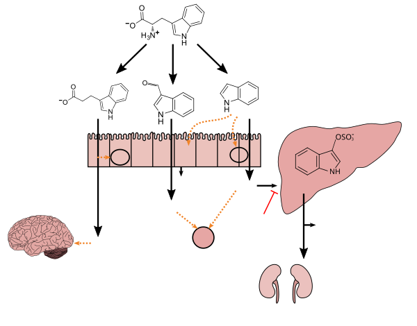
Tryptophan metabolism by human gastrointestinal microbiota 
This diagram shows the biosynthesis of bioactive compounds (indole and certain other derivatives) from tryptophan by bacteria in the gut. Indole is produced from tryptophan by bacteria that express tryptophanase. Clostridium sporogenes metabolizes tryptophan into indole and subsequently 3-indolepropionic acid (IPA), a highly potent neuroprotective antioxidant that scavenges hydroxyl radicals. IPA binds to the pregnane X receptor (PXR) in intestinal cells, thereby facilitating mucosal homeostasis and barrier function
 
Following absorption from the intestine and distribution to the brain, IPA confers a neuroprotective effect against cerebral ischemia and Alzheimer's disease. Lactobacillus species metabolize tryptophan into indole-3-aldehyde (I3A) which acts on the aryl hydrocarbon receptor (AhR) in intestinal immune cells, in turn increasing interleukin-22 (IL-22) production. Indole itself triggers the secretion of glucagon-like peptide-1 (GLP-1) in intestinal L cells and acts as a ligand for AhR. Indole can also be metabolized by the liver into indoxyl sulfate, a compound that is toxic in high concentrations and associated with vascular disease and renal dysfunction. AST-120 (activated charcoal), an intestinal sorbent that is taken by mouth, adsorbs indole, in turn decreasing the concentration of indoxyl sulfate in blood plasma.
 

Enteric nervous system

The enteric nervous system is one of the main divisions of the nervous system and consists of a mesh-like system of neurons that governs the function of the gastrointestinal system; it has been described as a "second brain" for several reasons. The enteric nervous system can operate autonomously. It normally communicates with the central nervous system (CNS) through the parasympathetic (e.g., via the vagus nerve) and sympathetic (e.g., via the prevertebral ganglia) nervous systems. However, vertebrate studies show that when the vagus nerve is severed, the enteric nervous system continues to function.

In vertebrates, the enteric nervous system includes efferent neurons, afferent neurons, and interneurons, all of which make the enteric nervous system capable of carrying reflexes in the absence of CNS input. The sensory neurons report on mechanical and chemical conditions. Through intestinal muscles, the motor neurons control peristalsis and churning of intestinal contents. Other neurons control the secretion of enzymes. The enteric nervous system also makes use of more than 30 neurotransmitters, most of which are identical to the ones found in CNS, such as acetylcholine, dopamine, and serotonin. More than 90% of the body's serotonin lies in the gut, as well as about 50% of the body's dopamine; the dual function of these neurotransmitters is an active part of gut–brain research.

The first of the gut–brain interactions was shown to be between the sight and smell of food and the release of gastric secretions, known as the cephalic phase, or cephalic response of digestion.

Gut–brain integration

The gut–brain axis, a bidirectional neurohumoral communication system, is important for maintaining homeostasis and is regulated through the central and enteric nervous systems and the neural, endocrine, immune, and metabolic pathways, and especially including the hypothalamic–pituitary–adrenal axis (HPA axis). That term has been expanded to include the role of the gut flora as part of the "microbiome-gut-brain axis", a linkage of functions including the gut flora.

Interest in the field was sparked by a 2004 study (Nobuyuki Sudo and Yoichi Chida) showing that germ-free mice (genetically homogeneous laboratory mice, birthed and raised in an antiseptic environment) showed an exaggerated HPA axis response to stress, compared to non-GF laboratory mice.

The gut flora can produce a range of neuroactive molecules, such as acetylcholine, catecholamines, γ-aminobutyric acid, histamine, melatonin, and serotonin, which are essential for regulating peristalsis and sensation in the gut. Changes in the composition of the gut flora due to diet, drugs, or disease correlate with changes in levels of circulating cytokines, some of which can affect brain function. The gut flora also release molecules that can directly activate the vagus nerve, which transmits information about the state of the intestines to the brain.

Likewise, chronic or acutely stressful situations activate the hypothalamic–pituitary–adrenal axis, causing changes in the gut flora and intestinal epithelium, and possibly having systemic effects. Additionally, the cholinergic anti-inflammatory pathway, signaling through the vagus nerve, affects the gut epithelium and flora. Hunger and satiety are integrated in the brain, and the presence or absence of food in the gut and types of food present also affect the composition and activity of gut flora.

That said, most of the work that has been done on the role of gut flora in the gut–brain axis has been conducted in animals, including the highly artificial germ-free mice. As of 2016, studies with humans measuring changes to gut flora in response to stress, or measuring effects of various probiotics, have generally been small and cannot be generalized; whether changes to gut flora are a result of disease, a cause of disease, or both in any number of possible feedback loops in the gut–brain axis, remains unclear.

The history of ideas about a relationship between the gut and the mind dates from the nineteenth century. The concepts of dyspepsia and neurasthenia gastrica referred to the influence of the gut on human emotions and thoughts.

Gut-brain-skin axis

A unifying theory that tied gastrointestinal mechanisms to anxiety, depression, and skin conditions such as acne was proposed as early as 1930. In a paper in 1930, it was proposed that emotional states might alter normal intestinal flora which could lead to increased intestinal permeability and therefore contribute to systemic inflammation. Many aspects of this theory have been validated since then. Gut microbiota and oral probiotics have been found to influence systemic inflammation, oxidative stress, glycemic control, tissue lipid content, and mood.

Research

Probiotics

A 2016 systematic review of laboratory animal studies and preliminary human clinical trials using commercially available strains of probiotic bacteria found that certain species of the Bifidobacterium and Lactobacillus genera (i.e., B. longum, B. breve, B. infantis, L. helveticus, L. rhamnosus, L. plantarum, and L. casei) had the most potential to be useful for certain central nervous system disorders.

Anxiety and mood disorders

As of 2018 work on the relationship between gut flora and anxiety disorders and mood disorders, as well as attempts to influence that relationship using probiotics or prebiotics (called "psychobiotics"), was at an early stage, with insufficient evidence to draw conclusions about a causal role for gut flora changes in these conditions, or about the efficacy of any probiotic or prebiotic treatment.

People with anxiety and mood disorders tend to have gastrointestinal problems; small studies have been conducted to compare the gut flora of people with major depressive disorder and healthy people, but those studies have had contradictory results.

Much interest was generated in the potential role of gut flora in anxiety disorders, and more generally in the role of gut flora in the gut–brain axis, by studies published in 2004 showing that germ-free mice have an exaggerated HPA axis response to stress caused by being restrained, which was reversed by colonizing their gut with a Bifidobacterium species. Studies looking at maternal separation for rats shows neonatal stress leads to long-term changes in the gut microbiota such as its diversity and composition, which also led to stress and anxiety-like behavior. Additionally, while much work had been done as of 2016 to characterize various neurotransmitters known to be involved in anxiety and mood disorders that gut flora can produce (for example, Escherichia, Bacillus, and Saccharomyces species can produce noradrenalin; Candida, Streptococcus, and Escherichia species can produce serotonin, etc.) the interrelationships and pathways by which the gut flora might affect anxiety in humans were unclear.

In one study, germ-free mice underwent fecal transplants with microbes from humans with or without major depressive disorder (MDD). Mice with microbes from humans with MDD displayed more behaviors associated with anxiety and depression than mice transplanted with microbes from humans without MDD. The taxonomic composition of microbiota between depressed patients and healthy patients, as well as between the respective mice, also differed. Germ-free mice in another study also displayed behaviors associated with anxiety and depression as compared to mice with normal microbiota, and had higher levels of corticosterone after exposure to behavioral tests. Using rodents in microbiome and mental health studies allows researchers to compare behavior and microbial composition of rodents to humans, ideally to elucidate therapeutic application for mental disorders.

Additionally, there is a link between the gut microbiome, mood disorders and anxiety, and sleep. The microbial composition of the gut microbiome changes depending on the time of day, meaning that throughout the day, the gut is exposed to varying metabolites produced by the microbes active during that time. These time-dependent microbial changes are associated with differences in the transcription of circadian clock genes involved in circadian rhythm. One mouse study showed that altering clock gene transcription by disrupting circadian rhythm, such as through sleep deprivation, potentially has a direct effect on the composition of the gut microbiome. Another study found that mice that could not produce the CLOCK protein, made by a clock gene, were more likely to develop depression. Stress and sleep disturbances can lead to greater gut mucosal permeability via activation of the HPA axis. This in turn causes immune inflammatory responses that contribute to the development of illnesses that cause depression and anxiety.

Autism

Around 70% of people with autism also have gastrointestinal problems, and autism is often diagnosed at the time that the gut flora becomes established, indicating that there may be a connection between autism and gut flora. Some studies have found differences in the gut flora of children with autism compared with children without autism – most notably elevations in the amount of Clostridium in the stools of children with autism compared with the stools of the children without – but these results have not been consistently replicated. Many of the environmental factors thought to be relevant to the development of autism would also affect the gut flora, leaving open the question of whether specific developments in the gut flora drive the development of autism or whether those developments happen concurrently. As of 2016, studies with probiotics had only been conducted with animals; studies of other dietary changes to treat autism have been inconclusive.

Parkinson's disease

As of 2015, one study had been conducted comparing the gut flora of people with Parkinson's disease to healthy controls; in that study people with Parkinson's had lower levels of Prevotellaceae and people with Parkinson's who had higher levels of Enterobacteriaceae had more clinically severe symptoms; the authors of the study drew no conclusions about whether gut flora changes were driving the disease or vice versa.

Endocrine disease

From Wikipedia, the free encyclopedia
 
Endocrine diseases
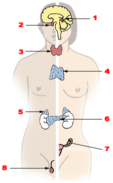
Illu endocrine system.png
Major endocrine glands. 1. Pineal gland 2. Pituitary gland 3. Thyroid gland 4. Thymus 5. Adrenal gland 6. Pancreas 7. Ovary 8. Testes

Endocrine diseases are disorders of the endocrine system. The branch of medicine associated with endocrine disorders is known as endocrinology.

Types of disease

Broadly speaking, endocrine disorders may be subdivided into three groups:

  1. Endocrine gland hyposecretion (leading to hormone deficiency)
  2. Endocrine gland hypersecretion (leading to hormone excess)
  3. Tumours (benign or malignant) of endocrine glands

Endocrine disorders are often quite complex, involving a mixed picture of hyposecretion and hypersecretion because of the feedback mechanisms involved in the endocrine system. For example, most forms of hyperthyroidism are associated with an excess of thyroid hormone and a low level of thyroid stimulating hormone.

List of diseases

Glucose homeostasis disorders

Thyroid disorders

Calcium homeostasis disorders and Metabolic bone disease

Pituitary gland disorders

Posterior pituitary

Anterior pituitary

Sex hormone disorders

Tumours of the endocrine glands not mentioned elsewhere

See also separate organs

Endocrine emergencies

In endocrinology, medical emergencies include diabetic ketoacidosis, hyperosmolar hyperglycemic state, hypoglycemic coma, acute adrenocortical insufficiency, phaeochromocytoma crisis, hypercalcemic crisis, thyroid storm, myxoedema coma and pituitary apoplexy.

Emergencies arising from decompensated pheochromocytomas or parathyroid adenomas are sometimes referred for emergency resection when aggressive medical therapies fail to control the patient's state, however the surgical risks are significant, especially blood pressure lability and the possibility of cardiovascular collapse after resection (due to a brutal drop in respectively catecholamines and calcium, which must be compensated with gradual normalization). It remains debated when emergency surgery is appropriate as opposed to urgent or elective surgery after continued attempts to stabilize the patient, notably in view of newer and more efficient medications and protocols.

Hormone

From Wikipedia, the free encyclopedia

Left: A hormone feedback loop in a female adult. (1) Follicle-Stimulating Hormone, (2) Luteinizing Hormone, (3) Progesterone, (4) Estrogen. Right: Auxin transport from leaves to roots in Arabidopsis thaliana

A hormone (from the Greek participle ὁρμῶν, "setting in motion") is any member of a class of signaling molecules in multicellular organisms, that are transported to distant organs to regulate physiology and / or behavior. Hormones are required for the correct development of animals, plants and fungi. The lax definition of a hormone (as a signalling molecule that acts distant from its site of production) means that many different classes of molecule can be defined as hormones. Among the substances that can be considered hormones, are eicosanoids (e.g. prostaglandins and thromboxanes), steroids (e.g. oestrogen and brassinosteroid), amino acid derivatives (e.g. epinephrine and auxin), protein / peptides (e.g. insulin and CLE peptides) and gases (e.g ethylene and nitrous oxide).

Hormones are used to communicate between organs and tissues. In vertebrates, hormones are responsible for the regulation of many physiological processes and behavioral activities such as digestion, metabolism, respiration, sensory perception, sleep, excretion, lactation, stress induction, growth and development, movement, reproduction, and mood manipulation. In plants, hormones modulate almost all aspects of development, from germination to senescence.

Hormones affect distant cells by binding to specific receptor proteins in the target cell, resulting in a change in cell function. When a hormone binds to the receptor, it results in the activation of a signal transduction pathway that typically activates gene transcription, resulting in increased expression of target proteins. Hormones can also act in rapid, non-genomic pathways that can be synergistic with genomic effects. Water-soluble hormones (such as peptides and amines) generally act on the surface of target cells via second messengers. Lipid soluble hormones, (such as steroids) generally pass through the plasma membranes of target cells (both cytoplasmic and nuclear) to act within their nuclei. A notable exception to this are brassinosteroids in plants, which despite being lipid soluble, still bind to their receptor at the cell surface.

In vertebrates, endocrine glands are specialized organs that secrete hormones into the endocrine signaling system. Hormone secretion occurs in response to specific biochemical signals and is often subject to negative feedback regulation. For instance, high blood sugar (serum glucose concentration) promotes insulin synthesis. Insulin then acts to reduce glucose levels and maintain homeostasis, leading to reduced insulin levels. Upon secretion water soluble hormones are readily transported through the circulatory system. Lipid-soluble hormones must bond to carrier plasma glycoproteins (e.g., thyroxine-binding globulin (TBG)) to form ligand-protein complexes. Some hormones are completely active when released into the bloodstream (as is the case for insulin and growth hormones), while others are prohormones that must be activated in specific cells through a series of activation steps that are commonly highly regulated. The endocrine system secretes hormones directly into the bloodstream, typically via fenestrated capillaries, whereas the exocrine system secretes its hormones indirectly using ducts. Hormones with paracrine function diffuse through the interstitial spaces to nearby target tissue.

Plants lack specialized organs for the secretion of hormones, although there is spacial distribution of hormone production. For example, the hormone auxin is produced mainly at the tips of young leaves and in the shoot apical meristem. The lack of specialised glands means that the main site of hormone production can change throughout the life of a plant, and the site of production is dependent on the plant's age and environment.

Introduction and overview

Hormonal signaling involves the following steps:

  1. Biosynthesis of a particular hormone in a particular tissue
  2. Storage and secretion of the hormone
  3. Transport of the hormone to the target cell(s)
  4. Recognition of the hormone by an associated cell membrane or intracellular receptor protein
  5. Relay and amplification of the received hormonal signal via a signal transduction process: This then leads to a cellular response. The reaction of the target cells may then be recognized by the original hormone-producing cells, leading to a down regulation in hormone production. This is an example of a homeostatic negative feedback loop.
  6. Breakdown of the hormone.

Hormone producing cells are typically of a specialized cell type, residing within a particular endocrine gland, such as the thyroid gland, ovaries, and testes. Hormones exit their cell of origin via exocytosis or another means of membrane transport. The hierarchical model is an oversimplification of the hormonal signaling process. Cellular recipients of a particular hormonal signal may be one of several cell types that reside within a number of different tissues, as is the case for insulin, which triggers a diverse range of systemic physiological effects. Different tissue types may also respond differently to the same hormonal signal.

Discovery

Arnold Adolph Berthold (1849)

Arnold Adolph Berthold was a German physiologist and zoologist, who, in 1849, had a question about the function of the testes. He noticed that in castrated roosters that they did not have the same sexual behaviors as roosters with their testes intact. He decided to run an experiment on male roosters to examine this phenomenon. He kept a group of roosters with their testes intact, and saw that they had normal sized wattles and combs (secondary sexual organs), a normal crow, and normal sexual and aggressive behaviors. He also had a group with their testes surgically removed, and noticed that their secondary sexual organs were decreased in size, had a weak crow, did not have sexual attraction towards females, and were not aggressive. He realized that this organ was essential for these behaviors, but he did not know how. To test this further, he removed one testis and placed it in the abdominal cavity. The roosters acted and had normal physical anatomy. He was able to see that location of the testes do not matter. He then wanted to see if it was a genetic factor that was involved in the testes that provided these functions. He transplanted a testis from another rooster to a rooster with one testis removed, and saw that they had normal behavior and physical anatomy as well. Berthold determined that the location or genetic factors of the testes do not matter in relation to sexual organs and behaviors, but that some chemical in the testes being secreted is causing this phenomenon. It was later identified that this factor was the hormone testosterone.

Charles and Francis Darwin (1880)

Although known primarily for his work on the Theory of Evolution, Charles Darwin was also keenly interested in plants. Through the 1870's, he and his son Francis studied the movement of plants towards light. They were able to show that light is perceived at the tip of a young stem (the coleoptile), whereas the bending occurs lower down the stem. They proposed that a 'transmissible substance' communicated the direction of light from the tip down to the stem. The idea of a 'transmissible substance' was initially dismissed by other plant biologists, but their work later led to the discovery of the first plant hormone. In the 1920's Dutch scientist Frits Warmolt Went and Russian scientist Nikolai Cholodny (working independently of each other) conclusively showed that asymmetric accumulation of a growth hormone was responsible for this bending. In 1933 this hormone was finally isolated by Kögl, Haagen-Smit and Erxleben and christened 'auxin'.

Bayliss and Starling (1902)

William Bayliss and Ernest Starling, a physiologist and biologist, respectively, wanted to see if the nervous system had an impact on the digestive system. They knew that the pancreas was involved in the secretion of digestive fluids after the passage of food from the stomach to the intestines, which they believed to be due to the nervous system. They cut the nerves to the pancreas in an animal model and discovered that it was not nerve impulses that controlled secretion from the pancreas. It was determined that a factor secreted from the intestines into the bloodstream was stimulating the pancreas to secrete digestive fluids. This factor was named secretin: a hormone, although the term hormone was not coined until 1905 by Starling.

Types of signaling

Hormonal effects are dependent on where they are released, as they can be released in different manners. Not all hormones are released from a cell and into the blood until it binds to a receptor on a target. The major types of hormone signaling are:

Chemical classes

As hormones are defined functionally, not structurally, they may have diverse chemical structures. Hormones occur in multicellular organisms (plants, animals, fungi, brown algae, and red algae). These compounds occur also in unicellular organisms, and may act as signaling molecules however there is no agreement that these molecules can be called hormones.

Vertebrates

Hormone types in Vertebrates
SN Types Description
1 Peptide Peptide hormones are made of a chain of amino acids that can range from just 3 to hundreds of amino acids. Examples include oxytocin and insulin. Their sequences are encoded in DNA and can be modified by alternative splicing and/or post-translational modification. They are packed in vesicles and are hydrophilic, meaning that they are soluble in water. Due to their hydrophilicity, they can only bind to receptors on the membrane, as travelling through the membrane is unlikely. However, some hormones can bind to intracellular receptors through an intracrine mechanism.
2 Amino acid Amino acid hormones are derived from amino acid, most commonly tyrosine. They are stored in vesicles. Examples include melatonin and thyroxine.
3 Steroid Steroid hormones are derived from cholesterol. Examples include the sex hormones estradiol and testosterone as well as the stress hormone cortisol. Steroids contain four fused rings. They are lipophilic and hence can cross membranes to bind to intracellular nuclear receptors.
4 Eicosanoid Eicosanoids hormones are derived from lipids such as arachidonic acid, lipoxins and prostaglandins. Examples include prostaglandin and thromboxane. These hormones are produced by cyclooxygenases and lipoxygenases. They are hydrophobic and act on membrane receptors.
Different types of hormones are secreted in the human body, with different biological roles and functions.

Invertebrates

Compared with vertebrates, insects and crustaceans possess a number of structurally unusual hormones such as the juvenile hormone, a sesquiterpenoid.

Plants

Examples include abscisic acid, auxin, cytokinin, ethylene, and gibberellin.

Receptors

The left diagram shows a steroid (lipid) hormone (1) entering a cell and (2) binding to a receptor protein in the nucleus, causing (3) mRNA synthesis which is the first step of protein synthesis. The right side shows protein hormones (1) binding with receptors which (2) begins a transduction pathway. The transduction pathway ends (3) with transcription factors being activated in the nucleus, and protein synthesis beginning. In both diagrams, a is the hormone, b is the cell membrane, c is the cytoplasm, and d is the nucleus.

Most hormones initiate a cellular response by initially binding to either cell membrane associated or intracellular receptors. A cell may have several different receptor types that recognize the same hormone but activate different signal transduction pathways, or a cell may have several different receptors that recognize different hormones and activate the same biochemical pathway.

Receptors for most peptide as well as many eicosanoid hormones are embedded in the plasma membrane at the surface of the cell and the majority of these receptors belong to the G protein-coupled receptor (GPCR) class of seven alpha helix transmembrane proteins. The interaction of hormone and receptor typically triggers a cascade of secondary effects within the cytoplasm of the cell, described as signal transduction, often involving phosphorylation or dephosphorylation of various other cytoplasmic proteins, changes in ion channel permeability, or increased concentrations of intracellular molecules that may act as secondary messengers (e.g., cyclic AMP). Some protein hormones also interact with intracellular receptors located in the cytoplasm or nucleus by an intracrine mechanism.

For steroid or thyroid hormones, their receptors are located inside the cell within the cytoplasm of the target cell. These receptors belong to the nuclear receptor family of ligand-activated transcription factors. To bind their receptors, these hormones must first cross the cell membrane. They can do so because they are lipid-soluble. The combined hormone-receptor complex then moves across the nuclear membrane into the nucleus of the cell, where it binds to specific DNA sequences, regulating the expression of certain genes, and thereby increasing the levels of the proteins encoded by these genes. However, it has been shown that not all steroid receptors are located inside the cell. Some are associated with the plasma membrane.

Effects in humans

Hormones have the following effects on the body:

A hormone may also regulate the production and release of other hormones. Hormone signals control the internal environment of the body through homeostasis.

Regulation

The rate of hormone biosynthesis and secretion is often regulated by a homeostatic negative feedback control mechanism. Such a mechanism depends on factors that influence the metabolism and excretion of hormones. Thus, higher hormone concentration alone cannot trigger the negative feedback mechanism. Negative feedback must be triggered by overproduction of an "effect" of the hormone.

Blood glucose levels are maintained at a constant level in the body by a negative feedback mechanism. When the blood glucose level is too high, the pancreas secretes insulin and when the level is too low, the pancreas then secretes glucagon. The flat line shown represents the homeostatic set point. The sinusoidal line represents the blood glucose level.

Hormone secretion can be stimulated and inhibited by:

  • Other hormones (stimulating- or releasing -hormones)
  • Plasma concentrations of ions or nutrients, as well as binding globulins
  • Neurons and mental activity
  • Environmental changes, e.g., of light or temperature

One special group of hormones is the tropic hormones that stimulate the hormone production of other endocrine glands. For example, thyroid-stimulating hormone (TSH) causes growth and increased activity of another endocrine gland, the thyroid, which increases output of thyroid hormones.

To release active hormones quickly into the circulation, hormone biosynthetic cells may produce and store biologically inactive hormones in the form of pre- or prohormones. These can then be quickly converted into their active hormone form in response to a particular stimulus.

Eicosanoids are considered to act as local hormones. They are considered to be "local" because they possess specific effects on target cells close to their site of formation. They also have a rapid degradation cycle, making sure they do not reach distant sites within the body.

Hormones are also regulated by receptor agonists. Hormones are ligands, which are any kinds of molecules that produce a signal by binding to a receptor site on a protein. Hormone effects can be inhibited, thus regulated, by competing ligands that bind to the same target receptor as the hormone in question. When a competing ligand is bound to the receptor site, the hormone is unable to bind to that site and is unable to elicit a response from the target cell. These competing ligands are called antagonists of the hormone.

Therapeutic use

Many hormones and their structural and functional analogs are used as medication. The most commonly prescribed hormones are estrogens and progestogens (as methods of hormonal contraception and as HRT), thyroxine (as levothyroxine, for hypothyroidism) and steroids (for autoimmune diseases and several respiratory disorders). Insulin is used by many diabetics. Local preparations for use in otolaryngology often contain pharmacologic equivalents of adrenaline, while steroid and vitamin D creams are used extensively in dermatological practice.

A "pharmacologic dose" or "supraphysiological dose" of a hormone is a medical usage referring to an amount of a hormone far greater than naturally occurs in a healthy body. The effects of pharmacologic doses of hormones may be different from responses to naturally occurring amounts and may be therapeutically useful, though not without potentially adverse side effects. An example is the ability of pharmacologic doses of glucocorticoids to suppress inflammation.

Hormone-behavior interactions

At the neurological level, behavior can be inferred based on: hormone concentrations; hormone-release patterns; the numbers and locations of hormone receptors; and the efficiency of hormone receptors for those involved in gene transcription. Not only do hormones influence behavior, but also behavior and the environment influence hormones. Thus, a feedback loop is formed. For example, behavior can affect hormones, which in turn can affect behavior, which in turn can affect hormones, and so on.

Three broad stages of reasoning may be used when determining hormone-behavior interactions:

  • The frequency of occurrence of a hormonally dependent behavior should correspond to that of its hormonal source
  • A hormonally dependent behavior is not expected if the hormonal source (or its types of action) is non-existent
  • The reintroduction of a missing behaviorally dependent hormonal source (or its types of action) is expected to bring back the absent behavior

Comparison with neurotransmitters

There are various clear distinctions between hormones and neurotransmitters:

  • A hormone can perform functions over a larger spatial and temporal scale than can a neurotransmitter.
  • Hormonal signals can travel virtually anywhere in the circulatory system, whereas neural signals are restricted to pre-existing nerve tracts
  • Assuming the travel distance is equivalent, neural signals can be transmitted much more quickly (in the range of milliseconds) than can hormonal signals (in the range of seconds, minutes, or hours). Neural signals can be sent at speeds up to 100 meters per second.
  • Neural signalling is an all-or-nothing (digital) action, whereas hormonal signalling is an action that can be continuously variable as dependent upon hormone concentration.

Neurohormones are a type of hormone that are produced by endocrine cells that receive input from neurons, or neuroendocrine cells. Both classic hormones and neurohormones are secreted by endocrine tissue; however, neurohormones are the result of a combination between endocrine reflexes and neural reflexes, creating a neuroendocrine pathway. While endocrine pathways produce chemical signals in the form of hormones, the neuroendocrine pathway involves the electrical signals of neurons. In this pathway, the result of the electrical signal produced by a neuron is the release of a chemical, which is the neurohormone. Finally, like a classic hormone, the neurohormone is released into the bloodstream to reach its target.

Binding proteins

Hormone transport and the involvement of binding proteins is an essential aspect when considering the function of hormones. There are several benefits with the formation of a complex with a binding protein: the effective half-life of the bound hormone is increased; a reservoir of bound hormones is created, which evens the variations in concentration of unbound hormones (bound hormones will replace the unbound hormones when these are eliminated).

Mandatory Palestine

From Wikipedia, the free encyclopedia https://en.wikipedia.org/wiki/Mandatory_Palestine   Palestine 1920–...