NC-AFM imaging of the molecular self-assembly process of 2-aminoterephthalic acid molecules on calcite(104).
STM image of self-assembled Br4-pyrene molecules on Au(111) surface (top) and its model (bottom; pink spheres are Br atoms).
Molecular self-assembly is the process by which molecules adopt a defined arrangement without guidance or management from an outside source. There are two types of self-assembly. These are intramolecular self-assembly and intermolecular
self-assembly. Commonly, the term molecular self-assembly refers to
intermolecular self-assembly, while the intramolecular analog is more
commonly called folding.
Molecular self-assembly allows the construction of challenging molecular topologies. One example is Borromean rings, interlocking rings wherein removal of one ring unlocks each of the other rings. DNA has been used to prepare a molecular analog of Borromean rings. More recently, a similar structure has been prepared using non-biological building blocks.
Biological systems
Molecular self-assembly underlies the construction of biologic macromolecular assemblies in living organisms, and so is crucial to the function of cells. It is exhibited in the self-assembly of lipids to form the membrane, the formation of double helical DNA through hydrogen bonding of the individual strands, and the assembly of proteins to form quaternary structures. Molecular self-assembly of incorrectly folded proteins into insoluble amyloid fibers is responsible for infectious prion-related neurodegenerative diseases. Molecular self-assembly of nanoscale structures plays a role in the growth of the remarkable β-keratinlamellae/setae/spatulae structures used to give geckos the ability to climb walls and adhere to ceilings and rock overhangs.
Nanotechnology
Molecular self-assembly is an important aspect of bottom-up approaches to nanotechnology.
Using molecular self-assembly the final (desired) structure is
programmed in the shape and functional groups of the molecules.
Self-assembly is referred to as a 'bottom-up' manufacturing technique in
contrast to a 'top-down' technique such as lithography where the desired final structure is carved from a larger block of matter. In the speculative vision of molecular nanotechnology,
microchips of the future might be made by molecular self-assembly. An
advantage to constructing nanostructure using molecular self-assembly
for biological materials is that they will degrade back into individual
molecules that can be broken down by the body.
DNA nanotechnology
DNA nanotechnology is an area of current research that uses the
bottom-up, self-assembly approach for nanotechnological goals. DNA
nanotechnology uses the unique molecular recognition properties of DNA and other nucleic acids to create self-assembling branched DNA complexes with useful properties.
DNA is thus used as a structural material rather than as a carrier of
biological information, to make structures such as complex 2D and 3D
lattices (both tile-based as well as using the "DNA origami" method) and three-dimensional structures in the shapes of polyhedra. These DNA structures have also been used as templates in the assembly of other molecules such as gold nanoparticles and streptavidin proteins.
Two-dimensional monolayers
The spontaneous assembly of a single layer of molecules at interfaces
is usually referred to as two-dimensional self-assembly. One of the
common examples of such assemblies are Langmuir-Blodgett
monolayers and multilayers of surfactants. Non-surface active molecules
can assemble into ordered structures as well. Early direct proofs
showing that non-surface active molecules can assemble into higher-order
architectures at solid interfaces came with the development of scanning tunneling microscopy and shortly thereafter.
Eventually two strategies became popular for the self-assembly of 2D
architectures, namely self-assembly following ultra-high-vacuum
deposition and annealing and self-assembly at the solid-liquid
interface.
The design of molecules and conditions leading to the formation of
highly-crystalline architectures is considered today a form of 2D crystal engineering at the nanoscopic scale.
Scanning probe microscope (SPM) is a branch of microscopy
that forms images of surfaces using a physical probe that scans the
specimen. SPM was founded in 1981, with the invention of the scanning tunneling microscope,
an instrument for imaging surfaces at the atomic level. The first
successful scanning tunneling microscope experiment was done by Binnig
and Rohrer. The key to their success was using a feedback loop to
regulate gap distance between the sample and the probe.
Many scanning probe microscopes can image several interactions
simultaneously. The manner of using these interactions to obtain an
image is generally called a mode.
The resolution varies somewhat from technique to technique, but
some probe techniques reach a rather impressive atomic resolution. This is due largely because piezoelectric actuators
can execute motions with a precision and accuracy at the atomic level
or better on electronic command. This family of techniques can be called
"piezoelectric techniques". The other common denominator is that the
data are typically obtained as a two-dimensional grid of data points,
visualized in false color as a computer image.
To form images, scanning probe microscopes raster scan
the tip over the surface. At discrete points in the raster scan a value
is recorded (which value depends on the type of SPM and the mode of
operation, see below). These recorded values are displayed as a heat map to produce the final STM images, usually using a black and white or an orange color scale.
Constant interaction mode
In
constant interaction mode (often referred to as "in feedback"), a
feedback loop is used to physically move the probe closer to or further
from the surface (in the z axis) under study to maintain a
constant interaction. This interaction depends on the type of SPM, for
scanning tunneling microscopy the interaction is the tunnel current, for
contact mode AFM or MFM it is the cantilever deflection, etc. The type of feedback loop used is usually a PI-loop, which is a PID-loop where the differential gain has been set to zero (as it amplifies noise). The z position of the tip (scanning plane is the xy-plane) is recorded periodically and displayed as a heat map. This is normally referred to as a topography image.
In this mode a second image, known as the ″error signal" or
"error image" is also taken, which is a heat map of the interaction
which was fed back on. Under perfect operation this image would be a
blank at a constant value which was set on the feedback loop. Under real
operation the image shows noise and often some indication of the
surface structure. The user can use this image to edit the feedback
gains to minimise features in the error signal.
If the gains are set incorrectly, many imaging artifacts are
possible. If gains are too low features can appear smeared. If the gains
are too high the feedback can become unstable and oscillate, producing
striped features in the images which are not physical.
Constant height mode
In constant height mode the probe is not moved in the z-axis
during the raster scan. Instead the value of the interaction under
study is recorded (i.e. the tunnel current for STM, or the cantilever
oscillation amplitude for amplitude modulated non-contact AFM). This
recorded information is displayed as a heat map, and is usually referred
to as a constant height image.
Constant height imaging is much more difficult than constant
interaction imaging as the probe is much more likely to crash into the
sample surface.
Usually before performing constant height imaging one must image in
constant interaction mode to check the surface has no large contaminants
in the imaging region, to measure and correct for the sample tilt, and
(especially for slow scans) to measure and correct for thermal drift of
the sample. Piezoelectric creep can also be a problem, so the microscope
often needs time to settle after large movements before constant height
imaging can be performed.
Constant height imaging can be advantageous for eliminating the possibility of feedback artifacts.
Probe tips
The
nature of an SPM probe depends entirely on the type of SPM being used.
The combination of tip shape and topography of the sample make up a SPM
image. However, certain characteristics are common to all, or at least most, SPMs.
Most importantly the probe must have a very sharp apex.
The apex of the probe defines the resolution of the microscope, the
sharper the probe the better the resolution. For atomic resolution
imaging the probe must be terminated by a single atom.
For many cantilever based SPMs (e.g. AFM and MFM), the entire cantilever and integrated probe are fabricated by acid [etching], usually from silicon nitride. Conducting probes, needed for STM and SCM among others, are usually constructed from platinum/iridium wire for ambient operations, or tungsten for UHV
operation. Other materials such as gold are sometimes used either for
sample specific reasons or if the SPM is to be combined with other
experiments such as TERS.
Platinum/iridium (and other ambient) probes are normally cut using
sharp wire cutters, the optimal method is to cut most of the way through
the wire and then pull to snap the last of the wire, increasing the
likelihood of a single atom termination. Tungsten wires are usually
electrochemically etched, following this the oxide layer normally needs
to be removed once the tip is in UHV conditions.
It is not uncommon for SPM probes (both purchased and
"home-made") to not image with the desired resolution. This could be a
tip which is too blunt or the probe may have more than one peak,
resulting in a doubled or ghost image. For some probes, in situ
modification of the tip apex is possible, this is usually done by either
crashing the tip into the surface or by applying a large electric
field. The latter is achieved by applying a bias voltage (of order 10V)
between the tip and the sample, as this distance is usually 1-3 Angstroms, a very large field is generated.
Advantages
The resolution of the microscopes is not limited by diffraction, only by the size of the probe-sample interaction volume (i.e., point spread function), which can be as small as a few picometres.
Hence the ability to measure small local differences in object height
(like that of 135 picometre steps on <100> silicon) is
unparalleled. Laterally the probe-sample interaction extends only across
the tip atom or atoms involved in the interaction.
The interaction can be used to modify the sample to create small structures (Scanning probe lithography).
Unlike electron microscope methods, specimens do not require a
partial vacuum but can be observed in air at standard temperature and
pressure or while submerged in a liquid reaction vessel.
Disadvantages
The
detailed shape of the scanning tip is sometimes difficult to determine.
Its effect on the resulting data is particularly noticeable if the
specimen varies greatly in height over lateral distances of 10 nm or
less.
The scanning techniques are generally slower in acquiring images,
due to the scanning process. As a result, efforts are being made to
greatly improve the scanning rate. Like all scanning techniques, the
embedding of spatial information into a time sequence opens the door to
uncertainties in metrology, say of lateral spacings and angles, which
arise due to time-domain effects like specimen drift, feedback loop
oscillation, and mechanical vibration.
The maximum image size is generally smaller.
Scanning probe microscopy is often not useful for examining buried solid-solid or liquid-liquid interfaces.
Visualization and analysis software
In
all instances and contrary to optical microscopes, rendering software
is necessary to produce images.
Such software is produced and embedded by instrument manufacturers but
also available as an accessory from specialized work groups or
companies.
The main packages used are freeware: Gwyddion, WSxM (developed by Nanotec) and commercial: SPIP (developed by Image Metrology), FemtoScan Online (developed by Advanced Technologies Center), MountainsMap SPM (developed by Digital Surf), TopoStitch (developed by Image Metrology).
The Great Hippocampus Question was a 19th-century scientific controversy about the anatomy of apes and human uniqueness. The dispute between Thomas Henry Huxley and Richard Owen became central to the scientific debate on human evolution that followed Charles Darwin's publication of On the Origin of Species. The name comes from the title of a satire the Reverend Charles Kingsley wrote about the arguments, which in modified form appeared as "the great hippopotamus test" in Kingsley's book for children, The Water-Babies, A Fairy Tale for a Land Baby. Together with other humorous skits on the topic, this helped to spread and popularise Darwin's ideas on evolution.
The key point that Owen asserted was that only humans had part of the brain then known as the hippocampus minor (now called the calcar avis),
and that this gave us our unique abilities. Careful dissection
eventually showed that apes and monkeys also have a hippocampus minor.
Background
In October 1836 Charles Darwin returned from the Beagle voyage with fossil collections which the anatomist Richard Owen described, contributing to the inception of Darwin's theory of natural selection. Darwin outlined his theory in an Essay of 1844, and discussed transmutation with his friend Joseph Dalton Hooker. He did not tell Owen, who as the up-and-coming "English Cuvier"
held the conventional belief that every species was uniquely created
and perfectly adapted. Owen's brilliance and political skills made him a
leading figure in the scientific establishment, developing ideas of
divine archetypes produced by vague secondary laws similar to a form of theistic evolution, while emphasising the differences separating man from ape. At the end of 1844 the anonymous book Vestiges of the Natural History of Creation brought wide public interest in transmutation of species
and the idea that humans were descended from apes, and after a slow
initial response, strong condemnation from the scientific establishment.
Darwin discussed his interest in transmutation with friends including Charles Lyell, and Hooker eventually read Darwin's Essay in 1847. When Thomas Henry Huxley savagely reviewed the latest edition of Vestiges in 1854, Darwin wrote to him, making friends while cautiously admitting to being "almost as unorthodox about species".
Huxley had become increasingly irritated by Owen's condescension and
manipulation, and having gained a teaching position at the school of
mining, began openly attacking Owen's work.
Hippocampus minor
In 1564 a prominent feature on the floor of the lateral ventricles of the brain was named the hippocampus by Aranzi as its curved shape on each side supposedly reminded him of a seahorse, the Hippocampus (though Mayer mistakenly used the term hippopotamus in 1779, and was followed by several others until 1829). At that same time a ridge on the occipital horn was named the calcar avis, but in 1786 this was renamed the hippocampus minor, with the hippocampus being called the hippocampus major.
The hippocampus minor is a small fold on the occipital horn towards the back of the brain (to the right) to the rear of the hippocampus major which forms a curved ridge on each side of the lower central area.
Richard Owen
presented several papers on the anatomical differences between apes and
humans, arguing that they had been created separately and stressing the
impossibility of apes being transmuted into men. In 1857 he went even further, presenting an authoritative paper to the Linnean Society of London on his anatomical studies of primate brains and asserting that humans were not merely a distinct biological order of primates, as had been accepted by great anatomists such as Carl Linnaeus and Georges Cuvier, but a separate sub-class of mammalia,
distinct from all the other primates and mammals generally. Owen
supported his argument with a figure by himself of a South American
monkey, a figure of a negro's brain by Friedrich Tiedemann, and of a chimpanzee's brain by Jacobus Schroeder van der Kolk and Willem Vrolik.
While Owen conceded the "all-pervading similitude of
structure—every tooth, every bone, strictly homologous" which made it
difficult for anatomists to determine the difference between man and
ape, he based his new classification on three characteristics which to
him distinguished mankind's "highest form of brain", the most important
being his claim that only the human brain has a hippocampus minor. To Owen in 1857, this feature together with the extent to which the "posterior lobe" projected beyond the cerebellum
and the presence of the posterior horn were how man "fulfills his
destiny as the supreme master of this earth and of the lower creation." Charles Darwin commented, "Owen's is a grand Paper; but I cannot swallow Man making a division as distinct from a Chimpanzee, as an ornithorhynchus from a Horse: I wonder what a Chimpanzee wd. say to this?". Owen repeated the paper as the Rede Lecture at the University of Cambridge on 10 May 1859 when he was the first to be given an honorary degree by the university.
To Thomas Henry Huxley
the claim about the hippocampus minor appeared to be a significant
blunder by Owen, and Huxley began systematically dissecting the brains
of monkeys, determined that "before I have done with that mendacious
humbug I will nail him out, like a kite to a barn door, an example to
all evil doers." He did not discuss this in public at this stage, but continued to attack Owen's other ideas, aiming to undermine Owen's status. At his 17 June 1858 Royal InstitutionCroonian Lecture
"On the Theory of the Vertebrate Skull", Huxley directly challenged
Owen's central idea of archetypes shown by homology, with Owen in the
audience. Huxley's aim was to overcome the domination of science by
wealthy clergymen led by Owen, in order to create a professional
salaried scientific civil service and make science secular.
Under Darwin's influence he took up transmutation as a way of dividing
science from theology, and in January 1859 argued that "it is as
respectable to be modified monkey as modified dirt".
Owen and Huxley debate human and ape brain structure
Following
publication of Darwin's theory, ape ancestry became a fashionable
talking point: in May 1861, an "alarmed flunkey" stammers in announcing
"Mr G-G-G-O-O-O-Rilla.
Huxley was among the friends rallying round the publication of Darwin's On the Origin of Species, and was sharpening his "beak and claws" to disembowel "the curs who will bark and yelp". Charles Kingsley
was sent a review copy, and told Darwin that he had "long since, from
watching the crossing of domesticated animals and plants, learnt to
disbelieve the dogma of the permanence of species."
Darwin was delighted that this "celebrated author and divine" had
"gradually learnt to see that it is just as noble a conception of the
Deity to believe that He created a few original forms capable of
self-development into other and needful forms, as to believe that He
required a fresh act of creation to supply the voids caused by the
action of His laws."
While reviews were by custom anonymous, their authors were usually known. Huxley's reviews of On the Origin of Species irritated Owen, whose own anonymous review in April praised himself and his own axiom of the continuous operation of the ordained becoming of living things, took offence at the way the creationist position had been depicted, and complained that his own pre-eminence had been ignored. Owen bitterly attacked Huxley, Hooker and Darwin, but also signalled acceptance of a kind of evolution as a teleological plan in a continuous "ordained becoming", with new species appearing by natural birth.
The dispute between Huxley and Owen over human uniqueness began in public at the 1860 Oxford evolution debate, during a meeting of the British Association for the Advancement of Science in Oxford on Thursday 28 June 1860. After Charles Daubeny's
paper "On the Final Causes of the Sexuality of Plants with Particular
Reference to Mr. Darwin's Work", the chairman asked Huxley for comments,
but he declined as he thought the public venue inappropriate. Owen then
spoke of facts which would enable the public to "come to some
conclusions ... of the truth of Mr. Darwin's theory", reportedly arguing
that "the brain of the gorilla was more different from that of man than
from that of the lowest primate particularly because only man had a
posterior lobe, a posterior horn, and a hippocampus minor." In response,
Huxley flatly but politely "denied altogether that the difference
between the brain of the gorilla and man was so great" in a "direct and
unqualified contradiction" of Owen, citing previous studies as well as
promising to provide detailed support for his position.
Anguish over the death of his son of scarlet fever
in September 1860 pushed Huxley to the brink, from which Kingsley
rescued him by a series of letters. Huxley put his fury over the death
into composing a paper which violently assaulted Owen's ideas and
professional reputation. It was published in January 1861 in the first
issue of Huxley's relaunched Natural History Review
magazine, and presented citations, quotations and letters from leading
anatomists to attack Owen's three claims, aiming to prove him "guilty of
wilful and deliberate falsehood" by citing Owen himself, and (with less
clear cut justification) the anatomists whose illustrations Owen had
used in the 1857 paper. While readily agreeing that the human brain
differed from that of apes in size, proportions and complexity of
convolutions, Huxley played the significance of these features down, and
argued that to a lesser extent these also differed between the
"highest" and "lowest" human races. Darwin congratulated Huxley on this "smasher" against the "canting humbug" Owen. From February to May Huxley delivered a very popular series of sixpenny lectures for working men at the School of Mines
where he taught, on "The Relation of Man to the Rest of the Animal
Kingdom". He told his wife that "My working men stick by me wonderfully,
the house being fuller than ever last night. By next Friday evening
they will all be convinced that they are monkeys."
Owen's illustration of the brains of The Gorilla and the Negro.
Gorillas became the topic of the day with the return of the explorer Paul Du Chaillu. Owen arranged for him to speak and display his collections on stage at a spectacular Royal Geographical Society meeting on 25 February, and followed this by giving a lecture at the Royal Institution on 19 March on the brains of The Gorilla and the Negro, asserting that the dispute was one of interpretation rather than fact,
and hedging his previous claim by stating that humans alone had a
hippocampus minor "as defined in human anatomy". This lecture was
published in the Athenæum on 23 March with unlabelled and inaccurate illustrations, and Huxley's response in the next issue a week later, Man and the Apes,
ridiculed Owen's use of these illustrations and failure to mention the
findings of anatomists that the three structures were present in
animals. In the following week's issue Owen's letter blamed "the Artist"
for the illustrations, but claimed that the argument was correct and
referred the reader to his 1858 paper. In the Athenæum
of 13 April Huxley responded to this repetition of the claim by writing
that "Life is too short to occupy oneself with the slaying of the slain
more than once."
Each Saturday, Darwin read the latest ripostes in the Athenæum.
Owen tried to smear Huxley by portraying him as an "advocate of man's
origins from a transmuted ape", and one of his contributions was titled
"Ape-Origin of Man as Tested by the Brain". This backfired, as Huxley
had already delighted Darwin by speculating on "pithecoid man" (ape-like
man), and was glad of the invitation to publicly turn the anatomy of
brain structure into a question of human ancestry. Darwin egged him on
from Down, writing "Oh Lord what a thorn you must be in the poor dear
man's side". Huxley told Darwin's friend Joseph Dalton Hooker,
"Owen occupied an entirely untenable position ... The fact is he made a
prodigious blunder in commencing the attack, and now his only chance is
to be silent and let people forget the exposure. I do not believe that
in the whole history of science there is a case of any man of reputation
getting himself into such a contemptible position. He will be the
laughing-stock of all the continental anatomists."
Public interest and satire
This very public slanging match attracted wide attention, and humorists were quick to take up the opportunity for satire. Punch featured the issue several times that year, notably on 18 May 1861 when a cartoon under the heading Monkeyana showed a standing gorilla with a sign parodying Josiah Wedgwood's
anti-slavery slogan "Am I Not A Man And A Brother?". This was
accompanied by a satirical poem by "Gorilla" at the zoo asking to be
told if he was "A man in ape's shape, An anthropoid ape, Or monkey
deprived of his tail?", and noting:
Am I A Man And A Brother?
Says Owen, you can see
The brain of Chimpanzee
Is always exceedingly small,
With the hindermost "horn"
Of extremity shorn,
And no "Hippocampus" at all.
It then recounts Huxley's ripostes, and:
Next Huxley replies,
That Owen he lies,
And garbles his Latin quotation;
That his facts are not new,
His mistakes not a few.
Detrimental to his reputation.
"To twice slay the slain,"
By dint of the Brain,
(Thus Huxley concludes his review)
Is but labour in vain,
Unproductive of gain.
And so I shall bid you "Adieu!"
— Gorilla (Sir Philip Egerton), Monkeyana.
The poem was actually by the eminent palaeontologist Sir Philip Egerton who, as a trustee of the Royal College of Surgeons and the British Museum,
acted as Owen's patron. When a delighted Huxley found out who the
author of the piece was, he thought it "speaks volumes for Owen's
perfect success in damning himself."
In the second issue of Huxley's Natural History Review, an article by George Rolleston on the orangutan brain showed the features that Owen claimed apes lacked, and when Owen responded in a letter to the Annals and Magazine of Natural History that the issue was a matter of definition rather than fact, Huxley made a public dissection of a spider monkey that had died at the zoo, to support his case. In the following issue John Marshall provided detailed measurements making the same point about the chimpanzee,
as well as explaining how a chimpanzee's brain could be distorted by
not being properly preserved and removed from the skull, so that it
would look like the one in Owen's illustration.
The Great Hippocampus Question
The debate continued in 1862. A detailed paper by William Henry Flower in the prestigious journal, the Philosophical Transactions of the Royal Society,
reviewed the earlier literature and presented his own studies based on
having dissected sixteen species of primates, including prosimians, monkeys and an orangutan.
Having stated at the outset that he had no opinion on transmutation or
the origin of humans, he refuted Owen's three claims, and went further,
stating that in relation to the mass of the brain, the hippocampus minor
was proportionately largest in the marmoset,
and proportionately smallest in mankind. The paper used terms recently
coined by Huxley, and Flower was one of his close colleagues. Huxley
presented more evidence against Owen in his Natural History Review. The Dutch anatomists Jacobus Schroeder van der Kolk and Willem Vrolik
found that Owen had repeatedly used their 1849 illustration of a
chimpanzee's brain to support his arguments, and to prevent the public
from being misled they dissected the brain of an orangutan that had died
in the Amsterdam zoo, reporting at a meeting of the Royal Netherlands Academy of Arts and Sciences
that the three features Owen claimed were unique to humans were present
in this ape. They admitted that their earlier illustration was
incorrect due to the way they had removed the brain for inspection, and
suggested that Owen had become "lost" and "fell into a trap" in debating
against Darwin. Huxley reprinted the report, in French, in his Review. His confrontations with Owen went on.
At the 1862 British Association meeting in Cambridge that year, Owen presented two papers opposing Darwin: one claimed that the adaptations of the Aye-aye
disproved evolution, and the second paper reiterated Owen's claims
about human brains being unique, as well as discussing the question of
whether apes have toes or thumbs. Huxley said Owen appeared to be "lying
& shuffling", and Huxley's allies presented successive attacks on
Owen. This was the first British Association annual meeting attended by Charles Kingsley,
and during the meeting he produced a privately printed satirical skit
on the argument, "a little squib for circulation among his friends"
written in the style of the then popular stage character Lord Dundreary,
a good natured but brainless aristocrat known for huge bushy sideburns
and for mangling proverbs or sayings in "Dundrearyisms". The skit was
titled Speech of Lord Dundreary in Section D, on Friday Last, On the Great Hippocampus Question.
We were very much delighted, and I
may say, quite interested, to find that we had all hippopotamuses in our
brains. Of course they're right, you know, because seeing's believing. Certainly,
I never felt one in mine; but perhaps it's dead, and so didn't stir,
and then of course, it don't count, you know. .... every one has brains
in his head, unless he's a skeleton; and it curled its tail round things
like a monkey, that I know, for I saw it with my own eyes. That was
Professor Rolleston's theory, you know. It was Professor Huxley said it
was in his tail–not Mr. Huxley's, of course, but the ape's: only apes
have no tails, so I don't quite see that. And then the other gentleman
who got up last, Mr. Flower, you know, he said that it was all over the
ape, everywhere. All over hippocampuses, from head to foot, poor beast,
like a dog all over ticks! I wonder why they don't rub bluestone into
the back of its neck, as one does to a pointer. Well, then. Where was I?
Oh! and Professor Owen said it wasn't in apes at all: but only in the
order bimana, that's you and me. Well, he know best. And they all know
best too, for they are monstrous clever fellows. So one must be right,
and all the rest wrong, or else one of them wrong, and all the rest
right–you see that? I wonder why they don't toss up about it.
Professor Huxley says there's a gulf between a man and an ape. I'm sure
I'm glad of it, especially if the ape bit; and Professor Owen says there
ain't. What? am I wrong, eh? Of course. Yes–beg a thousand pardons,
really now. Of course–Professor Owen says there is, and Professor Huxley
says there ain't. Well, a fellow can't recollect everything. But I say,
if there's a gulf, the ape might get over it and bite one after all.
— Charles Kingsley, Speech of Lord Dundreary in Section D, on Friday Last, On the Great Hippocampus Question.
The British Medical Journal
asked, "Is it not high time that the annual passage of barbed words
between Professor Owen and Professor Huxley, on the cerebral distinction
between men and monkeys, should cease? ... Continued on its present
footing, it becomes a hindrance and an injury to science, a joke for the
populace, and a scandal to the scientific world." The London Quarterly Review took up the joke, describing the confrontation of Owen with Huxley and his supporters Rolleston
and Flower dramatically: "Animation increased, 'decorous reticence' was
at an end, and all parties enjoyed the scene except the disputants.
Surely apes were never before so honoured, as to be the theme of the
warmest discussion in one of the two principal university towns in
England. Strange sight was this, that three or four most accomplished
anatomists were contending against each other like so many gorillas, and
either reducing man to a monkey, or elevating the monkey to the man!" In October the Medical Times and Gazette
reported Owen's presentation with full detail of the responses by
Huxley, Rolleston and Flower, as well as Owen's rebuttal. The dispute
continued in the next two issues of the magazine.
The great hippopotamus test
Richard Owen and Thomas Henry Huxley inspect a water baby in a large carboy, in Linley Sambourne's 1885 illustration
At about the same time as he was attending the Cambridge British Association meeting in 1862, instalments of Charles Kingsley's story for children The Water-Babies, A Fairy Tale for a Land Baby were being published in Macmillan's Magazine as a serial. Kingsley incorporated material modified from his skit about Dundreary's speech On the Great Hippocampus Question,
as well as other references to the protagonists, the British
Association, and notable scientists of the day. When the protagonist Tom
is turned into a water-baby by the fairies, the question is raised that
if there were water-babies, surely someone would have caught one and
"put it into spirits, or into the Illustrated News, or perhaps
cut it into two halves, poor dear little thing, and sent one to
Professor Owen, and one to Professor Huxley, to see what they would each
say about it." As for the suggestion that a water-baby is contrary to nature:
You must not say that this cannot
be, or that that is contrary to nature. You do not know what Nature is,
or what she can do; and nobody knows; not even Sir Roderick Murchison, or Professor Owen, or Professor Sedgwick, or Professor Huxley, or Mr. Darwin, or Professor Faraday, or Mr. Grove,
or any other of the great men whom good boys are taught to respect.
They are very wise men; and you must listen respectfully to all they
say: but even if they should say, which I am sure they never would,
“That cannot exist. That is contrary to nature,” you must wait a little,
and see; for perhaps even they may be wrong.
— Charles Kingsley, The Water Babies.
Keeping up an even-handed treatment, Kingsley introduced as a
character in the story Professor Ptthmllnsprts (Put-them-all-in-spirits)
as an amalgam of Owen and Huxley, satirising each in turn. Like the
very possessive Owen, the Professor was "very good to all the world as
long as it was good to him. Only one fault he had, which cock-robins
have likewise, as you may see if you look out of the nursery
window—that, when any one else found a curious worm, he would hop round
them, and peck them, and set up his tail, and bristle up his feathers,
just as a cock-robin would; and declare that he found the worm first;
and that it was his worm; and, if not, that then it was not a worm at
all." Like Huxley, "the professor had not the least notion of allowing
that things were true, merely because people thought them beautiful. ...
The professor, indeed, went further, and held that no man was forced to
believe anything to be true, but what he could see, hear, taste, or
handle." A paragraph on "the great hippopotamus test" opens with the
Professor, like Huxley, declaring "that apes had hippopotamus majors in
their brains just as men have", but then like Owen presenting the
argument that "If you have a hippopotamus major in your brain, you are
no ape".
He held very strange theories about
a good many things. He had even got up once at the British Association,
and declared that apes had hippopotamus majors in their brains just as
men have. Which was a shocking thing to say; for, if it were so, what
would become of the faith, hope, and charity of immortal millions? You
may think that there are other more important differences between you
and an ape, such as being able to speak, and make machines, and know
right from wrong, and say your prayers, and other little matters of that
kind; but that is a child’s fancy, my dear. Nothing is to be depended
on but the great hippopotamus test. If you have a hippopotamus major in
your brain, you are no ape, though you had four hands, no feet, and were
more apish than the apes of all aperies. But if a hippopotamus major is
ever discovered in one single ape’s brain, nothing will save your
great-great-great-great-great-great-great-great-great-great-great-greater-greatest-grandmother
from having been an ape too. No, my dear little man; always remember
that the one true, certain, final, and all-important difference between
you and an ape is, that you have a hippopotamus major in your brain, and
it has none; and that, therefore, to discover one in its brain will be a
very wrong and dangerous thing, at which every one will be very much
shocked, as we may suppose they were at the professor.—Though really,
after all, it don’t much matter; because—as Lord Dundreary and others
would put it—nobody but men have hippopotamuses in their brains; so, if a
hippopotamus was discovered in an ape’s brain, why it would not be one,
you know, but something else.
— Charles Kingsley, The Water Babies.
Then, presented with the awkward question, "But why are there not
water-babies?", the Professor in Huxley's characteristic voice answered
quite sharply: "Because there ain’t."
The Water-Babies was published in book form in 1863, and in the same year an even more satirical short play was published anonymously by George Pycroft. In A Report of a Sad Case Recently Tried before the Lord Mayor, Owen versus Huxley... the Great Bone Case,
the vulgarity of the behaviour of Owen and Huxley is parodied as them
being taken to court for brawling
in the streets and disturbing the peace. In court, they shout terms such
as "posterior cornu" and "hippocampus minor". In giving evidence,
Huxley states "Well, as I was saying, Owen and me is in the same trade;
and we both cuts up monkeys, and I finds something in the brains of
them. Hallo! says I, here's a hippocampus. No, there ain't says Owen.
Look here says I. I can't see it he says and he sets to werriting and
haggling about it, and goes and tells everybody, as what I finds ain't
there, and what he finds is".
Man's Place in Nature
Huxley's book on Man's Place in Nature used illustrations to show that humans and apes had the same basic anatomy.
Huxley expanded his lectures for working men into a book titled Evidence as to Man's Place in Nature, published in 1863. His intention was expressed in a letter to Charles Lyell which referred to the Monkeyana
poem of 1861: "I do not think you will find room to complain of any
want of distinctness in my definition of Owen's position touching the
Hippocampus question. I mean to give the whole history of the business
in a note, so that the paraphrase of Sir Ph. Egerton's line 'To which
Huxley replies that Owen he lies', shall be unmistakable." Darwin exclaimed, "Hurrah the monkey book has come".
A central part of the book provides a step by step explanation suitable
for newcomers to anatomy of how the brains of apes and humans are
fundamentally similar, with particular reference to both having a
posterior lobe, a posterior horn, and a hippocampus minor. The chapter
concludes that this close similarity between apes and mankind proves
that the original definition by Linnaeus of the biological Order of Primates
was correct to include both, and mentions that an explanation of humans
originating from apes is provided by Darwin's theory. The book also
includes six pages of small print giving "a succinct History of the
Controversy respecting the Cerebral Structure of Man and the Apes"
describing how Owen had "suppressed" and denied what Huxley had now
shown to be the truth regarding the hippocampus minor, posterior horn,
and posterior lobe, describing this as reflecting on Owen's "personal
veracity". Reviewers regarded the book as a polemic against Owen, and a
majority of them sided with Huxley.
Lyell's
book included an illustration showing the distorted image of a
chimpanzee brain used by Owen, and a correct view by another anatomist
showing the projection of the occipital lobe at the rear which Owen said was not present in apes.
Sir Charles Lyell's authoritative Geological Evidences of the Antiquity of Man
was also published in 1863, and included a detailed review of the
hippocampus question which gave solid and unambiguous support to
Huxley's arguments. In an attempt to refute Lyell's judgement, Owen
again defended his classification scheme, introducing a new claim that
the hippocampus minor was virtually absent in an "idiot". Then in 1866
Owen's book On the Anatomy of Vertebrates presented accurate
brain illustrations. In a long footnote, Owen cited himself and the
earlier literature to admit at last that in apes "all the homologous
parts of the human cerebral organ exist". However, he still believed
that this did not invalidate his classification of man in a separate
subclass. He now claimed that the structures concerned – the posterior
lobe, the posterior horn, and the hippocampus minor – were in apes only
"under modified form and low grades of development". He accused Huxley
and his allies of making "puerile", "ridiculous" and "disgraceful"
attacks on his scheme of classification.
The publicity surrounding the affair tarnished Owen's reputation.
While Owen had a laudable aim of finding an objective way of defining
the uniqueness of humanity and distinguishing their brain anatomy in a
qualitative way, not just a quantitative way, his obstinacy in refusing
to admit his errors in trying to find that difference led to his fall
from the pinnacle of British science. Huxley gained influence, and his X Club of like minded scientists used the journal Nature to promote evolution and naturalism, shaping much of late Victorian science. Even many of his supporters, including Charles Lyell and Alfred Russel Wallace,
thought that though humans shared a common ancestor with apes, the
higher mental faculties could not have evolved through a purely material
process. Darwin published his own explanation in 1871 in the Descent of Man.
Modern relevance
In a talk about biological systematics (classification) and cladistics given at the American Museum of Natural History in 1981, the paleontologistColin Patterson discussed an argument put in a paper by Ernst Mayr that humans could be distinguished from apes by the presence of Broca's area
in the brain. Patterson commented that this reminded him of "The Great
Hippocampus Question" as recorded in fiction by Kingsley, and as in fact
being a controversy between Huxley and Owen that "eventually as usual,
Huxley won."
Fluorescence and confocal microscopes operating principle
A fluorescence microscope is an optical microscope that uses fluorescence and phosphorescence instead of, or in addition to, scattering, reflection, and attenuation or absorption, to study the properties of organic or inorganic substances.
"Fluorescence microscope" refers to any microscope that uses
fluorescence to generate an image, whether it is a more simple set up
like an epifluorescence microscope or a more complicated design such as a
confocal microscope, which uses optical sectioning to get better resolution of the fluorescence image.
Principle
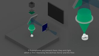
The specimen is illuminated with light of a specific wavelength (or wavelengths) which is absorbed by the fluorophores,
causing them to emit light of longer wavelengths (i.e., of a different
color than the absorbed light). The illumination light is separated from
the much weaker emitted fluorescence through the use of a spectral
emission filter. Typical components of a fluorescence microscope are a
light source (xenon arc lamp or mercury-vapor lamp are common; more advanced forms are high-power LEDs and lasers), the excitation filter, the dichroic mirror (or dichroic beamsplitter), and the emission filter
(see figure below). The filters and the dichroic beamsplitter are
chosen to match the spectral excitation and emission characteristics of
the fluorophore used to label the specimen.
In this manner, the distribution of a single fluorophore (color) is
imaged at a time. Multi-color images of several types of fluorophores
must be composed by combining several single-color images.
Most fluorescence microscopes in use are epifluorescence
microscopes, where excitation of the fluorophore and detection of the
fluorescence are done through the same light path (i.e. through the
objective). These microscopes are widely used in biology and are the
basis for more advanced microscope designs, such as the confocal microscope and the total internal reflection fluorescence microscope (TIRF).
Epifluorescence microscopy
Schematic of a fluorescence microscope.
The majority of fluorescence microscopes, especially those used in the life sciences,
are of the epifluorescence design shown in the diagram. Light of the
excitation wavelength illuminates the specimen through the objective lens. The fluorescence
emitted by the specimen is focused to the detector by the same
objective that is used for the excitation which for greater resolution
will need objective lens with higher numerical aperture.
Since most of the excitation light is transmitted through the specimen,
only reflected excitatory light reaches the objective together with the
emitted light and the epifluorescence method therefore gives a high
signal-to-noise ratio. The dichroic beamsplitter acts as a wavelength
specific filter, transmitting fluoresced light through to the eyepiece
or detector, but reflecting any remaining excitation light back towards
the source.
Light sources
Fluorescence microscopy requires intense, near-monochromatic, illumination which some widespread light sources, like halogen lamps cannot provide. Four main types of light source are used, including xenon arc lamps or mercury-vapor lamps with an excitation filter, lasers, supercontinuum sources, and high-power LEDs. Lasers are most widely used for more complex fluorescence microscopy techniques like confocal microscopy and total internal reflection fluorescence microscopy while xenon lamps, and mercury lamps, and LEDs with a dichroic excitation filter are commonly used for widefield epifluorescence microscopes. By placing two microlens arrays into the illumination path of a widefield epifluorescence microscope, highly uniform illumination with a coefficient of variation of 1-2% can be achieved.
Sample preparation
A sample of herringsperm stained with SYBR green in a cuvette illuminated by blue light in an epifluorescence microscope. The SYBR green in the sample binds to the herring sperm DNA and, once bound, fluoresces giving off green light when illuminated by blue light.
In order for a sample to be suitable for fluorescence microscopy it
must be fluorescent. There are several methods of creating a fluorescent
sample; the main techniques are labelling with fluorescent stains or,
in the case of biological samples, expression of a fluorescent protein. Alternatively the intrinsic fluorescence of a sample (i.e., autofluorescence) can be used.
In the life sciences fluorescence microscopy is a powerful tool which
allows the specific and sensitive staining of a specimen in order to
detect the distribution of proteins
or other molecules of interest. As a result, there is a diverse range
of techniques for fluorescent staining of biological samples.
Biological fluorescent stains
Many
fluorescent stains have been designed for a range of biological
molecules. Some of these are small molecules which are intrinsically
fluorescent and bind a biological molecule of interest. Major examples
of these are nucleic acid stains such as DAPI and Hoechst (excited by UV wavelength light) and DRAQ5 and DRAQ7 (optimally excited by red light) which all bind the minor groove of DNA, thus labeling the nuclei
of cells. Others are drugs, toxins, or peptides which bind specific
cellular structures and have been derivatised with a fluorescent
reporter. A major example of this class of fluorescent stain is phalloidin, which is used to stain actin fibers in mammalian cells. A new peptide, known as the Collagen Hybridizing Peptide, can also be conjugated with fluorophores and used to stain denatured collagen fibers.
Immunofluorescence is a technique which uses the highly specific binding of an antibody to its antigen
in order to label specific proteins or other molecules within the cell.
A sample is treated with a primary antibody specific for the molecule
of interest. A fluorophore can be directly conjugated to the primary
antibody. Alternatively a secondary antibody,
conjugated to a fluorophore, which binds specifically to the first
antibody can be used. For example, a primary antibody raised in a mouse
which recognises tubulin combined with a secondary anti-mouse antibody derivatised with a fluorophore could be used to label microtubules in a cell.
Fluorescent proteins
The modern understanding of genetics
and the techniques available for modifying DNA allow scientists to
genetically modify proteins to also carry a fluorescent protein
reporter. In biological samples this allows a scientist to directly make
a protein of interest fluorescent. The protein location can then be
directly tracked, including in live cells.
Limitations
Fluorophores lose their ability to fluoresce as they are illuminated in a process called photobleaching.
Photobleaching occurs as the fluorescent molecules accumulate chemical
damage from the electrons excited during fluorescence. Photobleaching
can severely limit the time over which a sample can be observed by
fluorescence microscopy. Several techniques exist to reduce
photobleaching such as the use of more robust fluorophores, by
minimizing illumination, or by using photoprotective scavenger chemicals.
Fluorescence microscopy with fluorescent reporter proteins has
enabled analysis of live cells by fluorescence microscopy, however cells
are susceptible to phototoxicity, particularly with short wavelength
light. Furthermore, fluorescent molecules have a tendency to generate
reactive chemical species when under illumination which enhances the
phototoxic effect.
Unlike transmitted and reflected light microscopy techniques
fluorescence microscopy only allows observation of the specific
structures which have been labeled for fluorescence. For example,
observing a tissue sample prepared with a fluorescent DNA stain by
fluorescence microscopy only reveals the organization of the DNA within
the cells and reveals nothing else about the cell morphologies.
Sub-diffraction techniques
The wave nature of light limits the size of the spot to which light can be focused due to the diffraction limit. This limitation was described in the 19th century by Ernst Abbe
and "limits an optical microscope's resolution to approximately half of
the wavelength of the light used." Fluorescence microscopy is central
to many techniques which aim to reach past this limit by specialized
optical configurations.
Several improvements in microscopy techniques have been invented
in the 20th century and have resulted in increased resolution and
contrast to some extent. However they did not overcome the diffraction
limit. In 1978 first theoretical ideas have been developed to break this
barrier by using a 4Pi microscope as a confocal laser scanning
fluorescence microscope where the light is focused ideally from all
sides to a common focus which is used to scan the object by
'point-by-point' excitation combined with 'point-by-point' detection.
However, the first experimental demonstration of the 4pi microscope took place in 1994. 4Pi microscopy maximizes the amount of available focusing directions by using two opposing objective lenses or two-photon excitation microscopy using redshifted light and multi-photon excitation.
Integrated correlative microscopy
combines a fluorescence microscope with an electron microscope. This
allows one to visualize ultrastructure and contextual information with
the electron microscope while using the data from the fluorescence
microscope as a labelling tool.
The first technique to really achieve a sub-diffraction resolution was STED microscopy, proposed in 1994. This method and all techniques following the RESOLFT
concept rely on a strong non-linear interaction between light and
fluorescing molecules. The molecules are driven strongly between
distinguishable molecular states at each specific location, so that
finally light can be emitted at only a small fraction of space, hence an
increased resolution.
As well in the 1990s another super resolution microscopy method
based on wide field microscopy has been developed. Substantially
improved size resolution of cellular nanostructures
stained with a fluorescent marker was achieved by development of SPDM
localization microscopy and the structured laser illumination (spatially
modulated illumination, SMI). Combining the principle of SPDM with SMI resulted in the development of the Vertico SMI microscope. Single molecule detection of normal blinking fluorescent dyes like green fluorescent protein
(GFP) can be achieved by using a further development of SPDM the
so-called SPDMphymod technology which makes it possible to detect and
count two different fluorescent molecule types at the molecular level
(this technology is referred to as two-color localization microscopy or
2CLM).
Alternatively, the advent of photoactivated localization microscopy
could achieve similar results by relying on blinking or switching of
single molecules, where the fraction of fluorescing molecules is very
small at each time. This stochastic response of molecules on the applied
light corresponds also to a highly nonlinear interaction, leading to
subdiffraction resolution.
Fluorescence micrograph gallery
A z-projection of an osteosarcoma cell, stained with phalloidin to
visualise actin filaments. The image was taken on a confocal microscope,
and the subsequent deconvolution was done using an experimentally
derived point spread function.
Epifluorescent imaging of the three components in a dividing human cancer cell. DNA is stained blue, a protein called INCENP is green, and the microtubules are red. Each fluorophore
is imaged separately using a different combination of excitation and
emission filters, and the images are captured sequentially using a
digital CCD camera, then overlaid to give a complete image.
Endothelial cells under the microscope. Nuclei are stained blue with
DAPI, microtubules are marked green by an antibody bound to FITC and
actin filaments are labeled red with phalloidin bound to TRITC. Bovine
pulmonary artery endothelial (BPAE) cells
3D dual-color super-resolution microscopy with Her2 and Her3 in
breast cells, standard dyes: Alexa 488, Alexa 568. LIMON microscopy
Human lymphocyte nucleus stained with DAPI with chromosome 13 (green) and 21 (red) centromere probes hybridized (Fluorescent in situ hybridization (FISH))
Yeast cell membrane visualized by some membrane proteins fused with
RFP and GFP fluorescent markers. Imposition of light from both of
markers results in yellow color.
Super-resolution microscopy: Single YFP molecule detection in a human
cancer cell. Typical distance measurements in the 15 nm range measured with a Vertico-SMI/SPDMphymod microscope
Super-resolution microscopy: Co-localization microscopy (2CLM) with
GFP and RFP fusion proteins (nucleus of a bone cancer cell) 120.000
localized molecules in a wide-field area (470 µm2) measured with a Vertico-SMI/SPDMphymod microscope
Fluorescence microscopy of DNA Expression in the Human Wild-Type and P239S Mutant Palladin.
Fluorescence microscopy images of sun flares pathology in a blood cell showing the affected areas in red.