Search This Blog

Saturday, September 18, 2021

Radiosurgery

From Wikipedia, the free encyclopedia

Radiosurgery
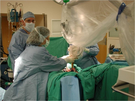
Intraoperative photograph showing a radiosurgery system.png
Intraoperative photograph showing a radiosurgery system being positioned. The patient in the photo is being treated for rectal cancer.
SpecialtyOncology
MedlinePlus007577
eMedicine1423298

Radiosurgery is surgery using radiation, that is, the destruction of precisely selected areas of tissue using ionizing radiation rather than excision with a blade. Like other forms of radiation therapy (also called radiotherapy), it is usually used to treat cancer. Radiosurgery was originally defined by the Swedish neurosurgeon Lars Leksell as "a single high dose fraction of radiation, stereotactically directed to an intracranial region of interest".

In stereotactic radiosurgery (SRS), the word "stereotactic" refers to a three-dimensional coordinate system that enables accurate correlation of a virtual target seen in the patient's diagnostic images with the actual target position in the patient. Stereotactic radiosurgery may also be called stereotactic body radiation therapy (SBRT) or stereotactic ablative radiotherapy (SABR) when used outside the central nervous system (CNS).

History

Stereotactic radiosurgery was first developed in 1949 by the Swedish neurosurgeon Lars Leksell to treat small targets in the brain that were not amenable to conventional surgery. The initial stereotactic instrument he conceived used probes and electrodes. The first attempt to supplant the electrodes with radiation was made in the early fifties, with x-rays. The principle of this instrument was to hit the intra-cranial target with narrow beams of radiation from multiple directions. The beam paths converge in the target volume, delivering a lethal cumulative dose of radiation there, while limiting the dose to the adjacent healthy tissue. Ten years later significant progress had been made, due in considerable measure to the contribution of the physicists Kurt Liden and Börje Larsson. At this time, stereotactic proton beams had replaced the x-rays. The heavy particle beam presented as an excellent replacement for the surgical knife, but the synchrocyclotron was too clumsy. Leksell proceeded to develop a practical, compact, precise and simple tool which could be handled by the surgeon himself. In 1968 this resulted in the Gamma Knife, which was installed at the Karolinska Institute and consisted of several cobalt-60 radioactive sources placed in a kind of helmet with central channels for irradiation with gamma rays. This prototype was designed to produce slit-like radiation lesions for functional neurosurgical procedures to treat pain, movement disorders, or behavioral disorders that did not respond to conventional treatment. The success of this first unit led to the construction of a second device, containing 179 cobalt-60 sources. This second Gamma Knife unit was designed to produce spherical lesions to treat brain tumors and intracranial arteriovenous malformations (AVMs). Additional units were installed in the 1980s all with 201 cobalt-60 sources.

In parallel to these developments, a similar approach was designed for a linear particle accelerator or Linac. Installation of the first 4 MeV clinical linear accelerator began in June 1952 in the Medical Research Council (MRC) Radiotherapeutic Research Unit at the Hammersmith Hospital, London. The system was handed over for physics and other testing in February 1953 and began to treat patients on 7 September that year. Meanwhile, work at the Stanford Microwave Laboratory led to the development of a 6-MV accelerator, which was installed at Stanford University Hospital, California, in 1956. Linac units quickly became favored devices for conventional fractionated radiotherapy but it lasted until the 1980s before dedicated Linac radiosurgery became a reality. In 1982, the Spanish neurosurgeon J. Barcia-Salorio began to evaluate the role of cobalt-generated and then Linac-based photon radiosurgery for the treatment of AVMs and epilepsy. In 1984, Betti and Derechinsky described a Linac-based radiosurgical system. Winston and Lutz further advanced Linac-based radiosurgical prototype technologies by incorporating an improved stereotactic positioning device and a method to measure the accuracy of various components. Using a modified Linac, the first patient in the United States was treated in Boston Brigham and Women's Hospital in February 1986.

21st century

Technological improvements in medical imaging and computing have led to increased clinical adoption of stereotactic radiosurgery and have broadened its scope in the 21st century. The localization accuracy and precision that are implicit in the word "stereotactic" remain of utmost importance for radiosurgical interventions and are significantly improved via image-guidance technologies such as the N-localizer and Sturm-Pastyr localizer that were originally developed for stereotactic surgery.

In the 21st century the original concept of radiosurgery expanded to include treatments comprising up to five fractions, and stereotactic radiosurgery has been redefined as a distinct neurosurgical discipline that utilizes externally generated ionizing radiation to inactivate or eradicate defined targets, typically in the head or spine, without the need for a surgical incision. Irrespective of the similarities between the concepts of stereotactic radiosurgery and fractionated radiotherapy the mechanism to achieve treatment is subtly different, although both treatment modalities are reported to have identical outcomes for certain indications. Stereotactic radiosurgery has a greater emphasis on delivering precise, high doses to small areas, to destroy target tissue while preserving adjacent normal tissue. The same principle is followed in conventional radiotherapy although lower dose rates spread over larger areas are more likely to be used (for example as in VMAT treatments). Fractionated radiotherapy relies more heavily on the different radiosensitivity of the target and the surrounding normal tissue to the total accumulated radiation dose. Historically, the field of fractionated radiotherapy evolved from the original concept of stereotactic radiosurgery following discovery of the principles of radiobiology: repair, reassortment, repopulation, and reoxygenation. Today, both treatment techniques are complementary, as tumors that may be resistant to fractionated radiotherapy may respond well to radiosurgery, and tumors that are too large or too close to critical organs for safe radiosurgery may be suitable candidates for fractionated radiotherapy.

Today, both Gamma Knife and Linac radiosurgery programs are commercially available worldwide. While the Gamma Knife is dedicated to radiosurgery, many Linacs are built for conventional fractionated radiotherapy and require additional technology and expertise to become dedicated radiosurgery tools. There is not a clear difference in efficacy between these different approaches. The major manufacturers, Varian and Elekta offer dedicated radiosurgery Linacs as well as machines designed for conventional treatment with radiosurgery capabilities. Systems designed to complement conventional Linacs with beam-shaping technology, treatment planning, and image-guidance tools to provide. An example of a dedicated radiosurgery Linac is the CyberKnife, a compact Linac mounted onto a robotic arm that moves around the patient and irradiates the tumor from a large set of fixed positions, thereby mimicking the Gamma Knife concept.

Clinical applications

When used outside the CNS it may be called stereotactic body radiation therapy (SBRT) or stereotactic ablative radiotherapy (SABR).

Central nervous system

Radiosurgery is performed by a multidisciplinary team of neurosurgeons, radiation oncologists and medical physicists to operate and maintain highly sophisticated, highly precise and complex instruments, including medical linear accelerators, the Gamma Knife unit and the Cyberknife unit. The highly precise irradiation of targets within the brain and spine is planned using information from medical images that are obtained via computed tomography, magnetic resonance imaging, and angiography.

Radiosurgery is indicated primarily for the therapy of tumors, vascular lesions and functional disorders. Significant clinical judgment must be used with this technique and considerations must include lesion type, pathology if available, size, location and age and general health of the patient. General contraindications to radiosurgery include excessively large size of the target lesion, or lesions too numerous for practical treatment. Patients can be treated within one to five days as outpatients. By comparison, the average hospital stay for a craniotomy (conventional neurosurgery, requiring the opening of the skull) is about 15 days. The radiosurgery outcome may not be evident until months after the treatment. Since radiosurgery does not remove the tumor but inactivates it biologically, lack of growth of the lesion is normally considered to be treatment success. General indications for radiosurgery include many kinds of brain tumors, such as acoustic neuromas, germinomas, meningiomas, metastases, trigeminal neuralgia, arteriovenous malformations, and skull base tumors, among others. Expansion of stereotactic radiotherapy to extracranial lesions is increasing, and includes metastases, liver cancer, lung cancer, pancreatic cancer, etc.

Mechanism of action

Planning CT scan with IV contrast in a patient with left cerebellopontine angle vestibular schwannoma

The fundamental principle of radiosurgery is that of selective ionization of tissue, by means of high-energy beams of radiation. Ionization is the production of ions and free radicals which are damaging to the cells. These ions and radicals, which may be formed from the water in the cell or biological materials, can produce irreparable damage to DNA, proteins, and lipids, resulting in the cell's death. Thus, biological inactivation is carried out in a volume of tissue to be treated, with a precise destructive effect. The radiation dose is usually measured in grays (one gray (Gy) is the absorption of one joule of energy per kilogram of mass). A unit that attempts to take into account both the different organs that are irradiated and the type of radiation is the sievert, a unit that describes both the amount of energy deposited and the biological effectiveness.

Risks

The New York Times reported in December 2010 that radiation overdoses had occurred with the linear accelerator method of radiosurgery, due in large part to inadequate safeguards in equipment retrofitted for stereotactic radiosurgery. In the U.S. the Food and Drug Administration (FDA) regulates these devices, whereas the Gamma Knife is regulated by the Nuclear Regulatory Commission.

This is evidence that immunotherapy may be useful for treatment of radiation necrosis following stereotactic radiotherapy.

Types of radiation source

The selection of the proper kind of radiation and device depends on many factors including lesion type, size, and location in relation to critical structures. Data suggest that similar clinical outcomes are possible with all of the various techniques. More important than the device used are issues regarding indications for treatment, total dose delivered, fractionation schedule and conformity of the treatment plan.

Gamma Knife

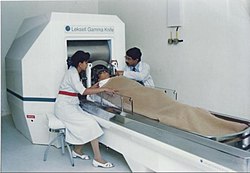
A doctor performing Gamma Knife Radiosurgery
 
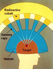
NRC graphic of the Leksell Gamma Knife

A Gamma Knife (also known as the Leksell Gamma Knife) is used to treat brain tumors by administering high-intensity gamma radiation therapy in a manner that concentrates the radiation over a small volume. The device was invented in 1967 at the Karolinska Institute in Stockholm, Sweden by Lars Leksell, Romanian-born neurosurgeon Ladislau Steiner, and radiobiologist Börje Larsson from Uppsala University, Sweden. The first Gamma Knife was brought to the United States through an arrangement between US neurosurgeon Robert Wheeler Rand and Leksell and was given to the University of California, Los Angeles (UCLA) in 1979.

A Gamma Knife typically contains 201 cobalt-60 sources of approximately 30 curies each (1.1 TBq), placed in a hemispheric array in a heavily shielded assembly. The device aims gamma radiation through a target point in the patient's brain. The patient wears a specialized helmet that is surgically fixed to the skull, so that the brain tumor remains stationary at the target point of the gamma rays. An ablative dose of radiation is thereby sent through the tumor in one treatment session, while surrounding brain tissues are relatively spared.

Gamma Knife therapy, like all radiosurgery, uses doses of radiation to kill cancer cells and shrink tumors, delivered precisely to avoid damaging healthy brain tissue. Gamma Knife radiosurgery is able to accurately focus many beams of gamma radiation on one or more tumors. Each individual beam is of relatively low intensity, so the radiation has little effect on intervening brain tissue and is concentrated only at the tumor itself.

Gamma Knife radiosurgery has proven effective for patients with benign or malignant brain tumors up to 4 cm (1.6 in) in size, vascular malformations such as an arteriovenous malformation (AVM), pain, and other functional problems. For treatment of trigeminal neuralgia the procedure may be used repeatedly on patients.

Acute complications following Gamma Knife radiosurgery are rare, and complications are related to the condition being treated.

Linear accelerator-based therapies

A linear accelerator (linac) produces x-rays from the impact of accelerated electrons striking a high z target, usually tungsten. The process is also referred to as "x-ray therapy" or "photon therapy." The emission head, or "gantry", is mechanically rotated around the patient in a full or partial circle. The table where the patient is lying, the "couch", can also be moved in small linear or angular steps. The combination of the movements of the gantry and of the couch allow the computerized planning of the volume of tissue that is going to be irradiated. Devices with a high energy of 6 MeV are the commonly used for the treatment of the brain, due to the depth of the target. The diameter of the energy beam leaving the emission head can be adjusted to the size of the lesion by means of collimators. They may be interchangeable orifices with different diameters, typically varying from 5 to 40 mm in 5 mm steps, or multileaf collimators, which consist of a number of metal leaflets that can be moved dynamically during treatment in order to shape the radiation beam to conform to the mass to be ablated. As of 2017 Linacs were capable of achieving extremely narrow beam geometries, such as 0.15 to 0.3 mm. Therefore, they can be used for several kinds of surgeries which hitherto had been carried out by open or endoscopic surgery, such as for trigeminal neuralgia. Long-term follow-up data has shown it to be as effective as radiofrequency ablation, but inferior to surgery in preventing the recurrence of pain.

The first such systems were developed by John R. Adler, a Stanford University professor of neurosurgery and radiation oncology, and Russell and Peter Schonberg at Schonberg Research, and commercialized under the brand name CyberKnife.

Proton beam therapy

Protons may also be used in radiosurgery in a procedure called Proton Beam Therapy (PBT) or proton therapy. Protons are extracted from proton donor materials by a medical synchrotron or cyclotron, and accelerated in successive transits through a circular, evacuated conduit or cavity, using powerful magnets to shape their path, until they reach the energy required to just traverse a human body, usually about 200 MeV. They are then released toward the region to be treated in the patient's body, the irradiation target. In some machines, which deliver protons of only a specific energy, a custom mask made of plastic is interposed between the beam source and the patient to adjust the beam energy to provide the appropriate degree of penetration. The phenomenon of the Bragg peak of ejected protons gives proton therapy advantages over other forms of radiation, since most of the proton's energy is deposited within a limited distance, so tissue beyond this range (and to some extent also tissue inside this range) is spared from the effects of radiation. This property of protons, which has been called the "depth charge effect" by analogy to the explosive weapons used in anti-submarine warfare, allows for conformal dose distributions to be created around even very irregularly shaped targets, and for higher doses to targets surrounded or backstopped by radiation-sensitive structures such as the optic chiasm or brainstem. The development of "intensity modulated" techniques allowed similar conformities to be attained using linear accelerator radiosurgery.

As of 2013 there was no evidence that proton therapy is better than any other types of treatment in most cases, except for a "handful of rare pediatric cancers". Critics, responding to the increasing number of very expensive PBT installations, spoke of a "medical arms race" and "crazy medicine and unsustainable public policy".


Neurosurgery

From Wikipedia, the free encyclopedia
 
Parkinson surgery.jpg
Stereotactic guided insertion of DBS electrodes in neurosurgery
Occupation
Activity sectors
Surgery
Description
Education required

or

or

Fields of
employment
Hospitals, Clinics

Neurosurgery or neurological surgery, known in common parlance as brain surgery, is the medical specialty concerned with the prevention, diagnosis, surgical treatment, and rehabilitation of disorders which affect any portion of the nervous system including the brain, spinal cord, central and peripheral nervous system, and cerebrovascular system.

Education and context

In different countries, there are different requirements for an individual to legally practice neurosurgery, and there are varying methods through which they must be educated. In most countries, neurosurgeon training requires a minimum period of seven years after graduating from medical school.

United States

In the United States, a neurosurgeon must generally complete four years of undergraduate education, four years of medical school, and seven years of residency (PGY-1-7). Most, but not all, residency programs have some component of basic science or clinical research. Neurosurgeons may pursue additional training in the form of a fellowship after residency, or, in some cases, as a senior resident in the form of an enfolded fellowship. These fellowships include pediatric neurosurgery, trauma/neurocritical care, functional and stereotactic surgery, surgical neuro-oncology, radiosurgery, neurovascular surgery, skull-base surgery, peripheral nerve and complex spinal surgery. Fellowships typically span one to two years. In the U.S., neurosurgery "is a small specialty, constituting only 0.5 percent of all physicians."

United Kingdom

In the United Kingdom, students must gain entry into medical school. MBBS qualification (Bachelor of Medicine, Bachelor of Surgery) takes four to six years depending on the student's route. The newly qualified physician must then complete foundation training lasting two years; this is a paid training program in a hospital or clinical setting covering a range of medical specialties including surgery. Junior doctors then apply to enter the neurosurgical pathway. Unlike most other surgical specialties, it currently has its own independent training pathway which takes around eight years (ST1-8); before being able to sit for consultant exams with sufficient amounts of experience and practice behind them. Neurosurgery remains consistently amongst the most competitive medical specialties in which to obtain entry.

History

Neurosurgery, or the premeditated incision into the head for pain relief, has been around for thousands of years, but notable advancements in neurosurgery have only come within the last hundred years.

Edinburgh Skull, trepanning showing hole in back of skull Wellcome M0009393.jpg

Ancient

The Incas appear to have practiced a procedure known as trepanation since before European colonization. During the Middle Ages in Al-Andalus from 936 to 1013 AD, Al-Zahrawi performed surgical treatments of head injuries, skull fractures, spinal injuries, hydrocephalus, subdural effusions and headache. In China, Hua Tuo created the first general anaesthesia called mafeisan, which he used on surgical procedures on the brain.

Modern

HochbergFig1.jpg

There was not much advancement in neurosurgery until late 19th early 20th century, when electrodes were placed on the brain and superficial tumors were removed.

History of electrodes in the brain: In 1878 Richard Caton discovered that electrical signals transmitted through an animal's brain. In 1950 Dr. Jose Delgado invented the first electrode that was implanted in an animal's brain, using it to make it run and change direction. In 1972 the cochlear implant, a neurological prosthetic that allowed deaf people to hear was marketed for commercial use. In 1998 researcher Philip Kennedy implanted the first Brain Computer Interface (BCI) into a human subject.

History of tumor removal: In 1879 after locating it via neurological signs alone, Scottish surgeon William Macewen (1848-1924) performed the first successful brain tumor removal. On November 25, 1884 after English physician Alexander Hughes Bennett (1848-1901) used Macewen's technique to locate it, English surgeon Rickman Godlee (1849-1925) performed the first primary brain tumor removal, which differs from Macewen's operation in that Bennett operated on the exposed brain, whereas Macewen operated outside of the "brain proper" via trepanation. On March 16, 1907 Austrian surgeon Hermann Schloffer became the first to successfully remove a pituitary tumor.

Modern surgical instruments

The main advancements in neurosurgery came about as a result of highly crafted tools. Modern neurosurgical tools, or instruments, include chisels, curettes, dissectors, distractors, elevators, forceps, hooks, impactors, probes, suction tubes, power tools, and robots. Most of these modern tools, like chisels, elevators, forceps, hooks, impactors, and probes, have been in medical practice for a relatively long time. The main difference of these tools, pre and post advancement in neurosurgery, were the precision in which they were crafted. These tools are crafted with edges that are within a millimeter of desired accuracy. Other tools such as hand held power saws and robots have only recently been commonly used inside of a neurological operating room. As an example, the University of Utah developed a device for computer-aided design / computer-aided manufacturing (CAD-CAM) which uses an image-guided system to define a cutting tool path for a robotic cranial drill.

Organised neurosurgery

World Academy of Neurological Surgery's conference

The World Federation of Neurosurgical Societies (WFNS), founded in 1955, in Switzerland, as a professional, scientific, non governmental organization, is composed of 130 member societies: consisting of 5 Continental Associations (AANS, AASNS, CAANS, EANS and FLANC), 6 Affiliate Societies, and 119 National Neurosurgical Societies, representing some 50,000 neurosurgeons worldwide. It has a consultative status in the United Nations. The official Journal of the Organization is World Neurosurgery. The other global organisations being the World Academy of Neurological Surgery (WANS) and the World Federation of Skull Base Societies (WFSBS).

Main divisions

General neurosurgery involves most neurosurgical conditions including neuro-trauma and other neuro-emergencies such as intracranial hemorrhage. Most level 1 hospitals have this kind of practice.

Specialized branches have developed to cater to special and difficult conditions. These specialized branches co-exist with general neurosurgery in more sophisticated hospitals. To practice advanced specialization within neurosurgery, additional higher fellowship training of one to two years is expected from the neurosurgeon. Some of these divisions of neurosurgery are:

  1. Vascular neurosurgery includes clipping of aneurysms and performing carotid endarterectomy (CEA).
  2. Stereotactic neurosurgery, functional neurosurgery, and epilepsy surgery (the latter includes partial or total corpus callosotomy – severing part or all of the corpus callosum to stop or lessen seizure spread and activity, and the surgical removal of functional, physiological and/or anatomical pieces or divisions of the brain, called epileptic foci, that are operable and that are causing seizures, and also the more radical and very, very rare partial or total lobectomy, or even hemispherectomy – the removal of part or all of one of the lobes, or one of the cerebral hemispheres of the brain; those two procedures, when possible, are also very, very rarely used in oncological neurosurgery or to treat very severe neurological trauma, such as stab or gunshot wounds to the brain)
  3. Oncological neurosurgery also called neurosurgical oncology; includes pediatric oncological neurosurgery; treatment of benign and malignant central and peripheral nervous system cancers and pre-cancerous lesions in adults and children (including, among others, glioblastoma multiforme and other gliomas, brain stem cancer, astrocytoma, pontine glioma, medulloblastoma, spinal cancer, tumors of the meninges and intracranial spaces, secondary metastases to the brain, spine, and nerves, and peripheral nervous system tumors)
  4. Skull base surgery
  5. Spinal neurosurgery
  6. Peripheral nerve surgery
  7. Pediatric neurosurgery (for cancer, seizures, bleeding, stroke, cognitive disorders or congenital neurological disorders)

Neuropathology

Neuropathology case V 03.jpg

Neuropathology is a specialty within the study of pathology focused on the disease of the brain, spinal cord, and neural tissue. This includes the central nervous system and the peripheral nervous system. Tissue analysis comes from either surgical biopsies or post mortem autopsies. Common tissue samples include muscle fibers and nervous tissue. Common applications of neuropathology include studying samples of tissue in patients who have Parkinson's disease, Alzheimer's disease, dementia, Huntington's disease, amyotrophic lateral sclerosis, mitochondria disease, and any disorder that has neural deterioration in the brain or spinal cord.

History

While pathology has been studied for millennia only within the last few hundred years has medicine focused on a tissue- and organ-based approach to tissue disease. In 1810, Thomas Hodgkin started to look at the damaged tissue for the cause. This was conjoined with the emergence of microscopy and started the current understanding of how the tissue of the human body is studied.

Neuroanesthesia

Neuroanesthesia is a field of anesthesiology which focuses on neurosurgery. Anesthesia is not used during the middle of an "awake" brain surgery. Awake brain surgery is where the patient is conscious for the middle of the procedure and sedated for the beginning and end. This procedure is used when the tumor does not have clear boundaries and the surgeon wants to know if they are invading on critical regions of the brain which involve functions like talking, cognition, vision, and hearing. It will also be conducted for procedures which the surgeon is trying to combat epileptic seizures.

History

Trepanning, an early form of neuroanesthesia, appears to have been practised by the Incas in South America. In these procedures coca leaves and datura plants were used to manage pain as the person had dull primitive tools cut open their skull. In 400 BC the physician Hippocrates made accounts of using different wines to sedate patients while trepanning. In 60 AD Dioscorides, a physician, pharmacologist, and botanist, detailed how mandrake, henbane, opium, and alcohol were used to put patients to sleep during trepanning. In 972 AD two brother surgeons in Paramara, now India, used "samohine" to sedate a patient while removing a small tumor and awoke the patient by pouring onion and vinegar in the patient's mouth. Since then, multiple cocktails have been derived in order to sedate a patient during a brain surgery. The most recent form of neuroanesthesia is the combination of carbon dioxide, hydrogen, and nitrogen. This was discovered in the 18th century by Humphry Davy and brought into the operating room by Astley Cooper.

Neurosurgery methods

Neurosurgery
ICD-10-PCS00-01
ICD-9-CM0105
MeSHD019635
OPS-301 code5-01...5-05

Neuroradiology methods are used in modern neurosurgery diagnosis and treatment. They include computer assisted imaging computed tomography (CT), magnetic resonance imaging (MRI), positron emission tomography (PET), magnetoencephalography (MEG), and stereotactic radiosurgery. Some neurosurgery procedures involve the use of intra-operative MRI and functional MRI.

In conventional open surgery the neurosurgeon opens the skull, creating a large opening to access the brain. Techniques involving smaller openings with the aid of microscopes and endoscopes are now being used as well. Methods that utilize small craniotomies in conjunction with high-clarity microscopic visualization of neural tissue offer excellent results. However, the open methods are still traditionally used in trauma or emergency situations.

Microsurgery is utilized in many aspects of neurological surgery. Microvascular techniques are used in EC-IC bypass surgery and in restoration carotid endarterectomy. The clipping of an aneurysm is performed under microscopic vision. Minimally-invasive spine surgery utilizes microscopes or endoscopes. Procedures such as microdiscectomy, laminectomy, and artificial disc replacement rely on microsurgery.

Using stereotaxy neurosurgeons can approach a minute target in the brain through a minimal opening. This is used in functional neurosurgery where electrodes are implanted or gene therapy is instituted with high level of accuracy as in the case of Parkinson's disease or Alzheimer's disease. Using the combination method of open and stereotactic surgery, intraventricular hemorrhages can potentially be evacuated successfully. Conventional surgery using image guidance technologies is also becoming common and is referred to as surgical navigation, computer-assisted surgery, navigated surgery, stereotactic navigation. Similar to a car or mobile Global Positioning System (GPS), image-guided surgery systems, like Curve Image Guided Surgery and StealthStation, use cameras or electromagnetic fields to capture and relay the patient’s anatomy and the surgeon’s precise movements in relation to the patient, to computer monitors in the operating room. These sophisticated computerized systems are used before and during surgery to help orient the surgeon with three-dimensional images of the patient’s anatomy including the tumor. Real-time functional brain mapping has been employed to identify specific functional regions using electrocorticography (ECoG)

Minimally invasive endoscopic surgery is commonly utilized by neurosurgeons when appropriate. Techniques such as endoscopic endonasal surgery are used in pituitary tumors, craniopharyngiomas, chordomas, and the repair of cerebrospinal fluid leaks. Ventricular endoscopy is used in the treatment of intraventricular bleeds, hydrocephalus, colloid cyst and neurocysticercosis. Endonasal endoscopy is at times carried out with neurosurgeons and ENT surgeons working together as a team.

Repair of craniofacial disorders and disturbance of cerebrospinal fluid circulation is done by neurosurgeons who also occasionally team up with maxillofacial and plastic surgeons. Cranioplasty for craniosynostosis is performed by pediatric neurosurgeons with or without plastic surgeons.

Neurosurgeons are involved in stereotactic radiosurgery along with radiation oncologists in tumor and AVM treatment. Radiosurgical methods such as Gamma knife, Cyberknife and Novalis Radiosurgery are used as well.

Endovascular surgical neuroradiology utilize endovascular image guided procedures for the treatment of aneurysms, AVMs, carotid stenosis, strokes, and spinal malformations, and vasospasms. Techniques such as angioplasty, stenting, clot retrieval, embolization, and diagnostic angiography are endovascular procedures.

A common procedure performed in neurosurgery is the placement of ventriculo-peritoneal shunt (VP shunt). In pediatric practice this is often implemented in cases of congenital hydrocephalus. The most common indication for this procedure in adults is normal pressure hydrocephalus (NPH).

Neurosurgery of the spine covers the cervical, thoracic and lumbar spine. Some indications for spine surgery include spinal cord compression resulting from trauma, arthritis of the spinal discs, or spondylosis. In cervical cord compression, patients may have difficulty with gait, balance issues, and/or numbness and tingling in the hands or feet. Spondylosis is the condition of spinal disc degeneration and arthritis that may compress the spinal canal. This condition can often result in bone-spurring and disc herniation. Power drills and special instruments are often used to correct any compression problems of the spinal canal. Disc herniations of spinal vertebral discs are removed with special rongeurs. This procedure is known as a discectomy. Generally once a disc is removed it is replaced by an implant which will create a bony fusion between vertebral bodies above and below. Instead, a mobile disc could be implanted into the disc space to maintain mobility. This is commonly used in cervical disc surgery. At times instead of disc removal a Laser discectomy could be used to decompress a nerve root. This method is mainly used for lumbar discs. Laminectomy is the removal of the lamina of the vertebrae of the spine in order to make room for the compressed nerve tissue.

Surgery for chronic pain is a sub-branch of functional neurosurgery. Some of the techniques include implantation of deep brain stimulators, spinal cord stimulators, peripheral stimulators and pain pumps.

Surgery of the peripheral nervous system is also possible, and includes the very common procedures of carpal tunnel decompression and peripheral nerve transposition. Numerous other types of nerve entrapment conditions and other problems with the peripheral nervous system are treated as well.

Conditions

Conditions treated by neurosurgeons include, but are not limited to:

Recovery

Post operative pain

Pain following brain surgery can be significant and may lengthen recovery, increase the amount of time a person stays in the hospital following surgery, and increase the risk of complications following surgery. Severe acute pain following brain surgery may also increase the risk of a person developing a chronic post-craniotomy headache. Approaches to treating pain in adults include treatment with nonsteroidal anti‐inflammatory drugs (NSAIDs), which have been shown to reduce pain for up to 24 hours following surgery. Low quality evidence supports the use of the medications dexmedetomidine, pregabalin or gabapentin to reduce post-operative pain. Low quality evidence also supports scalp blocks and scalp infiltration to reduce postoperative pain. Gabapentin or pregabalin may also decrease vomiting and nausea following surgery (very low quality medical evidence).

Notable neurosurgeons

See also

Stereotactic surgery

From Wikipedia, the free encyclopedia

Stereotactic surgery
Brain biopsy under stereotaxy.jpg
Brain biopsy using a needle mounted on a stereotactic instrument
Other namesStereotaxy
Specialtyneurosurgery

Stereotactic surgery is a minimally invasive form of surgical intervention that makes use of a three-dimensional coordinate system to locate small targets inside the body and to perform on them some action such as ablation, biopsy, lesion, injection, stimulation, implantation, radiosurgery (SRS), etc.

In theory, any organ system inside the body can be subjected to stereotactic surgery. However, difficulties in setting up a reliable frame of reference (such as bone landmarks, which bear a constant spatial relation to soft tissues) mean that its applications have been, traditionally and until recently, limited to brain surgery. Besides the brain, biopsy and surgery of the breast are done routinely to locate, sample (biopsy), and remove tissue. Plain X-ray images (radiographic mammography), computed tomography, and magnetic resonance imaging can be used to guide the procedure.

Another accepted form of "stereotactic" is "stereotaxic". The word roots are stereo-, a prefix derived from the Greek word στερεός (stereos, "solid"), and -taxis (a suffix of New Latin and ISV, derived from Greek taxis, "arrangement", "order", from tassein, "to arrange").

Procedure

Stereotactic surgery works on the basis of three main components:

  • A stereotactic planning system, including atlas, multimodality image matching tools, coordinates calculator, etc.
  • A stereotactic device or apparatus
  • A stereotactic localization and placement procedure

Modern stereotactic planning systems are computer based. The stereotactic atlas is a series of cross sections of anatomical structure (for example, a human brain), depicted in reference to a two-coordinate frame. Thus, each brain structure can be easily assigned a range of three coordinate numbers, which will be used for positioning the stereotactic device. In most atlases, the three dimensions are: latero-lateral (x), dorso-ventral (y) and rostro-caudal (z).

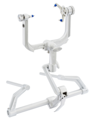
The stereotactic apparatus uses a set of three coordinates (x, y and z) in an orthogonal frame of reference (cartesian coordinates), or, alternatively, a cylindrical coordinates system, also with three coordinates: angle, depth and antero-posterior (or axial) location. The mechanical device has head-holding clamps and bars which puts the head in a fixed position in reference to the coordinate system (the so-called zero or origin). In small laboratory animals, these are usually bone landmarks which are known to bear a constant spatial relation to soft tissue. For example, brain atlases often use the external auditory meatus, the inferior orbital ridges, the median point of the maxilla between the incisive teeth. or the bregma (confluence of sutures of frontal and parietal bones), as such landmarks. In humans, the reference points, as described above, are intracerebral structures which are clearly discernible in a radiograph or tomograph. In newborn human babies, the "soft spot" where the coronal and sagittal sutures meet (known as the fontanelle) becomes the bregma when this gap closes.

Guide bars in the x, y and z directions (or alternatively, in the polar coordinate holder), fitted with high precision vernier scales allow the neurosurgeon to position the point of a probe (an electrode, a cannula, etc.) inside the brain, at the calculated coordinates for the desired structure, through a small trephined hole in the skull.

Currently, a number of manufacturers produce stereotactic devices fitted for neurosurgery in humans, for both brain and spine procedures, as well as for animal experimentation.

Types frame systems

  1. Simple Orthogonal System: The probe is directed perpendicular to a square base unit fixed to the skull. These provide three degrees of freedom by means of a carriage that moved orthogonally along the base plate or along a bar attached parallel to the base plate of the instrument. Attached to the carriage was a second track that extended across the head frame perpendicularly.
  2. Burr Hole Mounted System: This provides a limited range of possible intracranial target points with a fixed entry point. They provided two angular degrees of freedom and a depth adjustment. The surgeon could place the burr hole over nonessential brain tissue and utilize the instrument to direct the probe to the target point from the fixed entry point at the burr hole.
  3. Arc-Quadrant Systems: Probes are directed perpendicular to the tangent of an arc (which rotates about the vertical axis) and a quadrant (which rotates about the horizontal axis). The probe, directed to a depth equal to the radius of the sphere defined by the arc-quadrant, will always arrive at the center or focal point of that sphere.
  4. Arc-Phantom Systems: An aiming bow attaches to the head ring, which is fixed to the patient's skull, and can be transferred to a similar ring that contains a simulated target. In this system, the phantom target is moved on the simulator to 3D coordinates. After adjusting the probe holder on the aiming bow so that the probe touches the desired target on the phantom, the transferable aiming bow is moved from the phantom base ring to the base ring on the patient. The probe is then lowered to the determined depth in order to reach the target point deep in the patient's brain.

Treatment

Stereotactic radiosurgery

A doctor performing Gamma Knife Radiosurgery

Stereotactic radiosurgery utilizes externally generated ionizing radiation to inactivate or eradicate defined targets in the head or spine without the need to make an incision. This concept requires steep dose gradients to reduce injury to adjacent normal tissue while maintaining treatment efficacy in the target. As a consequence of this definition, the overall treatment accuracy should match the treatment planning margins of 1-2 millimeter or better. To use this paradigm optimally and treat patients with the highest possible accuracy and precision, all errors, from image acquisition over treatment planning to mechanical aspects of the delivery of treatment and intra-fraction motion concerns, must be systematically optimized. To assure quality of patient care the procedure involves a multidisciplinary team consisting of a radiation oncologist, medical physicist, and radiation therapist. Dedicated, commercially available stereotactic radiosurgery programs are provided by the irrespective Gamma Knife, CyberKnife, and Novalis Radiosurgery devices.

Stereotactic radiosurgery provides an efficient, safe, and minimal invasive treatment alternative for patients diagnosed with malignant, benign and functional indications in the brain and spine, including but not limited to both primary and secondary tumors. Stereotactic radiosurgery is a well-described management option for most metastases, meningiomas, schwannomas, pituitary adenomas, arteriovenous malformations, and trigeminal neuralgia, among others.

Irrespective of the similarities between the concepts of stereotactic radiosurgery and fractionated radiotherapy and although both treatment modalities are reported to have identical outcomes for certain indications, the intent of both approaches is fundamentally different. The aim of stereotactic radiosurgery is to destroy target tissue while preserving adjacent normal tissue, where fractionated radiotherapy relies on a different sensitivity of the target and the surrounding normal tissue to the total accumulated radiation. Historically, the field of fractionated radiotherapy evolved from the original concept of stereotactic radiosurgery following discovery of the principles of radiobiology: repair, reassortment, repopulation, and reoxygenation. Today, both treatment techniques are complementary as tumors that may be resistant to fractionated radiotherapy may respond well to radiosurgery and tumors that are too large or too close to critical organs for safe radiosurgery may be suitable candidates for fractionated radiotherapy.

A second, more recent evolution extrapolates the original concept of stereotactic radiosurgery to extra-cranial targets, most notably in the lung, liver, pancreas, and prostate. This treatment approach, entitled stereotactic body radiotherapy or SBRT, is challenged by various types of motion. On top of patient immobilization challenges and the associated patient motion, extra-cranial lesions move with respect to the patient's position due to respiration, bladder and rectum filling. Like stereotactic radiosurgery, the intent of stereotactic body radiotherapy is to eradicate a defined extra-cranial target. However, target motion requires larger treatment margins around the target to compensate for the positioning uncertainty. This in turn implies more normal tissue exposed to high doses, which could result in negative treatment side effects. As a consequence, stereotactic body radiotherapy is mostly delivered in a limited number of fractions, thereby blending the concept of stereotactic radiosurgery with the therapeutic benefits of fractionated radiotherapy. To monitor and correct target motion for accurate and precise patient positioning prior and during treatment, advanced image-guided technologies are commercially available and included in the radiosurgery programs offered by the CyberKnife and Novalis communities.

Parkinson's disease

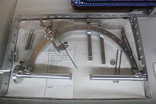
Frame for Stereotactic Thalamotomy on display at the Glenside Museum

Functional neurosurgery comprises treatment of several disorders such as Parkinson's disease, hyperkinesia, disorder of muscle tone, intractable pain, convulsive disorders and psychological phenomena. Treatment for these phenomena was believed to be located in the superficial parts of the CNS and PNS. Most of the interventions made for treatment consisted of cortical extirpation. To alleviate extra pyramidal disorders, pioneer Russell Meyers dissected or transected the head of the caudate nucleus in 1939, and part of the putamen and globus pallidus. Attempts to abolish intractable pain were made with success by transection of the spinothalamic tract at spinal medullary level and further proximally, even at mesencephalic levels.

In 1939-1941 Putnam and Oliver tried to improve Parkinsonism and hyperkinesias by trying a series of modifications of the lateral and antero-lateral cordotomies. Additionally, other scientists like Schurman, Walker, and Guiot made significant contributions to functional neurosurgery. In 1953, Cooper discovered by chance that ligation of the anterior chorioidal artery resulted in improvement of Parkinson's disease. Similarly, when Grood was performing an operation in a patient with Parkinson's, he accidentally lesioned the thalamus. This caused the patient's tremors to stop. From then on, thalamic lesions became the target point with more satisfactory results.

More recent clinical applications can be seen in surgeries used to treat Parkinson's disease, such as Pallidotomy or Thalamotomy (lesioning procedures), or Deep Brain Stimulation (DBS). During DBS, an electrode is placed into the thalamus, the pallidum of the subthalmamic nucleus, parts of brain that are involved in motor control, and are affected by Parkinson's disease. The electrode is connected to a small battery operated stimulator that is placed under the collarbone, where a wire runs beneath the skin to connect it to the electrode in the brain. The stimulator produces electrical impulses that affect the nerve cells around the electrode and should help alleviate tremors or symptoms that are associated with the affected area.

In Thalamotomy, a needle electrode is placed into the thalamus, and the patient must cooperate with tasks assigned to find the affected area- after this area of the thalamus is located, a small high frequency current is applied to the electrode and this destroys a small part of the thalamus. Approximately 90% of patients experience instantaneous tremor relief.

In Pallidotomy, an almost identical procedure to thalamotomy, a small part of the pallidum is destroyed and 80% of patients see improvement in rigidity and hypokinesia and a tremor relief or improvement comes weeks after the procedure.

History

The stereotactic method was first published in 1908 by two British scientists, Victor Horsley, a physician and neurosurgeon, and Robert H. Clarke, a physiologist and was built by Swift & Son; the two scientists stopped collaborating after the 1908 publication. The Horsley–Clarke apparatus used a Cartesian (three-orthogonal axis) system. That device is in the Science Museum, London; a copy was brought to the US by Ernest Sachs and is in the Department of Neurosurgery at UCLA. Clarke used the original to do research that led to publications of primate and cat brain atlases. There is no evidence it was ever used in a human surgery. The first stereotactic device designed for the human brain appears to have been an adaptation of the Horseley–Clarke frame built at Aubrey T. Mussen's behest by a London workshop in 1918, but it received little attention and does not appear to have been used on people. It was a frame made of brass.

The first stereotactic device used in humans was used by Martin Kirschner, for a method to treat trigeminal neuralgia by inserting an electrode into the trigeminal nerve and ablating it. He published this in 1933.

In 1947 and 1949, two neurosurgeons working at Temple University in Philadelphia, Ernest A. Spiegel (who had fled Austria when the Nazis took over) and Henry T. Wycis, published their work on a device similar to the Horsley–Clarke apparatus in using a cartesian system; it was attached to the patient's head with a plaster cast instead of screws. Their device was the first to be used for brain surgery; they used it for psychosurgery. They also created the first atlas of the human brain, and used intracranial reference points, generated by using medical images acquired with contrast agents.

The work of Spiegel and Wycis sparked enormous interest and research. In Paris, Jean Talairach collaborated with Marcel David, Henri Hacaen, and Julian de Ajuriaguerra on a stereotactic device, publishing their first work in 1949 and eventually developing the Talairach coordinates. In Japan, Hirotaro Narabayashi was doing similar work.

In 1949, Lars Leksell published a device that used polar coordinates instead of cartesian, and two years later he published work where he used his device to target a beam of radiation into a brain. Leksell's radiosurgery system is also used by the Gamma Knife device, and by other neurosurgeons, using linear accelerators, proton beam therapy and neutron capture therapy. Lars Leksell went on to commercialize his inventions by founding Elekta in 1972.

In 1979, Russell A. Brown proposed a device, now known as the N-localizer, that enables guidance of stereotactic surgery using tomographic images that are obtained via medical imaging technologies such as X-ray computed tomography (CT), magnetic resonance imaging (MRI), or positron emission tomography (PET). The N-localizer comprises a diagonal rod that spans two vertical rods to form an N-shape that allows tomographic images to be mapped to physical space. This device became almost universally adopted by the 1980s and is included in the Brown-Roberts-Wells (BRW), Kelly-Goerss, Leksell, Cosman-Roberts-Wells (CRW), Micromar-ETM03B, FiMe-BlueFrame, Macom, and Adeor-Zeppelin stereotactic frames and in the Gamma Knife radiosurgery system. An alternative to the N-localizer is the Sturm-Pastyr localizer that is included in the Riechert-Mundinger and Zamorano-Dujovny stereotactic frames.

Other localization methods also exist that do not make use of tomographic images produced by CT, MRI, or PET, but instead conventional radiographs.

The stereotactic method has continued to evolve, and at present employs an elaborate mixture of image-guided surgery that uses computed tomography, magnetic resonance imaging and stereotactic localization.

History in Latin America

In 1970, in the city of Buenos Aires, Argentina, Aparatos Especiales company, produced the first stereotactic system in Latin America. Antonio Martos Calvo, together with Jorge Candia and Jorge Olivetti (figure 38 A) through the request of the neurosurgeon Jorge Schvarcz (1942-2019), developed an equipment based on the principle of the Hitchcock stereotactic system. The patient was seated in an adapted chair with two telescopic arms attached at it base, which fixed the stereotactic frame preventing the patient's movement (Figure 38B). A double radiopaque ruler attached to the side of the frame made it possible to obtain the antero-posterior and latero-lateral X-ray images without the need of moving the radiopaque ruler (Figure 38B). The thermal coagulation lesion was performed using tungsten monopole electrodes of 1,5mm of diameter (without temperature control) with a 3mm active tip, utilizing an electrical bipolar coagulator. The lesion size was previously determined by testing the electrode in egg albumin. Coagulation size was the results of the of the electrical coagulator power regulation and the application time of the radiofrequency. The first surgery performed with this system was a Trigeminal Nucleotractothomy (Fig. 38 B,C). Jorge Schvarcz performed more than 700 functional surgeries until 1994 when, due to health problems he stopped exercising his profession. But the equipment developed kept improving on neurosurgery history. This was the beginning of the developing of technology to produce stereotactic devices in Latin America. This was the beginning of the first stereotactic manufacturer of Latin America - the Brazilian Micromar.

Research

Stereotactic surgery is sometimes used to aid in several different types of animal research studies. Specifically, it is used to target specific sites of the brain and directly introduce pharmacological agents to the brain which otherwise may not be able to cross the blood–brain barrier. In rodents, the main applications of stereotactic surgery are to introduce fluids directly to the brain or to implant cannulae and microdialysis probes. Site specific central microinjections are used when rodents do not need to be awake and behaving or when the substance to be injected has a long duration of action. For protocols in which rodents’ behaviors must be assessed soon after injection, stereotactic surgery can be used to implant a cannula through which the animal can be injected after recovery from the surgery. These protocols take longer than site-specific central injections in anesthetized mice because they require the construction of cannulae, wire plugs, and injection needles, but induce less stress in the animals because they allow for a recovery period for the healing of trauma induced to the brain before injection. Surgery can also be used for microdialysis protocols to implant and tether the dialysis probe and guide cannula.

See also

Buddhist cosmology

From Wikipedia, the free encyclopedia https://en.wikipedia.org/wiki/Bud...