Search This Blog

Monday, March 13, 2023

African trypanosomiasis

From Wikipedia, the free encyclopedia
African trypanosomiasis
Other namesSleeping sickness, African sleeping sickness
Trypanosoma sp. PHIL 613 lores.jpg
Trypanosoma forms in a blood smear
SpecialtyInfectious disease
SymptomsStage 1: Fevers, headaches, itchiness, joint pains
Stage 2: Trouble sleeping, confusion, poor coordination
Usual onset1–3 weeks post exposure
TypesTrypanosoma brucei gambiense (TbG), Trypanosoma brucei rhodesiense (TbR)
CausesTrypanosoma brucei spread by tsetse flies
Diagnostic methodBlood smear, lumbar puncture
MedicationFexinidazole, pentamidine, suramin, eflornithine, nifurtimox
PrognosisFatal without treatment
Frequency977 (2018)
Deaths3,500 (2015)

African trypanosomiasis, also known as African sleeping sickness or simply sleeping sickness, is an insect-borne parasitic infection of humans and other animals. It is caused by the species Trypanosoma brucei. Humans are infected by two types, Trypanosoma brucei gambiense (TbG) and Trypanosoma brucei rhodesiense (TbR). TbG causes over 98% of reported cases. Both are usually transmitted by the bite of an infected tsetse fly and are most common in rural areas.

Initially, the first stage of the disease is characterized by fevers, headaches, itchiness, and joint pains, beginning one to three weeks after the bite. Weeks to months later, the second stage begins with confusion, poor coordination, numbness, and trouble sleeping. Diagnosis is by finding the parasite in a blood smear or in the fluid of a lymph node. A lumbar puncture is often needed to tell the difference between first- and second-stage disease. If the disease is not treated quickly it can lead to death.

Prevention of severe disease involves screening the at-risk population with blood tests for TbG. Treatment is easier when the disease is detected early and before neurological symptoms occur. Treatment of the first stage has been with the medications pentamidine or suramin. Treatment of the second stage has involved eflornithine or a combination of nifurtimox and eflornithine for TbG. Fexinidazole is a more recent treatment that can be taken by mouth, for either stage of TbG. While melarsoprol works for both types, it is typically only used for TbR, due to serious side effects. Without treatment, sleeping sickness typically results in death.

The disease occurs regularly in some regions of sub-Saharan Africa with the population at risk being about 70 million in 36 countries. An estimated 11,000 people are currently infected with 2,800 new infections in 2015. In 2018 there were 977 new cases. In 2015 it caused around 3,500 deaths, down from 34,000 in 1990. More than 80% of these cases are in the Democratic Republic of the Congo. Three major outbreaks have occurred in recent history: one from 1896 to 1906 primarily in Uganda and the Congo Basin, and two in 1920 and 1970, in several African countries. It is classified as a neglected tropical disease. Other animals, such as cows, may carry the disease and become infected in which case it is known as Nagana or animal trypanosomiasis.

Signs and symptoms

African trypanosomiasis symptoms occur in two stages: the hemolymphatic stage and the neurological stage (the latter being characterised by parasitic invasion of the central nervous system). Neurological symptoms occur in addition to the initial features, however, and the two stages may be difficult to distinguish based on clinical features alone.

The disease has been reported to present with atypical symptoms in infected individuals who originate from non-endemic areas (e.g. travelers). The reasons for this are unclear and may be genetic. The low number of such cases may also have skewed findings. In such persons, the infection is said to present mainly as fever with gastrointestinal symptoms (e.g. diarrhoea and jaundice) with lymphadenopathy developing only rarely.

Trypanosomal chancre

Systemic disease is sometimes presaged by a trypanosomal chancre developing at the site of the infectious fly bite within 2 days of infection. The chancre is most commonly observed in T. b. rhodesiense infection, and only rarely in T. b. gambiense (however, in T. b. gambiense infection, chancres are more common in persons from non-endemic areas).

Hemolymphatic phase

Incubation period is 1–3 weeks for T. b. rhodesiense, and longer (but less precisely characterised) in T. b. gambiense infection. The first/initial stage, known as the hemolymphatic phase, is characterized by non-specific, generalised symptoms like: fever (intermittent), headaches (severe), joint pains, itching, weakness, malaise, fatigue, weight loss, lymphadenopathy, and hepatosplenomegaly.

Diagnosis may be delayed due to the vagueness of initial symptoms. The disease may also be mistaken for malaria (which may in fact occur as a co-infection).

Intermittent fever

Fever is intermittent, with attacks lasting from a day to a week, separated by intervals of a few days to a month or longer. Episodes of fever become less frequent over the course of the disease.

Lymphadenopathy

Invasion of the circulatory and lymphatic systems by the parasite is associated with severe swelling of lymph nodes, often to tremendous sizes. Posterior cervical lymph nodes are most commonly affected, however, axillary, inguinal, and epitrochlear lymph node involvement may also occur. Winterbottom's sign, the tell-tale swollen lymph nodes along the back of the neck, may appear. Winterbottom's sign is common in T. b. gambiense infection.

Other features

Those affected may additionally present with: skin rash, haemolytic anaemia, hepatomegaly and abnormal liver function, splenomegaly, endocrine disturbance, cardiac involvement (e.g. pericarditis, and congestive heart failure), and ophthalmic involvement.

Neurological phase

The second phase of the disease, the neurological phase (also called the meningoencephalic stage), begins when the parasite invades the central nervous system by passing through the blood–brain barrier. Progression to the neurological phase occurs after an estimated 21–60 days in case of T. b. rhodesiense infection, and 300–500 days in case of T. b. gambiense infection.

In actuality, the two phases overlap and are difficult to distinguish based on clinical features alone; determining the actual stage of the disease is achieved by examining the cerebrospinal fluid for the presence of the parasite.

Sleep disorders

Sleep-wake disturbances are a leading feature of neurological stage and gave the disease its common name African sleeping sickness. Infected individuals experience a disorganized and fragmented sleep-wake cycle. Those affected experience sleep inversion resulting in daytime sleep and somnolence, and nighttime periods of wakefulness and insomnia. Additionally, those affected also experience episodes of sudden sleepiness.

Neurological/neurocognitive symptoms

Neurological symptoms include: tremor, general muscle weakness, hemiparesis, paralysis of a limb, abnormal muscle tone, gait disturbance, ataxia, speech disturbances, paraesthesia, hyperaesthesia, anaesthesia, visual disturbance, abnormal reflexes, seizures, and coma. Parkinson-like movements might arise due to non-specific movement disorders and speech disorders.

Psychiatric/behavioural symptoms

Individuals may exhibit psychiatric symptoms which may sometimes dominate the clinical diagnosis and may include aggressiveness, apathy, irritability, psychotic reactions and hallucinations, anxiety, emotional lability, confusion, mania, attention deficit, and delirium.

Advanced/late disease and outcomes

Without treatment, the disease is invariably fatal, with progressive mental deterioration leading to coma, systemic organ failure, and death. An untreated infection with T. b. rhodesiense will cause death within months whereas an untreated infection with T. b. gambiense will cause death after several years. Damage caused in the neurological phase is irreversible.

Cause

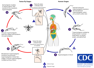
The life cycle of the Trypanosoma brucei parasites

Trypanosoma brucei gambiense accounts for the majority of African trypanosomiasis cases, with humans as the main reservoir needed for the transmission, while Trypanosoma brucei rhodesiense is mainly zoonotic, with the occasional human infection. African trypanosomiasis is dependent on the interaction of the parasite (trypanosome) with the tsetse flies (vector), as well as the host (human for Trypanosoma brucei gambiense, and animals for Trypanosoma brucei rhodesiense). The risk of contracting African trypanosomiasis is dependent on coming in contact with an infected tsetse fly.

Trypanosoma brucei

There are two subspecies of the parasite that are responsible for starting the disease in humans. Trypanosoma brucei gambiense causes the diseases in west and central Africa, whereas Trypanosoma brucei rhodesiense has a limited geographical range and is responsible for causing the disease in east and southern Africa. In addition, a third subspecies of the parasite known as Trypanosoma brucei brucei is responsible for affecting animals but not humans.

Humans are the main reservoir for T. b. gambiense but this species can also be found in pigs and other animals. Wild game animals and cattle are the main reservoir of T. b. rhodesiense. These parasites primarily infect individuals in sub-Saharan Africa because that is where the vector (tsetse fly) is located. The two human forms of the disease also vary greatly in intensity. T. b. gambiense causes a chronic condition that can remain in a passive phase for months or years before symptoms emerge and the infection can last about three years before death occurs.

T. b. rhodesiense is the acute form of the disease, and death can occur within months since the symptoms emerge within weeks and it is more virulent and faster developing than T. b. gambiense. Furthermore, trypanosomes are surrounded by a coat that is composed of variant surface glycoproteins (VSG). These proteins act to protect the parasite from any lytic factors that are present in human plasma. The host's immune system recognizes the glycoproteins present on the coat of the parasite leading to the production of different antibodies (IgM and IgG).

These antibodies will then act to destroy the parasites that circulate around the blood. However, from the several parasites present in the plasma, a small number of them will experience changes in their surface coats resulting in the formation of new VSGs. Thus, the antibodies produced by the immune system will no longer recognize the parasite leading to proliferation until new antibodies are created to combat the novel VSGs. Eventually, the immune system will no longer be able to fight off the parasite due to the constant changes in VSGs and infection will arise.

Vector

Type Trypanosoma Distribution Vector
Chronic T. brucei gambiense Western Africa G. palpalis

G. tachinoides

G. fuscipes

G. morsitans

Acute T. brucei rhodesiense Eastern Africa G. morsitans

G. swynnertoni

G. pallidipes

G. fuscipes

Drawing of a tsetse fly from 1880

The tsetse fly (genus Glossina) is a large, brown, biting fly that serves as both a host and vector for the trypanosome parasites. While taking blood from a mammalian host, an infected tsetse fly injects metacyclic trypomastigotes into skin tissue. From the bite, parasites first enter the lymphatic system and then pass into the bloodstream. Inside the mammalian host, they transform into bloodstream trypomastigotes, and are carried to other sites throughout the body, reach other body fluids (e.g., lymph, spinal fluid), and continue to replicate by binary fission.

The entire life cycle of African trypanosomes is represented by extracellular stages. A tsetse fly becomes infected with bloodstream trypomastigotes when taking a blood meal on an infected mammalian host. In the fly's midgut, the parasites transform into procyclic trypomastigotes, multiply by binary fission, leave the midgut, and transform into epimastigotes. The epimastigotes reach the fly's salivary glands and continue multiplication by binary fission.

The entire life cycle of the fly takes about three weeks. In addition to the bite of the tsetse fly, the disease can be transmitted by:

  • Mother-to-child infection: the trypanosome can sometimes cross the placenta and infect the fetus.
  • Laboratories: accidental infections, for example, through the handling of blood of an infected person and organ transplantation, although this is uncommon.
  • Blood transfusion
  • Sexual contact

Horse-flies (Tabanidae) and stable flies (Muscidae) possibly play a role in transmission of nagana (the animal form of sleeping sickness) and the human disease form.

Pathophysiology

Tryptophol is a chemical compound produced by the trypanosomal parasite in sleeping sickness which induces sleep in humans.

Diagnosis

Two areas from a blood smear from a person with African trypanosomiasis, thin blood smear stained with Giemsa: Typical trypomastigote stages (the only stages found in people), with a posterior kinetoplast, a centrally located nucleus, an undulating membrane, and an anterior flagellum. The two Trypanosoma brucei subspecies that cause human trypanosomiasis, T. b. gambiense and T. b. rhodesiense, are indistinguishable morphologically. The trypanosomes' length range is 14 to 33 µm, Source: CDC

The gold standard for diagnosis is identification of trypanosomes in a sample by microscopic examination. Samples that can be used for diagnosis include chancre fluid, lymph node aspirates, blood, bone marrow, and, during the neurological stage, cerebrospinal fluid. Detection of trypanosome-specific antibodies can be used for diagnosis, but the sensitivity and specificity of these methods are too variable to be used alone for clinical diagnosis. Further, seroconversion occurs after the onset of clinical symptoms during a T. b. rhodesiense infection, so is of limited diagnostic use.

Trypanosomes can be detected from samples using two different preparations. A wet preparation can be used to look for the motile trypanosomes. Alternatively, a fixed (dried) smear can be stained using Giemsa's or Field's technique and examined under a microscope. Often, the parasite is in relatively low abundance in the sample, so techniques to concentrate the parasites can be used prior to microscopic examination. For blood samples, these include centrifugation followed by examination of the buffy coat; mini anion-exchange/centrifugation; and the quantitative buffy coat (QBC) technique. For other samples, such as spinal fluid, concentration techniques include centrifugation followed by examination of the sediment.

Three serological tests are also available for detection of the parasite: the micro-CATT (card agglutination test for trypanosomiasis), wb-CATT, and wb-LATEX. The first uses dried blood, while the other two use whole blood samples. A 2002 study found the wb-CATT to be the most efficient for diagnosis, while the wb-LATEX is a better exam for situations where greater sensitivity is required.

Prevention

Capture devices for tsetse flies, on shore and on a boat in Africa. Efforts to prevent sleeping sickness.

Currently there are few medically related prevention options for African trypanosomiasis (i.e. no vaccine exists for immunity). Although the risk of infection from a tsetse fly bite is minor (estimated at less than 0.1%), the use of insect repellants, wearing long-sleeved clothing, avoiding tsetse-dense areas, implementing bush clearance methods and wild game culling are the best options to avoid infection available for local residents of affected areas.

In July 2000, a resolution was passed to form the Pan African Tsetse and Trypanosomiasis Eradication Campaign (PATTEC). The campaign works to eradicate the tsetse vector population levels and subsequently the protozoan disease, by use of insecticide-impregnated targets, fly traps, insecticide-treated cattle, ultra-low dose aerial/ground spraying (SAT) of tsetse resting sites and the sterile insect technique (SIT). The use of SIT in Zanzibar proved effective in eliminating the entire population of tsetse flies but was expensive and is relatively impractical to use in many of the endemic countries afflicted with African trypanosomiasis.

A pilot program in Senegal has reduced the tsetse fly population by as much as 99% by introducing male flies which have been sterilized by exposure to gamma rays.

Regular active surveillance, involving detection and prompt treatment of new infections, and tsetse fly control is the backbone of the strategy used to control sleeping sickness. Systematic screening of at-risk communities is the best approach, because case-by-case screening is not practical in endemic regions. Systematic screening may be in the form of mobile clinics or fixed screening centres where teams travel daily to areas of high infection rates. Such screening efforts are important because early symptoms are not evident or serious enough to warrant people with gambiense disease to seek medical attention, particularly in very remote areas. Also, diagnosis of the disease is difficult and health workers may not associate such general symptoms with trypanosomiasis. Systematic screening allows early-stage disease to be detected and treated before the disease progresses, and removes the potential human reservoir. A single case of sexual transmission of West African sleeping sickness has been reported.

Treatment

First stage

The treatment for first-stage disease is fexinidazole by mouth or pentamidine by injection for T. b. gambiense. Suramin by injection is used for T. b. rhodesiense.

Second stage

Fexinidazole may be used for the second stage of TbG, if the disease is not severe. Otherwise a regimen involving the combination of nifurtimox and eflornithine, nifurtimox-eflornithine combination treatment (NECT), or eflornithine alone appear to be more effective and result in fewer side effects. These treatments may replace melarsoprol when available. NECT has the benefit of requiring less injections of eflornithine.

Intravenous melarsoprol was previously the standard treatment for second-stage (neurological phase) disease and is effective for both types. Melarsoprol is the only treatment for second stage T. b. rhodesiense; however, it causes death in 5% of people who take it. Resistance to melarsoprol can occur.

Prognosis

If untreated, T. b. gambiense almost always results in death, with only a few individuals shown in a long-term 15 year follow-up to have survived after refusing treatment. T. b. rhodesiense, being a more acute and severe form of the disease, is consistently fatal if not treated. Disease progression greatly varies depending on disease form. For individuals which are infected by T. b. gambiense, which accounts for 98% of all of the reported cases, a person can be infected for months or even years without signs or symptoms until the advanced disease stage, where it is too late to be treated successfully. For individuals affected by T. b. rhodesiense, which accounts for 2% of all reported cases, symptoms appear within weeks or months of the infection. Disease progression is rapid and invades the central nervous system, causing death within a short amount of time.

Epidemiology

Deaths per 100,000 population due to African trypanosomiasis by country in 2002

In 2010, it caused around 9,000 deaths, down from 34,000 in 1990. As of 2000, the disability-adjusted life-years (9 to 10 years) lost due to sleeping sickness are 2.0 million. From 2010 to 2014, there was an estimated 55 million people at risk for gambiense African Trypanosomiasis and over 6 million people at risk for rhodesiense African trypanosomiasis. In 2014, the World Health Organization reported 3,797 cases of Human African Trypanosomiasis when the predicted number of cases were to be 5,000. The number of total reported cases in 2014 is an 86% reduction to the total number of cases reported in 2000.

The disease has been recorded as occurring in 37 countries, all in sub-Saharan Africa. It occurs regularly in southeast Uganda and western Kenya, and killed more than 48,000 Africans in 2008. The Democratic Republic of the Congo is the most affected country in the world, accounting for 75% of the Trypanosoma brucei gambiense cases. The population at risk being about 69 million with one third of this number being at a 'very high' to 'moderate' risk and the remaining two thirds at a 'low' to 'very low' risk. The number of people being affected by the disease has declined. At this rate, sleeping sickness elimination is a possibility. The World Health Organization plans to eradicate sleeping sickness by 2030.

Protozoan infection

From Wikipedia, the free encyclopedia
 
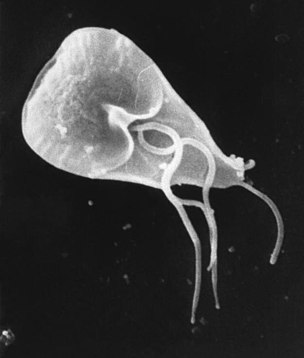
Giardia lamblia, an infectious protozoan

Protozoan infections are parasitic diseases caused by organisms formerly classified in the kingdom Protozoa. They are usually contracted by either an insect vector or by contact with an infected substance or surface and include organisms that are now classified in the supergroups Excavata, Amoebozoa, SAR, and Archaeplastida.

Protozoan infections are responsible for diseases that affect many different types of organisms, including plants, animals, and some marine life. Many of the most prevalent and deadly human diseases are caused by a protozoan infection, including African sleeping sickness, amoebic dysentery, and malaria.

The species originally termed "protozoa" are not closely related to each other and only have superficial similarities (eukaryotic, unicellular, motile, though with exceptions). The terms "protozoa" (and protist) are usually discouraged in the modern biosciences. However, this terminology is still encountered in medicine. This is partially because of the conservative character of medical classification and partially due to the necessity of making identifications of organisms based upon morphology.

Within the taxonomic classification, the four protist supergroups (Amoebozoa, Excavata, SAR, and Archaeplastida) fall under the domain Eukarya. Protists are an artificial grouping of over 64,000 different single-celled life forms. This means that it is difficult to define protists due to their extreme differences and uniqueness. Protists are a polyphyletic [(of a group of organisms) derived from more than one common evolutionary ancestor or ancestral group and therefore not suitable for placing in the same taxon] a collection of organisms and they are unicellular, which means that they lack the level of tissue organization which is present in more complex eukaryotes. Protists grow in a wide variety of moist habitats and a majority of them are free-living organisms. In these moist environments, plankton and terrestrial forms can also be found. Protists are chemoorganotrophic [organisms which oxidize the chemical bonds in organic compounds as their energy source and are responsible for recycling nitrogen and phosphorus. Parasites also are responsible for causing disease in humans and domesticated animals.

Protozoa are chemoorganotrophic protists and have three different ways of acquiring nutrients. The first method of acquiring nutrients is through saprotrophic nutrition. In saprotrophic nutrition, nutrients are obtained from dead organic matter through enzymatic degradation. The second method of acquiring nutrients is through osmotrophic nutrition. In osmotrophic nutrition, nutrients are obtained through absorbing soluble products. The third method of acquiring nutrients is through holozoic nutrition. In holozoic nutrition, solid nutrients are absorbed through phagocytosis.

Some protozoa are photoautotrophic protists. These protists include strict aerobes, and use photosystems I and II in order to carry out photosynthesis which produces oxygen.

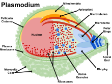
Diagram of Plasmodium structure

Mixotrophic protists obtain nutrients through organic and inorganic carbon compounds simultaneously.

All cells have a plasma membrane. In a protist, the plasma membrane is also known as the plasmalemma. Just below the plasma membrane, and in the inner fluid region, cytoplasm can be found. The pellicle structure in the protist is a thin layer of protein that helps provide the cell with some support and protection.  In addition to the plasma membrane, protists contain two different types of vacuoles. Contractile vacuoles help to maintain osmoregulation, and phagocytic vacuoles allow select protists to ingest food. In some protists, flagella or cilia may be present to help with motility and nutrient intake. The flagella or cilia create water currents that assist in feeding and respiration. Energy intake is necessary for protists’ survival. Aerobic chemoorganotrophic protists produce energy through the use of their mitochondria. The mitochondria then generate energy for the protist to keep up with cellular life functions. Photosynthetic protists produce energy through the use of their mitochondria and chloroplasts. Finally, anaerobic chemoorganotrophs produce energy through the use of hydrogenosomes, which are membrane-enclosed organelles that release molecular hydrogen (H2).

Encystment is when a protist becomes a dormant cyst with a cell wall; during encystment, the cyst has decreased complexity and metabolic activity relative to the protist. Encystment protects the protist from environmental changes, the cyst can be a site for nuclear reorganization and cell division, and it can act as a host cell in order to transfer parasitic species. Excystment is when a return to favorable conditions may cause a cyst to return to its original state. In parasitic protists, excystment may occur when the cyst is ingested by a new host.

Protists reproduce asexually or sexually. If the protists reproduce asexually, they do so through binary fission, multiple fission, budding, and fragmentation. If the protists reproduce sexually, they do so through a syngamy process where there is a fusion of the gametes. If this occurs in an individual it is recognized as autogamy. If this occurs between individuals, it is known as conjugation.

Supergroup Excavata


Excavata are considered primitive eukaryotes. They are characterized by a feeding groove with a posteriorly located flagella, which allows them to create a current that captures small food particles. The cytostome is the specialized structure that allows the protists this function. This supergroup Excavata includes the subgroups Diplomonads (Fornicata), Parabasalids, and Euglenozoans.

Diplomonads

Diplomonads used to be defined as Fornicata, but their characteristics remain the same despite their renaming. They are microaerophilic protists. Diplomonads were previously defined by the lack of a mitochondrion, but recent studies have found that they have a nonfunctional, mitochondrial remnant organelle called a mitosome. Most are harmless except for Giardia, Hexamita salmonis, and Histomonas meleagridis. Giardia causes diarrhea, Hexamita salmonis is a fish parasite, and Histomonas meleagridis is a turkey pathogen.

Giardia intestinalis is a human pathogen, which is transmitted by cyst-contaminated water. It causes epidemic diarrhea from contaminated water. One can tell one may be infected by the observation of cysts or trophozoites in stools and ELISA (enzyme-linked immunosorbent assay) test. To prevent contamination, avoid any possibly contaminated water, and if contaminated water is the only thing available to drink, a slow sand filter should be used. A study found that the chlorination of water and nutritional intervention had no effect on childhood giardia infection. Only handwashing and hygienic sanitation interventions reduced infection rates in children.

Hexamita salmonis is a common flagellated fish pathogen. Infected fish are weak and emaciated, and typically swim on their sides.

Histomonas meleagridis is a common bird pathogen that causes histomoniasis. Signs of histomoniasis include reduced appetite, drooping wings, unkempt feathers, and yellow fecal droppings.

Parabasilia

Most Parabasalia are flagellated endosymbionts of animals. They lack a distinct cytostome, which means they must use phagocytosis to engulf food. There are two subgroups: Trichonympha and Trichomonadida. Trichonympha are obligate mutualists of wood-eating insects such as termites. They secrete cellulase, which is used for digesting wood. The next subgroup, Trichomonadida, does not require oxygen and possesses hydrogenosomes. They only reproduce through asexual reproduction and some strains are human pathogens. There are three types of pathogenic parabasalia: Trichomonas foetus, Dientamoeba fragilis, and Trichomonas vaginalis. Trichomonas foetus causes spontaneous abortion in cattle, Dientamoeba fragilis causes diarrhea in humans, and Trichomonas vaginalis is a sexually transmitted disease.

Image of a cultured Tritrichomonas foetus

Trichomonas foetus is a parasite that resides in the urogenital tract of cattle and causes bovine trichomoniasis. Trichomoniasis is a sexually transmitted disease that causes infertility in heifers. Most infertility is caused by sudden embryonic death. Various imidazoles have been used to treat infected bulls, but none are safe and effective. Ipronidazole is probably most effective but it frequently causes sterile abscesses at injection sites.

Dientamoeba fragilis is a parasite that lives in the large intestine of humans. No one knows how D. fragilis is spread; one possibility is from swallowing contaminated water or food. Many people who are infected with this parasite show no signs of being infected. Sometimes the infection can be observed; the most common symptoms include diarrhea, stomach pains, loss of appetite, nausea, and fatigue.

Trichomonas vaginalis is a sexually transmitted disease. Men who are infected rarely show any symptoms (asymptomatic). Women who are infected usually show signs of soreness, inflammation, and redness around the vagina and a possible change in vaginal discharge. Trichomonas vaginalis can be treated with a course of antibiotics.

Euglenozoa

Most Euglenozoa are photoautotrophic, but some are chemoorganotrophs (saprophytic). They are commonly found in freshwater. The members of the phylum Euglenozoa have a pellicle for support, a red eye spot called a stigma to orient the cell toward light, chlorophyll a and b to assist in the process of photosynthesis, contractile vacuoles, and flagella.

Leishmaniasis lesion on adult human forearm

One major pathogen from the phylum Euglenozoa is Leishmania. Leishmania causes leishmaniasis. The symptoms of leishmaniasis include systemic and skin/membrane damage. Leishmania parasites spread by phlebotomine sand flies in the tropics, subtropics, and southern Europe. They may manifest cutaneously (cutaneous leishmaniasis) as skin sores with as scab a few weeks after the bite or internally (visceral leishmaniasis), affecting the organs, which can be life-threatening. Cutaneous leishmaniasis can spread to the mucus membranes and cause mucosal leishmaniasis even years after the initial infection. Cutaneous leishmaniasis heals on its own and leaves bad scars. Only FDA approved for visceral leishmaniasis is amphotericin B and oral miltefosine for cutaneous and mucosal leishmaniasis diagnosis- tissue specimen, bone marrow, blood tests detect antibody to parasite for visceral leishmaniasis.

Reduviid Bug

The second pathogen from this phylum is Trypanosoma cruzi. Trypanosoma cruzi causes Chagas disease and is transmitted by the reduviid bug, also known as the “kissing bug.”  Chagas disease is diagnosed using a physical exam and blood test. The only treatment includes antiparasitics only from the CDC, which are not FDA approved. Acute Chagas disease has a quick onset, the trypanosomes enter the bloodstream, they become amastigotes, and replicate. Acute Chagas disease can be treated using benznidazole or nifurtimox. Chronic chagas disease is asymptomatic and causes heart and gastrointestinal cells to be affected. Currently, there are only investigational treatments for this disease. Unfortunately, vaccines are not effective with Chagas disease due to antigenic variation. This pathogen causes damage to the nervous system.

African Sleeping Sickness is caused by Trypanosoma brucei rhodensiense and Trypanosoma brucei gambiense, and is transmitted by the tsetse fly. It is diagnosed by a physical exam and blood test. African sleeping sickness causes interstitial inflammation, lethargy, brain swelling, and death within one to three years. Drug therapy, using Eflornithine and Melarsoprol Pentamidine for T. gambiense and Suramin (Antrypol) for either Trypanosoma brucei rhodensiense and Trypanosoma brucei gambiense, or combinations of these medications, can help treat this disease, but vaccines can not be used due to antigenic variation.

Supergroup Amoebozoa

X-ray of colon infected with E. histolytica

Amoebozoa are characterized by the use of pseudopodia for movement and feeding. These protists reproduce by binary or multiple fission.

Entamoebida

Entamoebida lack mitochondria and possess mitosomes. Entamoeba histolytica is a pathogenic parasite known to cause amoebiasis, which is the third leading cause of parasitic deaths. It is diagnosed by the assessment of stool samples. Amoebiasis is caused by the ingestion of food or water contaminated with feces or other bodily wastes of an infected person, which contain cysts, the dormant form of the microbe. These cysts on reaching the terminal ileum region of the gastrointestinal tract give rise to a mass of proliferating cells, the trophozoite form of the parasite, by the process of excystation. Symptoms of this infection include diarrhea with blood and mucus, and can alternate between constipation and remission, abdominal pain, and fever. Symptoms can progress to ameboma, fulminant colitis, toxic megacolon, colonic ulcers, leading to perforation, and abscesses in vital organs like liver, lung, and brain. Amoebiasis can be treated with the administration of anti-amoebic compounds, this often includes the use of Metronidazole, Ornidazole, Chloroquine, Secnidazole, Nitazoxanide and Tinidazole. Tinidazole may be effective in curing children. The usage of conventional therapeutics to treat amoebiasis if often linked with substantial side effects, a threat to the efficacy of these therapeutics, further worsened by the development of drug resistance in the parasite. Amoebic meningoencephalitis and keratitis is a brain-eating amoeba caused by free-living Naeglaria and Acanthomoeba. One way this pathogen can be acquired is by soaking contact lenses in water instead of contact solution. This will result in progressive ulceration of the cornea. This pathogen can be diagnosed by demonstration of amoebae in clinical specimens. There is currently no drug therapy available for amoebic meningoencephalitis and keratitis.

Supergroup SAR

The supergroup SAR includes Stramenopiles, Alveolata and Rhizaria, and is distinguished by fine pseudopodia which can be branched, simple, or connected.

Stramenopila

Some members of Stramenopila are brown algae, diatoms, and water molds. An example of Stramenopila are Peronosporomycetes. The most well-known example of Peronosporomycetes is Phytophthora infestans. This organism caused the Great Famine of Ireland in the 1850s.

Alveolata

Alveolata is a large group, which includes Dinoflagellata, Ciliophora, and Apicomplexa.

Toxoplasmosis life cycle between humans and animals

Balantidium Coli (Balantidiasis) is an example of a member of the phylum Ciliophora. Balantidiasis is the only ciliate known to be capable of infecting humans, and swine are the primary reservoir host. Balantidiasis is opportunistic and rare in Western countries. Apicomplexans are parasites of animals and contain an arrangement of organelles called the apical complex. One example of an apicomplexan is Malaria. Five species of plasmodium cause malaria in animals. Malaria is transmitted by the bite of an infected female mosquito. Symptoms of malaria include: periodic chills and fever, anemia, and hypertrophy of the liver and spleen. Cerebral malaria can occur in children. In order to diagnose Malaria, doctors will look for parasites in Wright-or-Giemsa-stained red blood cells and serological tests. Treatment includes antimalarial drugs, however, resistance has been observed. New vaccines are being discovered to this day. Preventative measures that can be taken include sleeping with netting and using insecticide to prevent mosquitoes. Eimeria is another example of an apicomplexan pathogen. This pathogen causes cecal coccidiosis in chickens. Coccidiosis is a parasitic disease of the intestinal tract. This disease is treated by placing anticoccidials in the chickens’ feed. It also causes malabsorption, diarrhea, and sometimes bloody diarrhea in animals. Theileria parva & T. annulata are tick-borne parasites which cause fatal East Coast fever in cattle. East Coast fever is transmitted by the bite of the three-host tick Phipicephalus appendiculatus and results in respiratory failure and death in African cattle. Most hosts of P. appendiculatus succumb to pulmonary edema and die within three weeks of infection. The severity of the infection can be lessened by treatment with antiprotozoal drugs like buparvaquone. Toxoplasma causes toxoplasmosis and can be acquired from undercooked meat or cat feces containing Toxoplasma gondii. The majority of the 60 million Americans infected with T. gondii are asymptomatic. The group most vulnerable to this pathogen are the fetuses of mothers who have been infected with the parasite for the first time during pregnancy. This can result in damage to the fetus’s brain, eyes, and other organs. Treatment is available for pregnant women and the immunosuppressed. Cryptosporidiosis can be contracted through contact with water, food, soil, or surfaces contaminated with feces containing the Cryptosporidium. Immunocompromised people are the most susceptible. Cryptosporidiosis causes watery diarrhea and can resolve itself without medical intervention. It is diagnosed by examining stool samples, and diarrhea can be treated using Nitazoxanide.

Rhizaria

Plasmodiophorids and Halosporidians are two examples of parasitic Rhizaria. Plasmodiophorids cause infections in crops such as Spongospora subterranea. They cause powdery scabs and galls and disrupt growth. Halosporidians cause infections in marine invertebrates such as Mikrocytos mackini in Pacific oysters. Mikrocytos mackini are abscesses or green pustules on palps and mantles of certain molluscs.

Archaeplastida

The supergroup Archaeplastida includes red algae, green algae and land plants. Each of these three groups have multicellular species and the green and red algae have many single-celled species. The land plants are not considered protists.

Red algae are primarily multicellular, lack flagella, and range in size from microscopic, unicellular to large, multicellular forms. Some species of red algae contain phycoerythrins, photosynthetic accessory pigments that are red in color and outcompete the green tint of chlorophyll, making these species appear as varying shades of red. This group doesn’t include many pathogens.

Green algae exhibit similar features to the land plants, particularly in terms of chloroplast structure. The green algae are subdivided into the chlorophytes and charophytes. It is very rare for green algae to become parasitic.

Prototheca moriformis belongs to the subdivision Chloroplastida. P. moriformis is a green algae that lacks chlorophyll and has turned to parasitism. It is found in sewage and the soil. P. moriformis causes a disease called protothecosis. This disease mainly infects cattle and dogs. Cattle can be affected by prototheca enteritis and mastitis. Protothecosis is commonly seen in dogs; it enters the body through the mouth or nose and causes infection in the intestines. Treatment with amphotericin B has been reported.

Future Treatment

Scientists have been researching new ways to fight protozoan infections, including targeting channels and transporters involved in the diseases and finding the link between a persons microbiome and their ability to resist a protozoan infection

Pathogen transmission

From Wikipedia, the free encyclopedia
https://en.wikipedia.org/wiki/Pathogen_transmission

In medicine, public health, and biology, transmission is the passing of a pathogen causing communicable disease from an infected host individual or group to a particular individual or group, regardless of whether the other individual was previously infected. The term strictly refers to the transmission of microorganisms directly from one individual to another by one or more of the following means:

  • airborne transmission – very small dry and wet particles that stay in the air for long periods of time allowing airborne contamination even after the departure of the host. Particle size < 5 μm.
  • droplet transmission – small and usually wet particles that stay in the air for a short period of time. Contamination usually occurs in the presence of the host. Particle size > 5 μm.
  • direct physical contact – touching an infected individual, including sexual contact
  • indirect physical contact – usually by touching a contaminated surface, including soil (fomite)
  • fecal–oral transmission – usually from unwashed hands, contaminated food or water sources due to lack of sanitation and hygiene, an important transmission route in pediatrics, veterinary medicine and developing countries.

Transmission can also be indirect, via another organism, either a vector (e.g. a mosquito or fly) or an intermediate host (e.g. tapeworm in pigs can be transmitted to humans who ingest improperly cooked pork). Indirect transmission could involve zoonoses or, more typically, larger pathogens like macroparasites with more complex life cycles. Transmissions can be autochthonous (i.e. between two individuals in the same place) or may involve travel of the microorganism or the affected hosts.

Definition and related terms

An infectious disease agent can be transmitted in two ways: as horizontal disease agent transmission from one individual to another in the same generation (peers in the same age group) by either direct contact (licking, touching, biting), or indirect contact through air – cough or sneeze (vectors or fomites that allow the transmission of the agent causing the disease without physical contact) or by vertical disease transmission, passing the agent causing the disease from parent to offspring, such as in prenatal or perinatal transmission.

The term infectivity describes the ability of an organism to enter, survive and multiply in the host, while the infectiousness of a disease agent indicates the comparative ease with which the disease agent is transmitted to other hosts. Transmission of pathogens can occur by direct contact, through contaminated food, body fluids or objects, by airborne inhalation or through vector organisms.

Transmissibility is the probability of an infection, given a contact between an infected host and a noninfected host.

Community transmission means that the source of infection for the spread of an illness is unknown or a link in terms of contacts between patients and other people is missing. It refers to the difficulty in grasping the epidemiological link in the community beyond confirmed cases.

Local transmission means that the source of the infection has been identified within the reporting location (such as within a country, region or city).

Routes of transmission

The route of transmission is important to epidemiologists because patterns of contact vary between different populations and different groups of populations depending on socio-economic, cultural and other features. For example, low personal and food hygiene due to the lack of a clean water supply may result in increased transmission of diseases by the fecal-oral route, such as cholera. Differences in incidence of such diseases between different groups can also throw light on the routes of transmission of the disease. For example, if it is noted that polio is more common in cities in underdeveloped countries, without a clean water supply, than in cities with a good plumbing system, we might advance the theory that polio is spread by the fecal-oral route. Two routes are considered to be airborne: Airborne infections and droplet infections.

Airborne infection

"Airborne transmission refers to infectious agents that are spread via droplet nuclei (residue from evaporated droplets) containing infective microorganisms. These organisms can survive outside the body and remain suspended in the air for long periods of time. They infect others via the upper and lower respiratory tracts." The size of the particles for airborne infections need to be < 5 μm. It includes both dry and wet aerosols and thus requires usually higher levels of isolation since it can stay suspended in the air for longer periods of time. i.e., separate ventilation systems or negative pressure environments are needed to avoid general contamination. e.g., tuberculosis, chickenpox, measles.

Droplet infection

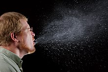
Droplet image captured under dark background on scattering illumination or tyndall effect
Respiratory droplets are released through talking, coughing, or sneezing.

A common form of transmission is by way of respiratory droplets, generated by coughing, sneezing, or talking. Respiratory droplet transmission is the usual route for respiratory infections. Transmission can occur when respiratory droplets reach susceptible mucosal surfaces, such as in the eyes, nose or mouth. This can also happen indirectly via contact with contaminated surfaces when hands then touch the face. Before drying, respiratory droplets are large and cannot remain suspended in the air for long, and are usually dispersed over short distances. The size of the particles for droplet infections are > 5 μm.

Organisms spread by droplet transmission include respiratory viruses such as influenza virus, parainfluenza virus, adenoviruses, rhinovirus, respiratory syncytial virus, human metapneumovirus, Bordetella pertussis, pneumococci, streptococcus pyogenes, diphtheria, rubella, and coronaviruses. Spread of respiratory droplets from the wearer can be reduced through wearing of a surgical mask.

Direct contact

Direct contact occurs through skin-to-skin contact, kissing, and sexual intercourse. Direct contact also refers to contact with soil or vegetation harboring infectious organisms. Additionally, while fecal–oral transmission is primarily considered an indirect contact route, direct contact can also result in transmission through feces.

Diseases that can be transmitted by direct contact are called contagious (contagious is not the same as infectious; although all contagious diseases are infectious, not all infectious diseases are contagious). These diseases can also be transmitted by sharing a towel (where the towel is rubbed vigorously on both bodies) or items of clothing in close contact with the body (socks, for example) if they are not washed thoroughly between uses. For this reason, contagious diseases often break out in schools, where towels are shared and personal items of clothing accidentally swapped in the changing rooms.

Some diseases that are transmissible by direct contact include athlete's foot, impetigo, syphilis, warts, and conjunctivitis.

Sexual

This refers to any disease that can be caught during sexual activity with another person, including vaginal or anal sex or (less commonly) through oral sex (see below). Transmission is either directly between surfaces in contact during intercourse (the usual route for bacterial infections and those infections causing sores) or from secretions (semen or the fluid secreted by the excited female) which carry infectious agents that get into the partner's blood stream through tiny tears in the penis, vagina or rectum (this is a more usual route for viruses). In this second case, anal sex is considerably more hazardous since the penis opens more tears in the rectum than the vagina, as the vagina is more elastic and more accommodating.

Some diseases transmissible by the sexual route include HIV/AIDS, chlamydia, genital warts, gonorrhea, hepatitis B, syphilis, herpes, and trichomoniasis.

Oral sexual

Sexually transmitted diseases such as HIV and hepatitis B are thought to not normally be transmitted through mouth-to-mouth contact, although it is possible to transmit some STDs between the genitals and the mouth, during oral sex. In the case of HIV this possibility has been established. It is also responsible for the increased incidence of herpes simplex virus 1 (which is usually responsible for oral infections) in genital infections and the increased incidence of the type 2 virus (more common genitally) in oral infections.

Oral

Diseases that are transmitted primarily by oral means may be caught through direct oral contact such as kissing, or by indirect contact such as by sharing a drinking glass or a cigarette. Diseases that are known to be transmissible by kissing or by other direct or indirect oral contact include all of the diseases transmissible by droplet contact and (at least) all forms of herpes viruses, namely Cytomegalovirus infections herpes simplex virus (especially HSV-1) and infectious mononucleosis.

Mother-to-child transmission

Brocky, Karoly - Mother and Child (1846-50)
 

This is from mother to child (more rarely father to child), often in utero, during childbirth (also referred to as perinatal infection) or during postnatal physical contact between parents and offspring. In mammals, including humans, it occurs also via breast milk (transmammary transmission). Infectious diseases that can be transmitted in this way include: HIV, hepatitis B and syphilis. Many mutualistic organisms are transmitted vertically.

Iatrogenic

Transmission due to medical procedures, such as touching a wound, an injection or transplantation of infected material. Some diseases that can be transmitted iatrogenically include: Creutzfeldt–Jakob disease by injection of contaminated human growth hormone, MRSA and many more.

Indirect contact

Indirect contact transmission, also known as vehicleborne transmission, involves transmission through contamination of inanimate objects. Vehicles that may indirectly transmit an infectious agent include food, water, biologic products such as blood, and fomites such as handkerchiefs, bedding, or surgical scalpels. A vehicle may passively carry a pathogen, as in the case of food or water may carrying hepatitis A virus. Alternatively, the vehicle may provide an environment in which the agent grows, multiplies, or produces toxin, such as improperly canned foods provide an environment that supports production of botulinum toxin by Clostridium botulinum.

Transmission by other organisms

A vector is an organism that does not cause disease itself but that transmits infection by conveying pathogens from one host to another.

Vectors may be mechanical or biological. A mechanical vector picks up an infectious agent on the outside of its body and transmits it in a passive manner. An example of a mechanical vector is a housefly, which lands on cow dung, contaminating its appendages with bacteria from the feces, and then lands on food prior to consumption. The pathogen never enters the body of the fly. In contrast, biological vectors harbor pathogens within their bodies and deliver pathogens to new hosts in an active manner, usually a bite. Biological vectors are often responsible for serious blood-borne diseases, such as malaria, viral encephalitis, Chagas disease, Lyme disease and African sleeping sickness. Biological vectors are usually, though not exclusively, arthropods, such as mosquitoes, ticks, fleas and lice. Vectors are often required in the life cycle of a pathogen. A common strategy used to control vector-borne infectious diseases is to interrupt the life cycle of a pathogen by killing the vector.

Fecal–oral

1940 US WPA poster encouraging modernized privies
 

In the fecal-oral route, pathogens in fecal particles pass from one person to the mouth of another person. Although it is usually discussed as a route of transmission, it is actually a specification of the entry and exit portals of the pathogen, and can operate across several of the other routes of transmission. Fecal–oral transmission is primarily considered as an indirect contact route through contaminated food or water. However, it can also operate through direct contact with feces or contaminated body parts, such as through anal sex. It can also operate through droplet or airborne transmission through the toilet plume from contaminated toilets.

Main causes of fecal–oral disease transmission include lack of adequate sanitation and poor hygiene practices - which can take various forms. Fecal oral transmission can be via foodstuffs or water that has become contaminated. This can happen when people do not adequately wash their hands after using the toilet and before preparing food or tending to patients.

The fecal-oral route of transmission can be a public health risk for people in developing countries who live in urban slums without access to adequate sanitation. Here, excreta or untreated sewage can pollute drinking water sources (groundwater or surface water). The people who drink the polluted water can become infected. Another problem in some developing countries, is open defecation which leads to disease transmission via the fecal-oral route.

Even in developed countries there are periodic system failures resulting in a sanitary sewer overflow. This is the typical mode of transmission for infectious agents such as cholera, hepatitis A, polio, Rotavirus, Salmonella, and parasites (e.g. Ascaris lumbricoides).

Tracking

Tracking the transmission of infectious diseases is called disease surveillance. Surveillance of infectious diseases in the public realm traditionally has been the responsibility of public health agencies, on an international, national, or local level. Public health staff relies on health care workers and microbiology laboratories to report cases of reportable diseases to them. The analysis of aggregate data can show the spread of a disease and is at the core of the specialty of epidemiology. To understand the spread of the vast majority of non-notifiable diseases, data either need to be collected in a particular study, or existing data collections can be mined, such as insurance company data or antimicrobial drug sales for example.

For diseases transmitted within an institution, such as a hospital, prison, nursing home, boarding school, orphanage, refugee camp, etc., infection control specialists are employed, who will review medical records to analyze transmission as part of a hospital epidemiology program, for example.

Because these traditional methods are slow, time-consuming, and labor-intensive, proxies of transmission have been sought. One proxy in the case of influenza is tracking of influenza-like illness at certain sentinel sites of health care practitioners within a state, for example. Tools have been developed to help track influenza epidemics by finding patterns in certain web search query activity. It was found that the frequency of influenza-related web searches as a whole rises as the number of people sick with influenza rises. Examining space-time relationships of web queries has been shown to approximate the spread of influenza and dengue.

Computer simulations of infectious disease spread have been used. Human aggregation can drive transmission, seasonal variation and outbreaks of infectious diseases, such as the annual start of school, bootcamp, the annual Hajj etc. Most recently, data from cell phones have been shown to be able to capture population movements well enough to predict the transmission of certain infectious diseases, like rubella.

Relationship with virulence and survival

Pathogens must have a way to be transmitted from one host to another to ensure their species' survival. Infectious agents are generally specialized for a particular method of transmission. Taking an example from the respiratory route, from an evolutionary perspective viruses or bacteria that cause their host to develop coughing and sneezing symptoms have a great survival advantage, as they are much more likely to be ejected from one host and carried to another. This is also the reason that many microorganisms cause diarrhea.

The relationship between virulence and transmission is complex and has important consequences for the long term evolution of a pathogen. Since it takes many generations for a microbe and a new host species to co-evolve, an emerging pathogen may hit its earliest victims especially hard. It is usually in the first wave of a new disease that death rates are highest. If a disease is rapidly fatal, the host may die before the microbe can be passed along to another host. However, this cost may be overwhelmed by the short-term benefit of higher infectiousness if transmission is linked to virulence, as it is for instance in the case of cholera (the explosive diarrhea aids the bacterium in finding new hosts) or many respiratory infections (sneezing and coughing create infectious aerosols).

Anything that reduces the rate of transmission of an infection carries positive externalities, which are benefits to society that are not reflected in a price to a consumer. This is recognized implicitly when vaccines are offered for free or at a cost to the patient less than the purchase price.

Beneficial microorganisms

The mode of transmission is also an important aspect of the biology of beneficial microbial symbionts, such as coral-associated dinoflagellates or human microbiota. Organisms can form symbioses with microbes transmitted from their parents, from the environment or unrelated individuals, or both.

Vertical transmission

Vertical transmission refers to acquisition of symbionts from parents (usually mothers). Vertical transmission can be intracellular (e.g. transovarial), or extracellular (for example through post-embryonic contact between parents and offspring). Both intracellular and extracellular vertical transmission can be considered a form of non-genetic inheritance or parental effect. It has been argued that most organisms experience some form of vertical transmission of symbionts. Canonical examples of vertically transmitted symbionts include the nutritional symbiont Buchnera in aphids (transovarially transmitted intracellular symbiont) and some components of the human microbiota (transmitted during passage of infants through the birth canal and also through breastfeeding).

Horizontal transmission

Some beneficial symbionts are acquired horizontally, from the environment or unrelated individuals. This requires that host and symbiont have some method of recognizing each other or each other's products or services. Often, horizontally acquired symbionts are relevant to secondary rather than primary metabolism, for example for use in defense against pathogens, but some primary nutritional symbionts are also horizontally (environmentally) acquired. Additional examples of horizontally transmitted beneficial symbionts include bioluminescent bacteria associated with bobtail squid and nitrogen-fixing bacteria in plants.

Mixed-mode transmission

Many microbial symbionts, including human microbiota, can be transmitted both vertically and horizontally. Mixed-mode transmission can allow symbionts to have the “best of both worlds” – they can vertically infect host offspring when host density is low, and horizontally infect diverse additional hosts when a number of additional hosts are available. Mixed-mode transmission make the outcome (degree of harm or benefit) of the relationship more difficult to predict, because the evolutionary success of the symbiont is sometimes but not always tied to the success of the host.

Citation signal

From Wikipedia, the free encyclopedia https://en.wikipedia.org/wiki/Cit...