Search This Blog

Wednesday, March 11, 2020

Stomach cancer

From Wikipedia, the free encyclopedia
 
Stomach cancer
Other namesGastric cancer
Adenocarcinoma of the stomach.jpg
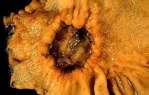
A stomach ulcer that was diagnosed as cancer on biopsy and surgically removed
SpecialtyOncology
SymptomsEarly: Heartburn, upper abdominal pain, nausea, loss of appetite.
Later: Weight loss, yellowing of the skin and whites of the eyes, vomiting, difficulty swallowing, blood in the stool
Usual onsetOver years
TypesGastric carcinomas, lymphoma, mesenchymal tumor
CausesHelicobacter pylori, genetics
Risk factorsSmoking, dietary factors such as pickled vegetables, obesity
Diagnostic methodBiopsy done during endoscopy
PreventionMediterranean diet, stopping smoking
TreatmentSurgery, chemotherapy, radiation therapy, targeted therapy
PrognosisFive-year survival rate:
< 10% (advanced cases),
32% (US), 71% (Japan)
Frequency3.5 million (2015)
Deaths783,000 (2018)

Stomach cancer, also known as gastric cancer, is a cancer that develops from the lining of the stomach. Most cases of stomach cancers are gastric carcinomas, which can be divided into a number of subtypes including gastric adenocarcinomas. Lymphomas and mesenchymal tumors may also develop in the stomach. Early symptoms may include heartburn, upper abdominal pain, nausea and loss of appetite. Later signs and symptoms may include weight loss, yellowing of the skin and whites of the eyes, vomiting, difficulty swallowing and blood in the stool among others. The cancer may spread from the stomach to other parts of the body, particularly the liver, lungs, bones, lining of the abdomen and lymph nodes.

The most common cause is infection by the bacterium Helicobacter pylori, which accounts for more than 60% of cases. Certain types of H. pylori have greater risks than others. Smoking, dietary factors such as pickled vegetables and obesity are other risk factors. About 10% of cases run in families, and between 1% and 3% of cases are due to genetic syndromes inherited from a person's parents such as hereditary diffuse gastric cancer. Most of the time, stomach cancer develops in stages over years. Diagnosis is usually by biopsy done during endoscopy. This is followed by medical imaging to determine if the disease has spread to other parts of the body. Japan and South Korea, two countries that have high rates of the disease, screen for stomach cancer.

A Mediterranean diet lowers the risk of stomach cancer, as does the stopping of smoking. There is tentative evidence that treating H. pylori decreases the future risk. If stomach cancer is treated early, it can be cured. Treatments may include some combination of surgery, chemotherapy, radiation therapy and targeted therapy. If treated late, palliative care may be advised. Some types of lymphoma can be cured by eliminating H. pylori. Outcomes are often poor, with a less than 10% five-year survival rate in the Western world for advanced cases. This is largely because most people with the condition present with advanced disease. In the United States, five-year survival is 31.5%, while in South Korea it is over 65% and Japan over 70%, partly due to screening efforts.

Globally, stomach cancer is the fifth leading type of cancer and the third leading cause of death from cancer, making up 7% of cases and 9% of deaths. In 2018, it newly occurred in 1.03 million people and caused 783,000 deaths. Before the 1930s, in much of the world, including most Western developed countries, it was the most common cause of death from cancer. Rates of death have been decreasing in many areas of the world since then. This is believed to be due to the eating of less salted and pickled foods as a result of the development of refrigeration as a method of keeping food fresh. Stomach cancer occurs most commonly in East Asia and Eastern Europe. It occurs twice as often in males as in females.

Signs and symptoms

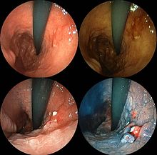
Endoscopic image of linitis plastica, a type of stomach cancer where the entire stomach is invaded, leading to a leather bottle-like appearance with blood coming out of it
 
Endoscopic images of the stomach cancer in early stage. Its histology was poorly differentiated adenocarcinoma with signet ring cells. Left above: normal, right above: FICE, left low: acetate stained, right low: AIM stained
 
Stomach cancer is often either asymptomatic (producing no noticeable symptoms) or it may cause only nonspecific symptoms (symptoms that may also be present in other related or unrelated disorders) in its early stages. By the time symptoms occur, the cancer has often reached an advanced stage (see below) and may have metastasized (spread to other, perhaps distant, parts of the body), which is one of the main reasons for its relatively poor prognosis. Stomach cancer can cause the following signs and symptoms:

Early cancers may be associated with indigestion or a burning sensation (heartburn). However, fewer than 1 in every 50 people referred for endoscopy due to indigestion has cancer. Abdominal discomfort and loss of appetite, especially for meat, can occur.

Gastric cancers that have enlarged and invaded normal tissue can cause weakness, fatigue, bloating of the stomach after meals, abdominal pain in the upper abdomen, nausea and occasional vomiting, diarrhea or constipation. Further enlargement may cause weight loss or bleeding with vomiting blood or having blood in the stool, the latter apparent as black discolouration (melena) and sometimes leading to anemia. Dysphagia suggests a tumour in the cardia or extension of the gastric tumour into the esophagus

These can be symptoms of other problems such as a stomach virus, gastric ulcer, or tropical sprue.

Causes

Gastric cancer occurs as a result of many factors. It occurs twice as commonly in males as females. Estrogen may protect women against the development of this form of cancer.

Infections

Helicobacter pylori infection is an essential risk factor in 65–80% of gastric cancers, but only 2% of people with Helicobacter infections develop stomach cancer. The mechanism by which H. pylori induces stomach cancer potentially involves chronic inflammation, or the action of H. pylori virulence factors such as CagA. It was estimated that Epstein–Barr virus is responsible for 84,000 cases per year. AIDS is also associated with elevated risk.

Smoking

Smoking increases the risk of developing gastric cancer significantly, from 40% increased risk for current smokers to 82% increase for heavy smokers. Gastric cancers due to smoking mostly occur in the upper part of the stomach near the esophagus. Some studies show increased risk with alcohol consumption as well.

Diet

Sequence of 123-iodine human scintiscans after an intravenous injection: (from left) after 30 minutes, 20 hours and 48 hours. A high and rapid concentration of radio-iodine is evident in gastric mucosa of the stomach, in salivary glands, oral mucosa and in the periencephalic and cerebrospinal fluid (left). In the thyroid gland, I-concentration is more progressive, also in the reservoir (from 1% after 30 minutes to 5.8 % after 48 hours, of the total injected dose).[33]

Dietary factors are not proven causes, and the association between stomach cancer and various foods and beverages is weak. Some foods including smoked foods, salt and salt-rich foods, red meat, processed meat, pickled vegetables, and bracken are associated with a higher risk of stomach cancer. Nitrates and nitrites in cured meats can be converted by certain bacteria, including H. pylori, into compounds that have been found to cause stomach cancer in animals.

Fresh fruit and vegetable intake, citrus fruit intake, and antioxidant intake are associated with a lower risk of stomach cancer. A Mediterranean diet is associated with lower rates of stomach cancer, as is regular aspirin use.

Obesity is a physical risk factor that has been found to increase the risk of gastric adenocarcinoma by contributing to the development of gastroesophageal reflux disease (GERD). The exact mechanism by which obesity causes GERD is not completely known. Studies hypothesize that increased dietary fat leading to increased pressure on the stomach and the lower esophageal sphincter, due to excess adipose tissue, could play a role, yet no statistically significant data has been collected. However, the risk of gastric cardia adenocarcinoma, with GERD present, has been found to increase more than 2 times for an obese person. There is a correlation between iodine deficiency and gastric cancer.

Genetics

About 10% of cases run in families and between 1% and 3% of cases are due to genetic syndromes inherited from a person's parents such as hereditary diffuse gastric cancer.

A genetic risk factor for gastric cancer is a genetic defect of the CDH1 gene known as hereditary diffuse gastric cancer (HDGC). The CDH1 gene, which codes for E-cadherin, lies on the 16th chromosome. When the gene experiences a particular mutation, gastric cancer develops through a mechanism that is not fully understood. This mutation is considered autosomal dominant meaning that half of a carrier's children will likely experience the same mutation. Diagnosis of hereditary diffuse gastric cancer usually takes place when at least two cases involving a family member, such as a parent or grandparent, are diagnosed, with at least one diagnosed before the age of 50. The diagnosis can also be made if there are at least three cases in the family, in which case age is not considered.

The International Cancer Genome Consortium is leading efforts to identify genomic changes involved in stomach cancer. A very small percentage of diffuse-type gastric cancers (see Histopathology below) arise from an inherited abnormal CDH1 gene. Genetic testing and treatment options are available for families at risk.

Other

Other risks include diabetes, pernicious anemia, chronic atrophic gastritis, Menetrier's disease (hyperplastic, hypersecretory gastropathy), and intestinal metaplasia.

Diagnosis

To find the cause of symptoms, the doctor asks about the patient's medical history, does a physical exam, and may order laboratory studies. The patient may also have one or all of the following exams:
  • Gastroscopic exam is the diagnostic method of choice. This involves insertion of a fibre optic camera into the stomach to visualise it.
  • Upper GI series (may be called barium roentgenogram).
  • Computed tomography or CT scanning of the abdomen may reveal gastric cancer. It is more useful to determine invasion into adjacent tissues or the presence of spread to local lymph nodes. Wall thickening of more than 1 cm that is focal, eccentric and enhancing favours malignancy.
In 2013, Chinese and Israeli scientists reported a successful pilot study of a breathalyzer-style breath test intended to diagnose stomach cancer by analyzing exhaled chemicals without the need for an intrusive endoscopy. A larger-scale clinical trial of this technology was completed in 2014.

Abnormal tissue seen in a gastroscope examination will be biopsied by the surgeon or gastroenterologist. This tissue is then sent to a pathologist for histological examination under a microscope to check for the presence of cancerous cells. A biopsy, with subsequent histological analysis, is the only sure way to confirm the presence of cancer cells.

Various gastroscopic modalities have been developed to increase yield of detected mucosa with a dye that accentuates the cell structure and can identify areas of dysplasia. Endocytoscopy involves ultra-high magnification to visualise cellular structure to better determine areas of dysplasia. Other gastroscopic modalities such as optical coherence tomography are being tested investigationally for similar applications.

A number of cutaneous conditions are associated with gastric cancer. A condition of darkened hyperplasia of the skin, frequently of the axilla and groin, known as acanthosis nigricans, is associated with intra-abdominal cancers such as gastric cancer. Other cutaneous manifestations of gastric cancer include tripe palms (a similar darkening hyperplasia of the skin of the palms) and the Leser-Trelat sign, which is the rapid development of skin lesions known as seborrheic keratoses.

Various blood tests may be done including a complete blood count (CBC) to check for anaemia, and a fecal occult blood test to check for blood in the stool.

Histopathology

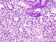
  • Gastric adenocarcinoma is a malignant epithelial tumour, originating from glandular epithelium of the gastric mucosa. Stomach cancers are about 90% adenocarcinomas. Histologically, there are two major types of gastric adenocarcinoma (Lauren classification): intestinal type or diffuse type. Adenocarcinomas tend to aggressively invade the gastric wall, infiltrating the muscularis mucosae, the submucosa and then the muscularis propria. Intestinal type adenocarcinoma tumour cells describe irregular tubular structures, harbouring pluristratification, multiple lumens, reduced stroma ("back to back" aspect). Often, it associates intestinal metaplasia in neighbouring mucosa. Depending on glandular architecture, cellular pleomorphism and mucosecretion, adenocarcinoma may present 3 degrees of differentiation: well, moderate and poorly differentiated. Diffuse type adenocarcinoma (mucinous, colloid, linitis plastica or leather-bottle stomach) tumour cells are discohesive and secrete mucus, which is delivered in the interstitium, producing large pools of mucus/colloid (optically "empty" spaces). It is poorly differentiated. If the mucus remains inside the tumour cell, it pushes the nucleus to the periphery: "signet-ring cell".
  • Around 5% of gastric cancers are lymphomas. These may include extranodal marginal zone B-cell lymphomas (MALT type) and to a lesser extent diffuse large B-cell lymphomas. MALT type make up about half of stomach lymphomas.
  • Carcinoid and stromal tumors may occur.

Staging

T stages of stomach cancer

If cancer cells are found in the tissue sample, the next step is to stage, or find out the extent of the disease. Various tests determine whether the cancer has spread and, if so, what parts of the body are affected. Because stomach cancer can spread to the liver, the pancreas, and other organs near the stomach as well as to the lungs, the doctor may order a CT scan, a PET scan, an endoscopic ultrasound exam, or other tests to check these areas. Blood tests for tumor markers, such as carcinoembryonic antigen (CEA) and carbohydrate antigen (CA) may be ordered, as their levels correlate to extent of metastasis, especially to the liver, and the cure rate.

Staging may not be complete until after surgery. The surgeon removes nearby lymph nodes and possibly samples of tissue from other areas in the abdomen for examination by a pathologist.
The clinical stages of stomach cancer are:
  • Stage 0. Limited to the inner lining of the stomach. Treatable by endoscopic mucosal resection when found very early (in routine screenings); otherwise by gastrectomy and lymphadenectomy without need for chemotherapy or radiation.
  • Stage I. Penetration to the second or third layers of the stomach (Stage 1A) or to the second layer and nearby lymph nodes (Stage 1B). Stage 1A is treated by surgery, including removal of the omentum. Stage 1B may be treated with chemotherapy (5-fluorouracil) and radiation therapy.
  • Stage II. Penetration to the second layer and more distant lymph nodes, or the third layer and only nearby lymph nodes, or all four layers but not the lymph nodes. Treated as for Stage I, sometimes with additional neoadjuvant chemotherapy.
  • Stage III. Penetration to the third layer and more distant lymph nodes, or penetration to the fourth layer and either nearby tissues or nearby or more distant lymph nodes. Treated as for Stage II; a cure is still possible in some cases.
  • Stage IV. Cancer has spread to nearby tissues and more distant lymph nodes, or has metastasized to other organs. A cure is very rarely possible at this stage. Some other techniques to prolong life or improve symptoms are used, including laser treatment, surgery, and/or stents to keep the digestive tract open, and chemotherapy by drugs such as 5-fluorouracil, cisplatin, epirubicin, etoposide, docetaxel, oxaliplatin, capecitabine or irinotecan.
Stomach cancer metastasized to the lungs

The TNM staging system is also used.

In a study of open-access endoscopy in Scotland, patients were diagnosed 7% in Stage I 17% in Stage II, and 28% in Stage III. A Minnesota population was diagnosed 10% in Stage I, 13% in Stage II, and 18% in Stage III. However, in a high-risk population in the Valdivia Province of southern Chile, only 5% of patients were diagnosed in the first two stages and 10% in stage III.

Prevention

Getting rid of H. pylori in those who are infected decreases the risk of stomach cancer, at least in those who are Asian. A 2014 meta-analysis of observational studies found that a diet high in fruits, mushrooms, garlic, soybeans, and green onions was associated with a lower risk of stomach cancer in the Korean population. Low doses of vitamins, especially from a healthy diet, decrease the risk of stomach cancer. A previous review of antioxidant supplementation did not find supporting evidence and possibly worse outcomes.

Management

Cancer of the stomach is difficult to cure unless it is found at an early stage (before it has begun to spread). Unfortunately, because early stomach cancer causes few symptoms, the disease is usually advanced when the diagnosis is made.

Treatment for stomach cancer may include surgery, chemotherapy, or radiation therapy. New treatment approaches such as immunotherapy or gene therapy and improved ways of using current methods are being studied in clinical trials.

Surgery

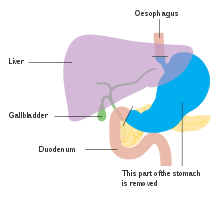
Anatomy before Roux-en-y surgery to resect stomach cancer.

Surgery remains the only curative therapy for stomach cancer. Of the different surgical techniques, endoscopic mucosal resection (EMR) is a treatment for early gastric cancer (tumor only involves the mucosa) that was pioneered in Japan and is available in the United States at some centers. In this procedure, the tumor, together with the inner lining of stomach (mucosa), is removed from the wall of the stomach using an electrical wire loop through the endoscope. The advantage is that it is a much smaller operation than removing the stomach. Endoscopic submucosal dissection (ESD) is a similar technique pioneered in Japan, used to resect a large area of mucosa in one piece. If the pathologic examination of the resected specimen shows incomplete resection or deep invasion by tumor, the patient would need a formal stomach resection. A 2016 Cochrane review found low quality evidence of no difference in short-term mortality between laparoscopic and open gastrectomy (removal of stomach), and that benefits or harms of laparoscopic gastrectomy cannot be ruled out. Post-operatively, up to 70% of people undergoing total gastrectomy develop complications such as dumping syndrome and reflux esophagitis. Construction of a "pouch", which serves as a "stomach substitute", reduced the incidence of dumping syndrome and reflux esophagitis by 73% and 63% respectively, and led to improvements in quality-of-life, nutritional outcomes, and body mass index.

Those with metastatic disease at the time of presentation may receive palliative surgery and while it remains controversial, due to the possibility of complications from the surgery itself and the fact that it may delay chemotherapy the data so far is mostly positive, with improved survival rates being seen in those treated with this approach.

Chemotherapy

The use of chemotherapy to treat stomach cancer has no firmly established standard of care. Unfortunately, stomach cancer has not been particularly sensitive to these drugs, and chemotherapy, if used, has usually served to palliatively reduce the size of the tumor, relieve symptoms of the disease and increase survival time. Some drugs used in stomach cancer treatment have included: 5-FU (fluorouracil) or its analog capecitabine, BCNU (carmustine), methyl-CCNU (semustine) and doxorubicin (Adriamycin), as well as mitomycin C, and more recently cisplatin and taxotere, often using drugs in various combinations. The relative benefits of these different drugs, alone and in combination, are unclear. Clinical researchers are exploring the benefits of giving chemotherapy before surgery to shrink the tumor, or as adjuvant therapy after surgery to destroy remaining cancer cells.

Targeted therapy

Recently, treatment with human epidermal growth factor receptor 2 (HER2) inhibitor, trastuzumab, has been demonstrated to increase overall survival in inoperable locally advanced or metastatic gastric carcinoma over-expressing the HER2/neu gene. In particular, HER2 is overexpressed in 13–22% of patients with gastric cancer. Of note, HER2 overexpression in gastric neoplasia is heterogeneous and comprises a minority of tumor cells (less than 10% of gastric cancers overexpress HER2 in more than 5% of tumor cells). Hence, this heterogeneous expression should be taken into account for HER2 testing, particularly in small samples such as biopsies, requiring the evaluation of more than one bioptic sample.

Radiation

Radiation therapy (also called radiotherapy) may be used to treat stomach cancer, often as an adjuvant to chemotherapy and/or surgery.

Lymphoma

Lymphoma of the MALT type can often be fully treated by treating an underlying H. pylori infection. This results in remission in about 80% of cases.

Prognosis

The prognosis of stomach cancer is generally poor, due to the fact the tumour has often metastasised by the time of discovery and the fact that most people with the condition are elderly (median age is between 70 and 75 years) at presentation. The average life expectancy after being diagnosed is around 24 months, and the five-year survival rate for stomach cancer is less than 10 percent.

Almost 300 genes are related to outcomes in stomach cancer with both unfavorable genes where high expression related to poor survival and favorable genes where high expression associated with longer survival times. Examples of poor prognosis genes include ITGAV and DUSP1.

Epidemiology

Stomach cancer deaths per million persons in 2012
  0–11
  12–16
  17–24
  25–33
  34–51
  52–76
  77–102
  103–128
  129–175
  176–400

Worldwide, stomach cancer is the fifth most-common cancer with 952,000 cases diagnosed in 2012. It is more common both in men and in developing countries. In 2012, it represented 8.5% of cancer cases in men, making it the fourth most-common cancer in men. Also in 2012, the number of deaths was 700,000 having decreased slightly from 774,000 in 1990, making it the third-leading cause of cancer-related death (after lung cancer and liver cancer).

Less than 5% of stomach cancers occur in people under 40 years of age with 81.1% of that 5% in the age-group of 30 to 39 and 18.9% in the age-group of 20 to 29.

In 2014, stomach cancer resulted in 0.61% of deaths (13,303 cases) in the U.S. In China, stomach cancer accounted for 3.56% of all deaths (324,439 cases). The highest rate of stomach cancer was in Mongolia, at 28 cases per 100,000 people.

In the United Kingdom, stomach cancer is the fifteenth most-common cancer (around 7,100 people were diagnosed with stomach cancer in 2011), and it is the tenth most-common cause of cancer-related deaths (around 4,800 people died in 2012).

Incidence and mortality rates of gastric cancer vary greatly in Africa. The GLOBOCAN system is currently the most widely used method to compare these rates between countries, but African incidence and mortality rates are seen to differ among countries, possibly due to the lack of universal access to a registry system for all countries. Variation as drastic as estimated rates from 0.3/100000 in Botswana to 20.3/100000 in Mali have been observed. In Uganda, the incidence of gastric cancer has increased from the 1960s measurement of 0.8/100000 to 5.6/100000. Gastric cancer, though present, is relatively low when compared to countries with high incidence like Japan and China. One suspected cause of the variation within Africa and between other countries is due to different strains of the Helicobacter pylori bacteria. The trend commonly-seen is that H. pylori infection increases the risk for gastric cancer. However, this is not the case in Africa, giving this phenomenon the name the “African enigma.” Although this bacteria is found in Africa, evidence has supported that different strains with mutations in the bacterial genotype may contribute to the difference in cancer development between African countries and others outside the continent. However, increasing access to health care and treatment measures have been commonly-associated with the rising incidence, particularly in Uganda.

Other animals

The stomach is a muscular organ of the gastrointestinal tract that holds food and begins the digestive process by secreting gastric juice. The most common cancers of the stomach are adenocarcinomas but other histological types have been reported. Signs vary but may include vomiting (especially if blood is present), weight loss, anemia, and lack of appetite. Bowel movements may be dark and tarry in nature. In order to determine whether cancer is present in the stomach, special X-rays and/or abdominal ultrasound may be performed. Gastroscopy, a test using an instrument called endoscope to examine the stomach, is a useful diagnostic tool that can also take samples of the suspected mass for histopathological analysis to confirm or rule out cancer. The most definitive method of cancer diagnosis is through open surgical biopsy. Most stomach tumors are malignant with evidence of spread to lymph nodes or liver, making treatment difficult. Except for lymphoma, surgery is the most frequent treatment option for stomach cancers but it is associated with significant risks.

Helicobacter pylori

From Wikipedia, the free encyclopedia
 
Helicobacter pylori
Other namesCampylobacter pylori
Immunohistochemical detection of Helicobacter (1) histopatholgy.jpg
Immunohistochemical staining of H. pylori (brown) from a gastric biopsy
Pronunciation
SpecialtyInfectious disease, gastroenterology
SymptomsNone, abdominal pain, nausea
ComplicationsStomach ulcer, stomach cancer
CausesHelicobacter pylori spread by fecal oral route
Diagnostic methodUrea breath test, fecal antigen assay, tissue biopsy
MedicationProton pump inhibitor, clarithromycin, amoxicillin, metronidazole
Frequency>50%

Helicobacter pylori, previously known as Campylobacter pylori, is a gram-negative, helically-shaped, microaerophilic bacterium usually found in the stomach. Its helical shape (from which the genus name, helicobacter, derives) is thought to have evolved in order to penetrate the mucoid lining of the stomach and thereby establish infection. The bacterium was first identified in 1982 by Australian doctors Barry Marshall and Robin Warren, who found that it was present in a person with chronic gastritis and gastric ulcers, conditions not previously believed to have a microbial cause. HP has been associated with the mucosa-associated lymphoid tissue in the stomach, esophagus, colon, rectum, or tissues around the eye (termed extranodal marginal zone B-cell lymphoma of the cited organ), and of lymphoid tissue in the stomach (termed diffuse large B-cell lymphoma).

Many investigators have proposed causal associations between H. pylori and a wide range of other diseases (e.g. idiopathic thrombocytopenic purpura, iron deficiency anemia, atherosclerosis, Alzheimer's disease, multiple sclerosis, coronary artery disease, periodontitis, Parkinson's disease, Guillain–Barré syndrome, rosacea, psoriasis, chronic urticaria, spot baldness, various autoimmune skin diseases, Henoch–Schönlein purpura, low blood levels of vitamin B12, autoimmune neutropenia, the antiphospholipid syndrome, plasma cell dyscrasias, central serous chorioretinitis, open angle glaucoma, blepharitis, diabetes mellitus, the metabolic syndrome, various types of allergies, non-alcoholic fatty liver disease, non-alcoholic steatohepatitis, hepatic fibrosis, and liver cancer). The bacterial infection has also been proposed to have protective effects for its hosts against infections by other pathogens, asthma, obesity, celiac disease, inflammatory bowel disease, rhinitis, atopic dermatitis, gastroesophageal reflux disease, and esophageal cancer. However, these deleterious and protective effects have frequently been based on correlative rather than direct relationship studies and have often been contradicted by other studies that show either the opposite or no effect on the cited disease. Consequently, many of these relationships are currently regarded as questionable and in need of more definitive studes. They are not considered further here.

Some studies suggest that H. pylori plays an important role in the natural stomach ecology, e.g. by influencing the type of bacteria that colonize the gastrointestinal tract. Other studies suggest that non-pathogenic strains of H. pylori may be beneficial, e.g., by normalizing stomach acid secretion, and may play a role in regulating appetite, since the bacterium's presence in the stomach results in a persistent but reversible reduction in the level of ghrelin, a hormone that increases appetite.

In general, over 50% of the world's population has H. pylori in their upper gastrointestinal tracts with this infection (or colonization) being more common in developing countries. In recent decades, however the prevalence of H. pylori colonization of the gastrointestinal tract has declined in many countries. This is attributed to improved socioeconomic conditions: in the United States of America, for example, the prevalence of H. pylori, as detected by endoscopy conducted on a referral population, fell from 65.8 to 6.8% over a recent 10-year period while over the same time period in some developing countries H. pylori colonization remained very common with prevalence levels as high as 80%. In all events, H. pylori infection is usually asymptomatic, being associated with overt disease (commonly gastritis or peptic ulcers rather than the relatively very rarely occurring cancers) in less than 20% of cases.

Signs and symptoms

Up to 90% of people infected with H. pylori never experience symptoms or complications. However, individuals infected with H. pylori have a 10 to 20% lifetime risk of developing peptic ulcers. Acute infection may appear as an acute gastritis with abdominal pain (stomach ache) or nausea. Where this develops into chronic gastritis, the symptoms, if present, are often those of non-ulcer dyspepsia: stomach pains, nausea, bloating, belching, and sometimes vomiting. Pain typically occurs when the stomach is empty, between meals, and in the early morning hours, but it can also occur at other times. Less common ulcer symptoms include nausea, vomiting, and loss of appetite. Bleeding in the stomach can also occur as evidenced by the passage of black stools; prolonged bleeding may cause anemia leading to weakness and fatigue. If bleeding is heavy, hematemesis, hematochezia, or melena may occur. Inflammation of the pyloric antrum, which connects the stomach to the duodenum, is more likely to lead to duodenal ulcers, while inflammation of the corpus (i.e. body of the stomach) is more likely to lead to gastric ulcers. Individuals infected with H. pylori may also develop colorectal or gastric polyps, i.e. a non-cancerous growth of tissue projecting from the mucous membranes of these organs. Usually, these polyps are asymptomatic but gastric polyps may be the cause of dyspepsia, heartburn, bleeding from the upper gastrointestinal tract, and, rarely, gastric outlet obstruction while colorectal polyps may be the cause of rectal bleeding, anemia, constipation, diarrhea, weight loss, and abdominal pain.

Individuals with chronic H. pylori infection have an increased risk of acquiring a cancer that is directly related to this infection. These cancers are stomach adenocarcinoma, less commonly diffuse large B-cell lymphoma of the stomach, or extranodal marginal zone B-cell lymphomas of the stomach, or, more rarely, of the colon, rectum, esophagus, or ocular adenexa (i.e. orbit, conjunctiva, and/or eyelids). The signs, symptoms, pathophysiology, and diagnoses of these cancers are given in the cited linkages.

Microbiology

Helicobacter pylori
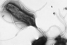
EMpylori.jpg
Scientific classification edit
Domain: Bacteria
Phylum: Proteobacteria
Class: Epsilonproteobacteria
Order: Campylobacterales
Family: Helicobacteraceae
Genus: Helicobacter
Species:
H. pylori
Binomial name
Helicobacter pylori
(Marshall et al. 1985) Goodwin et al., 1989

Morphology

Helicobacter pylori is a helix-shaped (classified as a curved rod, not spirochaete) Gram-negative bacterium about 3 μm long with a diameter of about 0.5μm. H. pylori can be demonstrated in tissue by Gram stain, Giemsa stain, haematoxylin–eosin stain, Warthin–Starry silver stain, acridine orange stain, and phase-contrast microscopy. It is capable of forming biofilms and can convert from spiral to a possibly viable but nonculturable coccoid form.

Helicobacter pylori has four to six flagella at the same location; all gastric and enterohepatic Helicobacter species are highly motile owing to flagella. The characteristic sheathed flagellar filaments of Helicobacter are composed of two copolymerized flagellins, FlaA and FlaB.

Physiology

Helicobacter pylori is microaerophilic—that is, it requires oxygen, but at lower concentration than in the atmosphere. It contains a hydrogenase that can produce energy by oxidizing molecular hydrogen (H2) made by intestinal bacteria. It produces oxidase, catalase, and urease.

H. pylori possesses five major outer membrane protein families. The largest family includes known and putative adhesins. The other four families are porins, iron transporters, flagellum-associated proteins, and proteins of unknown function. Like other typical Gram-negative bacteria, the outer membrane of H. pylori consists of phospholipids and lipopolysaccharide (LPS). The O antigen of LPS may be fucosylated and mimic Lewis blood group antigens found on the gastric epithelium. The outer membrane also contains cholesterol glucosides, which are present in few other bacteria.

Genome

Helicobacter pylori consists of a large diversity of strains, and hundreds of genomes have been completely sequenced. The genome of the strain "26695" consists of about 1.7 million base pairs, with some 1,576 genes. The pan-genome, that is a combined set of 30 sequenced strains, encodes 2,239 protein families (orthologous groups, OGs). Among them, 1248 OGs are conserved in all the 30 strains, and represent the universal core. The remaining 991 OGs correspond to the accessory genome in which 277 OGs are unique (i.e., OGs present in only one strain).

Transcriptome

In 2010, Sharma et al. presented a comprehensive analysis of transcription at single-nucleotide resolution by differential RNA-seq that confirmed the known acid induction of major virulence loci, such as the urease (ure) operon or the cag pathogenicity island (see below). More importantly, this study identified a total of 1,907 transcriptional start sites, 337 primary operons, and 126 additional suboperons, and 66 monocistrons. Until 2010, only about 55 transcriptional start sites (TSSs) were known in this species. Notably, 27% of the primary TSSs are also antisense TSSs, indicating that—similar to E. coliantisense transcription occurs across the entire H. pylori genome. At least one antisense TSS is associated with about 46% of all open reading frames, including many housekeeping genes. Most (about 50%) of the 5' UTRs are 20–40 nucleotides (nt) in length and support the AAGGag motif located about 6 nt (median distance) upstream of start codons as the consensus Shine–Dalgarno sequence in H. pylori.

Genes involved in virulence and pathogenesis

Study of the H. pylori genome is centered on attempts to understand pathogenesis, the ability of this organism to cause disease. About 29% of the loci have a colonization defect when mutated. Two of sequenced strains have an around 40-kb-long Cag pathogenicity island (a common gene sequence believed responsible for pathogenesis) that contains over 40 genes. This pathogenicity island is usually absent from H. pylori strains isolated from humans who are carriers of H. pylori, but remain asymptomatic.

The cagA gene codes for one of the major H. pylori virulence proteins. Bacterial strains with the cagA gene are associated with an ability to cause ulcers. The cagA gene codes for a relatively long (1186-amino acid) protein. The cag pathogenicity island (PAI) has about 30 genes, part of which code for a complex type IV secretion system. The low GC-content of the cag PAI relative to the rest of the Helicobacter genome suggests the island was acquired by horizontal transfer from another bacterial species. The serine protease HtrA also plays a major role in the pathogenesis of H. pylori. The HtrA protein enables the bacterium to transmigrate across the host cells' epithelium, and is also needed for the translocation of CagA. 

Pathophysiology

Adaptation to the stomach

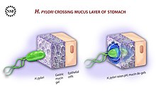
Diagram showing how H. pylori reaches the epithelium of the stomach
 
To avoid the acidic environment of the interior of the stomach (lumen), H. pylori uses its flagella to burrow into the mucus lining of the stomach to reach the epithelial cells underneath, where it is less acidic. H. pylori is able to sense the pH gradient in the mucus and move towards the less acidic region (chemotaxis). This also keeps the bacteria from being swept away into the lumen with the bacteria's mucus environment, which is constantly moving from its site of creation at the epithelium to its dissolution at the lumen interface.

H. pylori urease enzyme diagram

H. pylori is found in the mucus, on the inner surface of the epithelium, and occasionally inside the epithelial cells themselves. It adheres to the epithelial cells by producing adhesins, which bind to lipids and carbohydrates in the epithelial cell membrane. One such adhesin, BabA, binds to the Lewis b antigen displayed on the surface of stomach epithelial cells. H. pylori adherence via BabA is acid sensitive and can be fully reversed by decreased pH. It has been proposed that BabA's acid responsiveness enables adherence while also allowing an effective escape from unfavorable environment at pH that is harmful to the organism. Another such adhesin, SabA, binds to increased levels of sialyl-Lewis x antigen expressed on gastric mucosa.

In addition to using chemotaxis to avoid areas of low pH, H. pylori also neutralizes the acid in its environment by producing large amounts of urease, which breaks down the urea present in the stomach to carbon dioxide and ammonia. These react with the strong acids in the environment to produce a neutralized area around H. pylori. Urease knockout mutants are incapable of colonization. In fact, urease expression is not only required for establishing initial colonization but also for maintaining chronic infection.

Inflammation, gastritis and ulcer

Helicobacter pylori harms the stomach and duodenal linings by several mechanisms. The ammonia produced to regulate pH is toxic to epithelial cells, as are biochemicals produced by H. pylori such as proteases, vacuolating cytotoxin A (VacA) (this damages epithelial cells, disrupts tight junctions and causes apoptosis), and certain phospholipases. Cytotoxin associated gene CagA can also cause inflammation and is potentially a carcinogen.

Colonization of the stomach by H. pylori can result in chronic gastritis, an inflammation of the stomach lining, at the site of infection. Helicobacter cysteine-rich proteins (Hcp), particularly HcpA (hp0211), are known to trigger an immune response, causing inflammation. H. Pylori has been shown to increase the levels of COX2 in H. Pylori positive gastritis. Chronic gastritis is likely to underlie H. pylori-related diseases.

Ulcers in the stomach and duodenum result when the consequences of inflammation allow stomach acid and the digestive enzyme pepsin to overwhelm the mechanisms that protect the stomach and duodenal mucous membranes. The location of colonization of H. pylori, which affects the location of the ulcer, depends on the acidity of the stomach. In people producing large amounts of acid, H. pylori colonizes near the pyloric antrum (exit to the duodenum) to avoid the acid-secreting parietal cells at the fundus (near the entrance to the stomach). In people producing normal or reduced amounts of acid, H. pylori can also colonize the rest of the stomach.

The inflammatory response caused by bacteria colonizing near the pyloric antrum induces G cells in the antrum to secrete the hormone gastrin, which travels through the bloodstream to parietal cells in the fundus. Gastrin stimulates the parietal cells to secrete more acid into the stomach lumen, and over time increases the number of parietal cells, as well. The increased acid load damages the duodenum, which may eventually result in ulcers forming in the duodenum.

When H. pylori colonizes other areas of the stomach, the inflammatory response can result in atrophy of the stomach lining and eventually ulcers in the stomach. This also may increase the risk of stomach cancer.

Cag pathogenicity island

The pathogenicity of H. pylori may be increased by genes of the cag pathogenicity island; about 50–70% of H. pylori strains in Western countries carry it. Western people infected with strains carrying the cag PAI have a stronger inflammatory response in the stomach and are at a greater risk of developing peptic ulcers or stomach cancer than those infected with strains lacking the island. Following attachment of H. pylori to stomach epithelial cells, the type IV secretion system expressed by the cag PAI "injects" the inflammation-inducing agent, peptidoglycan, from their own cell walls into the epithelial cells. The injected peptidoglycan is recognized by the cytoplasmic pattern recognition receptor (immune sensor) Nod1, which then stimulates expression of cytokines that promote inflammation.

The type-IV secretion apparatus also injects the cag PAI-encoded protein CagA into the stomach's epithelial cells, where it disrupts the cytoskeleton, adherence to adjacent cells, intracellular signaling, cell polarity, and other cellular activities. Once inside the cell, the CagA protein is phosphorylated on tyrosine residues by a host cell membrane-associated tyrosine kinase (TK). CagA then allosterically activates protein tyrosine phosphatase/protooncogene Shp2. Pathogenic strains of H. pylori have been shown to activate the epidermal growth factor receptor (EGFR), a membrane protein with a TK domain. Activation of the EGFR by H. pylori is associated with altered signal transduction and gene expression in host epithelial cells that may contribute to pathogenesis. A C-terminal region of the CagA protein (amino acids 873–1002) has also been suggested to be able to regulate host cell gene transcription, independent of protein tyrosine phosphorylation. A great deal of diversity exists between strains of H. pylori, and the strain that infects a person can predict the outcome.

Cancer

Two related mechanisms by which H. pylori could promote cancer are under investigation. One mechanism involves the enhanced production of free radicals near H. pylori and an increased rate of host cell mutation. The other proposed mechanism has been called a "perigenetic pathway", and involves enhancement of the transformed host cell phenotype by means of alterations in cell proteins, such as adhesion proteins. H. pylori has been proposed to induce inflammation and locally high levels of TNF-α and/or interleukin 6 (IL-6). According to the proposed perigenetic mechanism, inflammation-associated signaling molecules, such as TNF-α, can alter gastric epithelial cell adhesion and lead to the dispersion and migration of mutated epithelial cells without the need for additional mutations in tumor suppressor genes, such as genes that code for cell adhesion proteins.

The strain of H. pylori a person is exposed to may influence the risk of developing gastric cancer. Strains of H. pylori that produce high levels of two proteins, vacuolating toxin A (VacA) and the cytotoxin-associated gene A (CagA), appear to cause greater tissue damage than those that produce lower levels or that lack those genes completely. These proteins are directly toxic to cells lining the stomach and signal strongly to the immune system that an invasion is under way. As a result of the bacterial presence, neutrophils and macrophages set up residence in the tissue to fight the bacteria assault.

Survival of Helicobacter pylori

The pathogenesis of H. pylori depends on its ability to survive in the harsh gastric environment characterized by acidity, peristalsis, and attack by phagocytes accompanied by release of reactive oxygen species. In particular, H. pylori elicits an oxidative stress response during host colonization. This oxidative stress response induces potentially lethal and mutagenic oxidative DNA adducts in the H. pylori genome.

Vulnerability to oxidative stress and oxidative DNA damage occurs commonly in many studied bacterial pathogens, including Neisseria gonorrhoeae, Hemophilus influenzae, Streptococcus pneumoniae, S. mutans, and H. pylori. For each of these pathogens, surviving the DNA damage induced by oxidative stress appears supported by transformation-mediated recombinational repair. Thus, transformation and recombinational repair appear to contribute to successful infection.

Transformation (the transfer of DNA from one bacterial cell to another through the intervening medium) appears to be part of an adaptation for DNA repair. H. pylori is naturally competent for transformation. While many organisms are competent only under certain environmental conditions, such as starvation, H. pylori is competent throughout logarithmic growth. All organisms encode genetic programs for response to stressful conditions including those that cause DNA damage. In H. pylori, homologous recombination is required for repairing DNA double-strand breaks (DSBs). The AddAB helicase-nuclease complex resects DSBs and loads RecA onto single-strand DNA (ssDNA), which then mediates strand exchange, leading to homologous recombination and repair. The requirement of RecA plus AddAB for efficient gastric colonization suggests, in the stomach, H. pylori is either exposed to double-strand DNA damage that must be repaired or requires some other recombination-mediated event. In particular, natural transformation is increased by DNA damage in H. pylori, and a connection exists between the DNA damage response and DNA uptake in H. pylori, suggesting natural competence contributes to persistence of H. pylori in its human host and explains the retention of competence in most clinical isolates.

RuvC protein is essential to the process of recombinational repair, since it resolves intermediates in this process termed Holliday junctions. H. pylori mutants that are defective in RuvC have increased sensitivity to DNA-damaging agents and to oxidative stress, exhibit reduced survival within macrophages, and are unable to establish successful infection in a mouse model. Similarly, RecN protein plays an important role in DSB repair in H. pylori. An H. pylori recN mutant displays an attenuated ability to colonize mouse stomachs, highlighting the importance of recombinational DNA repair in survival of H. pylori within its host.

Diagnosis

H. pylori colonized on the surface of regenerative epithelium (Warthin-Starry silver stain)

Colonization with H. pylori is not a disease in and of itself, but a condition associated with a number of disorders of the upper gastrointestinal tract. Testing for H. pylori is not routinely recommended. Testing is recommended if peptic ulcer disease or low-grade gastric MALT lymphoma is present, after endoscopic resection of early gastric cancer, for first-degree relatives with gastric cancer, and in certain cases of dyspepsia. Several methods of testing exist, including invasive and noninvasive testing methods.

Noninvasive tests for H. pylori infection may be suitable and include blood antibody tests, stool antigen tests, or the carbon urea breath test (in which the patient drinks 14C—or 13C-labelled urea, which the bacterium metabolizes, producing labelled carbon dioxide that can be detected in the breath). It is not known which non-invasive test is more accurate for diagnosing a H. pylori infection, and the clinical significance of the levels obtained with these tests are not clear. Some drugs can affect H. pylori urease activity and give false negatives with the urea-based tests.

An endoscopic biopsy is an invasive means to test for H. pylori infection. Low-level infections can be missed by biopsy, so multiple samples are recommended. The most accurate method for detecting H. pylori infection is with a histological examination from two sites after endoscopic biopsy, combined with either a rapid urease test or microbial culture.

Transmission

Helicobacter pylori is contagious, although the exact route of transmission is not known. Person-to-person transmission by either the oral–oral or fecal–oral route is most likely. Consistent with these transmission routes, the bacteria have been isolated from feces, saliva, and dental plaque of some infected people. Findings suggest H. pylori is more easily transmitted by gastric mucus than saliva. Transmission occurs mainly within families in developed nations, yet can also be acquired from the community in developing countries. H. pylori may also be transmitted orally by means of fecal matter through the ingestion of waste-tainted water, so a hygienic environment could help decrease the risk of H. pylori infection.

Prevention

Due to H. pylori's role as a major cause of certain diseases (particularly cancers) and its consistently increasing antibiotic resistance, there is a clear need for new therapeutic strategies to prevent or remove the bacterium from colonizing humans. Much work has been done on developing viable vaccines aimed at providing an alternative strategy to control H. pylori infection and related diseases. Researchers are studying different adjuvants, antigens, and routes of immunization to ascertain the most appropriate system of immune protection; however, most of the research only recently moved from animal to human trials. An economic evaluation of the use of a potential H. pylori vaccine in babies found its introduction could, at least in the Netherlands, prove cost-effective for the prevention of peptic ulcer and stomach adenocarcinoma. A similar approach has also been studied for the United States. Notwithstanding this proof-of-concept (i.e. vaccination protects children from acquisition of infection with H. pylori), as of late 2019 there have been no advanced vaccine candidates and only one vaccine in a Phase I clinical trial. Furthermore, development of a vaccine against H. pylori has not been a current priority of major pharmaceutical companies.

Many investigations have attempted to prevent the development of Helicobacter pylori-related diseases by eradicating the bacterium during an early stages of its infestation using antibiotic-based drug regimens. Studies find that such treatments, when effectively eradicating H. pylori from the stomach, reduce the inflammation and some of the histopathological abnormalities associated with the infestation. However studies disagree on the ability of these treatments to alleviate the more serious histopathological abnormalities in H. pylori infections, e.g. gastric atrophy and metaplasia, both of which are precursors to gastric adenocarcinoma. There is similar disagreement on the ability of antibiotic-based regiments to prevent gastric adenocarcinoma. A meta-analysis (i.e. a statistical analysis that combines the results of multiple randomized controlled trials) published in 2014 found that these regimens did not appear to prevent development of this adenocarcinoma. However, two subsequent prospective cohort studies conducted on high-risk individuals in China and Taiwan found that eradication of the bacterium produced a significant decrease in the number of individuals developing the disease. These results agreed with a retrospective cohort study done in Japan and published in 2016 as well as a meta-analysis, also published in 2016, of 24 studies conducted on individuals with varying levels of risk for developing the disease. These more recent studies suggest that the eradication of H. pylori infection reduces the incidence of H. pylori-related gastric adenocarcinoma in individuals at all levels of baseline risk. Further studies will be required to clarify this issue. In all events, studies agree that antibiotic-based regimens effectively reduce the occurrence of metachronous H. pylori-associated gastric adenocarcinoma. (Metachronus cancers are cancers that reoccur 6 months or later after resection of the original cancer.) It is suggested that antibiotic-based drug regimens be used after resecting H. pylori-associated gastric adenocarcinoma in order to reduce its metachronus reoccurrence.

Treatment

Gastritis

Superficial gastritis, either acute or chronic, is the most common manifestation of H. pylori infection. The signs and symptoms of this gastritis have been found to remit spontaneously in many individuals without resorting to Helicobacter pylori eradication protocols. The H. Pylori bacterial infection persists after remission in these cases. Various antibiotic plus proton pump inhibitor drug regimens are used to eradicate the bacterium and thereby successfully treat the disorder with triple-drug therapy consisting of clarithromycin, amoxicillin, and a proton-pump inhibitor given for 14–21 days often being considered first line treatment.

Peptic ulcers

Once H. pylori is detected in a person with a peptic ulcer, the normal procedure is to eradicate it and allow the ulcer to heal. The standard first-line therapy is a one-week "triple therapy" consisting of proton-pump inhibitors such as omeprazole and the antibiotics clarithromycin and amoxicillin. (The actions of proton pump inhibitors against H. pylori may reflect their direct bacteriostatic effect due to inhibition of the bacterium's P-type ATPase and/or urease.) Variations of the triple therapy have been developed over the years, such as using a different proton pump inhibitor, as with pantoprazole or rabeprazole, or replacing amoxicillin with metronidazole for people who are allergic to penicillin. In areas with higher rates of clarithromycin resistance, other options are recommended. Such a therapy has revolutionized the treatment of peptic ulcers and has made a cure to the disease possible. Previously, the only option was symptom control using antacids, H2-antagonists or proton pump inhibitors alone.

Antibiotic-resistant disease

An increasing number of infected individuals are found to harbor antibiotic-resistant bacteria. This results in initial treatment failure and requires additional rounds of antibiotic therapy or alternative strategies, such as a quadruple therapy, which adds a bismuth colloid, such as bismuth subsalicylate. For the treatment of clarithromycin-resistant strains of H. pylori, the use of levofloxacin as part of the therapy has been suggested.

Ingesting lactic acid bacteria exerts a suppressive effect on H. pylori infection in both animals and humans, and supplementing with Lactobacillus- and Bifidobacterium-containing yogurt improved the rates of eradication of H. pylori in humans. Symbiotic butyrate-producing bacteria which are normally present in the intestine are sometimes used as probiotics to help suppress H. pylori infections as an adjunct to antibiotic therapy. Butyrate itself is an antimicrobial which destroys the cell envelope of H. pylori by inducing regulatory T cell expression (specifically, FOXP3) and synthesis of an antimicrobial peptide called LL-37, which arises through its action as a histone deacetylase inhibitor.

The substance sulforaphane, which occurs in broccoli and cauliflower, has been proposed as a treatment. Periodontal therapy or scaling and root planing has also been suggested as an additional treatment.

Cancers

Extranodal marginal zone B-cell lymphomas

Extranodal marginal zone B-cell lymphomas are generally indolent malignancies. Recommended treatment of H. pylori-positive extranodal marginal zone B-cell lymphoma of the stomach, when localized (i.e. Ann Arbor stage I and II), employs one of the antibiotic-proton pump inhibitor regiments listed in the H. pylori eradication protocols. If the initial regimen fails to eradicate the pathogen, patients are treated with an alternate protocol. Eradication of the pathogen is successful in 70–95% of cases. Some 50-80% of patients who experience eradication of the pathogen develop within 3–28 months a remission and long-term clinical control of their lymphoma. Radiation therapy to the stomach and surrounding (i.e. peri-gastric) lymph nodes has also been used to successfully treat these localized cases. Patients with non-localized (i.e. systemic Ann Arbor stage III and IV) disease who are free of symptoms have been treated with watchful waiting or, if symptomatic, with the immunotherapy drug, rituximab, (given for 4 weeks) combined with the chemotherapy drug, chlorambucil, for 6–12 months; 58% of these patients attain a 58% progression-free survival rate at 5 years. Frail stage III/IV patients have been successfully treated with rituximab or the chemotherapy drug, cyclophosphamide, alone. Only rare cases of H. pylori-positive extranodal marginal zone B-cell lymphoma of the colon have been successfully treated with an antibiotic-proton pump inhibitor regimen; the currently recommended treatments for this disease are surgical resection, endoscopic resection, radiation, chemotherapy, or, more recently, rituximab. In the few reported cases of H. pylori-positive extranodal marginal zone B-cell lymphoma of the esophagus, localized disease has been successfully treated with antibiotic-proton pump inhibitor regimens; however, advanced disease appears less responsive or unresponsive to these regimens but partially responsive to rituximab. Antibiotic-proton pump inhibitor eradication therapy and localized radiation therapy have been used successfully to treat H.pylori-positive extranodal marginal zone B-cell lymphomas of the rectum; however radiation therapy has given slightly better results and therefore been suggested to be the disease' preferred treatment. The treatment of localized H. pylori-positive extranodal marginal zone B-cell lymphoma of the ocular adenexa with antibiotic/proton pump inhibitor regimens has achieved 2 year and 5 year failure-free survival rates of 67% and 55%, respectively, and a 5-year progression-free rate of 61%. However, the generally recognized treatment of choice for patients with systemic involvement uses various chemotherapy drugs often combined with rituximab.

Diffuse large B-cell lymphoma

Diffuse large B-cell lymphoma is a far more aggressive cancer than extranodal marginal zone B-cell lymphoma. Cases of this malignancy that are H. pylori-positive may be derived from the latter lymphoma and are less aggressive as well as more susceptible to treatment than H. pylori negative cases. Several recent studies strongly suggest that localized, early-stage diffuse Helicobacter pylori positive diffuse large B-cell lymphoma, when limited to the stomach, can be successfully treated with antibiotic-proton pump inhibitor regimens. However, these studies also agree that, given the aggressiveness of diffuse large B-cell lymphoma, patients treated with one of these H. pylori eradication regimes need to be carefully followed. If found unresponsive to or clinically worsening on these regimens, these patients should be switched to more conventional therapy such as chemotherapy (e.g. CHOP or a CHOP-like regimen), immunotherapy (e.g. rituximab), surgery, and/or local radiotherapy. H. pylori positive diffuse large B-cell lymphoma has been successfully treated with one or a combination of these methods.

Stomach adenocarcinoma

Helicobacter pylori is linked to the majority of gastric adenocarcinoma cases, particularly those that are located outside of the stomach's cardia (i.e. esophagus-stomach junction). The treatment for this cancer is highly aggressive with even localized disease being treated sequentially with chemotherapy and radiotherapy before surgical resection. Since this cancer, once developed, is independent of H. pylori infection, antibiotic-proton pump inhibitor regimens are not used in its treatment.

Prognosis

Helicobacter pylori colonizes the stomach and induces chronic gastritis, a long-lasting inflammation of the stomach. The bacterium persists in the stomach for decades in most people. Most individuals infected by H. pylori never experience clinical symptoms, despite having chronic gastritis. About 10–20% of those colonized by H. pylori ultimately develop gastric and duodenal ulcers. H. pylori infection is also associated with a 1–2% lifetime risk of stomach cancer and a less than 1% risk of gastric MALT lymphoma.

In the absence of treatment, H. pylori infection—once established in its gastric niche—is widely believed to persist for life. In the elderly, however, infection likely can disappear as the stomach's mucosa becomes increasingly atrophic and inhospitable to colonization. The proportion of acute infections that persist is not known, but several studies that followed the natural history in populations have reported apparent spontaneous elimination.

Mounting evidence suggests H. pylori has an important role in protection from some diseases. The incidence of acid reflux disease, Barrett's esophagus, and esophageal cancer have been rising dramatically at the same time as H. pylori's presence decreases. In 1996, Martin J. Blaser advanced the hypothesis that H. pylori has a beneficial effect by regulating the acidity of the stomach contents. The hypothesis is not universally accepted as several randomized controlled trials failed to demonstrate worsening of acid reflux disease symptoms following eradication of H. pylori. Nevertheless, Blaser has reasserted his view that H. pylori is a member of the normal flora of the stomach. He postulates that the changes in gastric physiology caused by the loss of H. pylori account for the recent increase in incidence of several diseases, including type 2 diabetes, obesity, and asthma. His group has recently shown that H. pylori colonization is associated with a lower incidence of childhood asthma.

Epidemiology

At least half the world's population is infected by the bacterium, making it the most widespread infection in the world. Actual infection rates vary from nation to nation; the developing world has much higher infection rates than the West (Western Europe, North America, Australasia), where rates are estimated to be around 25%.

The age when someone acquires this bacterium seems to influence the pathologic outcome of the infection. People infected at an early age are likely to develop more intense inflammation that may be followed by atrophic gastritis with a higher subsequent risk of gastric ulcer, gastric cancer, or both. Acquisition at an older age brings different gastric changes more likely to lead to duodenal ulcer. Infections are usually acquired in early childhood in all countries. However, the infection rate of children in developing nations is higher than in industrialized nations, probably due to poor sanitary conditions, perhaps combined with lower antibiotics usage for unrelated pathologies. In developed nations, it is currently uncommon to find infected children, but the percentage of infected people increases with age, with about 50% infected for those over the age of 60 compared with around 10% between 18 and 30 years. The higher prevalence among the elderly reflects higher infection rates in the past when the individuals were children rather than more recent infection at a later age of the individual. In the United States, prevalence appears higher in African-American and Hispanic populations, most likely due to socioeconomic factors. The lower rate of infection in the West is largely attributed to higher hygiene standards and widespread use of antibiotics. Despite high rates of infection in certain areas of the world, the overall frequency of H. pylori infection is declining. However, antibiotic resistance is appearing in H. pylori; many metronidazole- and clarithromycin-resistant strains are found in most parts of the world.

History

Helicobacter pylori migrated out of Africa along with its human host circa 60,000 years ago. Recent research states that genetic diversity in H. pylori, like that of its host, decreases with geographic distance from East Africa. Using the genetic diversity data, researchers have created simulations that indicate the bacteria seem to have spread from East Africa around 58,000 years ago. Their results indicate modern humans were already infected by H. pylori before their migrations out of Africa, and it has remained associated with human hosts since that time.

H. pylori was first discovered in the stomachs of patients with gastritis and ulcers in 1982 by Drs. Barry Marshall and Robin Warren of Perth, Western Australia. At the time, the conventional thinking was that no bacterium could live in the acid environment of the human stomach. In recognition of their discovery, Marshall and Warren were awarded the 2005 Nobel Prize in Physiology or Medicine.
Before the research of Marshall and Warren, German scientists found spiral-shaped bacteria in the lining of the human stomach in 1875, but they were unable to culture them, and the results were eventually forgotten. The Italian researcher Giulio Bizzozero described similarly shaped bacteria living in the acidic environment of the stomach of dogs in 1893. Professor Walery Jaworski of the Jagiellonian University in Kraków investigated sediments of gastric washings obtained by lavage from humans in 1899. Among some rod-like bacteria, he also found bacteria with a characteristic spiral shape, which he called Vibrio rugula. He was the first to suggest a possible role of this organism in the pathogenesis of gastric diseases. His work was included in the Handbook of Gastric Diseases, but it had little impact, as it was written in Polish. Several small studies conducted in the early 20th century demonstrated the presence of curved rods in the stomachs of many people with peptic ulcers and stomach cancers. Interest in the bacteria waned, however, when an American study published in 1954 failed to observe the bacteria in 1180 stomach biopsies.

Interest in understanding the role of bacteria in stomach diseases was rekindled in the 1970s, with the visualization of bacteria in the stomachs of people with gastric ulcers. The bacteria had also been observed in 1979, by Robin Warren, who researched it further with Barry Marshall from 1981. After unsuccessful attempts at culturing the bacteria from the stomach, they finally succeeded in visualizing colonies in 1982, when they unintentionally left their Petri dishes incubating for five days over the Easter weekend. In their original paper, Warren and Marshall contended that most stomach ulcers and gastritis were caused by bacterial infection and not by stress or spicy food, as had been assumed before.

Some skepticism was expressed initially, but within a few years multiple research groups had verified the association of H. pylori with gastritis and, to a lesser extent, ulcers. To demonstrate H. pylori caused gastritis and was not merely a bystander, Marshall drank a beaker of H. pylori culture. He became ill with nausea and vomiting several days later. An endoscopy 10 days after inoculation revealed signs of gastritis and the presence of H. pylori. These results suggested H. pylori was the causative agent. Marshall and Warren went on to demonstrate antibiotics are effective in the treatment of many cases of gastritis. In 1987, the Sydney gastroenterologist Thomas Borody invented the first triple therapy for the treatment of duodenal ulcers. In 1994, the National Institutes of Health stated most recurrent duodenal and gastric ulcers were caused by H. pylori, and recommended antibiotics be included in the treatment regimen.

The bacterium was initially named Campylobacter pyloridis, then renamed C. pylori in 1987 (pylori being the genitive of pylorus, the circular opening leading from the stomach into the duodenum, from the Ancient Greek word πυλωρός, which means gatekeeper.). When 16S ribosomal RNA gene sequencing and other research showed in 1989 that the bacterium did not belong in the genus Campylobacter, it was placed in its own genus, Helicobacter from the ancient Greek hělix/έλιξ "spiral" or "coil".

In October 1987, a group of experts met in Copenhagen to found the European Helicobacter Study Group (EHSG), an international multidisciplinary research group and the only institution focused on H. pylori. The Group is involved with the Annual International Workshop on Helicobacter and Related Bacteria, the Maastricht Consensus Reports (European Consensus on the management of H. pylori), and other educational and research projects, including two international long-term projects:
  • European Registry on H. pylori Management (Hp-EuReg) – a database systematically registering the routine clinical practice of European gastroenterologists.
  • Optimal H. pylori management in primary care (OptiCare) – a long-term educational project aiming to disseminate the evidence based recommendations of the Maastricht IV Consensus to primary care physicians in Europe, funded by an educational grant from United European Gastroenterology.

Research

Results from in vitro studies suggest that fatty acids, mainly polyunsaturated fatty acids, have a bactericidal effect against H. pylori, but their in vivo effects have not been proven.

Thailand

From Wikipedia, the free encyclopedia https://en.wikipedia.org/wiki/Thailand Thailand , officially the K...