Search This Blog

Wednesday, July 24, 2019

Cytosol

From Wikipedia, the free encyclopedia
 
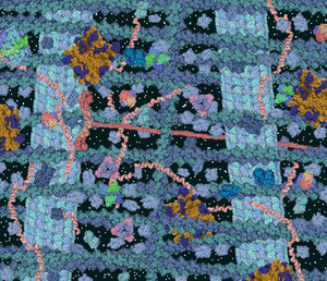
The cytosol is a crowded solution of many different types of molecules that fills much of the volume of cells.
 
Cell biology
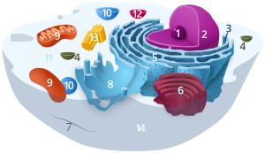
The animal cell
Animal Cell.svg

The cytosol, also known as intracellular fluid (ICF) or cytoplasmic matrix, or groundplasm, is the liquid found inside cells. It is separated into compartments by membranes. For example, the mitochondrial matrix separates the mitochondrion into many compartments. 

In the eukaryotic cell, the cytosol is surrounded by the cell membrane and is part of the cytoplasm, which also comprises the mitochondria, plastids, and other organelles (but not their internal fluids and structures); the cell nucleus is separate. The cytosol is thus a liquid matrix around the organelles. In prokaryotes, most of the chemical reactions of metabolism take place in the cytosol, while a few take place in membranes or in the periplasmic space. In eukaryotes, while many metabolic pathways still occur in the cytosol, others take place within organelles.

The cytosol is a complex mixture of substances dissolved in water. Although water forms the large majority of the cytosol, its structure and properties within cells is not well understood. The concentrations of ions such as sodium and potassium are different in the cytosol than in the extracellular fluid; these differences in ion levels are important in processes such as osmoregulation, cell signaling, and the generation of action potentials in excitable cells such as endocrine, nerve and muscle cells. The cytosol also contains large amounts of macromolecules, which can alter how molecules behave, through macromolecular crowding.

Although it was once thought to be a simple solution of molecules, the cytosol has multiple levels of organization. These include concentration gradients of small molecules such as calcium, large complexes of enzymes that act together and take part in metabolic pathways, and protein complexes such as proteasomes and carboxysomes that enclose and separate parts of the cytosol.

Definition

The term "cytosol" was first introduced in 1965 by H. A. Lardy, and initially referred to the liquid that was produced by breaking cells apart and pelleting all the insoluble components by ultracentrifugation. Such a soluble cell extract is not identical to the soluble part of the cell cytoplasm and is usually called a cytoplasmic fraction.

The term cytosol is now used to refer to the liquid phase of the cytoplasm in an intact cell. This excludes any part of the cytoplasm that is contained within organelles. Due to the possibility of confusion between the use of the word "cytosol" to refer to both extracts of cells and the soluble part of the cytoplasm in intact cells, the phrase "aqueous cytoplasm" has been used to describe the liquid contents of the cytoplasm of living cells.

Prior to this, other terms, including hyaloplasm, were used for the cell fluid, not always synonymously, as its nature was not very clear.

Properties and composition

Intracellular fluid content in humans
 
The proportion of cell volume that is cytosol varies: for example while this compartment forms the bulk of cell structure in bacteria, in plant cells the main compartment is the large central vacuole. The cytosol consists mostly of water, dissolved ions, small molecules, and large water-soluble molecules (such as proteins). The majority of these non-protein molecules have a molecular mass of less than 300 Da. This mixture of small molecules is extraordinarily complex, as the variety of molecules that are involved in metabolism (the metabolites) is immense. For example, up to 200,000 different small molecules might be made in plants, although not all these will be present in the same species, or in a single cell. Estimates of the number of metabolites in single cells such as E. coli and baker's yeast predict that under 1,000 are made.

Water

Most of the cytosol is water, which makes up about 70% of the total volume of a typical cell. The pH of the intracellular fluid is 7.4. while human cytosolic pH ranges between 7.0 - 7.4, and is usually higher if a cell is growing. The viscosity of cytoplasm is roughly the same as pure water, although diffusion of small molecules through this liquid is about fourfold slower than in pure water, due mostly to collisions with the large numbers of macromolecules in the cytosol. Studies in the brine shrimp have examined how water affects cell functions; these saw that a 20% reduction in the amount of water in a cell inhibits metabolism, with metabolism decreasing progressively as the cell dries out and all metabolic activity halting when the water level reaches 70% below normal.

Although water is vital for life, the structure of this water in the cytosol is not well understood, mostly because methods such as nuclear magnetic resonance spectroscopy only give information on the average structure of water, and cannot measure local variations at the microscopic scale. Even the structure of pure water is poorly understood, due to the ability of water to form structures such as water clusters through hydrogen bonds.

The classic view of water in cells is that about 5% of this water is strongly bound in by solutes or macromolecules as water of solvation, while the majority has the same structure as pure water. This water of solvation is not active in osmosis and may have different solvent properties, so that some dissolved molecules are excluded, while others become concentrated. However, others argue that the effects of the high concentrations of macromolecules in cells extend throughout the cytosol and that water in cells behaves very differently from the water in dilute solutions. These ideas include the proposal that cells contain zones of low and high-density water, which could have widespread effects on the structures and functions of the other parts of the cell. However, the use of advanced nuclear magnetic resonance methods to directly measure the mobility of water in living cells contradicts this idea, as it suggests that 85% of cell water acts like that pure water, while the remainder is less mobile and probably bound to macromolecules.

Ions

The concentrations of the other ions in cytosol are quite different from those in extracellular fluid and the cytosol also contains much higher amounts of charged macromolecules such as proteins and nucleic acids than the outside of the cell structure. 

Typical ion concentrations in mammalian cytosol and blood.
Ion  Concentration in cytosol (millimolar  Concentration in blood (millimolar
 Potassium   139   4 
 Sodium   12   145 
 Chloride   4   116 
 Bicarbonate   12   29 
 Amino acids in proteins   138   9 
 Magnesium   0.8   1.5 
 Calcium   <0 .0002="" nbsp="" span="">  1.8 

In contrast to extracellular fluid, cytosol has a high concentration of potassium ions and a low concentration of sodium ions. This difference in ion concentrations is critical for osmoregulation, since if the ion levels were the same inside a cell as outside, water would enter constantly by osmosis - since the levels of macromolecules inside cells are higher than their levels outside. Instead, sodium ions are expelled and potassium ions taken up by the Na⁺/K⁺-ATPase, potassium ions then flow down their concentration gradient through potassium-selection ion channels, this loss of positive charge creates a negative membrane potential. To balance this potential difference, negative chloride ions also exit the cell, through selective chloride channels. The loss of sodium and chloride ions compensates for the osmotic effect of the higher concentration of organic molecules inside the cell.

Cells can deal with even larger osmotic changes by accumulating osmoprotectants such as betaines or trehalose in their cytosol. Some of these molecules can allow cells to survive being completely dried out and allow an organism to enter a state of suspended animation called cryptobiosis. In this state the cytosol and osmoprotectants become a glass-like solid that helps stabilize proteins and cell membranes from the damaging effects of desiccation.

The low concentration of calcium in the cytosol allows calcium ions to function as a second messenger in calcium signaling. Here, a signal such as a hormone or an action potential opens calcium channels so that calcium floods into the cytosol. This sudden increase in cytosolic calcium activates other signalling molecules, such as calmodulin and protein kinase C. Other ions such as chloride and potassium may also have signaling functions in the cytosol, but these are not well understood.

Macromolecules

Protein molecules that do not bind to cell membranes or the cytoskeleton are dissolved in the cytosol. The amount of protein in cells is extremely high, and approaches 200 mg/ml, occupying about 20-30% of the volume of the cytosol. However, measuring precisely how much protein is dissolved in cytosol in intact cells is difficult, since some proteins appear to be weakly associated with membranes or organelles in whole cells and are released into solution upon cell lysis. Indeed, in experiments where the plasma membrane of cells were carefully disrupted using saponin, without damaging the other cell membranes, only about one quarter of cell protein was released. These cells were also able to synthesize proteins if given ATP and amino acids, implying that many of the enzymes in cytosol are bound to the cytoskeleton. However, the idea that the majority of the proteins in cells are tightly bound in a network called the microtrabecular lattice is now seen as unlikely.

In prokaryotes the cytosol contains the cell's genome, within a structure known as a nucleoid. This is an irregular mass of DNA and associated proteins that control the transcription and replication of the bacterial chromosome and plasmids. In eukaryotes the genome is held within the cell nucleus, which is separated from the cytosol by nuclear pores that block the free diffusion of any molecule larger than about 10 nanometres in diameter.

This high concentration of macromolecules in cytosol causes an effect called macromolecular crowding, which is when the effective concentration of other macromolecules is increased, since they have less volume to move in. This crowding effect can produce large changes in both the rates and the position of chemical equilibrium of reactions in the cytosol. It is particularly important in its ability to alter dissociation constants by favoring the association of macromolecules, such as when multiple proteins come together to form protein complexes, or when DNA-binding proteins bind to their targets in the genome.

Organization

Although the components of the cytosol are not separated into regions by cell membranes, these components do not always mix randomly and several levels of organization can localize specific molecules to defined sites within the cytosol.

Concentration gradients

Although small molecules diffuse rapidly in the cytosol, concentration gradients can still be produced within this compartment. A well-studied example of these are the "calcium sparks" that are produced for a short period in the region around an open calcium channel. These are about 2 micrometres in diameter and last for only a few milliseconds, although several sparks can merge to form larger gradients, called "calcium waves". Concentration gradients of other small molecules, such as oxygen and adenosine triphosphate may be produced in cells around clusters of mitochondria, although these are less well understood.

Protein complexes

Proteins can associate to form protein complexes, these often contain a set of proteins with similar functions, such as enzymes that carry out several steps in the same metabolic pathway. This organization can allow substrate channeling, which is when the product of one enzyme is passed directly to the next enzyme in a pathway without being released into solution. Channeling can make a pathway more rapid and efficient than it would be if the enzymes were randomly distributed in the cytosol, and can also prevent the release of unstable reaction intermediates. Although a wide variety of metabolic pathways involve enzymes that are tightly bound to each other, others may involve more loosely associated complexes that are very difficult to study outside the cell. Consequently, the importance of these complexes for metabolism in general remains unclear. 

Carboxysomes are protein-enclosed bacterial microcompartments within the cytosol. On the left is an electron microscope image of carboxysomes, and on the right a model of their structure.

Protein compartments

Some protein complexes contain a large central cavity that is isolated from the remainder of the cytosol. One example of such an enclosed compartment is the proteasome. Here, a set of subunits form a hollow barrel containing proteases that degrade cytosolic proteins. Since these would be damaging if they mixed freely with the remainder of the cytosol, the barrel is capped by a set of regulatory proteins that recognize proteins with a signal directing them for degradation (a ubiquitin tag) and feed them into the proteolytic cavity.

Another large class of protein compartments are bacterial microcompartments, which are made of a protein shell that encapsulates various enzymes. These compartments are typically about 100-200 nanometres across and made of interlocking proteins. A well-understood example is the carboxysome, which contains enzymes involved in carbon fixation such as RuBisCO.

Biomolecular condensates

Non-membrane bound organelles can form as biomolecular condensates, which arise by clustering, oligomerisation, or polymerisation of macromolecules to drive colloidal phase separation of the cytoplasm or nucleus.

Cytoskeletal sieving

Although the cytoskeleton is not part of the cytosol, the presence of this network of filaments restricts the diffusion of large particles in the cell. For example, in several studies tracer particles larger than about 25 nanometres (about the size of a ribosome) were excluded from parts of the cytosol around the edges of the cell and next to the nucleus. These "excluding compartments" may contain a much denser meshwork of actin fibres than the remainder of the cytosol. These microdomains could influence the distribution of large structures such as ribosomes and organelles within the cytosol by excluding them from some areas and concentrating them in others.

Function

The cytosol has no single function and is instead the site of multiple cell processes. Examples of these processes include signal transduction from the cell membrane to sites within the cell, such as the cell nucleus, or organelles. This compartment is also the site of many of the processes of cytokinesis, after the breakdown of the nuclear membrane in mitosis. Another major function of cytosol is to transport metabolites from their site of production to where they are used. This is relatively simple for water-soluble molecules, such as amino acids, which can diffuse rapidly through the cytosol. However, hydrophobic molecules, such as fatty acids or sterols, can be transported through the cytosol by specific binding proteins, which shuttle these molecules between cell membranes. Molecules taken into the cell by endocytosis or on their way to be secreted can also be transported through the cytosol inside vesicles, which are small spheres of lipids that are moved along the cytoskeleton by motor proteins.

The cytosol is the site of most metabolism in prokaryotes, and a large proportion of the metabolism of eukaryotes. For instance, in mammals about half of the proteins in the cell are localized to the cytosol. The most complete data are available in yeast, where metabolic reconstructions indicate that the majority of both metabolic processes and metabolites occur in the cytosol. Major metabolic pathways that occur in the cytosol in animals are protein biosynthesis, the pentose phosphate pathway, glycolysis and gluconeogenesis. The localization of pathways can be different in other organisms, for instance fatty acid synthesis occurs in chloroplasts in plants and in apicoplasts in apicomplexa.
Glycogen

From Wikipedia, the free encyclopedia
 
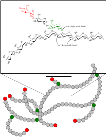
Schematic two-dimensional cross-sectional view of glycogen: A core protein of glycogenin is surrounded by branches of glucose units. The entire globular granule may contain around 30,000 glucose units.
 
A view of the atomic structure of a single branched strand of glucose units in a glycogen molecule.
 
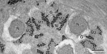
Glycogen (black granules) in spermatozoa of a flatworm; transmission electron microscopy, scale: 0.3 µm
 
Glycogen is a multibranched polysaccharide of glucose that serves as a form of energy storage in animals, fungi, and bacteria. The polysaccharide structure represents the main storage form of glucose in the body. 

Glycogen functions as one of two forms of long-term energy reserves, with the other form being triglyceride stores in adipose tissue (i.e., body fat). In humans, glycogen is made and stored primarily in the cells of the liver and skeletal muscle. In the liver, glycogen can make up 5–6% of the organ's fresh weight, and the liver of an adult weighing 70 kg can store roughly 100–120 grams of glycogen. In skeletal muscle, glycogen is found in a low concentration (1–2% of the muscle mass) and the skeletal muscle of an adult weighing 70 kg stores roughly 400 grams of glycogen. The amount of glycogen stored in the body—particularly within the muscles and liver—mostly depends on physical training, basal metabolic rate, and eating habits. Small amounts of glycogen are also found in other tissues and cells, including the kidneys, red blood cells, white blood cells, and glial cells in the brain. The uterus also stores glycogen during pregnancy to nourish the embryo.

Approximately 4 grams of glucose are present in the blood of humans at all times; in fasted individuals, blood glucose is maintained constant at this level at the expense of glycogen stores in the liver and skeletal muscle. Glycogen stores in skeletal muscle serve as a form of energy storage for the muscle itself; however, the breakdown of muscle glycogen impedes muscle glucose uptake, thereby increasing the amount of blood glucose available for use in other tissues. Liver glycogen stores serve as a store of glucose for use throughout the body, particularly the central nervous system. The human brain consumes approximately 60% of blood glucose in fasted, sedentary individuals.

Glycogen is the analogue of starch, a glucose polymer that functions as energy storage in plants. It has a structure similar to amylopectin (a component of starch), but is more extensively branched and compact than starch. Both are white powders in their dry state. Glycogen is found in the form of granules in the cytosol/cytoplasm in many cell types, and plays an important role in the glucose cycle. Glycogen forms an energy reserve that can be quickly mobilized to meet a sudden need for glucose, but one that is less compact than the energy reserves of triglycerides (lipids). As such it is also found as storage reserve in many parasitic protozoa.

Structure

1,4-α-glycosidic linkages in the glycogen oligomer
 
1,4-α-glycosidic and 1,6-glycosidic linkages in the glycogen oligomer
 
Glycogen is a branched biopolymer consisting of linear chains of glucose residues with an average chain length of approximately 8–12 glucose units. Glucose units are linked together linearly by α(1→4) glycosidic bonds from one glucose to the next. Branches are linked to the chains from which they are branching off by α(1→6) glycosidic bonds between the first glucose of the new branch and a glucose on the stem chain.

Due to the way glycogen is synthesised, every glycogen granule has at its core a glycogenin protein.

Glycogen in muscle, liver, and fat cells is stored in a hydrated form, composed of three or four parts of water per part of glycogen associated with 0.45 millimoles (18 mg) of potassium per gram of glycogen.

Glucose is an osmotic molecule, and can have profound effects on osmotic pressure in high concentrations possibly leading to cell damage or death if stored in the cell without being modified.[3] Glycogen is a non-osmotic molecule, so it can be used as a solution to storing glucose in the cell without disrupting osmotic pressure.

Functions

Liver

As a meal containing carbohydrates or protein is eaten and digested, blood glucose levels rise, and the pancreas secretes insulin. Blood glucose from the portal vein enters liver cells (hepatocytes). Insulin acts on the hepatocytes to stimulate the action of several enzymes, including glycogen synthase. Glucose molecules are added to the chains of glycogen as long as both insulin and glucose remain plentiful. In this postprandial or "fed" state, the liver takes in more glucose from the blood than it releases. 

After a meal has been digested and glucose levels begin to fall, insulin secretion is reduced, and glycogen synthesis stops. When it is needed for energy, glycogen is broken down and converted again to glucose. Glycogen phosphorylase is the primary enzyme of glycogen breakdown. For the next 8–12 hours, glucose derived from liver glycogen is the primary source of blood glucose used by the rest of the body for fuel.
Glucagon, another hormone produced by the pancreas, in many respects serves as a countersignal to insulin. In response to insulin levels being below normal (when blood levels of glucose begin to fall below the normal range), glucagon is secreted in increasing amounts and stimulates both glycogenolysis (the breakdown of glycogen) and gluconeogenesis (the production of glucose from other sources).

Muscle

Muscle cell glycogen appears to function as an immediate reserve source of available glucose for muscle cells. Other cells that contain small amounts use it locally, as well. As muscle cells lack glucose-6-phosphatase, which is required to pass glucose into the blood, the glycogen they store is available solely for internal use and is not shared with other cells. This is in contrast to liver cells, which, on demand, readily do break down their stored glycogen into glucose and send it through the blood stream as fuel for other organs.

History

Glycogen was discovered by Claude Bernard. His experiments showed that the liver contained a substance that could give rise to reducing sugar by the action of a "ferment" in the liver. By 1857, he described the isolation of a substance he called "la matière glycogène", or "sugar-forming substance". Soon after the discovery of glycogen in the liver, A. Sanson found that muscular tissue also contains glycogen. The empirical formula for glycogen of (C
6
H
10
O
5
)n was established by Kekulé in 1858.

Metabolism

Synthesis

Glycogen synthesis is, unlike its breakdown, endergonic—it requires the input of energy. Energy for glycogen synthesis comes from uridine triphosphate (UTP), which reacts with glucose-1-phosphate, forming UDP-glucose, in a reaction catalysed by UTP—glucose-1-phosphate uridylyltransferase. Glycogen is synthesized from monomers of UDP-glucose initially by the protein glycogenin, which has two tyrosine anchors for the reducing end of glycogen, since glycogenin is a homodimer. After about eight glucose molecules have been added to a tyrosine residue, the enzyme glycogen synthase progressively lengthens the glycogen chain using UDP-glucose, adding α(1→4)-bonded glucose to the reducing end of the glycogen chain.

The glycogen branching enzyme catalyzes the transfer of a terminal fragment of six or seven glucose residues from a nonreducing end to the C-6 hydroxyl group of a glucose residue deeper into the interior of the glycogen molecule. The branching enzyme can act upon only a branch having at least 11 residues, and the enzyme may transfer to the same glucose chain or adjacent glucose chains.

Breakdown

Glycogen is cleaved from the nonreducing ends of the chain by the enzyme glycogen phosphorylase to produce monomers of glucose-1-phosphate: 

Action of Glycogen Phosphorylase on Glycogen

In vivo, phosphorolysis proceeds in the direction of glycogen breakdown because the ratio of phosphate and glucose-1-phosphate is usually greater than 100. Glucose-1-phosphate is then converted to glucose 6-phosphate (G6P) by phosphoglucomutase. A special debranching enzyme is needed to remove the α(1-6) branches in branched glycogen and reshape the chain into a linear polymer. The G6P monomers produced have three possible fates:

Clinical relevance

Disorders of glycogen metabolism

The most common disease in which glycogen metabolism becomes abnormal is diabetes, in which, because of abnormal amounts of insulin, liver glycogen can be abnormally accumulated or depleted. Restoration of normal glucose metabolism usually normalizes glycogen metabolism, as well. 

In hypoglycemia caused by excessive insulin, liver glycogen levels are high, but the high insulin levels prevent the glycogenolysis necessary to maintain normal blood sugar levels. Glucagon is a common treatment for this type of hypoglycemia. 

Various inborn errors of metabolism are caused by deficiencies of enzymes necessary for glycogen synthesis or breakdown. These are collectively referred to as glycogen storage diseases.

Glycogen depletion and endurance exercise

Long-distance athletes, such as marathon runners, cross-country skiers, and cyclists, often experience glycogen depletion, where almost all of the athlete's glycogen stores are depleted after long periods of exertion without sufficient carbohydrate consumption. This phenomenon is referred to as "hitting the wall". 

Glycogen depletion can be forestalled in three possible ways. First, during exercise, carbohydrates with the highest possible rate of conversion to blood glucose (high glycemic index) are ingested continuously. The best possible outcome of this strategy replaces about 35% of glucose consumed at heart rates above about 80% of maximum. Second, through endurance training adaptations and specialized regimens (e.g. fasting low-intensity endurance training), the body can condition type I muscle fibers to improve both fuel use efficiency and workload capacity to increase the percentage of fatty acids used as fuel, sparing carbohydrate use from all sources. Third, by consuming large quantities of carbohydrates after depleting glycogen stores as a result of exercise or diet, the body can increase storage capacity of intramuscular glycogen stores. This process is known as carbohydrate loading. In general, glycemic index of carbohydrate source does not matter since muscular insulin sensitivity is increased as a result of temporary glycogen depletion.

When experiencing glycogen debt, athletes often experience extreme fatigue to the point that it is difficult to move. As a reference, the very best professional cyclists in the world will usually finish a 4- to 5-hr stage race right at the limit of glycogen depletion using the first three strategies.

When athletes ingest both carbohydrate and caffeine following exhaustive exercise, their glycogen stores tend to be replenished more rapidly; however, the minimum dose of caffeine at which there is a clinically significant effect on glycogen repletion has not been established.

Endurance running hypothesis

From Wikipedia, the free encyclopedia
 
The endurance running hypothesis is the hypothesis that the evolution of certain human characteristics can be explained as adaptations to long-distance running. The hypothesis suggests that endurance running played an important role for early hominins in obtaining food. Researchers have proposed that endurance running began as an adaptation for scavenging and later for persistence hunting.

Anatomical and physiological adaptations

Running vs. walking

Much research has been geared towards the mechanics of how bipedal walking has evolved in the genus Homo. However, little research has been conducted to examine how the specific adaptations for running emerged, and how they influenced human evolution. 

The bit of research that has focused on human running provides much evidence for bodily function and structures that improve running only, and are not used in walking. This suggests that running was an adaptation, not that it came about as a byproduct of walking. 

Running and walking incorporated different biomechanisms. Walking requires an "inverted pendulum" where the body's center of mass is shifted over the extended leg, to exchange potential and kinetic energy with each step. Running involves a "mass spring" mechanism to exchange potential and kinetic energy, with the use of tendons and ligaments. Tendons and ligaments are elastic tissues that store energy. They are stretched and then release energy as they recoil. This mass spring mechanism becomes less energetically costly at faster speeds and is therefore more efficient than the inverted pendulum of walking mechanics when traveling at greater speeds. Tendons and ligaments, however, do not provide these benefits in walking. 

Although the mass spring mechanism can be more energetically favorable at higher speeds, it also results in an increase in ground reaction forces and is less stable because there is more movement and pitching of the limbs and core of the body. Ground forces and body pitching movement is less of an issue in the walking gait, where the position of the body's center of mass varies less, making walking an inherently more stable gait. In response to the destabilization of the running gait, the human body appears to have evolved adaptations to increase stabilization, as well as for the mass-spring mechanism in general. These adaptations, described below, are all evidence for selection for endurance running.

Skeletal evidence

Many researchers compare the skeletal structures of early hominins such as Australopithecus to those of Homo in order to identify structural differences that may be significant to endurance running. 

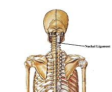
Nuchal ligament: Because the head is decoupled from the shoulders, early Homo needed a way to stabilize the head. The nuchal ligament is an important evolved feature in head stabilization. It starts at the midline of the occiput and connects to the upper trapezius. This ligament is also important in terms of archaeological findings, because it leaves a small indentation and ridge in the skull, allowing researchers to see if various species had a nuchal ligament. The ability to see traces of ligaments in archaeological findings is rare because they degrade quickly and often leave no trace. In the case of the nuchal ligament, a trace of its existence is left with the presence of the skull ridge. Because neither Australopithecus nor Pan had the skull ridge, it has been concluded that this feature is unique to Homo. Because the nuchal ligament is only activated while running, the amount of running can be inferred from the rugosity of the muscle insertions. In the case of Homo Erectus and Neanderthals, very strong nuchal ligament markings are present, but are less marked in modern humans, indicating a decrease in running behavior.

Nuchal ligament of Homo sapiens
 
Shoulder and head stabilization: The human skeleton is different from early hominins as there is less of a connection between the pectoral girdle parts of the shoulders and upper back and head, which would be advantageous for climbing but would hinder the movements of the upper body needed to counter leg movement and therefore stabilize the body and head when running. This stabilization is unnecessary in walking.

Limb length and mass: Homo has longer legs relative to body mass, which helps to decrease the energetic costs of running, as time in contact with the ground increases. There is also a decrease in mass of distal parts of limbs of humans, which is known to decrease metabolic costs in endurance running, but has little effect on walking. Additionally, the mass of the upper body limbs in Homo has decreased considerably, relative to total body mass, which is important to reduce the effort of stabilizing the arms in running.

Joint surface: Humans have evolved to absorb great shock and force on the skeletal structure while running. The impact force on the body can reach up to 3–4 times body weight in endurance running, putting the skeletal structure under great stress. To reduce this stress humans have increased joint surfaces relative to body mass to spread force over larger surface areas, particularly in the lower body. This adaptation, which allows humans to absorb great shock and force applied to the skeleton, is not seen in australopithecine skeletal structures.

Plantar arch: The plantar arch in the human foot has an elastic spring function that generates energy for running but not walking. Fossils of the australopithecine foot show only partial arch, suggesting less of a spring capacity. For the plantar arch spring mechanism to function fully, there must also be restricted rotation in the hind and front parts of the foot. This restriction comes from projected toe bone and compacted mid-foot joint structures in humans, which does not become present until Homo habilis.

Calcaneal tuber and Achilles tendon: Studies have explored the calcaneal tuber, the posterior half of the calcaneus bone, as a correlate for Achilles tendon length and have found correlation between calcaneal tuber length and Achilles tendon length. Because shorter calcaneal tuber length leads to greater Achilles stretch, more kinetic energy is converted to elastic energy, translating into better overall running economy. Comparisons between Neanderthals and modern humans reveal that this adaptation was absent in Neanderthals, leading researchers to conclude that endurance running capabilities may have been enhanced in anatomically modern humans.
Shorter toes: Human toes are straight and extremely short in relation to body size compared to other animals. In running, the toes support 50 to 75% of body mass in humans. Impulse and mechanical work increase in humans as toe length increases, showing that it is energetically favorable to have shorter toes. The costs of shorter toes are decreased gripping capabilities and power output. However, the efficiency benefits seem to outweigh these costs, as the toes of A. afarensis remains were shorter than great apes, but 40% longer than modern humans, meaning that there is a trend toward shorter toes as the primate species moves away from tree-dwelling. This 40% increase in toe length would theoretically induce a flexor impulse 2.5 times that of modern humans, which would require twice as much mechanical work to stabilize.

Stabilization

Semicircular canal: The semicircular canal, a series of three interconnected tubes within each ear, is important for sensing angular rotations of the head and thus plays a crucial role in maintaining balance and sensing and coordinating movement. Comparative studies have shown that animals with larger semicircular canals are able to sense a greater range of head movements and therefore have greater speed and agility. Evolutionarily, greatly reduced semicircular canal diameters are evident in Neanderthals but expanded in modern humans, suggesting that this adaptation was selected for in response to increased endurance running.

Vestibulo-ocular reflexes (VORs): VORs are enabled by muscles in the eye, which sense angular accelerations of the head and adjust eye movements to stabilize these images. This was an important adaptation for running because it allowed Homo to see more clearly during the rough pitching motion that occurs during running.

Gluteals: The gluteus maximus in Homo erectus is significantly larger than that of Australopithecus. It is suited to stabilize the trunk while running, but gluteals of that size and strength are not necessary for walking. 

Iliac spine: Homo has expanded areas on the sacrum and posterior iliac spine for greater muscle attachment. These areas are used to stabilize the trunk and reduce the body's forward pitch caused by running strides.

Increased efficiency

Thermoregulation

In addition to advances in skeletal structure and stabilization, adaptations that led to increased efficiency in dissipation of heat were instrumental in the evolution of endurance running in Homo. The duration for which an animal can run is determined by its capacity to release more heat than is produced to avoid lethal temperatures. 

The majority of mammals, including humans, rely on evaporative cooling to maintain body temperature. Most medium-to-large mammals rely on panting, while humans rely on sweating, to dissipate heat. Advantages to panting include cooler skin surface, little salt loss, and heat loss by forced convection instead of reliance on wind or other means of convection. On the other hand, sweating is advantageous in that evaporation occurs over a much larger surface area (the skin), and it is independent of respiration, thus is a much more flexible mode of cooling during intense activity such as running. Because human sweat glands are under a higher level of neuronal control than those of other species, they allow for the excretion of more sweat per unit surface area than any other species. Heat dissipation of later hominins was also enhanced by the reduction in body hair. By ridding themselves of an insulating fur coat, running humans are better able to dissipate the heat generated by exercise. 

In addition to improved thermoregulation, hominins have evolved an enhanced method of respiration consistent with the demands of running. Due to their orientation, respiration in quadrupedal mammals is affected by skeletal and muscular stresses generated through the motion of running. The bones and muscles of the chest cavity are not only responsible for shock absorption, but are also subjected to continuous compression and expansion during the running cycle. Because of this movement, quadrupeds are restricted to one breath per locomotor cycle, and thus must coordinate their running gait and respiration rate. This tight coordination then translates into another restriction: a specific running speed that is most energetically favorable. The upright orientation of bipedal hominins, however, frees them from this respiration-gait restriction. Because their chest cavities are not directly compressed or involved in the motion of running, hominins are able to vary their breathing patterns with gait. This flexibility in respiration rate and running gait contributes to hominins having a broader range of energetically favorable running speeds.

Storage and utilization of energy

During periods of prolonged exercise, animals are dependent on a combination of two sources of fuel: glycogen stored in the muscles and liver, and fat. Because glycogen is more easily oxidized than fat, it is depleted first. However, over longer periods of time, energy demands require that fat stores be utilized as fuel. This is true for all mammals, but hominins, and later modern humans, have an advantage of being able to alter their diet to meet these prolonged energy demands. 

In addition to flexibility in the utilization of energy, hominins have evolved larger thyroid and adrenal glands which enable them to utilize the energy in carbohydrates and fatty acids more readily and efficiently. These organs are responsible for releasing hormones including epinephrine, norepinephrine, adrenocorticotropic hormone (ACTH), glucagon, and thyroxine. Larger glands allows for greater production of these key hormones and ultimately, maximized utilization of stored fuel.

Taken together, the flexibility in diet and the enhanced usage of fuel heightens the previously mentioned finding that, unlike quadrupeds, hominins do not have a single energetically optimal running speed. For quadrupeds, increasing running speed means increasing the demand for oxygen and fuel. Due to skeletal structure and bipedalism, hominins are free to run energetically over a broader range of speeds and gaits, while maintaining a constant energy consumption rate of approximately 4.1 MJ per 15 km. Thus their utilization of energy is greatly enhanced.

Endurance running and scavenging

All of the aforementioned adaptations enabled Homo to scavenge for food more effectively. Endurance running could have been used as a means of gaining access to distant carcasses or food stores faster than other scavengers and/or carnivores. Scavenging may have taken one or both of two forms: opportunistic scavenging and strategic scavenging.

Early Homo almost certainly scavenged opportunistically. Scavenging is considered opportunistic when one "come[s] across carcasses in the course of [their] daily foraging activities".

Strategic scavenging involves a planned search for carcasses. This style of scavenging would have benefitted from endurance running much more than opportunistic scavenging. Strategic scavenging would have involved the use of long range cues, such as birds circling overhead. Endurance running would have been advantageous in this setting because it allowed hominins to reach the carcass more quickly. Selection pressures would have been very high for strategic scavenging, because hominins were diurnal, while their major competitors (hyenas, lions, etc.) were not. Thus, they would have had to make sure to capitalize on daytime carcasses. Selection pressure also came from the weakness of Homo. Because they were very weak, they were unlikely to drive off any large competition at the carcass. This fact led to an even higher need for a way to reach the carcass before these competitors.

Endurance running and persistence hunting

Persistence hunting is "a form of pursuit hunting in which [the hunter uses] endurance running during the midday heat to drive [prey] into hyperthermia and exhaustion so they can easily be killed". Many question persistence hunting's plausibility when bow and arrow and other technologies were so much more efficient. However, in the Early Stone Age (ESA), spears were only sharpened wood, and hominins had not begun using tools. The lack of spearheads or bows meant they could only hunt from very close range—between 6 and 10 meters. Hominins thus must have developed a way to stab prey from close range without causing serious bodily harm to themselves. Persistence hunting makes killing an animal easier by first bringing it to exhaustion, so that it can no longer retaliate violently.

Persistence hunters work by hunting in the middle of the day, when it is hottest. Hunters choose a single target prey and chase it at a speed between its trot and gallop, which is extremely inefficient for the animal. The hunter then continues pursuing over a period of hours, during which he may lose sight of the animal. In this case, the hunter must use tracks and an understanding of the animal to continue the chase. The prey eventually overheats and becomes unable to continue fleeing. Homo, which does not overheat as quickly because of its superior thermoregulation capabilities, is then able to stab the prey while it is incapacitated and cannot attack.

Tracking and running

Due to the complexity of following a fleeing animal, tracking methods must have been a prerequisite for the use of endurance running in persistence hunting. Scientists posit that early tracking methods were developed in open, sparsely vegetated terrain such as the Kalahari Desert in southern Africa. This "systemic tracking" involves simply following the footprints of animals and was most likely used for tracking grassland species on soft terrain. Skeletal remains suggest that during the Middle Stone Age, hominins used systemic tracking to scavenge for medium-sized animals in vegetation cover, but for hunting antelope in more open grasslands. From the Middle Stone Age into the Later Stone Age, tracking methods developed into what is termed "speculative tracking". When tracks could not easily be found and followed, Homo predicted where tracks were most likely to be found and interpreted other signs to locate prey. This advanced method of tracking allowed for the exploitation of prey in a variety of terrains, making endurance running for persistence hunting more plausible. 

The process of tracking can last many hours and even days in the case of very large mammals. Often times, the hunter(s) will have to run after the animal to keep up. The skeletal parameters of the tibia of early modern humans and Neanderthals have been compared with runners, and it surprisingly shows that these individuals were running even more than cross-country runners today. Particularly, European Neanderthals, the Skhul and Qafzeh hominins, and Late Stone Age Khoisan score very high compared to runners. This is consistent with modern observations of Khoisan, who routinely spend hours running after animals that have been shot with arrows.

Examples of persistence hunters

Although exact dates and methods of persistence hunting are difficult to study, several recent accounts of persistence hunting have been recorded. Tribes in the Kalahari Desert in Botswana have been known to employ endurance running to scavenge and hunt prey. In the open country, the !Xo and /Gwi tribes run down slow-moving animals such as aardvark and porcupines, while during the hotter part of the day, they target animals such as eland, kudu, gemsbok, hartebeest, duiker, steenbok, cheetah, caracal, and African wildcats. In addition to these existing African tribes, it has been suggested that the Tarahumara people in Mexico and the Paiute people and Navajo in the American Southwest, used persistence hunting to capture prey including deer and pronghorn. The Aborigines in Australia are known to have hunted kangaroo in similar ways. Due to the increased availability of weapons, nutrition, tracking devices, and motor vehicles, one may argue that persistence hunting is no longer an effective method of hunting animals for food. However, there are examples of the practice occurring in modern times: the !Xo and /Gwi in the central Kalahari, still practice persistence hunting and have developed advanced methods of doing so. Similarly, the Russian Lykov family that lived in isolation for 40 years also used persistence hunting due to a lack of weapons.

Seasonal differences

In particular, these two tribes maximize the efficiency of persistence hunting by targeting specific species during different seasons. In the rainy season, prime targets include steenbok, duiker, and gemsbok, as wet sand opens their hooves and stiffens their joints. Hunting in the early rainy season is particularly advantageous because dry leaves form "rocks" in the animals' stomachs, resulting in diarrhea. Stiff joints and suboptimal digestion make the prey weaker and more available targets. In contrast, in the dry season, hunters run down kudu, eland, and red hartebeest because these species tire more easily in the loose sand. Hunters say that the best time to practice persistence hunting is near the end of the dry season when animals are poorly nourished and therefore more easily run to exhaustion.[7] By targeting the most vulnerable prey during each season, the hunters maximize the advantages of endurance running.

Conditions/parameters

  1. Persistence hunting must be performed during the day when it is hot, so that the animal will overheat.
  2. Homo must have been able to track the animal, as they would have lost sight of it during the chase.
  3. Such a long hunt requires high amounts of dietary sources of water, salt, and glycogen.
  4. Although the success rate of recorded persistence hunts is very high (approximately 50%), unsuccessful hunts are very costly. Therefore, there would have had to be a social system in which individuals share food, so unsuccessful hunters could borrow food from others when necessary.

In culture and folklore

In the oral traditions of the Hadza, an isolated aboriginal people of hunter-gatherers living in Tanzania, the Tlaatlanebe in their folk history's second epoch practiced this. 

In the first epoch, the world was inhabited by large hairy humanoids called Akakaanebe ("ancestors"), who did not yet possess tools or fire. They simply "stared" at game until it fell dead, referring to either scavenging or early persistence hunting without weapons, or a combination of the two. They did not build houses but slept under trees.

The Tlaatlanebe of the second epoch, however, were large but without hair and lived in caves. As animals had grown more wary of humans due to earlier hunting, they now had to be chased and hunted with dogs.

Criticisms

While there is evidence supporting selection on human morphology to improve endurance running ability, there is some dispute over whether the ecological benefits of scavenging and persistence hunting foraging behaviors were the driving force behind this development.

The majority of the arguments opposing persistence hunting and scavenging behaviors are linked to the fact that the paleohabitat and paleoecology of early Homo were not conducive to these behaviors. It is thought that the earliest members of Homo lived in African savanna-woodlands. This environment consisted of open grassland, as well as parts with dense vegetation—an intermediate between forest and open savannas. The presence of such tree covering would reduce visibility and so require tracking skills. This causes problems for the hypothesis of persistence hunting and running to aid scavenging.

Against persistence hunting

Ungulates are known from archaeological evidence to have been the main prey of the early Homo, and given their great speed, they would have easily been able to outrun early hominins. Ungulate speed, coupled with the variable visibility of the savanna-woodland, meant that hunting by endurance running required the ability to track prey. Pickering and Bunn argue that tracking is part of a sophisticated cognitive skill set that early hominins would not have had, and that even if they were following a trail of blood left by an injured ungulate—which may have been in their cognitive capacity—the ability to craft penetrating projectile technology was absent in early hominins.

It has been suggested that modern hunters in Africa do not use persistence hunting as a foraging method, and most often give up a chase where the trail they were following ends in vegetation. The rare groups of hunters who do occasionally participate in persistence hunting are able to do so because of the extremely hot and open environments. In these groups, a full day of rest and recovery is required after a hunt, indicating the great toll persistence hunts take on the body, making them rare undertakings.

Finally, in critique of Liebenberg's research on modern day persistence hunting, it was revealed that the majority of the hunts initiated were prompted for filming rather than spontaneous, and that few of these hunts were successful. The hunts that were successful involved external factors such as the hunters being able to stop and refill water bottles.

A response to these criticisms has been formulated by Lieberman et al., noting that it is unclear how humans could have grown to occupy a new niche as a diurnal social carnivore without persistence hunting, as the weapons preferred in modern hunter-gatherer tribes would not have been available at the time.

Against scavenging

The proposed benefit of endurance running in scavenging is the ability of early hominins to outcompete other scavengers in reaching food sources. However paleoanthropological studies suggest that the savanna-woodland habitat caused a very low competition environment. Due to low visibility, carcasses were not easily located by mammalian carnivores, resulting in less competition.

Delayed-choice quantum eraser

From Wikipedia, the free encyclopedia https://en.wikipedia.org/wiki/Delayed-choice_quantum_eraser A delayed-cho...