Search This Blog

Tuesday, May 26, 2020

Doxorubicin

From Wikipedia, the free encyclopedia
 
Doxorubicin
Doxorubicin.svg
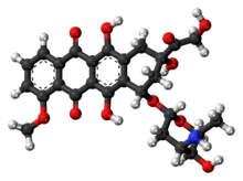
Doxorubicin 3D ball.png
Clinical data
Pronunciation/ˌdɒksəˈrbɪsɪn/
Trade namesAdriamycin, Caelyx, Myocet, others
AHFS/Drugs.comMonograph
MedlinePlusa682221
License data
Pregnancy
category
  • AU: D
  • US: D (Evidence of risk)
Routes of
administration
intravenous, intravesical
ATC code
Legal status
Legal status
  • AU: S4 (Prescription only)
  • CA: ℞-only
  • UK: POM (Prescription only)
  • US: ℞-only
  • In general: ℞ (Prescription only)
Pharmacokinetic data
Bioavailability5% (by mouth)
Protein binding75%
MetabolismLiver
Elimination half-lifeTriphasic; 12 minutes, 3.3 hours, 30 hours. Mean: 1–3 hours
ExcretionUrine (5–12%), faeces (40–50%)
Identifiers
CAS Number
PubChem CID
IUPHAR/BPS
DrugBank
ChemSpider
UNII
KEGG
ChEBI
ChEMBL
CompTox Dashboard (EPA)
ECHA InfoCard100.041.344 Edit this at Wikidata
Chemical and physical data
FormulaC27H29NO11
Molar mass543.525 g·mol−1
3D model (JSmol)

Doxorubicin, sold under the brand name Adriamycin among others, is a chemotherapy medication used to treat cancer. This includes breast cancer, bladder cancer, Kaposi's sarcoma, lymphoma, and acute lymphocytic leukemia. It is often used together with other chemotherapy agents. Doxorubicin is given by injection into a vein.

Common side effects include hair loss, bone marrow suppression, vomiting, rash, and inflammation of the mouth. Other serious side effects may include allergic reactions such as anaphylaxis, heart damage, tissue damage at the site of injection, radiation recall, and treatment-related leukemia. People often experience red discoloration of the urine for a few days. Doxorubicin is in the anthracycline and antitumor antibiotic family of medications. It works in part by interfering with the function of DNA.

Doxorubicin was approved for medical use in the United States in 1974. It is on the World Health Organization's List of Essential Medicines, the safest and most effective medicines needed in a health system. The wholesale cost in the developing world is about US$3.88–32.79 per 50 mg vial. In the United Kingdom this amount costs the NHS about £100.12. Versions that are pegylated and in liposomes are also available; however, are more expensive. Doxorubicin was originally made from the bacteria Streptomyces peucetius.

Medical use

Doxorubicin is commonly used to treat some leukemias and Hodgkin's lymphoma, as well as cancers of the bladder, breast, stomach, lung, ovaries, thyroid, soft tissue sarcoma, multiple myeloma, and others. Commonly used doxorubicin-containing regimens are AC (Adriamycin, cyclophosphamide), TAC (taxotere, AC), ABVD (Adriamycin, bleomycin, vinblastine, dacarbazine), BEACOPP, CHOP (cyclophosphamide, hydroxydaunorubicin, vincristine, prednisone) and FAC (5-fluorouracil, adriamycin, cyclophosphamide).

Doxil (see below) is used primarily for the treatment of ovarian cancer where the disease has progressed or recurred after platinum-based chemotherapy, or for the treatment of AIDS-related Kaposi's sarcoma.

Liposomal form

There is a pegylated (polyethylene glycol coated) liposome-encapsulated form of doxorubicin, sold as Doxil. It was developed to treat Kaposi's sarcoma, an AIDS-related cancer that causes lesions to grow under the skin, in the lining of the mouth, nose and throat, or in other organs. The polyethylene glycol coating results in preferential concentration of doxorubicin in the skin. However, this also results in a side effect called palmar plantar erythrodysesthesia (PPE), more commonly known as hand-foot syndrome.

Following administration of this form of doxorubicin, small amounts of the drug can leak from capillaries in the palms of the hands and soles of the feet. The result of this leakage is redness, tenderness, and peeling of the skin that can be uncomfortable and even painful. In clinical testing at 50 mg/m2 dosing every 4 weeks, half of people developed hand-foot syndrome. The rate of this side effect limits the dose of this formulation that can be given as compared with plain doxorubicin in the same treatment regimen, thereby limiting potential substitution. Substitution would be desirable because liposome-encapsulated doxorubicin is less cardiotoxic than unencapsulated doxorubicin. This form is also approved by the FDA for treatment of ovarian cancer and multiple myeloma.

A non-pegylated liposomal doxorubicin, called Myocet, is approved in Europe and Canada for treatment of metastatic breast cancer in combination with cyclophosphamide, but has not been approved by the FDA for use in the United States. Unlike Doxil, the Myocet liposome does not have a polyethylene glycol coating, and therefore does not result in the same rate of hand-foot syndrome. The minimization of this side effect may allow for one-for-one (1:1) substitution with doxorubicin in the same treatment regimen, thereby improving safety with no loss of efficacy. Like Doxil, the liposomal encapsulation of the doxorubicin limits the cardiotoxicity. In theory, by limiting the cardiotoxicity of doxorubicin through liposomal encapsulation, it can be used safely in concurrent combination with other cardiotoxic chemotherapy drugs, such as trastuzumab. There is an FDA black box warning that trastuzumab cannot be used in concurrent combination with doxorubicin, only in sequential combination. Though concurrent combination of trastuzumab and doxorubicin in clinical studies found superior tumor response, the combination resulted in unacceptable cardiotoxicity, including risk of cardiac failure manifesting as congestive heart failure (CHF). Published phase II study results have shown that Myocet, trastuzumab, and paclitaxel can safely be used concurrently without the cardiac risk, as measured by reduction in LVEF function, while still achieving superior tumor response. This finding is the basis for the ongoing phase III trial for FDA approval.

Side effects

Cardiotoxicity

The most dangerous side effect of doxorubicin is dilated cardiomyopathy, leading to congestive heart failure. The rate of cardiomyopathy is dependent on its cumulative dose, with an incidence about 4% when the dose of doxorubicin is 500–550 mg/m2, 18% when the dose is 551–600 mg/m2 and 36% when the dose exceeds 600 mg/m2. There are several ways in which doxorubicin is believed to cause cardiomyopathy, including oxidative stress, downregulation of genes for contractile proteins, and p53-mediated apoptosis.

Doxorubicin-induced cardiomyopathy typically results in dilated cardiomyopathy, with all four cardiac chambers being enlarged. This results in both systolic and diastolic dysfunction. Eventually, heart failure can result, which carries a 50% mortality rate. There is no effective treatment against established cardiomyopathy caused by the drug as of 2010. The drug dexrazoxane may be used to decrease the risk of doxorubicin's cardiotoxicity in certain cases.

Other

Another common and potentially fatal complication of doxorubicin is typhlitis, an acute life-threatening infection of the bowel. Additionally, some people may develop PPE, characterized by skin eruptions on the palms of the hand or soles of the feet, swelling, pain, and erythema. Due to these side effects and its red color, doxorubicin has earned the nickname "red devil" or "red death."

Chemotherapy can cause reactivation of hepatitis B, and doxorubicin-containing regimens are no exception.

Doxorubicin and several chemotherapeutic drugs (including cyclophosphamide) cause dyspigmentation. Other groups of drugs that cause this problem include antimalarials, amiodarone, heavy metals (but not iron), tetracyclines, and antipsychotics.

Biosynthesis

Doxorubicin (DXR) is a 14-hydroxylated version of daunorubicin, the immediate precursor of DXR in its biosynthetic pathway. 

Daunorubicin is more abundantly found as a natural product because it is produced by a number of different wild type strains of Streptomyces. In contrast, only one known non-wild type species, Streptomyces peucetius subspecies cesius ATCC 27952, was initially found to be capable of producing the more widely used doxorubicin. This strain was created by Arcamone et al. in 1969 by mutating a strain producing daunorubicin, but not DXR, at least in detectable quantities. Subsequently, Hutchinson's group showed that under special environmental conditions, or by the introduction of genetic modifications, other strains of Streptomyces can produce doxorubicin. His group also cloned many of the genes required for DXR production, although not all of them have been fully characterized. In 1996, Strohl's group discovered, isolated and characterized dox A, the gene encoding the enzyme that converts daunorubicin into DXR.

By 1999, they produced recombinant dox A, a cytochrome P450 oxidase, and found that it catalyzes multiple steps in DXR biosynthesis, including steps leading to daunorubicin. This was significant because it became clear that all daunorubicin-producing strains have the necessary genes to produce DXR, the much more therapeutically important of the two. Hutchinson's group went on to develop methods to improve the yield of DXR, from the fermentation process used in its commercial production, not only by introducing dox A encoding plasmids, but also by introducing mutations to deactivate enzymes that shunt DXR precursors to less useful products, for example baumycin-like glycosides. Some triple mutants, that also over-expressed dox A, were able to double the yield of DXR. This is of more than academic interest, because at that time DXR cost about $1.37 million per kg and current production in 1999 was 225 kg per annum.

More efficient production techniques have brought the price down to $1.1 million per kg for the nonliposomal formulation. Although DXR can be produced semi-synthetically from daunorubicin, the process involves electrophilic bromination and multiple steps, and the yield is poor. Since daunorubicin is produced by fermentation, it would be ideal if the bacteria could complete DXR synthesis more effectively.

Mechanism of action

Diagram of two doxorubicin molecules intercalating DNA, from PDB: 1D12​.
 
Doxorubicin interacts with DNA by intercalation and inhibition of macromolecular biosynthesis. This inhibits the progression of topoisomerase II, an enzyme which relaxes supercoils in DNA for transcription. Doxorubicin stabilizes the topoisomerase II complex after it has broken the DNA chain for replication, preventing the DNA double helix from being resealed and thereby stopping the process of replication. It may also increase quinone type free radical production, hence contributing to its cytotoxicity.

The planar aromatic chromophore portion of the molecule intercalates between two base pairs of the DNA, while the six-membered daunosamine sugar sits in the minor groove and interacts with flanking base pairs immediately adjacent to the intercalation site, as evidenced by several crystal structures.

By intercalation, doxorubicin can also induce histone eviction from transcriptionally active chromatin. As a result, DNA damage response, epigenome and transcriptome are deregulated in doxorubicin-exposed cells.

History

In the 1950s, an Italian research company, Farmitalia Research Laboratories, began an organized effort to find anticancer compounds from soil-based microbes. A soil sample was isolated from the area surrounding the Castel del Monte, a 13th-century castle. A new strain of Streptomyces peucetius, which produced a red pigment, was isolated, and an antibiotic from this bacterium was effective against tumors in mice. Since a group of French researchers discovered the same compound at about the same time, the two teams named the compound daunorubicin, combining the name Dauni, a pre-Roman tribe that occupied the area of Italy where the compound was isolated, with the French word for ruby, rubis, describing the color. Clinical trials began in the 1960s, and the drug was successful in treating acute leukemia and lymphoma. However, by 1967, it was recognized that daunorubicin could lead to fatal cardiac toxicity.

Researchers at Farmitalia soon discovered that changes in biological activity could be made by minor changes in the structure of the compound. A strain of Streptomyces was mutated using N-nitroso-N-methyl urethane, and this new strain produced a different, red-colored antibiotic. They named this new compound Adriamycin, after the Adriatic Sea, and the name was later changed to doxorubicin to conform to the established naming convention. Doxorubicin showed better activity than daunorubicin against mouse tumors, and especially solid tumors. It also showed a higher therapeutic index, yet the cardiotoxicity remained.

Doxorubicin and daunorubicin together can be thought of as prototype compounds for the anthracyclines. Subsequent research has led to many other anthracycline antibiotics, or analogs, and there are now over 2,000 known analogs of doxorubicin. By 1991, 553 of them had been evaluated in the screening program at the National Cancer Institute (NCI). In 2016 GPX-150 was granted orphan drug designation by US FDA.

Society and culture

Names

It is also known as hydroxydaunorubicin and hydroxydaunomycin.

It is sold under a number of different brand names, including Adriamycin PFS, Adriamycin RDF, or Rubex.

Formulations

Doxorubicin is photosensitive, and containers are often covered by an aluminum bag and/or brown wax paper to prevent light from affecting it. Doxorubicin is also available in liposome-encapsulated forms as Doxil (pegylated form), Myocet (nonpegylated form), and Caelyx, although these forms must also be given by intravenous injection.

Shortage

Between 2011 and March 2014, Doxil was in limited supply. In 2011, Doxil became available only in very limited supply due to production problems with the third-party manufacturer. Johnson & Johnson (JNJ), through its subsidiary Janssen Products, LP, had been receiving its Doxil supply from contract manufacturer Ben Venue Laboratories (located in Bedford, Ohio), a unit of Boehringer Ingelheim GmbH of Germany. The problems began when Ben Venue temporarily shut down their manufacturing facility due to quality control issues.

In February 2012, to address the Doxil shortage, the US Food and Drug Administration (FDA) allowed for the temporary importation of Lipodox, which contains the same active ingredient as Doxil and is made by Sun Pharma Global FZE (Sun), a subsidiary of India's Sun Pharmaceutical Industries Ltd. The agency said it intends to continue allowing the importation of Lipodox until Sun has made enough generic Doxil to meet demand.

The FDA approved the first generic version of Doxil, made by Sun, in February 2013. It will be available in 20 milligram and 50 milligram vials.

Research

Combination therapy experiments with sirolimus (rapamycin) and doxorubicin have shown promise in treating Akt-positive lymphomas in mice.

Recent animal research coupling a murine monoclonal antibody with doxorubicin has created an immunoconjugate that was able to eliminate HIV-1 infection in mice. Current treatment with antiretroviral therapy (ART) still leaves pockets of HIV within the host. The immunoconjugate could potentially provide a complementary treatment to ART to eradicate antigen-expressing T cells.

Antimalarial activity

There is some evidence for antimalarial activity for doxorubicin and similar compounds. In 2009, a compound similar in structure to doxorubicin was found to inhibit plasmepsin II, an enzyme unique to the malarial parasite Plasmodium falciparum. The pharmaceutical company GlaxoSmithKline (GSK) later identified doxorubicin in a set of compounds that inhibit parasite growth 

Fluorescence

Doxorubicin is also known to be fluorescent. This has often been used to characterize doxorubicin concentrations, and has opened the possibility of using the molecule as a theranostic agent. However, there are significant limitations, as doxorubicin's fluorescence spectrum is known to depend on a variety of factors, including the pH of the environment, solvent dielectric constant and others. Doxorubicin fluorescence is quenched by binding to DNA, and shielded by micelle encapsulation. It is also known to self-quench at high concentrations. In contrast, histone binding amplifies fluorescence.

Gold nanoparticles in chemotherapy

From Wikipedia, the free encyclopedia
 
Gold nanoparticles

Gold nanoparticles in chemotherapy and radiotherapy is the use of colloidal gold in therapeutic treatments, often for cancer or arthritis. Gold nanoparticle technology shows promise in the advancement of cancer treatments. Some of the properties that gold nanoparticles possess, such as small size, non-toxicity and non-immunogenicity make these molecules useful candidates for targeted drug delivery systems. With tumor-targeting delivery vectors becoming smaller, the ability to by-pass the natural barriers and obstacles of the body becomes more probable. To increase specificity and likelihood of drug delivery, tumor specific ligands may be grafted onto the particles along with the chemotherapeutic drug molecules, to allow these molecules to circulate throughout the tumor without being redistributed into the body.

Physical properties

Solutions of gold nanoparticles of various sizes. The size difference causes the difference in colors.

Size

Gold nanoparticles range in size depending on which therapy they are being used for. In photothermal cancer therapy, many gold nanoparticle molecules are used in each test and they must all be uniform in size. Including PEG coating, the nanoparticles measured to be ~130 nm in diameter. Gold nanoparticles that act as drug delivery systems in conjugation with chemotherapeutic drugs typically range in size from 10 to 100 nm.

Surface area plays a very important role in drug delivery and per mg of gold, as diameters decrease, the surface areas needed to transport drugs increase to the point where a single 1mL volume of 1.8 nm spherical gold nanoparticles have the same surface area as a cell phone.

Drug vectorization requires greater specificity, and are synthesized within the single digit measurements ranging from 3-7 nm.

Antibacterial treatments are testing different sizes for cell type targeting; 10, 20 and 40 nm.

Color

Due to the ability to tune the size and absorption of AuNPs, these molecules can vary in the colors they emit. Colors of AuNP solutions typically range from vibrant red to pale blue. These colors play a necessary role in the synthesis of AuNPs as indicators of reduction.

Synthesis

Other synthesis may include cell type targeting. A tumor consists of a multitude of cell types, and thus targeting a single type of cell is ineffective and potentially dangerous. At most, this type of targeting would only have a minor effect on killing the tumor. Tumors are constantly changing and thus phenotype targeting is rendered useless. Two main problems persist: how to get to the target and how to destroy a variety of cells.

Treatments

Photothermal cancer therapy

A direct method of accessing and destroying tumour cells can be accomplished by photothermal cancer therapy or photodynamic therapy (PDT). This procedure is known to treat small tumours that are difficult to access and avoids the drawbacks (adverse effects) of conventional methods, including the unnecessary destruction of healthy tissues. The cells are destroyed by exposure to light, rupturing membranes causing the release of digestive enzymes. AuNPs have high absorption cross sections requiring only minimal input of irradiation energy. Human breast carcinoma cells infused with metal nanoparticles in vitro have been shown to have an increase in morbidity with exposure to near infrared (NIR). Short term exposure in vivo (4–6 minutes) to NIR had undergone the same effect. Hirsch et al observed that extreme heating in tumours would cause irreversible tissue damage including coagulation, cell shrinkage and loss of nuclear straining. Results of their in vivo nanoshell therapy of mice revealed penetration of the tumor ~5mm.The metal particles were tuned to high absorption and scattering, resulting in effective conversion of light into heat covering a large surface area. The El-Sayed group studied AuNP effects in vitro and in vivo. They determined that the NIR wavelengths were converted into heat on the picosecond timescale, allowing for short exposure of CW to minimize possible exposure to healthy cells. In vitro, photothermal therapy was used in oral epithelial cell lines, (HSC 313 and HOC 3 Clone 8) and one benign epithelial cell line (HaCaT). El-Sayed et al found that the malignant cells that had undergone incubation in AuNPs conjugated with anti-epithelial growth factor receptor (EGFR) required half the energy to destroy a cell than a benign cell. Their material included gold coated silica nanoshells that could selectively absorb NIR waves. The particles were tuned by varying the thickness of the Au shell and changing the size of the silica core. In exposing these particles to NIR, the efficacy of Au was measured through the decrease of EFGR in oral squamous carcinoma cells. There are various biotechnological advances for in vivo delivery of drugs. To effectively target the malignant cells, the AuNPs were conjugated by polyethylene glycol, a process known as PEGylation. This masks the foreign particles from the immune system such that it arrives at its destination and increases circulation time in the system. Antibody conjugation lines the surface of the nanoparticle with cell markers to limit spread only to malignant cells. In vivo testing of mice that developed murine colon carcinoma tumour cells. They were injected with the solution of AuNPs that were allowed to spread after 6 hours. Surrounding cells were swabbed with PEG and exposed to laser treatment for detection of abnormal heating indicating areas where Au nanoshells may have gathered. The injected area was also swabbed with PEG to maximize light penetration.

Despite the unquestionable success of gold nanorods or nanoshells as photothermal agents in preclinical research, they have yet to obtain the approval for clinical use because their size is above the renal excretion threshold. In 2019, the first NIR-absorbing plasmonic ultrasmall-in-nano architecture has been reported, and jointly combine: (i) an efficient photothermal conversion suitable for multiple hyperthermia treatments, and (ii) renal excretion of the building blocks after the therapeutic action.

Radiofrequency therapy

X-ray radiography procedures involves the diagnosis of cancer cells through the process of image acquisition. These techniques rely on the absorption of x-rays on the exposed tissue in order to improve image quality. In certain radiological procedures such as Radiofrequency therapy, a contrast agent is injected into the targeted cancer tissue and result in increased x-ray attenuation.

Radiofrequency therapy treatment involves the destruction of tumor cancer tissue cells through the differential heating of cancer tissue by radio-frequency diathermy. This differential heating is a result of the blood supply in the body carrying away the heat and cooling the heated tissue. 

Gold nanoparticles are excellent absorbers of x-rays, due to its high atomic number of 197Au. This allows for a higher mass of the element, providing for a greater area of x-ray absorption. By acting as a contrast agent and injected into cancerous tumor cells, it would result in a higher dose of the cancerous tissue being exposed during radiotherapy treatment. Additionally gold nanoparticles are more efficiently removed from cells of healthy tissue, in comparison with cancer cells - a feature that makes them a promising radiosensitizers

Angiogenesis therapy

Angiogenesis is a process involving the formation of new blood vessels from pre-existing vessels. It involves the degradation of the extracellular matrix, activation, migration, proliferation, and differentiation of endothelial cells into vessels. It is said to play a large part in the growth and spread of cancer cells.

The process of angiogenesis involves the use of both promoters and inhibitors, balancing the process by only forming new blood vessels when needed. Examples of promoters include Vascular Endothelial Growth Factor (VEGF) and fibroblast growth factor (FGF) Examples of inhibitors include Vascular Endothelial Growth Factor Receptor 1, etc.

Tumor progression occurs as a result of the transition from a tumor in the dormant proliferation stage to the active stage as a result of oxygen and nutrients. This active stage leads to a state of cellular hypoxia, which causes an increased regulation of pro-angiogenesis proteins such as VEGF. This results in the spreading of inflammatory proteins and cancer cells alongside the newly created blood vessels.

AuNPs have the ability to inhibit angiogenesis by directly coordinating to heparin binding growth factors. They inhibit phosphorylation of proteins responsible for angiogenesis in a dose dependent matter. At concentrations 335-670 nM, almost complete inhibition of phosphorylation was observed. As a consequence of angiogenesis, rheumatoid arthritis has been found to develop due to the greater ability to spread inflammatory proteins. Through the inhibition of angiogenesis, the reduction of rheumatoid arthritis is prevalent. In addition, angiogenic inhibitors have a critical limitation due to the instability of biological conditions and high dosage required. To counter this, an emerging strategy for the development of therapies targeting tumor-associated angiogenesis through the use of nanotechnology and anti-angiogenic agents was developed, known as anti-angiogenic therapy. This approach solved the limitation instability by speeding up the delivery of angiogenesis inhibitors.

Gold nanoparticles display anti-angiogenic properties by inhibiting the function of pro-angiogenic heparin-binding growth factors (HG – GFs), with prime examples being the vascular endothelial growth factor 165 (VEGF165) and the basic fibroblast growth factor (bFGF) - both of which are pro-angiogenic promoters. Studies by Rochelle R. Arvizo, et al. have shown that the use of AuNPs of various size and surface charge plays an important role in its inhibitory effects.

In today’s biological fields, the use of nanotechnology has allowed for the indirect use of AuNPs to deliver DNA to mammalian cells; thereby reducing tumor agents and increasing efficiency of electron transfer by modulating the activity of glucose oxidase. Current ongoing research by the Mayo Clinic laboratories includes the examination of AuNPs as messengers to deliver reagents capable of manipulating the angiogenic response in vivo.

Current angiogenic inhibitors used today which are approved by the USFDA to treat cancer is Ayastin, Nexavar, Sutent and Affinitor.

Anti-bacterial therapy

Gold nanoparticles are used as bacteria targeting particles in antibacterial therapy. The therapy targets bacteria with light absorbing gold nanoparticles (10 nm, 20 nm, 40 nm) conjugated with specific antibodies, thus selectively kill bacteria using laser.

Studies has shown the effectiveness of this method on killing Staphylococcus aureus, which is significant human pathogen responsible for a wide range of diseases such as skin and wound infections, toxic shock syndrome, septic arthritis, endocarditis, and osteomyelitis. In this system, the bacteria damage is caused by inducing strong laser which leads to overheating effects accompanied by the bubble-formation phenomena around clustered gold nanoparticles.

The selective targeting of S. aureus was performed using a monoclonal antibody to one of the major surface-clustered proteins, protein A (spa), which is linked to the peptidoglycan portion of the cell wall. Monoclonal antibodies ensure the targeting of the specific cell, which is essential to this mechanism. Killing efficiency depends on local overheating effects accompanied by the bubble-formation phenomena, the bubble formation would enhance the PT killing effect.Better heating efficiency results from an enhanced ability to confine the nanosecond laser-pulse within the nanocluster’s size. Overlapping of bubbles from different nanoparticles within the nanoclusters decreases the bubble-formation threshold. An increase in the cluster’s average local absorption and its potential redshifting (from 525 nm for a single gold spherical nanoparticle to 700–800 nm for nanoclusters) in response to plasmon-plasmon resonance.

Drug vectorization

Another way in which AuNPs can be used in cancer therapy is as agents for targeted drug delivery. Research shows that AuNPs can be easily functionalized and conjugated with a variety of molecules, including chemotherapeutic drugs such as Doxorubicin. One major complication with the current methods of treating cancer with chemotherapy is that treatment is not optimized to specifically target cancer cells and the widespread distribution of chemotherapeutic drugs throughout the body can cause harmful side effects such as naseua, hair loss, and cardiotoxicity. Since many of the characteristics of AuNPs allow them to target cancer cells specifically and accumulate within tumor cells, these molecules can act as tumor-targeting drug delivery systems. Once within the tumor microenvironment, these complexes dissociate and release the chemotherapeutic, allowing the drug to take effect and eventually cause apoptosis.

Gold nanoparticles have their advantages in drug vectorization. They can pack several different sizes and types of dendrimers and several different types of ligands in order to effectively treat different types of cancers. For example, research shows that 80~90% of breast cancer’s tumor cells have estrogen receptors and 60~70% of prostate cancer’s tumor cells have androgen receptors. These significant amount of hormone receptors play a role in intermolecular actions. This role is now used by targeting and therapeutic ligands on gold nanoparticles to target tissue-selective anti-tumor drug delivery. In order to have multiple targeting and therapeutic ligands bind with gold nanoparticles, the gold nanoparticles must first undergo polymer stabilization. Then, anti-estrogen molecules with thiolated PEG are bound to gold nanoparticles via Au-S bonds, forming thiolate protected gold nanoparticles.

PEGylated gold nanoparticles

Docetaxel is packed into PEGylated gold nanoparticles Docetaxel is an anti-mitotic chemotherapy medicine which is showing great performance in clinical trials. Docetaxel was approved by FDA, to treat several different kinds of cancer. i.e. breast cancer(include locally advanced or metastatic).

Market approval

A Pilot Study of AuroLase™ Therapy (gold nano shells) in refractory and/or recurrent tumors of the head and neck was completed in 2009 and two trials are currently using AuroLase™ therapy for the treatment of primary/metastatic lung cancer and for prostate cancer. Other gold nanoparticles on the market are mostly for synthesis of nanoparticle complexes in research. Nanocomposix specializes in the production of various sizes of nanoparticles, controlled by varying the concentrations of reducing reagent and HAuCl4.

Sigma Aldrich offers six different sizes of spherical gold nanoparticles and have developed gold nanourchins for similar usage. The surface causes a red shift in the surface plasmon peak as compared to spherical gold nanoaprticles.

Nanopartz offers gold nanoparticles and gold nanorods for preclinical in vivo therapeutics that have been used extensively in preclinical therapeutics including photothermal hyperthermia and chemotherapeutic drug delivery. The pilot study using the Ntracker  gold nanorods was completed in 2012 and was used on seven canines with varying degrees of solid cancer tumors. The results showed significant loading of the gold nanorods after intravenous injection into the cancer tumors and significant heating of the tumors from an external laser.

Adverse effects and limitations

Shape

Depending on the shape of the molecule, the absorbance will vary, i.e. spherical particles will absorb wavelengths in the NIR region with a relatively low absorbance compared to long rods. Chan et al observed that 50 nm spherical nanoparticles were taken up more efficiently than both larger and smaller particles of the same shape. In regards to size, the spheres were taken up more efficiently than the rods. Ability of greater uptake of nanoshells into the cell will localize in the perinuclear membrane and accumulate to deliver toxic effects.

Charge

Electrostatic interactions were also investigated by Rotello et al by conjugating AuNPs with anionic and cationic functional groups. Their results showed that toxicity was more established in AuNPs conjugated with cationic functional groups as a consequence of electrostatic interactions with the anionic cell membrane.

Concentration

The concentrations of gold nanoparticles in biological systems for practical usage range from 1-100 nanoparticles per cell. High concentrations may lead to adverse effects for cell structure and function, which may not appear non-toxic in assays but preparation of the particles have been found to produce abnormal effects in the cell. If large concentrations quickly clear the blood vessels, the nanoshells may accumulate in major organs (mainly the liver and spleen). Residual concentrations of these particles were also found in kidneys, lungs, muscle, brain, and bone of mice after 28 days. The concentration of the solution injected intravenously 2.4*1011 nanoshells/mL. Even without complete clearance from the system, the nanoshells did not cause any physiological complications in the mice. Su et al observed a correlation with the concentration of Au3Cu and cell damage. Cells were incubated in concentrations of 0.001 and 200 mg mL−1 Au3Cu. They concluded a 15% cell viability and dose dependent cell damage. Reduction in cell viability was detected in vivo experiments; also related to dosage. Cytotoxicity is not a major concern in the usage of AuNPs, as they localize in the vesicles and cytoplasm as opposed to the nucleus. Thus, no complications spawned due to their aggregation in these parts of the cell.

Heating

Two key factors to consider when irradiating gold nanoparticles in cancer cells are the lattice cooling rate and lattice heat content. The lattice cooling rate is how fast heat in the particle is distributed to its surroundings. If the cooling rate for a particle is too low, the lattice heat content can be increased with moderate energy radiation (40 µJ/fs with 100-fs laser at 800 nm) to the point where gold nanorods can be melted to create spherical nanoparticles which become photothermally inactive. This decomposition has been shown using gold nanorods coated with phosphatidylcholine ligands in HeLa cells using a pulsed laser and were no longer useful for treatment due to their low NIR radiation absorbance. High energy laser pulses have also been shown to fragment nanorods into smaller particles. While these structural changes induced by laser pulses could be used to deactivate the photothermal effects of these particles after treatment, the resulting spherical particles or other particle fragments could lead to complications during or after treatment when gold nanoparticles are used for clinical treatment and imaging of cancer cells.

A limitation of photothermal chemotherapy using gold nanoparticles involves the choice of laser when conducting treatment. Pulsed lasers offer very selective treatment of cancer cells within a small, localized area, but can lead to potential destruction of particles and have a low heating efficiency due to heat lost during the single pulse excitation. Continuous wave lasers have a higher heating efficiency and work better in heating larger areas with lower risk of destroying the nanoparticles being heated. However, treatment with continuous wave lasers are much longer compared to treatment with a pulsed laser. A limitation of photothermal therapy with respect to the laser used is the depth of the tumor being treated. Most lasers used to induce tumor ablation using gold nanoparticles can only reach several centimeters into soft tissue, making it impossible to reach tumors farther in the body. Finding a way to carry out therapy in cells farther into the body without damaging surrounding cells is essential to making this technique viable as a cancer treatment in the future.

Toxicity

Toxic precursors

Studies in human leukemia cells revealed that prolonged exposure in AuNPs did not harm the cells, even at ~100 μM of Au. Rather they reduced the amount of reactive oxygen species in the cell. However, precursors to AuNP synthesis (CTAB and HAuCl4) were found to be toxic at small concentrations (10 μM); free CTAB especially. Studies in HeLa cells by Niidome et al further support this statement by examining the correlation with the removal of excess CTAB and cell viability rose to 90%.

Toxicity of nanoparticles in vivo and in vitro

After using nanoparticles for photothermal therapy, it has been shown in vitro that high concentrations of reactive oxygen species (ROS) are formed within the treated cancer cells. While these species are not of concern to the dead cancer cells, they can cause oxidative stress in surrounding healthy cells if enough ROS are created leading to healthy cell death. This oxidative stress can be passivated using polymers as reducing agents (after degradation of the nanoparticle) and damage from ROS can be reduced using targeted uptake of the nanoparticles to the cancer cells. The mechanism for the oxidative stress caused by nanoparticles in the body is still the subject of study and provides a possible limitation when using gold nanoparticles with radiation within the body.

While there are many in vitro studies of gold nanoparticles used for chemotherapy, in vivo studies are both rare and often report conflicting results. For example, one in vivo study has shown that 13-nm gold nanoparticles circulated in the bloodstream often “accumulate in the liver and spleen and…have long blood circulation times." Also, nanoparticles from 8 to 37 nanometers have been shown to cause abnormal symptoms leading to death in mice due to medical complications in the spleen, liver, and lungs. Yet, other studies have shown that 20 nm gold nanoparticles can pass into the retina without causing any cytotoxic effects and nanoparticles of 13 nm diameter were not toxic in the body. Many argue that these results differ due to different concentrations on nanoparticles used for these experiments and requires further research.

Biosafety and biokinetics investigations on biodegradable ultrasmall-in-nano architectures have demonstrated that gold nanoparticles are able to avoid metal accumulation in organisms through escaping by the renal pathway.

Part of the issue with these studies is the lack of reliable methods for determining the uptake of gold nanoparticles in vivo without examining the tumor site post-mortem. Gold nanoparticle uptake in cells is often carried out by examining the organs of injected mice post-mortem. This technique cannot be replicated during clinical trials, so new methods need to be developed to determine the uptake of cells to avoid higher concentrations of gold nanoparticles in the body leading to toxic effects. One recently suggested method to counter this limitation is radiolabeling. The uptake of thiolated gold nanoparticles has recently been monitored using 111In-labeled polymer shells that surround the gold nanoparticle and shows a possible way around this problem, but these polymer shells can be removed from the particle making a more stable labeling system required for these kinds of studies.

Other uses

The ligand used to decrease aggregation of gold nanorods.
 
Gold nanoparticles may be used in an indirectly therapeutic way. The issue of angiogenesis describes the formation of new blood vessels, which not only increased spread of cancerous cells, but may proliferate the spread of proteins responsible for rheumatoid arthritis. As AuNPs reduce angiogenesis, rheumatoid arthritis is reduced as a result. Chamberland et al studied the use of anti-TNF conjugated gold nanorods (AuNRs) ex vivo in rat tail joints to reduce the effect of rheumatoid arthritis. They observed the effects of the drug delivery system via PAT technology. The properties of the AuNRs found to be the most efficient had measurements of 45 x 15 nm with an absorption peak of 660 nm. This tuning allowed for better contrast between the targeted areas and intra-articular tissue. Thus, the etanercept conjugated AuNRs were seen to increase the light sensitivity. The imaging technique provides greater opportunities for sensitive in vivo drug tracking in biothechnology.

HIV

Several valences of AuNPs were found to inhibit HIV fusion. 2-nm AuNP-mercaptobenzoic acid were conjugated to a derivative of a known CCR5 antagonist, which is a small molecule that antagonize CCR5 receptor, and CCR5 is commonly used by HIV to enter the cell. The CCR5 antagonist would bind to CCR5, leaving no spots for HIV to bind. This will ultimately lead to an effect that restrict HIV infection.


Prepared AuNPs-Hepatitis B virus (HBV) DNA gene probes could be used to detect HBV DNA directly. The detection-visualized fluorescence-based method is highly sensitive, simple, low cost, which could potentially apply to multi-gene detection chips. The probe used here is essentially a biosensor, to specifically detect a certain material.


A successful application of the AuNP-nanoprobe colorimetric method to clinical diagnosis reported by Baptista et al. was the sensitive detection in clinical samples of Mycobacterium tuberculosis, the cause of human tuberculosis.

Health and safety hazards of nanomaterials

From Wikipedia, the free encyclopedia
 
The health and safety hazards of nanomaterials include the potential toxicity of various types of nanomaterials, as well as fire and dust explosion hazards. Because nanotechnology is a recent development, the health and safety effects of exposures to nanomaterials, and what levels of exposure may be acceptable, are subjects of ongoing research. Of the possible hazards, inhalation exposure appears to present the most concern, with animal studies showing pulmonary effects such as inflammation, fibrosis, and carcinogenicity for some nanomaterials. Skin contact and ingestion exposure, and dust explosion hazards, are also a concern.

Guidance has been developed for hazard controls that are effective in reducing exposures to safe levels, including substitution with safer forms of a nanomaterial, engineering controls such as proper ventilation, and personal protective equipment as a last resort. For some materials, occupational exposure limits have been developed to determine a maximum safe airborne concentration of nanomaterials, and exposure assessment is possible using standard industrial hygiene sampling methods. An ongoing occupational health surveillance program can also help to protect workers.

Background

Three greyscale microscope images arranged horizontally. The left two show agglomerations of black spots on a grey background, while the right one shows a mass of tangled fibers.
Optical micrographs of several nanomaterials present in aerosol particles. From left, silver nanoparticles, nickel nanoparticles, and multiwalled carbon nanotubes

Nanotechnology is the manipulation of matter at the atomic scale to create materials, devices, or systems with new properties or functions, with potential applications in energy, healthcare, industry, communications, agriculture, consumer products, and other sectors. Nanomaterials have at least one primary dimension of less than 100 nanometers, and often have properties different from those of their bulk components that are technologically useful. The classes of materials of which nanoparticles are typically composed include elemental carbon, metals or metal oxides, and ceramics. According to the Woodrow Wilson Center, the number of consumer products or product lines that incorporate nanomaterials increased from 212 to 1317 from 2006 to 2011. Worldwide investment in nanotechnology increased from $432 million in 1997 to about $4.1 billion in 2005.

Because nanotechnology is a recent development, the health and safety effects of exposures to nanomaterials, and what levels of exposure may be acceptable, is not yet fully understood. Research concerning the handling of nanomaterials is underway, and guidance for some nanomaterials has been developed. As with any new technology, the earliest exposures are expected to occur among workers conducting research in laboratories and pilot plants, making it important that they work in a manner that is protective of their safety and health.

A risk management system is composed of three parts. Hazard identification involves determining what health and safety concerns are present for both the nanomaterial and its corresponding bulk material, based on a review of safety data sheets, peer-reviewed literature, and guidance documents on the material. For nanomaterials, toxicity hazards are the most important, but dust explosion hazards may also be relevant. Exposure assessment involves determining actual routes of exposure in a specific workplace, including a review of which areas and tasks are most likely to cause exposure. Exposure control involves putting procedures in places to minimize or eliminate exposures according to the hierarchy of hazard controls. Ongoing verification of hazard controls can occur through monitoring of airborne nanomaterial concentrations using standard industrial hygiene sampling methods, and an occupational health surveillance program may be instituted.

Hazards

Toxicity

Respiratory

A greyscale microscope image showing a rigid rod extending from both sides of a mottled cellular mass
A scanning electron microscope image of bundles of multiwalled carbon nanotube piercing an alveolar epithelial cell.
 
Inhalation exposure is the most common route of exposure to airborne particles in the workplace. The deposition of nanoparticles in the respiratory tract is determined by the shape and size of particles or their agglomerates, and they are deposited in the alveolar compartment to a greater extent than larger respirable particles. Based on animal studies, nanoparticles may enter the bloodstream from the lungs and translocate to other organs, including the brain. The inhalation risk is affected by the dustiness of the material, the tendency of particles to become airborne in response to a stimulus. Dust generation is affected by the particle shape, size, bulk density, and inherent electrostatic forces, and whether the nanomaterial is a dry powder or incorporated into a slurry or liquid suspension.

Animal studies indicate that carbon nanotubes and carbon nanofibers can cause pulmonary effects including inflammation, granulomas, and pulmonary fibrosis, which were of similar or greater potency when compared with other known fibrogenic materials such as silica, asbestos, and ultrafine carbon black. Some studies in cells or animals have shown genotoxic or carcinogenic effects, or systemic cardiovascular effects from pulmonary exposure. Although the extent to which animal data may predict clinically significant lung effects in workers is not known, the toxicity seen in the short-term animal studies indicate a need for protective action for workers exposed to these nanomaterials. As of 2013, further research was needed in long-term animal studies and epidemiologic studies in workers. No reports of actual adverse health effects in workers using or producing these nanomaterials were known as of 2013. Titanium dioxide (TiO2) dust is considered a lung tumor risk, with ultrafine (nanoscale) particles having an increased mass-based potency relative to fine TiO2, through a secondary genotoxicity mechanism that is not specific to TiO2 but primarily related to particle size and surface area.

Dermal

Some studies suggest that nanomaterials could potentially enter the body through intact skin during occupational exposure. Studies have shown that particles smaller than 1 μm in diameter may penetrate into mechanically flexed skin samples, and that nanoparticles with varying physicochemical properties were able to penetrate the intact skin of pigs. Factors such as size, shape, water solubility, and surface coating directly affect a nanoparticle’s potential to penetrate the skin. At this time, it is not fully known whether skin penetration of nanoparticles would result in adverse effects in animal models, although topical application of raw SWCNT to nude mice has been shown to cause dermal irritation, and in vitro studies using primary or cultured human skin cells have shown that carbon nanotubes can enter cells and cause release of pro-inflammatory cytokines, oxidative stress, and decreased viability. It remains unclear, however, how these findings may be extrapolated to a potential occupational risk. In addition, nanoparticles may enter the body through wounds, with particles migrating into the blood and lymph nodes.

Gastrointestinal

Ingestion can occur from unintentional hand-to-mouth transfer of materials; this has been found to happen with traditional materials, and it is scientifically reasonable to assume that it also could happen during handling of nanomaterials. Ingestion may also accompany inhalation exposure because particles that are cleared from the respiratory tract via the mucociliary escalator may be swallowed.

Fire and explosion

A pentagon with each edge colored a different shade of blue and labeled with one of the five requirements for an explosion: fuel, orxygen, ignition, dispersion, and confinement
The explosion pentagon is a representation of the five requirements for a dust explosion.

There is concern that engineered carbon nanoparticles, when manufactured on an industrial scale, could pose a dust explosion hazard, especially for processes such as mixing, grinding, drilling, sanding, and cleaning. Knowledge remains limited about the potential explosivity of materials when subdivided down to the nanoscale. The explosion characteristics of nanoparticles are highly dependent on the manufacturer and the humidity.

For microscale particles, as particle size decreases and the specific surface area increases, the explosion severity increases. However, for dusts of organic materials such as coal, flour, methylcellulose, and polyethylene, severity ceases to increase as the particle size is reduced below ∼50 μm. This is because decreasing particle size primarily increases the volatilization rate, which becomes rapid enough that that gas phase combustion becomes the rate limiting step, and further decrease in particle size will not increase the overall combustion rate. While the minimum explosion concentration does not vary significantly with nanoparticle size, the minimum ignition energy and temperature have been found to decrease with particle size.

Metal-based nanoparticles exhibit more severe explosions than do carbon nanomaterials, and their chemical reaction pathway is qualitatively different. Studies on aluminum nanoparticles and titanium nanoparticles indicate that they are explosion hazards. One study found that the likelihood of an explosion but not its severity increases significantly for nanoscale metal particles, and they can spontaneously ignite under certain conditions during laboratory testing and handling.

High-resistivity powders can accumulate electric charge causing a spark hazard, and low-resistivity powders can build up in electronics causing a short circuit hazard, both of which can provide an ignition source. In general, powders of nanomaterials have higher resistivity than the equivalent micron-scale powders, and humidity decreases their resistivity. One study found powders of metal-based nanoparticles to be mid- to high-resistivity depending on humidity, while carbon-based nanoparticles were found to be low-resistivity regardless of humidity. Powders of nanomaterials are unlikely to present an unusual fire hazard as compared to their cardboard or plastic packaging, as they are usually produced in small quantities, with the exception of carbon black. However, the catalytic properties of nanoparticles and nanostructured porous materials may cause untended catalytic reactions that, based on their chemical composition, would not otherwise be anticipated.

Radioactivity

Engineered radioactive nanoparticles have applications in medical diagnostics, medical imaging, toxicokinetics, and environmental health, and are being investigated for applications in nuclear medicine. Radioactive nanoparticles present special challenges in operational health physics and internal dosimetry that are not present for vapors or larger particles, as the nanoparticles' toxicokinetics depend on their physical and chemical properties including size, shape, and surface chemistry. In some cases, the inherent physicochemical toxicity of the nanoparticle itself may lead to lower exposure limits than those associated with the radioactivity alone, which is not the case with most radioactive materials. In general, however, most elements of a standard radiation protection program are applicable to radioactive nanomaterials, and many hazard controls for nanomaterials will be effective with the radioactive versions.

Hazard controls

An inverted triangle consisting of five colored horizontal levels, each containing one tee five hazard control methods: elimination, substitution, engineering controls, administrative controls, and personal protective equipment
The hierarchy of hazard controls contains methods for controlling exposures to hazards. Methods listed towards the top potentially more effective than those at the bottom at reducing the risk of illness or injury.
 
Controlling exposures to hazards is the fundamental method of protecting workers. The hierarchy of hazard control is a framework that encompasses a succession of control methods to reduce the risk of illness or injury. In decreasing order of effectiveness, these are elimination of the hazard, substitution with another material or process that is a lesser hazard, engineering controls that isolate workers from the hazard, administrative controls that change workers' behavior to limit the quantity or duration of exposure, and personal protective equipment worn on the workers' body.

Prevention through design is the concept of applying control methods to minimize hazards early in the design process, with an emphasis on optimizing employee health and safety throughout the life cycle of materials and processes. It increases the cost-effectiveness of occupational safety and health because hazard control methods are integrated early into the process, rather than needing to disrupt existing procedures to include them later. In this context, adopting hazard controls earlier in the design process and higher on the hierarchy of controls leads to faster time to market, improved operational efficiency, and higher product quality.

Elimination and substitution

A microscope image of a ball made of agglomerated stringlike particles
An aerosol droplet containing nanomaterials ejected from a vial during sonication. Eliminating or limiting sonication and other handling processes reduces inhalation hazards.
 
Elimination and substitution are the most desirable approaches to hazard control, and are most effective early in the design process. Nanomaterials themselves often cannot be eliminated or substituted with conventional materials because their unique properties are necessary to the desired product or process. However, it may be possible to choose properties of the nanoparticle such as size, shape, functionalization, surface charge, solubility, agglomeration, and aggregation state to improve their toxicological properties while retaining the desired functionality. Other materials used incidentally in the process, such as solvents, are also amenable to substitution.

In addition to the materials themselves, procedures used to handle them can be improved. For example, using a nanomaterial slurry or suspension in a liquid solvent instead of a dry powder will reduce dust exposure. Reducing or eliminating steps that involve transfer of powder or opening packages containing nanomaterials also reduces aerosolization and thus the potential hazard to the worker. Reducing agitation procedures such as sonication, and reducing the temperature of reactors to minimize release of nanomaterials in exhaust, also reduce hazards to workers.

Engineering controls

A light green metal enclosure with a partially opened glass sash at front
A fume hood is an engineering control using local exhaust ventilation combined with an enclosure.
 
A white mat on a floor extesively soiled with soot-colored footprints
A sticky mat in a nanomaterials production facility. Ideally, other engineering controls should lessen the amount of dust collecting on the floor and being tracked onto the sticky mat, unlike this example.
 
Engineering controls are physical changes to the workplace that isolate workers from hazards by containing them in an enclosure, or removing contaminated air from the workplace through ventilation and filtering. They are used when hazardous substances and processes cannot be eliminated or replaced with less hazardous substitutes. Well-designed engineering controls are typically passive, in the sense of being independent of worker interactions, which reduces the potential for worker behavior to impact exposure levels. The initial cost of engineering controls can be higher than administrative controls or personal protective equipment, but the long-term operating costs are frequently lower and can sometimes provide cost savings in other areas of the process. The type of engineering control optimal for each situation is influenced by the quantity and dustiness of the material as well as the duration of the task.

Ventilation systems can be local or general. General exhaust ventilation operates on an entire room through a building's HVAC system. It is inefficient and costly as compared to local exhaust ventilation, and is not suitable by itself for controlling exposure, although it can provide negative room pressure to prevent contaminants from exiting the room. Local exhaust ventilation operates at or near the source of contamination, often in conjunction with an enclosure. Examples of local exhaust systems include fume hoods, gloveboxes, biosafety cabinets, and vented balance enclosures. Exhaust hoods lacking an enclosure are less preferable, and laminar flow hoods are not recommended because they direct air outwards towards the worker. Several control verification techniques can be used with ventilation systems, including pitot tubes, hot-wire anemometers, smoke generators, tracer-gas leak testing, and standardized testing and certification procedures.

Examples of non-ventilation engineering controls include placing equipment that may release nanomaterials in a separate room, and placing walk-off sticky mats at room exits. Antistatic devices can be used when handling nanomaterials to reduce their electrostatic charge, making them less likely to disperse or adhere to clothing. Standard dust control methods such as enclosures for conveyor systems, using a sealed system for bag filling, and water spray application are effective at reducing respirable dust concentrations.

Administrative controls

Administrative controls are changes to workers' behavior to mitigate a hazard. They include training on best practices for safe handling, storage, and disposal of nanomaterials, proper awareness of hazards through labeling and warning signage, and encouraging a general safety culture. Administrative controls can complement engineering controls should they fail, or when they are not feasible or do not reduce exposures to an acceptable level. Some examples of good work practices include cleaning work spaces with wet-wiping methods or a HEPA-filtered vacuum cleaner instead of dry sweeping with a broom, avoiding handling nanomaterials in a free particle state, storing nanomaterials in containers with tightly closed lids. Normal safety procedures such as hand washing, not storing or consuming food in the laboratory, and proper disposal of hazardous waste are also administrative controls. Other examples are limiting the time workers are handling a material or in a hazardous area, and exposure monitoring for the presence of nanomaterials.

Personal protective equipment

A man wearing a white lab coat reachess over a beaker containing white powder on a balance
A worker weighing carbon nanotubes. The worker is using personal protective equipment including a respirator, but is not using local engineering controls such as a fume hood.

Personal protective equipment (PPE) must be worn on the worker's body and is the least desirable option for controlling hazards. It is used when other controls are not effective, have not been evaluated, or while doing maintenance or in emergency situations such as spill response. PPE normally used for typical chemicals are also appropriate for nanomaterials, including wearing long pants, long-sleeve shirts, and closed-toed shoes, and the use of safety gloves, goggles, and impervious laboratory coats. Nitrile gloves are preferred because latex gloves do not provide protection from most chemical solvents and may present an allergy hazard. Face shields are not an acceptable replacement for goggles because they do not protect against unbound dry materials. Woven cotton lab coats are not recommended for nanomaterials, as they can become contaminated with nanomaterials and release them later. Donning and removing PPE in a changing room prevents contamination of outside areas.

Respirators are another form of PPE. Respirator filters with a NIOSH air filtration rating of N95 or P100 have been shown to be effective at capturing nanoparticles, although leakage between the respirator seal and the skin may be more significant, especially with half-mask respirators. Surgical masks are not effective against nanomaterials. Smaller nanoparticles of size 4–20 nm are captured more efficiently by filters than larger ones of size 30–100 nm, because Brownian motion results in the smaller particles being more likely to contact a filter fiber. In the United States, the Occupational Safety and Health Administration requires fit testing and medical clearance for use of respirators, and the Environmental Protection Agency requires the use of full face respirators with N100 filters for multi-walled carbon nanotubes not embedded in a solid matrix, if exposure is not otherwise controlled.

Industrial hygiene

Occupational exposure limits

An occupational exposure limit (OEL) is an upper limit on the acceptable concentration of a hazardous substance in workplace air. As of 2016, quantitative OELs have not been determined for most nanomaterials. The U.S. National Institute for Occupational Safety and Health has determined non-regulatory recommended exposure limits (RELs) of 1.0 μg/m3 for carbon nanotubes and carbon nanofibers as background-corrected elemental carbon as an 8-hour time-weighted average (TWA) respirable mass concentration, and 300 μg/m3 for ultrafine titanium dioxide as TWA concentrations for up to 10 hr/day during a 40-hour work week. A properly tested, half-face particulate respirator will provide protection at exposure concentrations 10 times the REL, while an elastomeric full facepiece respirator with P100 filters will provide protection at 50 times the REL. Agencies and organizations from other countries, including the British Standards Institute and the Institute for Occupational Safety and Health in Germany, have established OELs for some nanomaterials, and some companies have supplied OELs for their products.

In the absence of OELs, a control banding scheme may be used. Control banding is a qualitative strategy that uses a rubric to place hazards into one of four categories, or "bands", and each of which has a recommended level of hazard controls. Organizations including GoodNanoGuide, Lawrence Livermore National Laboratory, and Safe Work Australia have developed control banding tools that are specific for nanomaterials. The GoodNanoGuide control banding scheme is based only on exposure duration, whether the material is bound, and the extent of knowledge of the hazards. The LANL scheme assigns points for 15 different hazard parameters and 5 exposure potential factors. Alternatively, the "As Low As Reasonably Achievable" concept may be used.

Exposure assessment

Four small pieces of machinery connected by clear tubes sitting on a table
Equipment used for area sampling of airborne nanomaterials. The instruments shown here include a condensation particle counter, aerosol photometer, and two air sampling pumps for filter-based analysis.
 
Exposure assessment is a set of methods used to monitor contaminant release and exposures to workers. These methods include personal sampling, where samplers are located in the personal breathing zone of the worker, often attached to a shirt collar to be as close to the nose and mouth as possible; and area/background sampling, where they are placed at static locations. Assessment generally use both particle counters, which monitor the real-time quantity of nanomaterials and other background particles; and filter-based samples, which can be used to identify the nanomaterial, usually using electron microscopy and elemental analysis.

Not all instruments used to detect aerosols are suitable for monitoring occupational nanomaterial emissions because they may not be able to detect smaller particles, or may be too large or difficult to ship to a workplace. Suitable particle counters can detect a wide range of particle sizes, as nanomaterials may aggregate in the air. It is recommended to simultaneously test adjacent work areas to establish a background concentration, as direct reading instruments cannot distinguish the target nanomaterial from incidental background nanoparticles from motor or pump exhaust or heating vessels.

While mass-based metrics are traditionally used to characterize toxicological effects of exposure to air contaminants, as of 2013 it was unclear which metrics are most important with regard to engineered nanomaterials. Animal and cell-culture studies have shown that size and shape are the two major factors in their toxicological effects. Surface area and surface chemistry also appeared to be more important than mass concentration.

The NIOSH Nanomaterial Exposure Assessment Technique (NEAT 2.0) is a sampling strategy to determine exposure potential for engineered nanomaterials. It includes filter-based and area samples, as well as a comprehensive assessment of emissions at processes and job tasks to better understand peak emission periods. Evaluation of worker practices, ventilation efficacy, and other engineering exposure control systems and risk management strategies serve to allow for a comprehensive exposure assessment. The NIOSH Manual of Analytical Methods includes guidance on electron microscopy of filter samples of carbon nanotubes and nanofibers, and additionally some NIOSH methods developed for other chemicals can be used for off-line analysis of nanomaterials, including their morphology and geometry, elemental carbon content (relevant for carbon-based nanomaterials), and elemental makeup. Efforts to create reference materials are ongoing.

Occupational health surveillance

Occupational health surveillance involves the ongoing systematic collection, analysis, and dissemination of exposure and health data on groups of workers, for the purpose of preventing disease and evaluating the effectiveness of intervention programs. It encompasses both medical surveillance and hazard surveillance. A basic medical surveillance program contains a baseline medical evaluation and periodic follow-up examinations, post-incident evaluations, worker training, and identification of trends or patterns from medical screening data.

The related topic of medical screening focuses on the early detection of adverse health effects for individual workers, to provide an opportunity for intervention before disease processes occur. Screening may involve obtaining and reviewing an occupational history, medical examination, and medical testing. As of 2016, there were no specific screening tests or health evaluations to identify health effects in people that are caused solely by exposure to engineered nanomaterials. However, any medical screening recommendations for the bulk material that a nanoparticle is made of still apply, and in 2013 NIOSH concluded that the toxicologic evidence on carbon nanotubes and carbon nanofibers had advanced enough to make specific recommendations for the medical surveillance and screening of exposed workers. Medical screening and resulting interventions represent secondary prevention and do not replace primary prevention efforts based on direct hazard controls to minimize employee exposures to nanomaterials.

Emergency preparedness

It is recommended that a nanomaterial spill kit be assembled prior to an emergency and include barricade tape, nitrile or other chemically impervious gloves, an elastomeric full-facepiece respirator with P100 or N100 filters (fitted appropriately to the responder), adsorbent materials such as spill mats, disposable wipes, sealable plastic bags, walk-off sticky mats, a spray bottle with deionized water or another appropriate liquid to wet dry powders, and a HEPA-filtered vacuum. It is considered unsafe to use compressed air, dry sweeping, and vacuums without a HEPA filter to clear dust.

Regulation

United States

The Food and Drug Administration regulates nanomaterials under the Federal Food, Drug, and Cosmetic Act when used as food additives, drugs, or cosmetics. The Consumer Product Safety Commission requires testing and certification of many consumer products for compliance with consumer product safety requirements, and cautionary labeling of hazardous substances under the Federal Hazardous Substances Act.

The General Duty Clause of the Occupational Safety and Health Act requires all employers to keep their workplace free of serious recognized hazards. The Occupational Safety and Health Administration also has recording and reporting requirements for occupational injuries and illness under 29 C.F.R. 1904 for businesses with more than 10 employees, and protection and communication regulations under 29 C.F.R. 1910. Companies producing new products containing nanomaterials must use the Hazard Communication Standard to create safety data sheets containing 16 sections for downstream users such as customers, workers, disposal services, and others. This may require toxicological or other testing, and all data or information provided must be vetted by properly controlled testing The ISO/TR 13329 standard provides guidance specifically on the preparation of safety data sheets for nanomaterials. The National Institute for Occupational Safety and Health does not issue regulations, but conducts research and makes recommendations to prevent worker injury and illness. State and local governments may have additional regulations.

The Environmental Protection Agency (EPA) regulates nanomaterials under the Toxic Substances Control Act, and has permitted limited manufacture of new chemical nanomaterials through the use of consent orders or Significant New Use Rules (SNURs). In 2011 EPA issued a SNUR on multi-walled carbon nanotubes, codified as 40 C.F.R. 721.10155. Other statutes falling in the EPA's jurisdiction may apply, such as Federal Insecticide, Fungicide, and Rodenticide Act (if bacterial claims are being made), Clean Air Act, or Clean Water Act. EPA regulates nanomaterials under the same provisions as other hazardous chemical substances.

Other countries

In the European Union, nanomaterials classified by the European Commission as hazardous chemical substances are regulated under the European Chemical Agency's Registration, Evaluation, Authorisation, and Restriction of Chemicals (REACH) regulation, as well as the Classification, Labeling, and Packaging (CLP) regulations. Under the REACH regulation, companies have the responsibility of collecting information on the properties and uses of substances that they manufacture or import at or above quantities of 1 ton per year, including nanomaterials. There are special provisions for cosmetics that contain nanomaterials, and for biocidal materials under the Biocidal Products Regulation (BPR) when at least 50% of their primary particles are nanoparticles.

In the United Kingdom, powders of nanomaterials may fall under the Chemicals (Hazard Information and Packaging for Supply) Regulations 2002, as well as the Dangerous Substances and Explosive Atmosphere Regulations 2002 if they are capable of fueling a dust explosion.

Delayed-choice quantum eraser

From Wikipedia, the free encyclopedia https://en.wikipedia.org/wiki/Delayed-choice_quantum_eraser A delayed-cho...