Search This Blog

Thursday, June 20, 2019

Bone marrow

From Wikipedia, the free encyclopedia

Bone marrow
Details
SystemImmune system
Identifiers
LatinMedulla ossium
MeSHD001853
TAA13.1.01.001
FMA9608

Bone marrow is a semi-solid tissue which may be found within the spongy or cancellous portions of bones. In birds and mammals, bone marrow is the primary site of new blood cell production or hematopoiesis. It is composed of hematopoietic cells, marrow adipose tissue, and supportive stromal cells. In adult humans, bone marrow is primarily located in the ribs, vertebrae, sternum, and bones of the pelvis. On average, bone marrow constitutes 4% of the total body mass of humans; in an adult having 65 kilograms of mass (143 lb), bone marrow typically accounts for approximately 2.6 kilograms (5.7 lb).

Human marrow produces approximately 500 billion blood cells per day, which join the systemic circulation via permeable vasculature sinusoids within the medullary cavity. All types of hematopoietic cells, including both myeloid and lymphoid lineages, are created in bone marrow; however, lymphoid cells must migrate to other lymphoid organs (e.g. thymus) in order to complete maturation.

Bone marrow transplants can be conducted to treat severe diseases of the bone marrow, including certain forms of cancer such as leukemia. Additionally, bone marrow stem cells have been successfully transformed into functional neural cells, and can also potentially be used to treat illnesses such as inflammatory bowel disease.

Structure

The composition of marrow is dynamic, as the mixture of cellular and non-cellular components (connective tissue) shifts with age and in response to systemic factors. In humans, marrow is colloquially characterized as "red" or "yellow" marrow (Latin: medulla ossium rubra, Latin: medulla ossium flava, respectively) depending on the prevalence of hematopoetic cells vs fat cells. While the precise mechanisms underlying marrow regulation are not understood, compositional changes occur according to stereotypical patterns. For example, a newborn baby's bones exclusively contain hematopoietically active "red" marrow, and there is a progressive conversion towards "yellow" marrow with age. In adults, red marrow is found mainly in the central skeleton, such as the pelvis, sternum, cranium, ribs, vertebrae and scapulae, and variably found in the proximal epiphyseal ends of long bones such as the femur and humerus. In circumstances of chronic hypoxia, the body can convert yellow marrow back to red marrow to increase blood cell production.

Hematopoietic components

Hematopoietic precursor cells: promyelocyte in the center, two metamyelocytes next to it and band cells from a bone marrow aspirate.
 
At the cellular level, the main functional component of bone marrow includes the progenitor cells which are destined to mature into blood and lymphoid cells. Marrow contains hematopoietic stem cells which give rise to the three classes of blood cells that are found in circulation: white blood cells (leukocytes), red blood cells (erythrocytes), and platelets (thrombocytes).

Cellular constitution of the red bone marrow parenchyma
Group Cell type Average
fraction
Reference
range
Myelopoietic
cells
Myeloblasts 0.9% 0.2–1.5
Promyelocytes 3.3% 2.1–4.1
Neutrophilic myelocytes 12.7% 8.2–15.7
Eosinophilic myelocytes 0.8% 0.2–1.3
Neutrophilic metamyelocytes 15.9% 9.6–24.6
Eosinophilic metamyelocytes 1.2% 0.4–2.2
Neutrophilic band cells 12.4% 9.5–15.3
Eosinophilic band cells 0.9% 0.2–2.4
Segmented neutrophils 7.4% 6.0–12.0
Segmented eosinophils 0.5% 0.0–1.3
Segmented basophils and mast cells 0.1% 0.0–0.2
Erythropoietic
cells
Pronormoblasts 0.6% 0.2–1.3
Basophilic normoblasts 1.4% 0.5–2.4
Polychromatic normoblasts 21.6% 17.9–29.2
Orthochromatic normoblast 2.0% 0.4–4.6
Other cell
types
Megakaryocytes < 0.1% 0.0-0.4
Plasma cells 1.3% 0.4-3.9
Reticular cells 0.3% 0.0-0.9
Lymphocytes 16.2% 11.1-23.2
Monocytes 0.3% 0.0-0.8

Stroma

The stroma of the bone marrow includes all tissue not directly involved in the marrow's primary function of hematopoiesis. Stromal cells may be indirectly involved in hematopoiesis, providing a microenvironment that influences the function and differentiation of hematopoeietic cells. For instance, they generate colony stimulating factors, which have a significant effect on hematopoiesis. Cell types that constitute the bone marrow stroma include:

Function

Mesenchymal stem cells

The bone marrow stroma contains mesenchymal stem cells (MSCs), also known as marrow stromal cells. These are multipotent stem cells that can differentiate into a variety of cell types. MSCs have been shown to differentiate, in vitro or in vivo, into osteoblasts, chondrocytes, myocytes, marrow adipocytes and beta-pancreatic islets cells.

Bone marrow barrier

The blood vessels of the bone marrow constitute a barrier, inhibiting immature blood cells from leaving the marrow. Only mature blood cells contain the membrane proteins, such as aquaporin and glycophorin, that are required to attach to and pass the blood vessel endothelium. Hematopoietic stem cells may also cross the bone marrow barrier, and may thus be harvested from blood.

Lymphatic role

The red bone marrow is a key element of the lymphatic system, being one of the primary lymphoid organs that generate lymphocytes from immature hematopoietic progenitor cells. The bone marrow and thymus constitute the primary lymphoid tissues involved in the production and early selection of lymphocytes. Furthermore, bone marrow performs a valve-like function to prevent the backflow of lymphatic fluid in the lymphatic system.

Compartmentalization

Biological compartmentalization is evident within the bone marrow, in that certain cell types tend to aggregate in specific areas. For instance, erythrocytes, macrophages, and their precursors tend to gather around blood vessels, while granulocytes gather at the borders of the bone marrow.

As food

Animal bone marrow has been used in cuisine worldwide for millennia, such as the famed Milanese Ossobuco.

Clinical significance

Disease

The normal bone marrow architecture can be damaged or displaced by aplastic anemia, malignancies such as multiple myeloma, or infections such as tuberculosis, leading to a decrease in the production of blood cells and blood platelets. The bone marrow can also be affected by various forms of leukemia, which attacks its hematologic progenitor cells. Furthermore, exposure to radiation or chemotherapy will kill many of the rapidly dividing cells of the bone marrow, and will therefore result in a depressed immune system. Many of the symptoms of radiation poisoning are due to damage sustained by the bone marrow cells. 

To diagnose diseases involving the bone marrow, a bone marrow aspiration is sometimes performed. This typically involves using a hollow needle to acquire a sample of red bone marrow from the crest of the ilium under general or local anesthesia.

Application of stem cells in therapeutics

Bone marrow derived stem cells have a wide array of application in regenerative medicine.

Imaging

Medical imaging may provide a limited amount of information regarding bone marrow. Plain film x-rays pass through soft tissues such as marrow and do not provide visualization, although any changes in the structure of the associated bone may be detected. CT imaging has somewhat better capacity for assessing the marrow cavity of bones, although with low sensitivity and specificity. For example, normal fatty "yellow" marrow in adult long bones is of low density (-30 to -100 Hounsfield units), between subcutaneous fat and soft tissue. Tissue with increased cellular composition, such as normal "red" marrow or cancer cells within the medullary cavity will measure variably higher in density.

MRI is more sensitive and specific for assessing bone composition. MRI enables assessment of the average molecular composition of soft tissues, and thus provides information regarding the relative fat content of marrow. In adult humans, "yellow" fatty marrow is the dominant tissue in bones, particularly in the (peripheral) appendicular skeleton. Because fat molecules have a high T1-relaxivity, T1-weighted imaging sequences show "yellow" fatty marrow as bright (hyperintense). Furthermore, normal fatty marrow loses signal on fat-saturation sequences, in a similar pattern to subcutaneous fat. 

When "yellow" fatty marrow becomes replaced by tissue with more cellular composition, this change is apparent as decreased brightness on T1-weighted sequences. Both normal "red" marrow and pathologic marrow lesions (such as cancer) are darker than "yellow" marrow on T1-weight sequences, although can often be distinguished by comparison with the MR signal intensity of adjacent soft tissues. Normal "red" marrow is typically equivalent or brighter than skeletal muscle or intervertebral disc on T1-weighted sequences.

Fatty marrow change, the inverse of red marrow hyperplasia, can occur with normal aging, though it can also be seen with certain treatments such as radiation therapy. Diffuse marrow T1 hypointensity without contrast enhancement or cortical discontinuity suggests red marrow conversion or myelofibrosis. Falsely normal marrow on T1 can be seen with diffuse multiple myeloma or leukemic infiltration when the water to fat ratio is not sufficiently altered, as may be seen with lower grade tumors or earlier in the disease process.

Histology

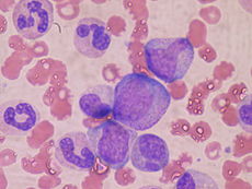
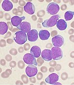
A Wright's-stained bone marrow aspirate smear from a patient with leukemia.
 
Bone marrow examination is the pathologic analysis of samples of bone marrow obtained via biopsy and bone marrow aspiration. Bone marrow examination is used in the diagnosis of a number of conditions, including leukemia, multiple myeloma, anemia, and pancytopenia. The bone marrow produces the cellular elements of the blood, including platelets, red blood cells and white blood cells. While much information can be gleaned by testing the blood itself (drawn from a vein by phlebotomy), it is sometimes necessary to examine the source of the blood cells in the bone marrow to obtain more information on hematopoiesis; this is the role of bone marrow aspiration and biopsy. 

The ratio between myeloid series and erythroid cells is relevant to bone marrow function, and also to diseases of the bone marrow and peripheral blood, such as leukemia and anemia. The normal myeloid-to-erythroid ratio is around 3:1; this ratio may increase in myelogenous leukemias, decrease in polycythemias, and reverse in cases of thalassemia.

Donation and transplantation

A bone marrow harvest in progress.
 
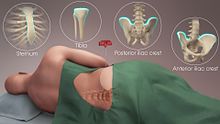
The preferred sites for the procedure
 
In a bone marrow transplant, hematopoietic stem cells are removed from a person and infused into another person (allogenic) or into the same person at a later time (autologous). If the donor and recipient are compatible, these infused cells will then travel to the bone marrow and initiate blood cell production. Transplantation from one person to another is conducted for the treatment of severe bone marrow diseases, such as congenital defects, autoimmune diseases or malignancies. The patient's own marrow is first killed off with drugs or radiation, and then the new stem cells are introduced. Before radiation therapy or chemotherapy in cases of cancer, some of the patient's hematopoietic stem cells are sometimes harvested and later infused back when the therapy is finished to restore the immune system.

Bone marrow stem cells can be induced to become neural cells to treat neurological illnesses, and can also potentially be used for the treatment of other illnesses, such as inflammatory bowel disease. In 2013, following a clinical trial, scientists proposed that bone marrow transplantation could be used to treat HIV in conjunction with antiretroviral drugs; however, it was later found that HIV remained in the bodies of the test subjects.

Harvesting

The stem cells are typically harvested directly from the red marrow in the iliac crest, often under general anesthesia. The procedure is minimally invasive and does not require stitches afterwards. Depending on the donor's health and reaction to the procedure, the actual harvesting can be an outpatient procedure, or can require 1–2 days of recovery in the hospital.

Another option is to administer certain drugs that stimulate the release of stem cells from the bone marrow into circulating blood. An intravenous catheter is inserted into the donor's arm, and the stem cells are then filtered out of the blood. This procedure is similar to that used in blood or platelet donation. In adults, bone marrow may also be taken from the sternum, while the tibia is often used when taking samples from infants. In newborns, stem cells may be retrieved from the umbilical cord.

Fossil record

Bone marrow may have first evolved in Eusthenopteron, a species of prehistoric fish with close links to early tetrapods.
 
The earliest fossilised evidence of bone marrow was discovered in 2014 in Eusthenopteron, a lobe-finned fish which lived during the Devonian period approximately 370 million years ago. Scientists from Uppsala University and the European Synchrotron Radiation Facility used X-ray synchrotron microtomography to study the fossilised interior of the skeleton's humerus, finding organised tubular structures akin to modern vertebrate bone marrow. Eusthenopteron is closely related to the early tetrapods, which ultimately evolved into the land-dwelling mammals and lizards of the present day.
Liquid breathing

From Wikipedia, the free encyclopedia

Liquid breathing is a form of respiration in which a normally air-breathing organism breathes an oxygen-rich liquid (such as a perfluorocarbon), rather than breathing air

This requires certain physical properties such as respiratory gas solubility, density, viscosity, vapor pressure, and lipid solubility which some, but not all, perfluorochemicals (perfluorocarbon) have. Thus, it is critical to choose the appropriate PFC for a specific biomedical application, such as liquid ventilation, drug delivery or blood substitutes. The physical properties of PFC liquids vary substantially; however, the one common property is their high solubility for respiratory gases. In fact, these liquids carry more oxygen and carbon dioxide than blood.

In theory, liquid breathing could assist in the treatment of patients with severe pulmonary or cardiac trauma, especially in pediatric cases. Liquid breathing has also been proposed for use in deep diving and space travel. Despite some recent advances in liquid ventilation, a standard mode of application has not yet been established.

Approaches

Physicochemical properties (37 °C at 1 atm) of 18 perfluorochemical liquids used for biomedical applications. This table characterizes the most significant physical properties related to systemic physiology and their range of properties.
Gas solubility
Oxygen 33–66 mL / 100 mL PFC
Carbon dioxide 140–166 mL / 100 mL PFC
Vapor pressure 0.2–400 torr
Density 1.58–2.0 g/mL
Viscosity 0.8–8.0 cS

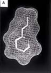
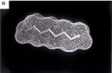
Computer models of three perfluorochemical molecules used for biomedical applications and for liquid ventilation studies: a) FC-75, b) perflubron, and c) perfluorodecalin.
 
Because liquid breathing is still a highly experimental technique, there are several proposed approaches.

Total liquid ventilation

Although total liquid ventilation (TLV) with completely liquid-filled lungs can be beneficial, the complex liquid-filled tube system required is a disadvantage compared to gas ventilation—the system must incorporate a membrane oxygenator, heater, and pumps to deliver to, and remove from the lungs tidal volume aliquots of conditioned perfluorocarbon (PFC). One research group led by Thomas H. Shaffer has maintained that with the use of microprocessors and new technology, it is possible to maintain better control of respiratory variables such as liquid functional residual capacity and tidal volume during TLV than with gas ventilation. Consequently, the total liquid ventilation necessitates a dedicated liquid ventilator similar to a medical ventilator except that it uses a breathable liquid. Many prototypes are used for animal experimentation, but experts recommend continued development of a liquid ventilator toward clinical applications. Specific preclinical liquid ventilator (Inolivent) is currently under joint development in Canada and France. The main application of this liquid ventilator is the ultra-fast induction of therapeutic hypothermia after cardiac arrest. This has been demonstrated to be more protective than slower cooling method after experimental cardiac arrest.

Partial liquid ventilation

In contrast, partial liquid ventilation (PLV) is a technique in which a PFC is instilled into the lung to a volume approximating functional residual capacity (approximately 40% of total lung capacity). Conventional mechanical ventilation delivers tidal volume breaths on top of it. This mode of liquid ventilation currently seems technologically more feasible than total liquid ventilation, because PLV could utilise technology currently in place in many neonatal intensive-care units (NICU) worldwide.

The influence of PLV on oxygenation, carbon dioxide removal and lung mechanics has been investigated in several animal studies using different models of lung injury. Clinical applications of PLV have been reported in patients with acute respiratory distress syndrome (ARDS), meconium aspiration syndrome, congenital diaphragmatic hernia and respiratory distress syndrome (RDS) of neonates. In order to correctly and effectively conduct PLV, it is essential to
  1. properly dose a patient to a specific lung volume (10–15 ml/kg) to recruit alveolar volume
  2. redose the lung with PFC liquid (1–2 ml/kg/h) to oppose PFC evaporation from the lung.
If PFC liquid is not maintained in the lung, PLV can not effectively protect the lung from biophysical forces associated with the gas ventilator. 

New application modes for PFC have been developed.

Partial liquid ventilation (PLV) involves filling the lungs with a fluid. This fluid is perfluorocarbon, also called Liquivent or Perflubron. The liquid has some unique properties. It has a very low surface tension, similar to surfactant, a substance that is produced in the lungs to prevent the alveoli from collapsing and sticking together during exhalation. It also has a high density, oxygen readily diffuses through it, and it may have some anti-inflammatory properties. In PLV, the lungs are filled with the liquid, the patient is then ventilated with a conventional ventilator using a protective lung ventilation strategy. This is called partial liquid ventilation. The hope is that the liquid will help the transport of oxygen to parts of the lung that are flooded and filled with debris, help remove this debris and open up more alveoli improving lung function. The study of PLV involves comparison to protocolized ventilator strategy designed to minimize lung damage.

PFC vapor

Vaporization of perfluorohexane with two anesthetic vaporizers calibrated for perfluorohexane has been shown to improve gas exchange in oleic acid-induced lung injury in sheep.

Predominantly PFCs with high vapor pressure are suitable for vaporization.

Aerosol-PFC

With aerosolized perfluorooctane, significant improvement of oxygenation and pulmonary mechanics was shown in adult sheep with oleic acid-induced lung injury. 

In surfactant-depleted piglets, persistent improvement of gas exchange and lung mechanics was demonstrated with Aerosol-PFC. The aerosol device is of decisive importance for the efficacy of PFC aerosolization, as aerosolization of PF5080 (a less purified FC77) has been shown to be ineffective using a different aerosol device in surfactant-depleted rabbits. Partial liquid ventilation and Aerosol-PFC reduced pulmonary inflammatory response.

Proposed uses

Diving

Gas pressure increases with depth, rising 1 bar (14.5 psi (100 kPa)) every 10 meters to over 1,000 bar at the bottom of the Mariana Trench. Diving becomes more dangerous as depth increases, and deep diving presents many hazards. All surface-breathing animals are subject to decompression sickness, including aquatic mammals and free-diving humans. Breathing at depth can cause nitrogen narcosis and oxygen toxicity. Holding the breath while ascending after breathing at depth can cause air embolisms, burst lung, and collapsed lung

Special breathing gas mixes such as trimix or heliox ameliorate the risk of decompression illness but do not eliminate it. Heliox further eliminates the risk of nitrogen narcosis but introduces the risk of helium tremors below about 500 feet (150 m). Atmospheric diving suits maintain body and breathing pressure at 1 bar, eliminating most of the hazards of descending, ascending, and breathing at depth. However, the rigid suits are bulky, clumsy, and very expensive. 

Liquid breathing offers a third option, promising the mobility available with flexible dive suits and the reduced risks of rigid suits. With liquid in the lungs, the pressure within the diver's lungs could accommodate changes in the pressure of the surrounding water without the huge gas partial pressure exposures required when the lungs are filled with gas. Liquid breathing would not result in the saturation of body tissues with high pressure nitrogen or helium that occurs with the use of non-liquids, thus would reduce or remove the need for slow decompression

A significant problem, however, arises from the high viscosity of the liquid and the corresponding reduction in its ability to remove CO2. All uses of liquid breathing for diving must involve total liquid ventilation (see above). Total liquid ventilation, however, has difficulty moving enough liquid to carry away CO2, because no matter how great the total pressure is, the amount of partial CO2 gas pressure available to dissolve CO2 into the breathing liquid can never be much more than the pressure at which CO2 exists in the blood (about 40 mm of mercury (Torr)).

At these pressures, most fluorocarbon liquids require about 70 mL/kg minute-ventilation volumes of liquid (about 5 L/min for a 70 kg adult) to remove enough CO2 for normal resting metabolism. This is a great deal of fluid to move, particularly as liquids are more viscous and denser than gases, (for example water is about 850 times the density of air). Any increase in the diver's metabolic activity also increases CO2 production and the breathing rate, which is already at the limits of realistic flow rates in liquid breathing. It seems unlikely that a person would move 10 liters/min of fluorocarbon liquid without assistance from a mechanical ventilator, so "free breathing" may be unlikely. However, it has been suggested that a liquid breathing system could be combined with a CO2 scrubber connected to the diver's blood supply; a US patent has been filed for such a method.

Medical treatment

Computer-generated model of perflubron and gentamicin molecules in liquid suspension for pulmonary administration
 
The most promising area for the use of liquid ventilation is in the field of pediatric medicine. The first medical use of liquid breathing was treatment of premature babies and adults with acute respiratory distress syndrome (ARDS) in the 1990s. Liquid breathing was used in clinical trials after the development by Alliance Pharmaceuticals of the fluorochemical perfluorooctyl bromide, or perflubron for short. Current methods of positive-pressure ventilation can contribute to the development of lung disease in pre-term neonates, leading to diseases such as bronchopulmonary dysplasia. Liquid ventilation removes many of the high pressure gradients responsible for this damage. Furthermore, perfluorocarbons have been demonstrated to reduce lung inflammation, improve ventilation-perfusion mismatch and to provide a novel route for the pulmonary administration of drugs.

In order to explore drug delivery techniques that would be useful for both partial and total liquid ventilation, more recent studies have focused on PFC drug delivery using a nanocrystal suspension. The first image is a computer model of a PFC liquid (perflubron) combined with gentamicin molecules. 

The second image shows experimental results comparing both plasma and tissue levels of gentamicin after an intratracheal (IT) and intravenous (IV) dose of 5 mg/kg in a newborn lamb during gas ventilation. Note that the plasma levels of the IV dose greatly exceed the levels of the IT dose over the 4 hour study period; whereas, the lung tissue levels of gentamicin when delivered by an intratracheal (IT) suspension, uniformly exceed the intravenous (IV) delivery approach after 4 hours. Thus, the IT approach allows more effective delivery of the drug to the target organ while maintaining a safer level systemically. Both images represent the in-vivo time course over 4 hours. Numerous studies have now demonstrated the effectiveness of PFC liquids as a delivery vehicle to the lungs.

Comparison of IT and IV administration of gentamicin.
 
Clinical trials with premature infants, children and adults were conducted. Since the safety of the procedure and the effectiveness were apparent from an early stage, the US Food and Drug Administration (FDA) gave the product "fast track" status (meaning an accelerated review of the product, designed to get it to the public as quickly as is safely possible) due to its life-saving potential. Clinical trials showed that using perflubron with ordinary ventilators improved outcomes as much as using high frequency oscillating ventilation (HFOV). But because perflubron was not better than HFOV, the FDA did not approve perflubron, and Alliance is no longer pursuing the partial liquid ventilation application. Whether perflubron would improve outcomes when used with HFOV or has fewer long-term consequences than HFOV remains an open question. 

In 1996 Mike Darwin and Steven B. Harris proposed using cold liquid ventilation with perfluorocarbon to quickly lower the body temperature of victims of cardiac arrest and other brain trauma to allow the brain to better recover. The technology came to be called gas/liquid ventilation (GLV), and was shown able to achieve a cooling rate of 0.5 °C per minute in large animals. It has not yet been tried in humans. 

Most recently, hypothermic brain protection has been associated with rapid brain cooling. In this regard, a new therapeutic approach is the use of intranasal perfluorochemical spray for preferential brain cooling. The nasopharyngeal (NP) approach is unique for brain cooling due to anatomic proximity to the cerebral circulation and arteries. Based on preclinical studies in adult sheep, it was shown that independent of region, brain cooling was faster during NP-perfluorochemical versus conventional whole body cooling with cooling blankets. To date, there have been four human studies including a completed randomized intra-arrest study (200 patients). Results clearly demonstrated that prehospital intra-arrest transnasal cooling is safe, feasible and is associated with an improvement in cooling time.

Space travel

Liquid immersion provides a way to reduce the physical stress of G forces. Forces applied to fluids are distributed as omnidirectional pressures. Because liquids cannot be practically compressed, they do not change density under high acceleration such as performed in aerial maneuvers or space travel. A person immersed in liquid of the same density as tissue has acceleration forces distributed around the body, rather than applied at a single point such as a seat or harness straps. This principle is used in a new type of G-suit called the Libelle G-suit, which allows aircraft pilots to remain conscious and functioning at more than 10 G acceleration by surrounding them with water in a rigid suit. 

Acceleration protection by liquid immersion is limited by the differential density of body tissues and immersion fluid, limiting the utility of this method to about 15 to 20 G. Extending acceleration protection beyond 20 G requires filling the lungs with fluid of density similar to water. An astronaut totally immersed in liquid, with liquid inside all body cavities, will feel little effect from extreme G forces because the forces on a liquid are distributed equally, and in all directions simultaneously. However effects will be felt because of density differences between different body tissues, so an upper acceleration limit still exists. 

Liquid breathing for acceleration protection may never be practical because of the difficulty of finding a suitable breathing medium of similar density to water that is compatible with lung tissue. Perfluorocarbon fluids are twice as dense as water, hence unsuitable for this application.

Examples in fiction

Literary works

  • Alexander Beliaev's 1928 science fiction novel Amphibian Man is based on a scientist and a maverick surgeon, who makes his son, Ichthyander (etymology: "fish" + "man") a life-saving transplant – a set of shark gills. There is a film based on the novel.
  • L. Sprague de Camp's 1938 short story "The Merman" hinges on an experimental process to make lungs function as gills, thus allowing a human being to "breathe" under water.
  • Hal Clement's 1973 novel Ocean on Top portrays a small underwater civilization living in a 'bubble' of oxygenated fluid denser than seawater.
  • Joe Haldeman's 1975 novel The Forever War describes liquid immersion and breathing in great detail as a key technology to allow space travel and combat with acceleration up to 50 G.
  • In the Star Trek: The Next Generation novel The Children of Hamlin (1988) the crew of the Enterprise-D encounter an alien race whose ships contain a breathable liquid environment.
  • Peter Benchley's 1994 novel White Shark centers around a Nazi scientist's experimental attempts to create an amphibious human, whose lungs are surgically modified to breathe underwater, and trained to reflexively do so after being flooded with a fluorocarbon solution.
  • Ben Bova's novel Jupiter (2000) features a craft in which the crew are suspended in a breathable liquid that allows them to survive in the high-pressure environment of Jupiter's atmosphere.
  • In Scott Westerfeld's sci-fi novel The Risen Empire (2003), the lungs of soldiers performing insertion from orbit are filled with an oxygen-rich polymer gel with embedded pseudo-alveoli and a rudimentary artificial intelligence.
  • The novel Mechanicum (2008) by Graham McNeill, Book 9 in the Horus Heresy book series, describes physically crippled Titan (gigantic war machine) pilots encased in nutrient fluid tanks. This allows them to continue operating beyond the limits normally imposed by the body.
  • In the 2009 novel The Lost Symbol by Dan Brown, Robert Langdon (the protagonist) is completely submerged in breathable liquid mixed with hallucinogenic chemicals and sedatives as a torture and interrogation technique by Mal'akh (the antagonist). He goes through a near death experience when he inhales the liquid and blacks out, losing control over his body, but is soon revived.
  • In Greg van Eekhout's 2014 novel California Bones, two characters are put into tanks filled with liquid: "They were given no breathing apparatus, but the water in the tank was rich with perfluorocarbon, which carried more oxygen than blood."
  • In author A.L. Mengel's science fiction novel The Wandering Star (2016), several characters breathe oxygenated fluid during a dive to explore an underwater city. They submerge in high pressure "bubbles" filled with the perfluorocarbon fluid.

Films and television

  • The aliens in the Gerry Anderson UFO series (1970-1971) use liquid-breathing spacesuits.
  • The 1989 film The Abyss by James Cameron features a character using liquid breathing to dive thousands of feet without compressing. The Abyss also features a scene with a rat submerged in and breathing fluorocarbon liquid, filmed in real life.
  • In the 1995 anime Neon Genesis Evangelion, the cockpits of the titular mecha are filled with a fictional oxygenated liquid called LCL which is required for the pilot to mentally sync with an Evangelion, as well as providing direct oxygenation of their blood, and dampening the impacts from battle. Once the cockpit is flooded the LCL is ionized, bringing its density, opacity, and viscosity close to that of air.
  • In the movies Event Horizon (1997) and Mission to Mars (2000), a character is depicted as being immersed in apparent breathable fluid before a high-acceleration launch.
  • In season 1, episode 13 of Seven Days (1998-2001) chrononaut Frank Parker is seen breathing a hyper-oxygenated perfluorocarbon liquid that is pumped through a sealed full body suit that he is wearing. This suit and liquid combination allow him to board a Russian submarine through open ocean at a depth of almost 1000 feet. Upon boarding the submarine he removes his helmet, expels the liquid from his lungs and is able to breathe air again.
  • In an episode of the Adult Swim cartoon series Metalocalypse (2006-2013), the other members of the band submerge guitarist Toki in a "liquid oxygen isolation chamber" while recording an album in the Mariana Trench.
  • In an episode of the Syfy Channel show Eureka (2006-2012), Sheriff Jack Carter is submerged in a tank of "oxygen rich plasma" to be cured of the effects of a scientific accident.
  • In the anime series Aldnoah.Zero (2014-2015), episode 5 shows that Slaine Troyard was in a liquid-filled capsule when he crashed. Princess Asseylum witnessed the crash, helped him to get out of the capsule, then used CPR on him to draw out the liquid from his lungs.

Video games

Immune tolerance

From Wikipedia, the free encyclopedia

Immune tolerance, or immunological tolerance, or immunotolerance, is a state of unresponsiveness of the immune system to substances or tissue that have the capacity to elicit an immune response in given organism. It is induced by prior exposure to that specific antigen and contrasts with conventional immune-mediated elimination of foreign antigens. Tolerance is classified into central tolerance or peripheral tolerance depending on where the state is originally induced—in the thymus and bone marrow (central) or in other tissues and lymph nodes (peripheral). The mechanisms by which these forms of tolerance are established are distinct, but the resulting effect is similar.

Immune tolerance is important for normal physiology. Central tolerance is the main way the immune system learns to discriminate self from non-self. Peripheral tolerance is key to preventing over-reactivity of the immune system to various environmental entities (allergens, gut microbes, etc.). Deficits in central or peripheral tolerance also cause autoimmune disease, resulting in syndromes such as systemic lupus erythematosus, rheumatoid arthritis, type 1 diabetes, autoimmune polyendocrine syndrome type 1 (APS-1), and immunodysregulation polyendocrinopathy enteropathy X-linked syndrome (IPEX), and potentially contribute to asthma, allergy, and inflammatory bowel disease. And immune tolerance in pregnancy is what allows a mother animal to gestate a genetically distinct offspring with an alloimmune response muted enough to prevent miscarriage.

Tolerance, however, also has its negative tradeoffs. It allows for some pathogenic microbes to successfully infect a host and avoid elimination. In addition, inducing peripheral tolerance in the local microenvironment is a common survival strategy for a number of tumors that prevents their elimination by the host immune system.

Historical background

The phenomenon of immune tolerance was first described by Ray D. Owens in 1945, who noted that dizygotic twin cattle sharing a common placenta also shared a stable mixture of each other's red blood cells (though not necessarily 50/50), and retained that mixture throughout life. Although Owens did not use the term immune tolerance, his study showed the body could be tolerant of these foreign tissues. This observation was experimentally validated by Rupert E. Billingham and Peter Medawar in 1953, who showed by injecting foreign cells into fetal or neonatal mice, they could become accepting of future grafts from the same foreign donor. However, they were not thinking of the immunological consequences of their work at the time: as Medawar explains:
"We did not set out with the idea in mind of studying the immunological consequences of the phenomenon described by Owen; on the contrary, we had been goaded by Dr. H.P. Donald into trying to devise a foolproof method of distinguishing monozygotic from dizygotic twins... ."
However, these discoveries, and the host of allograft experiments and observations of twin chimerism they inspired, were seminal for the theories of immune tolerance formulated by Sir Frank McFarlane Burnet and Frank Fenner, who were the first to propose the deletion of self-reactive lymphocytes to establish tolerance, now termed clonal deletion. Burnet and Medawar were ultimately credited for "the discovery of acquired immune tolerance" and shared the Nobel Prize in Physiology or Medicine in 1960.

Definitions and usage

In their Nobel Lecture, Medawar and Burnet define immune tolerance as "a state of indifference or non-reactivity towards a substance that would normally be expected to excite an immunological response." Other more recent definitions have remained more or less the same. The 8th edition of Janeway's Immunobiology defines tolerance as "immunologically unresponsive…to another's tissues.".

Immune tolerance encompasses the range of physiological mechanisms by which the body reduces or eliminates an immune response to particular agents. It is used to describe the phenomenon underlying discrimination of self from non-self, suppressing allergic responses, allowing chronic infection instead of rejection and elimination, and preventing attack of fetuses by the maternal immune system. Typically, a change in the host, not the antigen, is implied. Though some pathogens can evolve to become less virulent in host-pathogen coevolution, tolerance does not refer to the change in the pathogen, but can be used to describe the changes in host physiology. Immune tolerance also does not usually refer to artificially induced immunosuppression by corticosteroids, lymphotoxic chemotherapy agents, sublethal irradiation, etc. Nor does it refer to other types of non-reactivity such as immunological paralysis. In the latter two cases, the host's physiology is handicapped but not fundamentally changed. 

Immune tolerance is formally differentiated into central or peripheral; however, alternative terms such as "natural" or "acquired" tolerance have at times been used to refer to establishment of tolerance by physiological means or by artificial, experimental, or pharmacological means. These two methods of categorization are sometimes confused, but are not equivalent—central or peripheral tolerance may be present naturally or induced experimentally. This difference is important to keep in mind.

Central tolerance

Central tolerance refers to the tolerance established by deleting autoreactive lymphocyte clones before they develop into fully immunocompetent cells. It occurs during lymphocyte development in the thymus and bone marrow for T and B lymphocytes, respectively. In these tissues, maturing lymphocytes are exposed to self-antigens presented by medullary thymic epithelial cells and thymic dendritic cells, or bone marrow cells. Self-antigens are present due to endogenous expression, importation of antigen from peripheral sites via circulating blood, and in the case of thymic stromal cells, expression of proteins of other non-thymic tissues by the action of the transcription factor AIRE

Those lymphocytes that have receptors that bind strongly to self-antigens are removed by induction of apoptosis of the autoreactive cells, or by induction of anergy, a state of non-activity. Weakly autoreactive B cells may also remain in a state of immunological ignorance where they simply do not respond to stimulation of their B cell receptor. Some weakly self-recognizing T cells are alternatively differentiated into natural regulatory T cells (nTreg cells), which act as sentinels in the periphery to calm down potential instances of T cell autoreactivity (see peripheral tolerance below).

The deletion threshold is much more stringent for T cells than for B cells since T cells alone can cause direct tissue damage. Furthermore, it is more advantageous for the organism to let its B cells recognize a wider variety of antigen so it can produce antibodies against a greater diversity of pathogens. Since the B cells can only be fully activated after confirmation by more self-restricted T cells that recognize the same antigen, autoreactivity is held in check.

This process of negative selection ensures that T and B cells that could initiate a potent immune response to the host's own tissues are eliminated while preserving the ability to recognize foreign antigens. It is the step in lymphocyte education that is key for preventing autoimmunity (entire process detailed here). Lymphocyte development and education is most active in fetal development, but continues throughout life as immature lymphocytes are generated, slowing as the thymus degenerates and the bone marrow shrinks in adult life.

Peripheral tolerance

Peripheral tolerance develops after T and B cells mature and enter the peripheral tissues and lymph nodes. It is established by a number of partly overlapping mechanisms that mostly involve control at the level of T cells, especially CD4+ helper T cells, which orchestrate immune responses and give B cells the confirmatory signals they need in order to produce antibodies. Inappropriate reactivity toward normal self-antigen that was not eliminated in the thymus can occur, since the T cells that leave the thymus are relatively but not completely safe. Some will have receptors (TCRs) that can respond to self-antigens that
  • are present in such high concentration outside the thymus that they can bind to "weak" receptors.
  • the T cell did not encounter in the thymus (such as, tissue-specific molecules like those in the islets of Langerhans, brain, or spinal cord not expressed by AIRE in thymic tissues).
Those self-reactive T cells that escape intrathymic negative selection in the thymus can inflict cell injury unless they are deleted or effectively muzzled in the peripheral tissue chiefly by nTreg cells (see central tolerance above).

Appropriate reactivity toward certain antigens can also be quieted by induction of tolerance after repeated exposure, or exposure in a certain context. In these cases, there is a differentiation of naïve CD4+ helper T cells into induced Treg cells (iTreg cells) in the peripheral tissue or nearby lymphoid tissue (lymph nodes, mucosal-associated lymphoid tissue, etc.). This differentiation is mediated by IL-2 produced upon T cell activation, and TGF-β from any of a variety of sources, including tolerizing dendritic cells (DCs), other antigen presenting cells, or in certain conditions surrounding tissue.

Treg cells are not the only cells that mediate peripheral tolerance. Other regulatory immune cells include T cell subsets similar to but phenotypically distinct from Treg cells, including TR1 cells that make IL-10 but do not express Foxp3, TGF-β-secreting TH3 cells, as well as other less well-characterized cells that help establish a local tolerogenic environment. B cells also express CD22, a non-specific inhibitor receptor that dampens B cell receptor activation. A subset of B regulatory cells that makes IL-10 and TGF-β also exists. Some DCs can make Indoleamine 2,3-dioxygenase (IDO) that depletes the amino acid tryptophan needed by T cells to proliferate and thus reduce responsiveness. DCs also have the capacity to directly induce anergy in T cells that recognize antigen expressed at high levels and thus presented at steady-state by DCs. In addition, FasL expression by immune privileged tissues can result in activation-induced cell death of T cells.

nTreg vs. iTreg cells

The involvement of T cells, later classified as Treg cells, in immune tolerance was recognized in 1995 when animal models showed that CD4+ CD25+ T cells were necessary and sufficient for the prevention of autoimmunity in mice and rats. Initial observations showed removal of the thymus of a newborn mouse resulted in autoimmunity, which could be rescued by transplantation of CD4+ T cells. A more specific depletion and reconstitution experiment established the phenotype of these cells as CD4+ and CD25+. Later in 2003, experiments showed that Treg cells were characterized by the expression of the Foxp3 transcription factor, which is responsible for the suppressive phenotype of these cells.

It was assumed that, since the presence of the Treg cells originally characterized was dependent on the neonatal thymus, these cells were thymically derived. By the mid-2000s, however, evidence was accruing of conversion of naïve CD4+ T cells to Treg cells outside of the thymus. These were later defined as induced or iTreg cells to contrast them with thymus-derived nTreg cells. Both types of Treg cells quieten autoreactive T cell signaling and proliferation by cell-contact-dependent and -independent mechanisms including:
  • Contact-dependent:
  • Contact-independent
  • Secretion of TGF-β, which sensitizes cells to suppression and promotes Treg-like cell differentiation
  • Secretion of IL-10
  • Cytokine absorption leading to cytokine deprivation-mediated apoptosis
nTreg cells and iTreg cells, however, have a few important distinguishing characteristics that suggest they have different physiological roles:
  • nTreg cells develop in the thymus; iTreg cells develop outside the thymus in chronically inflamed tissue, lymph nodes, spleen, and gut-associated lymphoid tissue (GALT).
  • nTreg cells develop from Foxp3- CD25+ CD4+ cells while iTreg cells develop from Foxp3+ CD25- CD4- cells (both become Foxp3+ CD25+CD4+).
  • nTreg cells, when activated, require CD28 costimulation, while iTreg cells require CTLA-4 costimulation.
  • nTreg cells are specific, modestly, for self-antigen while iTreg cells recognize allergens, commensal bacteria, tumor antigens, alloantigens, and self-antigens in inflamed tissue.

Tolerance in physiology and medicine

Allograft tolerance

Immune recognition of non-self-antigens typically complicates transplantation and engrafting of foreign tissue from an organism of the same species (allografts), resulting in graft reaction. However, there are two general cases in which an allograft may be accepted. One is when cells or tissue are grafted to an immune-privileged site that is sequestered from immune surveillance (like in the eye or testes) or has strong molecular signals in place to prevent dangerous inflammation (like in the brain). The second is when a state of tolerance has been induced, either by previous exposure to the antigen of the donor in a manner that causes immune tolerance rather than sensitization in the recipient, or after chronic rejection. Long-term exposure to a foreign antigen from fetal development or birth may result in establishment of central tolerance, as was observed in Medawar's mouse-allograft experiments. In usual transplant cases, however, such early prior exposure is not possible. Nonetheless, a few patients can still develop allograft tolerance upon cessation of all exogenous immunosuppressive therapy, a condition referred to as operational tolerance. CD4+ Foxp3+ Treg cells, as well as CD8+ CD28- regulatory T cells that dampen cytotoxic responses to grafted organs, are thought to play a role. In addition, genes involved in NK cell and γδT cell function associated with tolerance have been implicated for liver transplant patients. The unique gene signatures of these patients implies their physiology may be predisposed toward immune tolerance.

Fetal development

The fetus has a different genetic makeup than the mother, as it also translates its father's genes, and is thus perceived as foreign by the maternal immune system. Women who have borne multiple children by the same father typically have antibodies against the father's red blood cell and major histocompatibility complex (MHC) proteins. However, the fetus usually is not rejected by the mother, making it essentially a physiologically tolerated allograft. It is thought that the placental tissues which interface with maternal tissues not only try to escape immunological recognition by downregulating identifying MHC proteins but also actively induce a marked peripheral tolerance. Placental trophoblast cells express a unique Human Leukocyte Antigen (HLA-G) that inhibits attack by maternal NK cells. These cells also express IDO, which represses maternal T cell responses by amino acid starvation. Maternal T cells specific for paternal antigens are also suppressed by tolerogenic DCs and activated iTregs or cross-reacting nTregs. Some maternal Treg cells also release soluble fibrinogen-like proteins 2 (sFGL2), which suppresses the function of DCs and macrophages involved in inflammation and antigen presentation to reactive T cells. These mechanisms altogether establish an immune-privileged state in the placenta that protects the fetus. A break in this peripheral tolerance results in miscarriage and fetal loss.

The microbiome

The skin and digestive tract of humans and many other organisms is colonized with an ecosystem of microorganisms that is referred to as the microbiome. Though in mammals a number of defenses exist to keep the microbiota at a safe distance, including a constant sampling and presentation of microbial antigens by local DCs, most organisms do not react against commensal microorganisms and tolerate their presence. Reactions are mounted, however, to pathogenic microbes and microbes that breach physiological barriers. Peripheral mucosal immune tolerance, in particular mediated by iTreg cells and tolerogenic antigen-presenting cells, is thought to be responsible for this phenomenon. In particular, specialized gut CD103+ DCs that produce both TGF-β and retinoic acid efficiently promotes the differentiation of iTreg cells in the gut lymphoid tissue. Foxp3- TR1 cells that make IL-10 are also enriched in the intestinal lining. Break in this tolerance is thought to underlie the pathogenesis of inflammatory bowel diseases like Crohn's disease and ulcerative colitis.

Oral tolerance and hypersensitivity

Oral tolerance refers to a specific type of peripheral tolerance induced by antigens given by mouth and exposed to the gut mucosa and its associated lymphoid tissues. The hypo-responsiveness induced by oral exposure is systemic, and can reduce hypersensitivity reactions in certain cases. Records from 1829 indicate that American Indians would reduce contact hypersensitivity from poison ivy by consuming leaves of related Rhus species; however, contemporary attempts to use oral tolerance to ameliorate autoimmune diseases like rheumatoid arthritis and other hypersensitivity reactions have been mixed. The systemic effects of oral tolerance may be explained by the extensive recirculation of immune cells primed in one mucosal tissue in another mucosal tissue, allowing extension of mucosal immunity. The same probably occurs for cells mediating mucosal immune tolerance. 

Oral tolerance may depend on the same mechanisms of peripheral tolerance that limit inflammation to bacterial antigens in the microbiome, since both involve the gut-associated lymphoid tissue. It may also have evolved to prevent hypersensitivity reactions to food proteins. It is of immense immunological importance, since it is a continuous natural immunologic event driven by exogenous antigen. 

Allergy and hypersensitivity reactions in general are traditionally thought of as misguided or excessive reactions by the immune system, possibly due to broken or underdeveloped mechanisms of peripheral tolerance. Usually, Treg cells, TR1, and Th3 cells at mucosal surfaces suppress type 2 CD4 helper cells, mast cells, and eosinophils, which mediate allergic response. Deficits in Treg cells or their localization to mucosa have been implicated in asthma and atopic dermatitis. Attempts have been made to reduce hypersensitivity reactions by oral tolerance and other means of repeated exposure. Repeated administration of the allergen in slowly increasing doses, subcutaneously or sublingually appears to be effective for allergic rhinitis. Repeated administration of antibiotics, which can form haptens to cause allergic reactions, can also reduce antibiotic allergies in children.

The tumor microenvironment

Immune tolerance is an important means by which growing tumors, which have mutated proteins and altered antigen expression, prevent elimination by the host immune system. It is well recognized that tumors are a complex and dynamic population of cells composed of transformed cells as well as stromal cells, blood vessels, tissue macrophages, and other immune infiltrates. These cells and their interactions all contribute to the changing tumor microenvironment, which the tumor largely manipulates to be immunotolerant so as to avoid elimination. There is an accumulation of metabolic enzymes that suppress T cell proliferation and activation, including IDO and arginase, and high expression of tolerance-inducing ligands like FasL, PD-1, CTLA-4, and B7. Pharmacologic monoclonal antibodies targeted against some of these ligands has been effective in treating cancer. Tumor-derived vesicles known as exosomes have also been implicated promoting differentiation of iTreg cells and myeloid derived suppressor cells (MDSCs), which also induce peripheral tolerance. In addition to promoting immune tolerance, other aspects of the microenvironment aid in immune evasion and induction of tumor-promoting inflammation.

Evolution

Though the exact evolutionary rationale behind the development of immunological tolerance is not completely known, it is thought to allow organisms to adapt to antigenic stimuli that will consistently be present instead of expending considerable resources fighting it off repeatedly. Tolerance in general can be thought of as an alternative defense strategy that focuses on minimizing impact of an invader on host fitness, instead of on destroying and eliminating the invader. Such efforts may have a prohibitive cost on host fitness. In plants, where the concept was originally used, tolerance is defined as a reaction norm of host fitness over a range of parasite burdens, and can be measured from the slope of the line fitting these data. Immune tolerance may constitute one aspect of this defense strategy, though other types of tissue tolerance have been described.

Schematic of the reaction norm of tolerance. Organisms of genotype 2 are considered more tolerant to the pathogen than organisms of genotype 1.
 
The advantages of immune tolerance in particular may be seen in experiments with mice infected with malaria, in which more tolerant mice have higher fitness at greater pathogen burdens. In addition, development of immune tolerance would have allowed organisms to reap the benefits of having a robust commensal microbiome, such as increased nutrient absorption and decreased colonization by pathogenic bacteria. 

Though it seems that the existence of tolerance is mostly adaptive, allowing an adjustment of the immune response to a level appropriate for the given stressor, it comes with important evolutionary disadvantages. Some infectious microbes take advantage of existing mechanisms of tolerance to avoid detection and/or elimination by the host immune system. Induction of regulatory T cells, for instance, has been noted in infections with Helicobacter pylori, Listeria monocytogenes, Brugia malayi, and other worms and parasites. Another important disadvantage of the existence of tolerance may be susceptibility to cancer progression. Treg cells inhibit anti-tumor NK cells. The injection of Treg cells specific for a tumor antigen also can reverse experimentally-mediated tumor rejection based on that same antigen. The prior existence of immune tolerance mechanisms due to selection for its fitness benefits facilitates its utilization in tumor growth.

Tradeoffs between immune tolerance and resistance

Immune tolerance contrasts with resistance. Upon exposure to a foreign antigen, either the antigen is eliminated by the standard immune response (resistance), or the immune system adapts to the pathogen, promoting immune tolerance instead. 

Resistance typically protects the host at the expense of the parasite, while tolerance reduces harm to the host without having any direct negative effects on the parasite. Each strategy has its unique costs and benefits for host fitness:


Costs Benefits
Elimination (resistance)
  • Pain, swelling, and disruption of tissue function by inflammation.
  • Tissue damage by inflammatory mediators (immunopathology)
  • High energy cost
  • Risk of autoimmunity, hypersensitivity, allergy
  • Reduces pathogen burden
  • Neutralizes toxins and eliminates dangerous organisms
  • Prevents parasitism
Tolerance
  • Direct damage by pathogen (toxins, digestion, etc.)
  • Energy and resources lost to pathogen
  • Reduced tissue damage from immune response
  • Less selection pressure on pathogens for resistance
  • Promotes commensalism
  • Lower energy cost

Evolution works to optimize host fitness, so whether elimination or tolerance occurs depends on which would benefit the organism most in a given scenario. If the antigen is from a rare, dangerous invader, the costs of tolerating its presence are high and it is more beneficial to the host to eliminate it. Conversely, if experience (of the organism or its ancestors) has showed that the antigen is innocuous, then it would be more beneficial to tolerate the presence of the antigen rather than pay the costs of inflammation. 

Despite having mechanisms for both immune resistance and tolerance, any one organism may be overall more skewed toward a tolerant or resistant phenotype depending on individual variation in both traits due to genetic and environmental factors. In mice infected with malaria, different genetic strains of mice fall neatly along a spectrum of being more tolerant but less resistant or more resistant but less tolerant. Patients with autoimmune diseases also often have a unique gene signature and certain environmental risk factors that predispose them to disease. This may have implications for current efforts to identify why certain individuals may be disposed to or protected against autoimmunity, allergy, inflammatory bowel disease, and other such diseases.

Brønsted–Lowry acid–base theory

From Wikipedia, the free encyclopedia https://en.wikipedia.org/wiki/Br%C3%B8nsted%E2%80%93Lowry_acid%E2%80%93base_theory The B...