Search This Blog

Tuesday, July 5, 2022

Visual memory

From Wikipedia, the free encyclopedia
 
Close up of the human eye, the main organ of visual sensation

Visual memory describes the relationship between perceptual processing and the encoding, storage and retrieval of the resulting neural representations. Visual memory occurs over a broad time range spanning from eye movements to years in order to visually navigate to a previously visited location. Visual memory is a form of memory which preserves some characteristics of our senses pertaining to visual experience. We are able to place in memory visual information which resembles objects, places, animals or people in a mental image. The experience of visual memory is also referred to as the mind's eye through which we can retrieve from our memory a mental image of original objects, places, animals or people. Visual memory is one of several cognitive systems, which are all interconnected parts that combine to form the human memory. Types of palinopsia, the persistence or recurrence of a visual image after the stimulus has been removed, is a dysfunction of visual memory.

Neuroanatomy

In humans, areas specialized for visual object recognition in the ventral stream have a more inferior location in the temporal cortex, whereas areas specialized for the visual-spatial location of objects in the dorsal stream have a more superior location in the parietal cortex. However, these two streams hypothesis, although useful, are a simplification of the visual system because the two streams maintain intercommunication along their entire rostral course.

Posterior parietal cortex

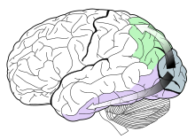
Posterior parietal cortex (light green) is shown at the posterior area of the parietal lobe

The posterior parietal cortex is a portion of the parietal lobe, which manipulates mental images, and integrates sensory and motor portions of the brain.

A majority of experiments highlights a role of human posterior parietal cortex in visual working memory and attention. We therefore have to establish a clear separation of visual memory and attention from processes related to the planning of goal-directed motor behaviors.

We can only hold in mind a minute fraction of the visual scene. These mental representations are stored in visual short-term memory. Activity in the posterior parietal cortex is tightly correlated with the limited amount of scene information that can be stored in visual short-term memory. These results suggest that the posterior parietal cortex is a key neural locus of our impoverished mental representation of the visual world.

The posterior cortex might act as a capacity-limited store for the representation of the visual scene, the frontal/prefrontal cortex might be necessary for the consolidation and/or maintenance of this store, especially during extended retention intervals.

Visual cortex

The dorsal stream (green) and ventral stream (purple) are both actively involved in visual memory. Both pathways originate in the visual cortex.

There is a visual cortex in each hemisphere of the brain, much of which is located in the Occipital lobe. The left hemisphere visual cortex receives signals mainly from the right visual field and the right visual cortex mainly from the left visual field, although each cortex receives a considerable amount of information from the ipsilateral visual field as well. The visual cortex also receives information from subcortical regions, such as the lateral geniculate body, located in the thalamus. However, ample evidence indicates that object identity and location are preferentially processed in ventral (occipito-temporal) and dorsal (occipito-parietal) cortical visual streams, respectively. Comparison of rCBF during performance of the two tasks again revealed differences between the ventral and dorsal pathways.

Dorsal stream pathway

The dorsal stream pathway is mainly involved in the visual-spatial location of objects in the external world, and it is also known colloquially as the 'where' pathway. The dorsal stream pathway is also involved in the guidance of movements (e.g., reaching for an object in space), and is therefore implicated in the analysis of the movement of objects in addition to their spatial locations.

The dorsal stream pathway begins with purely visual information in the occipital lobe, and then this information is transferred to the parietal lobe for spatial awareness functions. Specifically, the posterior parietal cortex is essential for "the perception and interpretation of spatial relationships, accurate body image, and the learning of tasks involving coordination of the body in space."

Ventral stream pathway

The ventral stream pathway is mainly involved in object recognition, and is known colloquially as the 'what' pathway. It has connections to the medial temporal lobe (which is involved in the storage of long-term memories), the limbic system (which regulates emotions), and the dorsal stream pathway (which is involved in the visual-spatial locations and motions of objects). Therefore, the ventral stream pathway not only deals with the recognition of objects in the external world, but also the emotional judgement and analysis of these objects.

The ventral stream pathway begins with purely visual information in the primary visual cortex (occipital lobe), and then this information is transferred to the temporal lobe.

Occipital lobes

Located at the back of the brain, the occipital lobes receive and process visual information. The occipital lobes also process colors and shapes. Whereas the right occipital lobe interprets images from the left visual space, the left occipital lobe interprets images from the right visual space. Damage to the occipital lobes can permanently damage visual perception.

Occipital lobe injury complications

Damage to the occipital lobe is characterized by loss of visual capability and the inability to identify colors both important processes in visual memory.

Short term visual memory

Visual short term memory is the capacity for holding a small amount of visual information in mind in an active, readily available state for a short period of time (usually no more than 30 seconds). Although visual short term memory is essential for the execution of a wide array of perceptual and cognitive functions, and is supported by an extensive network of brain regions, its storage capacity is severely limited.

Visual short-term memory storage is mediated by distinctive posterior brain mechanisms, such that capacity is determined both by a fixed number of objects and by object complexity.

Long term visual memory

Recall of the patterns from long term visual memory is associated with rCBF increases in different areas of the prefrontal cortex and the anterior cingulate cortex. The retrieval of long term visual memories is associated with activation of both anterior and posterior temporal cortices. Posterior temporal cortical regions are more associated with retrieval of category-specific aspects of visual memory, whereas anterior regions of the temporal cortex are more associated with category-independent visual memory.

Methods of study

Benton visual retention test

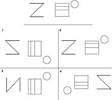
An example of the Benton Visual Retention Test. The target stimulus is presented at the top, after a delay participants are asked to recall the correct target stimulus from the list of design cards.

The Benton Visual Retention Test is an assessment of visual perception, and visual memory abilities. More than 50 years of proven clinical use is the staple of the Benton Visual Retention Test. This test has proven its sensitivity to reading disabilities, nonverbal learning disabilities, traumatic brain injury, attention-deficit disorder, alzheimer's, and other forms of dementia. During testing participants are presented with 10 cards for 10 seconds with unique designs on each. After the time has passed participants are asked to immediately reproduce the designs from each card using their visual memory. In the second stage participants are asked to copy each of the 10 card designs while the cards are in view. The participants results from each task are then assessed and placed into six categories; omissions, distortions, preservations, rotations, misplacements, and sizing errors. The further the participant's scores varies from the averages provided in the Benton Visual Retention Test manual the worse the participant is assessed to be on visual memory ability. The Benton Visual Retention Test has proved to be a generalizable test with the ability to be accurately administered to participants aged 8-adult, and no gender effect. Some studies have suggested a significant gender and education interaction indicating that an age-associated decline in visual memory performance may be more prominent for those individuals with a lower education level.

Neuroimaging tests

An example of a colored geometrical pattern a subject would encode, store, and recall while performing a visual memory neuroimaging test.

Neuroimaging studies focus on the neural networks involved in visual memory using methods designed to activate brain areas involved in encoding, storage, and recall. These studies involve the use of one or multiple types of brain imaging techniques designed to measure timing or activation within the brain. The data collected from neuroimaging studies gives researchers the ability to visualize which brain regions are activated in specific cognitive visual memory tasks. With the use of brain imaging devices researchers able to further investigate memory performance above and beyond standard tests based on exact response times, and activation.

Control condition

The subject's resting brain activation level is first determined in order to form a control or 'baseline' to measure from. Subjects are blindfolded and instructed to lay motionless while simultaneously eliminating any visual imagery present in their mind's eye. These instructions are intended to minimize the activation of brain regions involved in visual memory to form a true resting brain state. After the scan is complete a control has been formed which can be compared with activated regions of the brain while performing visual memory tasks.

Activation condition

During encoding, participants are typically exposed to 1–10 visual patterns while connected to a brain imaging device. As the subject encodes the visual patterns researchers are able to directly view the activation of areas involved in visual memory encoding. During recall subjects again need to have all visual stimuli removed by means of a dark room or blindfolding to avoid interfering activation of other visual areas in the brain. Subjects are asked to recall each image clearly in their mind's eye. While recalling the images researchers are able view the areas activated by the visual memory task. Comparing the control 'baseline' state to the activated areas during the visual memory task allows researchers to view which areas are used during visual memory.

Current theories

Visuo-spatial sketch pad

The visuo-spatial sketchpad is part of Baddeley and Hitch's model of working memory. It is responsible for temporarily storing visual and spatial information, which is currently being used or encoded. It is thought of as a three-dimensional cognitive map, which contains spatial features about where the person is and visual images of the area, or an object being concentrated on. It is used in tasks such as mental image manipulation where a person imagines how a real object would look if it were changed in some way (rotated, flipped, moved, change of colour, etc.). It is also responsible for representing how vivid an image is. A vivid image is one which you have a high potential for retrieving its sensory details. The visuo-spatial sketchpad is responsible for holding onto the visual and spatial qualities of a vivid image in your working memory, and the degree of vividness is directly affected by the limits of the sketchpad.

Eidetic and photographic memories

Eidetic memory is an ability to recall images, sounds, or objects in memory with high precision for a few minutes without using mnemonics. It occurs in a small number of children and generally is not found in adults.

The popular culture concept of photographic memory—where, for example, someone can briefly look at a page of text and then recite it perfectly from memory—is not the same as seeing eidetic images, and photographic memory has never been demonstrated to exist.

Iconic memory

Iconic memory is the visual part of the sensory memory system. Iconic memory is responsible for visual priming, because it works very quickly and unconsciously. Iconic memory decays very quickly, but contains a very vivid image of the surrounding stimuli.

Spatial memory

Spatial memory is a person's knowledge of the space around them, and their whereabouts in it. It also encompasses all memories of areas and places, and how to get to and from them. Spatial memory is distinct from object memory and involves different parts of the brain. Spatial memory involves the dorsal parts of the brain and more specifically the hippocampus. However many times both types of memory are used together, such as when trying to remember where you put a lost object. A classic test of spatial memory is the Corsi block-tapping task, where an instructor taps a series of blocks in a random order and the participant attempts to imitate them. The number of blocks they can tap before performance breaks down on average is called their Corsi span. Spatial memory is always being used whenever a person is moving any part of their body; therefore it is generally more vulnerable to decay than object memory is.

Object memory

Object memory involves processing features of an object or material such as texture, color, size, and orientation. It is processed mainly in the ventral regions of the brain. A few studies have shown that on average most people can recall up to four items each with a set of four different visual qualities. It is a separate system from spatial memory and is not affected by interference from spatial tasks.

Accuracy

Visual memory is not always accurate and can be misled by outside conditions. This can be seen in studies carried out by Elizabeth Loftus and Gary Wells. In one study by Wells, individuals were exposed to misleading information after witnessing an event; they were then tested on their ability to remember details from this event. Their findings included: when given misinformation that contradicts the witnessed event they were less able to recall those details; and whether misinformation was given before or after the witnessed event did not seem to matter. Furthermore, visual memory can be subjected to various memory errors which will affect accuracy.

Visual memory in education

"We do not store and retrieve words based on visual memory." "Our phonological filing system is the basis for word memory/word recognition." -Dr. Kilpatrick (Equipped for Reading Success).

Visual memory, in an academic environment, entails work with pictures, symbols, numbers, letters, and especially words. Students must be able to look at a word, form an image of that word in their minds and be able to recall the appearance of the word later. When teachers introduce a new vocabulary word, generally they write it on the chalkboard, have the children spell it, read it and then use it in a sentence. The word is then erased from the chalkboard. Students with good visual memory will recognize that same word later in their readers or other texts and will be able to recall the appearance of the word to spell it.

Children who have not developed their visual memory skills cannot readily reproduce a sequence of visual stimuli. They frequently experience difficulty in remembering the overall visual appearance of words or the letter sequence of words for reading and spelling.

Factors affecting visual memory

Sleep

Findings surrounding sleep and visual memory have been mixed. Studies have reported performance increases after a bout of sleep compared with the same period of waking. The implications of this are that there is a slow, offline process during sleep that strengthens and enhances the memory trace. Further studies have found that quiet rest has shown the same learning benefits as sleep. Replay has been found to occur during post-training quiet wakefulness as well as sleep. In a recent study where a visual search task was administered quiet rest or sleep is found to be necessary for increasing the amount of associations between configurations and target locations that can be learned within a day. Reactivation in sleep was only observed after extensive training of rodents on familiar tasks. It rapidly dissipates; it also makes up a small proportion of total recorded activity in sleep. It has also been found that there are gender differences between males and females in regards to visual memory and sleep. In a study done testing sleep and memory for pictures it was found that daytime sleep contributed to retention of source memory rather than item memory in females, females did not have recollection or familiarity influenced by daytime sleep, whereas males undergoing daytime sleep had a trend towards increased familiarity. The reasons for this may be linked to different memory traces resulting from different encoding strategies, as well as with different electrophysiological changes during daytime sleep.

Brain damage

Brain damage is another factor that has been found to have an effect on visual memory. Memory impairment affects both novel and familiar experiences. Poor memory after damage to the brain is usually considered to result from information being lost or rendered inaccessible. With such impairment it is assumed that it must be due to the incorrect interpretation of previously encountered information as being novel. In experiments testing rats’ object recognition memory it was found that memory impairment can be the opposite, that there was a tendency to treat novel experiences as familiar. A possible solution for this impairment could be the use of a visual-restriction procedure that reduces interference.

Age

Studies have shown that with aging, in terms of short-term visual memory, viewing time and task complexity affect performance. When there is a delay or when the task is complex recall declines. In a study conducted to measure whether visual memory in older adults with age-related visual decline was caused by memory performance or visual functioning, the following were examined: relationships among age, visual activity, and visual and verbal memory in 89 community dwelling volunteers aged 60–87 years. The findings were that the effect of vision was not specific to visual memory. Therefore, vision was found to be correlated with general memory function in older adults and is not modality specific.

As we age performance in regards to spatial configurations deteriorates. In a task to store and combine two different spatial configurations to form a novel one young people out-performed the elderly. Vision also has an effect on performance. Sighted participants outperformed the visually impaired regardless of testing modality. This suggests that vision tends to shape the general supramodal mechanisms of memory.

Alcohol

Studies have shown that there is an effect of alcohol on visual memory. In a recent study visual working memory and its neutral correlates was assessed in university students who partake in binge drinking, the intermittent consumption of large amounts of alcohol. The findings revealed that there may be binge-drinking related functional alteration in recognition working memory processes. This suggests that impaired prefrontal cortex function may occur at an early age in binge drinkers.

Another study conducted in 2004 examined the level of response to alcohol and brain response during visual working memory. This study looked at the neural correlated of the low level of response to alcohol using functional magnetic resonance imaging during a challenging visual memory task. The results were that young people who report having needed more alcohol to feel the effects showed higher levels of brain response during visual working memory, this suggests that the individual's capacity to adjust to cognitive processing decreases, they are less able to adjust cognitive processing to contextual demands.

Dysfunction of visual memory

Hallucinatory palinopsia, which is a dysfunction of visual memory, is caused by posterior visual pathway cortical lesions and seizures, most commonly in the non-dominant parietal lobe. Focal hyperactivity causes persistent activation of a visual cortex-hippocampal neuronal circuit which encodes an object or scene that is already in visual memory. "All of the hallucinatory palinopsia symptoms occur concomitantly in a patient with one lesion, which supports current evidence that objects, features, and scenes are all units of visual memory, perhaps at different levels of processing. This alludes to neuroanatomical integration in visual memory creation and storage." Studying the excitability alterations associated with palinopsia in migraineurs could provide insight on mechanisms of encoding visual memory.

One common group of people that have visual memory problems are children with reading disabilities. It was often thought that disabilities are caused by failure to perceive the letters of a written word in the right order. However, studies show it is more likely that it is caused by a failure to encode and process the correct order of letters within the word. This means that the child perceives the word just as anyone else would, however their brains do not appear to hold onto the visual characteristics of the word. Although initially it was found that children with reading disabilities had comparable visual memory to those without difficulty, a more specific part of the visual memory system has been found to cause reading disabilities.

These parts are the sustained and transient visual processing systems. The sustained system is responsible for fine detail such as word and letter recognition and is very important in encoding words in their correct order. The transient system is responsible for controlling eye movements, and processing the larger visual environment around us. When these two processes do not work in synchronization this can cause reading disabilities. This has been tested by having children with and without reading disabilities perform on tasks related to the transient systems, where the children with reading disabilities did very poorly. It has also been found in postmortem examinations of the brains of people with reading disabilities that they have fewer neurons and connections in the areas representing the transient visual systems. However, there is debate over whether this is the only reason for reading disabilities, scotopic sensitivity syndrome, deficits in verbal memory and orthographic knowledge are other proposed factors.

Deficits in visual memory can also be caused by disease and/or trauma to the brain. These can lead to the patient losing their spatial memory, and/or their visual memory for specific things. For example, a patient “L.E.” suffered brain damage and her ability to draw from memory was severely diminished, whilst her spatial memory remained normal. Other patients represent the opposite, where memory for colors and shapes is unaffected but spatial memory for previously known places is greatly impaired. These case studies show that these two types of visual memory are located in different parts of the brain and are somewhat unrelated in terms of functioning in daily life.

Spatial memory

From Wikipedia, the free encyclopedia
 
Spatial memory is required to navigate through an environment.

In cognitive psychology and neuroscience, spatial memory is a form of memory responsible for the recording and recovery of information needed to plan a course to a location and to recall the location of an object or the occurrence of an event. Spatial memory is necessary for orientation in space. Spatial memory can also be divided into egocentric and allocentric spatial memory. A person's spatial memory is required to navigate around a familiar city. A rat's spatial memory is needed to learn the location of food at the end of a maze. In both humans and animals, spatial memories are summarized as a cognitive map.

Spatial memory has representations within working, short-term memory and long-term memory. Research indicates that there are specific areas of the brain associated with spatial memory. Many methods are used for measuring spatial memory in children, adults, and animals.

Short-term spatial memory

Short-term memory (STM) can be described as a system allowing one to temporarily store and manage information that is necessary to complete complex cognitive tasks. Tasks which employ short-term memory include learning, reasoning, and comprehension. Spatial memory is a cognitive process that enables a person to remember different locations as well as spatial relations between objects. This allows one to remember where an object is in relation to another object; for instance, allowing someone to navigate through a familiar city. Spatial memories are said to form after a person has already gathered and processed sensory information about her or his environment.

Spatial working memory

Working memory (WM) can be described as a limited capacity system that allows one to temporarily store and process information. This temporary store enables one to complete or work on complex tasks while being able to keep information in mind. For instance, the ability to work on a complicated mathematical problem utilizes one's working memory.

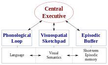
One influential theory of WM is the Baddeley and Hitch multi-component model of working memory. The most recent version of this model suggests that there are four subcomponents to WM: phonological loop, the visuo-spatial sketchpad, the central executive, and the episodic buffer. One component of this model, the visuo-spatial sketchpad, is likely responsible for the temporary storage, maintenance, and manipulation of both visual and spatial information.

Baddeley and Hitch's multi-component model of working memory.

In contrast to the multi-component model, some researchers believe that STM should be viewed as a unitary construct. In this respect, visual, spatial, and verbal information are thought to be organized by levels of representation rather than the type of store to which they belong. Within the literature, it is suggested that further research into the fractionation of STM and WM be explored. However, much of the research into the visuo-spatial memory construct have been conducted in accordance to the paradigm advanced by Baddeley and Hitch.

The role of the central executive

Research into the exact function of the visuo-spatial sketchpad has indicated that both spatial short-term memory and working memory are dependent on executive resources and are not entirely distinct. For instance, performance on a working memory but not on a short-term memory task was affected by articulatory suppression suggesting that impairment on the spatial task was caused by the concurrent performance on a task that had extensive use of executive resources. Results have also found that performances were impaired on STM and WM tasks with executive suppression. This illustrates how, within the visuo-spatial domain, both STM and WM require similar utility of the central executive.

Additionally, during a spatial visualisation task (which is related to executive functioning and not STM or WM) concurrent executive suppression impaired performance indicating that the effects were due to common demands on the central executive and not short-term storage. The researchers concluded with the explanation that the central executive employs cognitive strategies enabling participants to both encode and maintain mental representations during short-term memory tasks.

Although studies suggest that the central executive is intimately involved in a number of spatial tasks, the exact way in which they are connected remains to be seen.

Long-term spatial memory

Spatial memory recall is built upon a hierarchical structure. People remember the general layout of a particular space and then "cue target locations" within that spatial set. This paradigm includes an ordinal scale of features that an individual must attend to in order to inform his or her cognitive map. Recollection of spatial details is a top-down procedure that requires an individual to recall the superordinate features of a cognitive map, followed by the ordinate and subordinate features. Two spatial features are prominent in navigating a path: general layout and landmark orienting (Kahana et al., 2006). People are not only capable of learning about the spatial layout of their surroundings, but they can also piece together novel routes and new spatial relations through inference.

A cognitive map is "a mental model of objects' spatial configuration that permits navigation along optimal path between arbitrary pairs of points." This mental map is built upon two fundamental bedrocks: layout, also known as route knowledge, and landmark orientation. Layout is potentially the first method of navigation that people learn to utilize; its workings reflect our most basic understandings of the world.

Hermer and Spelke (1994) determined that when toddlers begin to walk, around eighteen months, they navigate by their sense of the world's layout. McNamara, Hardy and Hirtle identified region membership as a major building block of anyone's cognitive map (1989). Specifically, region membership is defined by any kind of boundary, whether physical, perceptual or subjective (McNamara et al., 1989). Boundaries are among the most basic and endemic qualities in the world around us. These boundaries are nothing more than axial lines which are a feature that people are biased towards when relating to space; for example, one axial line determinant is gravity (McNamara & Shelton, 2001; Kim & Penn, 2004). Axial lines aid everyone in apportioning our perceptions into regions. This parceled world idea is further supported items by the finding that items that get recalled together are more likely than not to also be clustered within the same region of one's larger cognitive map. Clustering shows that people tend to chunk information together according to smaller layouts within a larger cognitive map.

Boundaries are not the only determinants of layout. Clustering also demonstrates another important property of relation to spatial conceptions, which is that spatial recall is a hierarchical process. When someone recalls an environment or navigates terrain, that person implicitly recalls the overall layout at first. Then, due to the concept's "rich correlational structure", a series of associations become activated. Eventually, the resulting cascade of activations will awaken the particular details that correspond with the region being recalled. This is how people encode many entities from varying ontological levels, such as the location of a stapler; in a desk; which is in the office.

One can recall from only one region at a time (a bottleneck). A bottleneck in a person's cognitive navigational system could be an issue. For instance, if there were a need for a sudden detour on a long road trip. Lack of experience in a locale, or simply sheer size, can disorient one's mental layout, especially in a large and unfamiliar place with many overwhelming stimuli. In these environments, people are still able to orient themselves, and find their way around using landmarks. This ability to "prioritize objects and regions in complex scenes for selection (and) recognition" was labeled by Chun and Jiang in 1998. Landmarks give people guidance by activating "learned associations between the global context and target locations." Mallot and Gillner (2000) showed that subjects learned an association between a specific landmark and the direction of a turn, thereby furthering the relationship between associations and landmarks. Shelton and McNamara (2001) succinctly summed up why landmarks, as markers, are so helpful: "location...cannot be described without making reference to the orientation of the observer."

People use both the layout of a particular space and the presence of orienting landmarks in order to navigate. Psychologists have yet to explain whether layout affects landmarks or if landmarks determine the boundaries of a layout. Because of this, the concept suffers from a chicken and the egg paradox. McNamara has found that subjects use "clusters of landmarks as intrinsic frames of reference," which only confuses the issue further.

People perceive objects in their environment relative to other objects in that same environment. Landmarks and layout are complementary systems for spatial recall, but it is unknown how these two systems interact when both types of information are available. As a result, people have to make certain assumptions about the interaction between the two systems. For example, cognitive maps are not "absolute" but rather, as anyone can attest, are "used to provide a default...(which) modulated according to...task demands." Psychologists also think that cognitive maps are instance based, which accounts for "discriminative matching to past experience."

This field has traditionally been hampered by confounding variables, such as cost and the potential for previous exposure to an experimental environment. Technological advancements, including those in virtual reality technology, have made findings more accessible. Virtual reality affords experimenters the luxury of extreme control over their test environment. Any variable can be manipulated, including things that would not be possible in reality.

Virtual reality

During a 2006 study researchers designed three different virtual towns, each of which had its own "unique road layout and a unique set of five stores." However, the overall footprint of the different maps was exactly the same size, "80 sq. units." In this experiment, participants had to partake in two different sets of trials.

A study conducted at the University of Maryland compared the effect of different levels of immersion on spatial memory recall. In the study, 40 participants used both a traditional desktop and a head-mounted display to view two environments, a medieval town, and an ornate palace, where they memorized two sets of 21 faces presented as 3D portraits. After viewing these 21 faces for 5 minutes, followed by a brief rest period, the faces in the virtual environments were replaced with numbers and participants recalled which face was at each location. The study found on average, those who used the head-mounted display recalled the faces 8.8% more accurately, and with a greater confidence. The participants state that leveraging their innate vestibular and proprioceptive senses with the head-mounted display and mapping aspects of the environment relative to their body, elements that are absent with the desktop, was key to their success.

Spatial expertise

Within the literature, there is evidence that experts in a particular field are able to perform memory tasks in accordance with their skills at an exceptional level. The level of skill displayed by experts may exceed the limits of the normal capacity of both STM and WM. Because experts have an enormous amount of prelearned and task-specific knowledge, they may be able to encode information in a more efficient way.

An interesting study investigating taxi drivers' memory for streets in Helsinki, Finland, examined the role of prelearned spatial knowledge. This study compared experts to a control group to determine how this prelearned knowledge in their skill domain allows them to overcome the capacity limitations of STM and WM. The study used four levels of spatial randomness:

  • Route Order – spatially continuous route
  • Route Random – spatially continuous list presented randomly
  • Map Order – street names forming a straight line on the map, but omitting intermediate streets
  • Map Random – streets on map presented in random order
Yellow taxi cabs in New York city

The results of this study indicate that the taxi drivers' (experts') recall of streets was higher in both the route order condition and the map order condition than in the two random conditions. This indicates that the experts were able to use their prelearned spatial knowledge to organize the information in such a way that they surpassed STM and WM capacity limitations. The organization strategy that the drivers employed is known as chunking. Additionally, the comments made by the experts during the procedure point towards their use of route knowledge in completing the task. To ensure that it was in fact spatial information that they were encoding, the researchers also presented lists in alphabetical order and semantic categories. However, the researchers found that it was in fact spatial information that the experts were chunking, allowing them to surpass the limitations of both visuo-spatial STM and WM.

Animal research

Certain species of paridae and corvidae (such as the black-capped chickadee and the scrub jay) are able to use spatial memory to remember where, when and what type of food they have cached. Studies on rats and squirrels have also suggested that they are able to use spatial memory to locate previously hidden food. Experiments using the radial maze have allowed researchers to control for a number of variables, such as the type of food hidden, the locations where the food is hidden, the retention interval, as well as any odor cues that could skew results of memory research. Studies have indicated that rats have memory for where they have hidden food and what type of food they have hidden. This is shown in retrieval behavior, such that the rats are selective in going more often to the arms of the maze where they have previously hidden preferred food than to arms with less preferred food or where no food was hidden.

The evidence for the spatial memory of some species of animals, such as rats, indicates that they do use spatial memory to locate and retrieve hidden food stores.

A study using GPS tracking to see where domestic cats go when their owners let them outside reported that cats have substantial spatial memory. Some of the cats in the study demonstrated exceptional long term spatial memory. One of them, usually traveling no further than 200 m (660 ft) to 250 m (820 ft) from its home, unexpectedly traveled some 1,250 m (4,100 ft) from its home. Researchers initially thought this to be a GPS malfunction, but soon discovered that the cat's owners went out of town that weekend, and that the house the cat went to was the owner's old house. The owners and the cat had not lived in that house for well over a year.

Visual–spatial distinction

Logie (1995) proposed that the visuo-spatial sketchpad is broken down into two subcomponents, one visual and one spatial. These are the visual cache and the inner scribe, respectively. The visual cache is a temporary visual store including such dimensions as color and shape. Conversely, the inner scribe is a rehearsal mechanism for visual information and is responsible for information concerning movement sequences. Although a general lack of consensus regarding this distinction has been noted in the literature, there is a growing amount of evidence that the two components are separate and serve different functions.

Visual memory is responsible for retaining visual shapes and colors (i.e., what), whereas spatial memory is responsible for information about locations and movement (i.e., where). This distinction is not always straightforward since part of visual memory involves spatial information and vice versa. For example, memory for object shapes usually involves maintaining information about the spatial arrangement of the features which define the object in question.

In practice, the two systems work together in some capacity but different tasks have been developed to highlight the unique abilities involved in either visual or spatial memory. For example, the visual patterns test (VPT) measures visual span whereas the Corsi Blocks Task measures spatial span. Correlational studies of the two measures suggest a separation between visual and spatial abilities, due to a lack of correlation found between them in both healthy and brain damaged patients.

Support for the division of visual and spatial memory components is found through experiments using the dual-task paradigm. A number of studies have shown that the retention of visual shapes or colors (i.e., visual information) is disrupted by the presentation of irrelevant pictures or dynamic visual noise. Conversely, the retention of location (i.e., spatial information) is disrupted only by spatial tracking tasks, spatial tapping tasks, and eye movements. For example, participants completed both the VPT and the Corsi Blocks Task in a selective interference experiment. During the retention interval of the VPT, the subject viewed irrelevant pictures (e.g., avant-garde paintings). The spatial interference task required participants to follow, by touching the stimuli, an arrangement of small wooden pegs which were concealed behind a screen. Both the visual and spatial spans were shortened by their respective interference tasks, confirming that the Corsi Blocks Task relates primarily to spatial working memory.

Measurement

There are a variety of tasks psychologists use to measure spatial memory on adults, children and animal models. These tasks allow professionals to identify cognitive irregularities in adults and children and allows researchers to administer varying types of drugs and/or lesions in participants and measure the consequential effects on spatial memory.

The Corsi block tapping task

The Corsi block-tapping test, also known as the Corsi span rest, is a psychological test commonly used to determine the visual-spatial memory span and the implicit visual-spatial learning abilities of an individual. Participants sit with nine wooden 3x3-cm blocks fastened before them on a 25- x 30-cm baseboard in a standard random order. The experiment taps onto the blocks a sequence pattern which participants must then replicate. The blocks are numbered on the experimenters' side to allow for efficient pattern demonstration. The sequence length increases each trial until the participant is no longer able to replicate the pattern correctly. The test can be used to measure both short-term and long-term spatial memory, depending on the length of time between test and recall.

The test was created by Canadian neuropsychologist Phillip Corsi, who modeled it after Hebb's digit span task by replacing the numerical test items with spatial ones. On average, most participants achieve a span of five items on the Corsi span test and seven on the digit span task.

Visual pattern span

The visual pattern span is similar to the Corsi block tapping test but regarded as a more pure test of visual short-term recall. Participants are presented with a series of matrix patterns that have half their cells colored and the other half blank. The matrix patterns are arranged in a way that is difficult to code verbally, forcing the participant to rely on visual spatial memory. Beginning with a small 2 x 2 matrix, participants copy the matrix pattern from memory into an empty matrix. The matrix patterns are increased in size and complexity at a rate of two cells until the participant's ability to replicate them breaks down. On average, participants' performance tends to break down at sixteen cells.

Pathway span task

This task is designed to measure spatial memory abilities in children. The experimenter asks the participant to visualize a blank matrix with a little man. Through a series of directional instructions such as forwards, backwards, left or right, the experimenter guides the participant's little man on a pathway throughout the matrix. At the end, the participant is asked to indicate on a real matrix where the little man that he or she visualized finished. The length of the pathway varies depending on the level of difficulty (1-10) and the matrices themselves may vary in length from 2 x 2 cells to 6 x 6.

Dynamic mazes

Dynamic mazes are intended for measuring spatial ability in children. With this test, an experimenter presents the participant with a drawing of a maze with a picture of a man in the center. While the participant watches, the experimenter uses his or her finger to trace a pathway from the opening of the maze to the drawing of the man. The participant is then expected to replicate the demonstrated pathway through the maze to the drawing of the man. Mazes vary in complexity as difficulty increases.

Radial arm maze

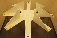
Simple Radial Maze

First pioneered by Olton and Samuelson in 1976, the radial arm maze is designed to test the spatial memory capabilities of rats. Mazes are typically designed with a center platform and a varying number of arms branching off with food placed at the ends. The arms are usually shielded from each other in some way but not to the extent that external cues cannot be used as reference points.

In most cases, the rat is placed in the center of the maze and needs to explore each arm individually to retrieve food while simultaneously remembering which arms it has already pursued. The maze is set up so the rat is forced to return to the center of the maze before pursuing another arm. Measures are usually taken to prevent the rat from using its olfactory senses to navigate such as placing extra food throughout the bottom of the maze.

Morris water navigation task

The Morris water navigation task is a classic test for studying spatial learning and memory in rats and was first developed in 1981 by Richard G. Morris for whom the test is named. The subject is placed in a round tank of translucent water with walls that are too high for it to climb out and water that is too deep for it to stand in. The walls of the tank are decorated with visual cues to serve as reference points. The rat must swim around the pool until by chance it discovers just below the surface the hidden platform onto which it can climb.

Typically, rats swim around the edge of the pool first before venturing out into the center in a meandering pattern before stumbling upon the hidden platform. However, as time spent in the pool increases experience, the amount of time needed to locate the platform decreases, with veteran rats swimming directly to the platform almost immediately after being placed in the water.

Physiology

Hippocampus

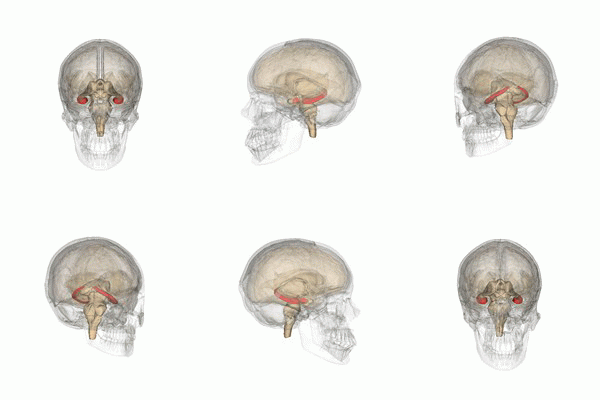
rotating 3D animation of the human hippocampus in skull.
Hippocampus shown in red

The hippocampus provides animals with a spatial map of their environment. It stores information regarding non-egocentric space (egocentric means in reference to one's body position in space) and therefore supports viewpoint independence in spatial memory. This means that it allows for viewpoint manipulation from memory. It is important for long-term spatial memory of allocentric space (reference to external cues in space). Maintenance and retrieval of memories are thus relational or context dependent. The hippocampus makes use of reference and working memory and has the important role of processing information about spatial locations.

Blocking plasticity in this region results in problems in goal-directed navigation and impairs the ability to remember precise locations. Amnesic patients with damage to the hippocampus cannot learn or remember spatial layouts, and patients having undergone hippocampal removal are severely impaired in spatial navigation.

Monkeys with lesions to this area cannot learn object-place associations and rats also display spatial deficits by not reacting to spatial change. In addition, rats with hippocampal lesions were shown to have temporally ungraded (time-independent) retrograde amnesia that is resistant to recognition of a learned platform task only when the entire hippocampus is lesioned, but not when it is partially lesioned. Deficits in spatial memory are also found in spatial discrimination tasks.

Brain slice showing areas CA1 and CA3 in hippocampus.
Brain slice showing areas CA1 and CA3 in the hippocampus

Large differences in spatial impairment are found among the dorsal and ventral hippocampus. Lesions to the ventral hippocampus have no effect on spatial memory, while the dorsal hippocampus is required for retrieval, processing short-term memory and transferring memory from the short term to longer delay periods. Infusion of amphetamine into the dorsal hippocampus has also been shown to enhance memory for spatial locations learned previously. These findings indicate that there is a functional dissociation between the dorsal and ventral hippocampus.

Hemispheric differences within the hippocampus are also observed. A study on London taxi drivers, asked drivers to recall complex routes around the city as well as famous landmarks for which the drivers had no knowledge of their spatial location. This resulted in an activation of the right hippocampus solely during recall of the complex routes which indicates that the right hippocampus is used for navigation in large scale spatial environments.

The hippocampus is known to contain two separate memory circuits. One circuit is used for recollection-based place recognition memory and includes the entorhinal-CA1 system, while the other system, consisting of the hippocampus trisynaptic loop (entohinal-dentate-CA3-CA1) is used for place recall memory and facilitation of plasticity at the entorhinal-dentate synapse in mice is sufficient to enhance place recall.

Place cells are also found in the hippocampus.

Posterior parietal cortex

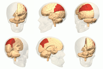
rotating 3D animation of the parietal lobe in human skull.
Parietal lobe shown in red

The parietal cortex encodes spatial information using an egocentric frame of reference. It is therefore involved in the transformation of sensory information coordinates into action or effector coordinates by updating the spatial representation of the body within the environment. As a result, lesions to the parietal cortex produce deficits in the acquisition and retention of egocentric tasks, whereas minor impairment is seen among allocentric tasks.

Rats with lesions to the anterior region of the posterior parietal cortex reexplore displaced objects, while rats with lesions to the posterior region of the posterior parietal cortex displayed no reaction to spatial change.

Parietal cortex lesions are also known to produce temporally ungraded retrograde amnesia.

Entorhinal cortex

medial view of the right cerebral hemisphere showing the entorhinal cortex near the base of the temporal lobe.
Medial view of the right cerebral hemisphere showing the entorhinal cortex in red at the base of the temporal lobe

The dorsalcaudal medial entorhinal cortex (dMEC) contains a topographically organized map of the spatial environment made up of grid cells. This brain region thus transforms sensory input from the environment and stores it as a durable allocentric representation in the brain to be used for path integration.

The entorhinal cortex contributes to the processing and integration of geometric properties and information in the environment. Lesions to this region impair the use of distal but not proximal landmarks during navigation and produces a delay-dependent deficit in spatial memory that is proportional to the length of the delay. Lesions to this region are also known to create retention deficits for tasks learned up to 4 weeks but not 6 weeks prior to the lesions.

Memory consolidation in the entorhinal cortex is achieved through extracellular signal-regulated kinase activity.

Prefrontal cortex

medial view of the right cerebral hemisphere showing the location of the prefrontal cortex at the front of the brain and more specifically the medial prefrontal cortex and ventromedial prefrontal cortex.
Medial view of the cerebral hemisphere showing the location of the prefrontal cortex and more specifically the medial and ventromedial prefrontal cortex in purple

The medial prefrontal cortex processes egocentric spatial information. It participates in the processing of short-term spatial memory used to guide planned search behavior and is believed to join spatial information with its motivational significance. The identification of neurons that anticipate expected rewards in a spatial task support this hypothesis. The medial prefrontal cortex is also implicated in the temporal organization of information.

Hemisphere specialization is found in this brain region. The left prefrontal cortex preferentially processes categorical spatial memory including source memory (reference to spatial relationships between a place or event), while the right prefrontal cortex preferentially processes coordinate spatial memory including item memory (reference to spatial relationships between features of an item).

Lesions to the medial prefrontal cortex impair the performance of rats on a previously trained radial arm maze, but rats can gradually improve to the level of the controls as a function of experience. Lesions to this area also cause deficits on delayed nonmatching-to-positions tasks and impairments in the acquisition of spatial memory tasks during training trials.

Retrosplenial cortex

The retrosplenial cortex is involved in the processing of allocentric memory and geometric properties in the environment. Inactivation of this region accounts for impaired navigation in the dark and it may be involved in the process of path integration.

Lesions to the retrosplenial cortex consistently impair tests of allocentric memory, while sparing egocentric memory. Animals with lesions to the caudal retrosplenial cortex show impaired performance on a radial arm maze only when the maze is rotated to remove their reliance on intramaze cues.

medial surface of the cerebral hemisphere indicating locations of Brodmann's areas.
Medial view of the cerebral hemisphere. The retrosplenial cortex encompasses Brodmann areas 26, 29, and 30. The perirhinal cortex contains Brodmann area 35 and 36 (not shown)

In humans, damage to the retrosplenial cortex results in topographical disorientation. Most cases involve damage to the right retrosplenial cortex and include Brodmann area 30. Patients are often impaired at learning new routes and at navigating through familiar environments. However, most patients usually recover within 8 weeks.

The retrosplenial cortex preferentially processes spatial information in the right hemisphere.

Perirhinal cortex

The perirhinal cortex is associated with both spatial reference and spatial working memory. It processes relational information of environmental cues and locations.

Lesions in the perirhinal cortex account for deficits in reference memory and working memory, and increase the rate of forgetting of information during training trials of the Morris water maze. This accounts for the impairment in the initial acquisition of the task. Lesions also cause impairment on an object location task and reduce habituation to a novel environment.

Neuroplasticity

Spatial memories are formed after an animal gathers and processes sensory information about its surroundings (especially vision and proprioception). In general, mammals require a functioning hippocampus (particularly area CA1) in order to form and process memories about space. There is some evidence that human spatial memory is strongly tied to the right hemisphere of the brain.

Spatial learning requires both NMDA and AMPA receptors, consolidation requires NMDA receptors, and the retrieval of spatial memories requires AMPA receptors. In rodents, spatial memory has been shown to covary with the size of a part of the hippocampal mossy fiber projection.

The function of NMDA receptors varies according to the subregion of the hippocampus. NMDA receptors are required in the CA3 of the hippocampus when spatial information needs to be reorganized, while NMDA receptors in the CA1 are required in the acquisition and retrieval of memory after a delay, as well as in the formation of CA1 place fields. Blockade of the NMDA receptors prevents induction of long-term potentiation and impairs spatial learning.

The CA3 of the hippocampus plays an especially important role in the encoding and retrieval of spatial memories. The CA3 is innervated by two afferent paths known as the perforant path (PPCA3) and the dentate gyrus (DG)-mediated mossy fibers (MFs). The first path is regarded as the retrieval index path while the second is concerned with encoding.

Disorders/deficits

Topographical disorientation

Topographical disorientation (TD) is a cognitive disorder that results in the individual being unable to orient his or herself in the real or virtual environment. Patients also struggle with spatial-information dependent tasks. These problems could possibly be the result of a disruption in the ability to access one's cognitive map, a mental representation of the surrounding environment or the inability to judge objects' location in relation to one's self.

Developmental topographical disorientation (DTD) is diagnosed when patients have shown an inability to navigate even familiar surroundings since birth and show no apparent neurological causes for this deficiency such as lesioning or brain damage. DTD is a relatively new disorder and can occur in varying degrees of severity.

A study was done to see if topographical disorientation had an effect on individuals who had mild cognitive impairment (MCI). The study was done by recruiting forty-one patients diagnosed with MCI and 24 healthy control individuals. The standards that were set for this experiment were:

  1. Subjective cognitive complaint by the patient or his/her caregiver.
  2. Normal general cognitive function above the 16th percentile on the Korean version of the Mini-Mental State Examination (K-MMSE).
  3. Normal activities of daily living (ADL) assessed both clinically and on a standardized scale (as described below).
  4. Objective cognitive decline below the 16th percentile on neuropsychological tests.
  5. Exclusion of dementia.

TD was assessed clinically in all participants. Neurological and neuropsychological evaluations were determined by a magnetic imaging scan which was performed on each participant. Voxel-based morphometry was used to compare patterns of gray-matter atrophy between patients with and without TD, and a group of normal controls. The outcome of the experiment was that they found TD in 17 out of the 41 MCI patients (41.4%). The functional abilities were significantly impaired in MCI patients with TD compared to in MCI patients without TD and that the presence of TD in MCI patients is associated with loss of gray matter in the medial temporal regions, including the hippocampus.

Hippocampal damage and schizophrenia

Research with rats indicates that spatial memory may be adversely affected by neonatal damage to the hippocampus in a way that closely resembles schizophrenia. Schizophrenia is thought to stem from neurodevelopmental problems shortly after birth.

Rats are commonly used as models of schizophrenia patients. Experimenters create lesions in the ventral hippocampal area shortly after birth, a procedure known as neonatal ventral hippocampal lesioning(NVHL). Adult rats who with NVHL show typical indicators of schizophrenia such as hypersensitivity to psychostimulants, reduced social interactions and impaired prepulse inhibition, working memory and set-shifting. Similar to schizophrenia, impaired rats fail to use environmental context in spatial learning tasks such as showing difficulty completing the radial arm maze and the Moris water maze.

GPS (Global Positioning System)

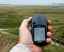
Example of a hand held GPS

Recent research on spatial memory and wayfinding in an article by Ishikawa et al. in 2008 revealed that using a GPS moving map device reduces an individual's navigation abilities when compared to other participants who were using maps or had previous experience on the route with a guide. GPS moving map devices are frequently set up to allow the user to only see a small detailed close-up of a particular segment of the map which is constantly updated. In comparison, maps usually allow the user to see the same view of the entire route from departure to arrival. Other research has shown that individuals who use GPS travel more slowly overall compared to map users who are faster. GPS users stop more frequently and for a longer period of time whereas map users and individuals using past experience as a guide travel on more direct routes to reach their goal.

NEIL1

Endonuclease VIII-like 1 (NEIL1) is a DNA repair enzyme that is widely expressed throughout the brain. NEIL1 is a DNA glycosylase that initiates the first step in base excision repair by cleaving bases damaged by reactive oxygen species and then introducing a DNA strand break via an associated lyase reaction. This enzyme recognizes and removes oxidized DNA bases including formamidopyrimidine, thymine glycol, 5-hydroxyuracil and 5-hydroxycytosine. NEIL1 promotes short-term spatial memory retention. Mice lacking NEIL1 have impaired short-term spatial memory retention in a water maze test.

Learning difficulties

Nonverbal learning disability is characterized by normal verbal abilities but impaired visuospatial abilities. Problem areas for children with nonverbal learning disability are arithmetic, geometry, and science. Impairments in spatial memory are linked to nonverbal learning disorder and other learning difficulties.

Arithmetic word problems involve written text containing a set of data followed by one or more questions and require the use of the four basic arithmetic operations (addition, subtraction, multiplication, or division). Researchers suggest that successful completion of arithmetic word problems involves spatial working memory (involved in building schematic representations) which facilitates the creation of spatial relationships between objects. Creating spatial relationships between objects is an important part of solving word problems because mental operations and transformations are required.

Researchers investigated the role of spatial memory and visual memory in the ability to complete arithmetic word problems. Children in the study completed the Corsi block task (forward and backward series) and a spatial matrix task, as well as a visual memory task called the house recognition test. Poor problem-solvers were impaired on the Corsi block tasks and the spatial matrix task, but performed normally on the house recognition test when compared to normally achieving children. The experiment demonstrated that poor problem solving is related specifically to deficient processing of spatial information.

Sleep

Sleep has been found to benefit spatial memory, by enhancing hippocampal-dependent memory consolidation. Hippocampal areas activated in route-learning are reactivated during subsequent sleep (NREM sleep in particular). It was demonstrated in a particular study that the actual extent of reactivation during sleep correlated with the improvement in route retrieval and therefore memory performance the following day. The study established the idea that sleep enhances the systems-level process of consolidation that consequently enhances/improves behavioral performance. A period of wakefulness has no effect on stabilizing memory traces, in comparison to a period of sleep. Sleep after the first post-training night, i.e. on the second night, does not benefit spatial memory consolidation further. Therefore, sleeping in the first post-training night e.g. after learning a route, is most important.

Sleep deprivation and sleep has also been a researched association. Sleep deprivation hinders memory performance improvement due to an active disruption of spatial memory consolidation. As a result, spatial memory is enhanced by a period of sleep.

Progressivism in the United States

From Wikipedia, the free encyclopedia ...