Search This Blog

Wednesday, June 23, 2021

Anticancer gene

From Wikipedia, the free encyclopedia

Anticancer genes are genes that, when ectopically overexpressed, specifically destroy tumour cells without harming normal, untransformed cells. This cellular destruction can be due to a variety of mechanisms, such as apoptosis, mitotic catastrophe followed by apoptosis or necrosis, and autophagy. Anticancer genes emerged from studies on cancer cells in the late 1990s. Currently, there have been 291 anticancer genes discovered in the human genome. In order to be classified as an anticancer gene, the gene must have base substitutions leading to missense amino-acid changes, deletions, or insertions leading to frameshifts that alter the protein the gene codes for, increases and decreases in copy-number increases, or gene rearrangements leading to their deregulation.

Anticancer genes as therapeutics

Cancer is classified as a group of diseases, all of which are characterized by uncontrolled cell proliferation. In normal functioning cells, apoptosis is induced to avoid these proliferative events. However, these processes may continue on to become cancer in the event the processes become dysregulated. Epidemiological studies have shown cancer to be a leading cause of death worldwide  (Figure 1). Current advancements in therapeutics have led to a substantial increase in patient survival rates.

Common anticancer gene examples

APOPTIN

History

Apoptin was the first anticancer gene to be isolated. This gene comes from the single, circular minus-strand DNA found in the Chicken Anemia Virus (CAV) genome. This virus belongs to the Gyrovirus genus, and is currently being studied as a new cancer therapeutic and diagnostic tool. This protein, also known as viral protein 3 (VP3) was isolated from chickens, and has been shown to cause PCD in transformed human cells.

Action

This protein encoded for by Apoptin has the specific capability of attacking transforming cells while leaving untransformed cells unharmed. Independent of p53, Apoptin induces apoptosis through an intrinsic, mitochondrial pathway. And unlike other PCD pathways, the pathway of Apoptin is independent of death receptors. In normal functioning cells, this 13.6-kDa protein resides in the cytoplasm, yet in cancerous cells, it travels to the nucleus via phosphorylation at the Thr-108 position via the mitogenic cyclin dependent kinase (CDK2). Additionally, this protein does not act alone. Several Apoptin-interacting molecules are needed in order for Apoptin to be fully functional. These molecules include, but not limited to, DNA, clyclinA-CDK2, and fas-associated death domain protein (FADD). Current apoptin therapeutic agents have been used to treat Lewis lung carcinomas, and osteosarcomas with future implications in treating liver cancers.

Brevinin-2R

History

Figure 2 Depiction of Rana ridibunda.

Brevinin-2R is a peptide product isolated from the skin of the frog Rana ridibunda (Figure 2). This non-hemolytic defensin has been shown to have preferential cytotoxicity towards various cancer cells including B-cell lymphoma, colon carcinomas, lung carcinomas, and breast adenocarcinoma. Currently, this peptide and two of its analogs, Brevinin-2R-C and Brevinin-2R-D, are being explored for cancer drug development. A phylogenetic analysis shows that Brevinin-2 is segregated into three major clades: A, B and C, where clade A contains the Brevining-2R homolog.

Action

This 25 amino acid peptide, in contrast to the majority of peptides within the Brevinin family, has low hemolytic action. Not only does the peptide have a reduced hemolytic action, it also is semi-selective towards cancer cells and leaves non cancerous cells largely unharmed. This peptide works as to prevent the progression of cancer by arresting the cell cycle at the G2/M phase, resulting in an induction of apoptosis.

This defensin traditionally works as a part of the innate immune system, working as an antimicrobial defense. However, this peptide is currently being studied as an anticancer peptide. Brevinin-2R works to trigger cell death by reducing the mitochondrial membrane potential resulting in lower cellular ATP levels while simultaneously increasing the concentration of reactive oxygen species. Currently and somewhat unrelated, Brevinin-2R is being considered for diabetic treatments. In treating type II diabetes, or diabetes mellitus, Brevinins have been shown to promote insulin release. Finally, these peptides even have the capability to increase the rate of tissue regeneration, as seen with the frog in which Brevinin-2R was isolated from.

E4orf4

History

Early region 4 open-reading-frame 4 (E4orf4) is an adenovirus protein of 14kDa which regulates growth in all stages of the adenovirus (Ad) infection. E4orf4 partners mainly with protein phosphatase 2A (PP2A) and Src kinases to induce cell death. Modeling of this protein reveals that it is likely made up of 3 α-helices with N- and C-terminal loops. It has a small stretch of amino acids in positions 66–75, which are highly basic, and likely are a place of nuclear and nucleolar targeting, as well as a place for Src kinases to bind.

Action

E4orf4 is an important regulator of adenoviruses. Additionally, outside of the context of the virus, it causes programmed cell death both in the context of a healthy cellular environment, and cancer. E4orf4 is a key regulator of Ad by down-regulating both viral and cellular genes, which plays an important role in regulating the proliferation of the virus. In turn, the down-regulation also impacts the alternative splicing of the viral RNA and protein translation. In the absence of a viral infection, E4orf4 induces apoptosis in a p53 and caspase-independent manner; however, there is still communication between this pathway and the caspase-dependent apoptosis pathway. In the context of cancer, E4orf4 is even more efficient at inducing cell death than in healthy cells, which could be an important finding for potential cancer therapies. It has been discovered that the mechanisms behind the function of E4orf4 is closely associated with several other proteins including the B55 subunit of PP2A. E4orf4 binds to PP2A to reduce the phosphorylation of the DNA damage response (DDR) proteins. Consequently, this reduces the function of DDR and limits DNA repair. Many cancer cells have defects in the DDR pathways and targeting these cells with E4orf4 can potentially destroy the remaining DDR pathways, resulting in cancer cell death.

The main mechanism behind the specificity of cancer cell targeting by E4orf4 is unknown but there are multiple hypotheses that scientists are considering: 1) The activation of the oncogenic state causes dormant apoptotic signals to be initiated and cause cell death to be more easily achieved by different signals. 2) There has been some indication that cancer cells become addicted to oncogenic pathways. E4orf4 may inhibit these pathways, causing cell death in cancer cells, but not normal cells. 3) E4orf4 may use oncogenes that have been activated in cancer cells, including Src, to cause cell death. 4) Cancer cells have disrupted cell cycle checkpoints and E4orf4 can take advantage of this by disrupting checkpoints in mitosis. 5) A Drosophila model demonstrated that E4orf4 can inhibit classical apoptosis in healthy tissues. It has been considered that this function of E4orf4 is lost in cancer cells causing a more effective killing of cells. 6) E4orf4 has been shown to cause structural changes in mitochondria, which could impact metabolic reprogramming and may affect cancer and healthy cells differently.

Figure 3: Crystal Structure of Calcium-bound α-lactalbumin.

HAMLET

History

HAMLET is known as an anticancer protein complex found in breast milk. One of the two molecules of this complex is multimeric alpha lactalbumin (MAL) (Figure 3), which was first discovered during a study in 1995 that investigated how breast milk affects bacteria transformed with lung cancer. This study found that transformed cells were selected for apoptosis at a much higher rate than the untransformed, healthy cells. A later study in 2000, ascertained that oleic acid, a C18:1 fatty acid, is a cofactor that binds to MAL forming HAMLET. This complex, in a partially unfolded state, then displays apoptotic activity in cancer cells.

Action

Apoptosis, or programed cell death, can occur through activation of three different pathways, intrinsic, extrinsic, or tumor necrosis factor. HAMLET proceeds by both a multifaceted intrinsic pathway and the caspase cascade, a subsection of the TNF pathway, through targeting many different cell components. First, after uptake by the cell, HAMLET proceeds to the mitochondria and depolarize the membranes at cytochrome c. Consequently, mitochondria dependent apoptosis factors are released as well as the caspase cascade is activated. Second, proteasomes are targeted by HAMLET through a mechanism that is less understood. Research does suggest that HAMLET directly binds to the proteasome leading to its inhibition. Third, HAMLET has been found to target the nucleus, specifically histones. HAMLET irreversibly binds to histones leading to the inactivation of transcription and chromatin condensation, which inevitably causes apoptosis. Lastly, studies show that cells treated by HAMLET exhibit behaviors common to macroautophagy. This includes presence of cytoplasmic vacuoles, double-membrane vesicles, and a dose-dependent decrease in ATP levels.

MDA-7

History

Melanoma differentiation associated gene-7 (mda-7), and also known as IL-24, was discovered in the mid-1900s using subtraction hybridization. mda-7 is classified in the interleukin IL-10 family because of similar structure and amino acid sequence to other interleukins in that class, the chromosomal location (human chromosome 1q32-33), and the shared properties it has with cytokines. Protein structural studies reveal that it is a dimer and glycolsylated. It has been found that its expression is either not present or present at very low levels in tumor cells, including advanced stage melanoma and metastatic disease, compared to normal non-transformed cells. Multiple studies within the past 15 years have demonstrated that increasing mda-7 expression in tumor cells results in growth arrest and cell death in many different cell lines. When mda-7 is over-expressed in normal cells, no change in growth or cell viability is detected. mda-7 is also considered a radio-sensitizing cytokine because it generates a reactive oxygen species and causes stress in endoplasmic reticulum. mda-7 has been used in several clinical trials because of its ability to induce apoptosis, prevent tumor angiogenesis, cause immune-regulation, and increase radiation lethality. It was seen in one Phase I clinical trial that injecting mda-7 via an adenovirus directly into a tumor resulted in safe tumor regulation and immune activation.

Action

mda-7 interacts with two of the type II cytokine hetero-dymeric receptor complexes IL-20R1/IL-20R2 and IL-22R1/IL-20R2. It has been seen that in some contexts, mda-7 activates STAT transcription factors. However, the STAT pathway is not always activated and is not required for mda-7 cell growth arrest and cell death. mda-7 can be placed into tumor cell lines via transfection or adenovirus-transduction; it has been seen that following this, apoptosis is induced only in the tumor cells and results in no toxicity in the healthy cells. Its function as a tumor suppressor is not fully understood, but it has been observed that in the context of melanoma, mda-7 expression is drastically decreased. While there are no official studies published backing this claim, it is thought that mda-7 could potentially act as a paracrine factor, be involved in signaling short-range, and immune function in skin. mda-7 is also thought to have a pro-inflammatory purpose. It is also possible that mda-7 induces cytokine secretion, which causes antigen-presenting cells to present tumor antigens, resulting in an immune response against tumors. It has also been discovered that mda-7, and its translated protein MDA-7, interacts with kinases including serine/threonine protein kinase (PKR). Further studies will need to be performed to better understand the mechanisms of mda-7 action.

NOXA

History

Figure 4: Depiction of T-Cells, T-Helper Cells, and B-Cells (CD4+) working to illicit an immune response.

Noxa, isolated from mice, is a member of the Bcl-2 family and is able to regulate cell death through a variety of intracellular stress signals. Having been discovered nearly three decades ago in 1990 by Hijikata et al., this gene product was isolated this protein from an adult T-cell leukemia (ATL) library This gene, and its protein in which it encodes for, has been studied as a potential therapeutic in chronic lymphocytic leukemia (CLL), the most common leukemia found in adults in the Western world. In humans, the Noxa homologue is known as APR/PMAIP1.

Action

Upon receiving intrinsic death signals, the gene NOXA encodes for the protein Noxa through a three-exon transcript. This protein binds to anti-apoptotic proteins resulting in these proteins' inhibition. As a p53 inducible gene, NOXA is transcribed and translated to Noxa in response to DNA damage and hypoxia induced apoptosis. A constitutive gene found in the brain, thymus, spleen, and several other organs, it initiates apoptosis through Bax-mediated mitochondrial-dysfunction through the inhibition of the Bcl2 family's antiapoptotic members. Through gene knockout studies, it was shown that double deficient Noxa there was no spontaneous tumor development as commonly observed with knockout of p53. Noxa has been shown to be involved in the maintenance of memory CD4+ T Th1/Th2 cell homeostasis where in the absence of Noxa, Th2 memory T-cell death results.[20]

NS1

History

In the 1960s rodent parvovirus was discovered by Dr. Helene Toolan to have an oncosuppressive activity. However, the specific gene found in the parvovirus genome, which is called NS1, that causes the oncosuppressive activity was not characterized until later. NS1 is a small protein (only 672 amino acids) with 5 distinct domains that exert different functions that inevitably lead to apoptosis and cell death. NS1 activates cell death through two different pathways, apoptosis/lysosomal-like programed cell death and necrosis/cytolysis.

Action

NS1 is considered a regulatory protein due to its activity in transcription, translation, and protein-protein interactions, which allows the parvovirus to replicate unhindered. However, scientists are primarily interested in utilizing its cytolytic activity since this has been proven to be active in cancerous cells. The first way NS1 propagates cell death through cytolysis is by interrupting the cell cycle at the S/G2 junction, causing a stress response in the cell. Specifically, NS1 interacts with many molecules and compounds important in the transition and inhibits their activity. When NS1 expression reaches a certain threshold, the triggered stress response finally causes caspase 3/9-mediated programmed cell death. Another way that NS1 causes cytolysis is through degradation of the cytoskeleton of the cell. NS1 specifically targets and degrades the microfilament tropomyosin using casein kinase II, actin filaments through activation of actin-severing protein gelsolin, and vimentin through an unknown mechanism. The last NS1-mediated mechanism of cytolysis involves the depolarization of the mitochondria. This results in the release of many reactive oxygen species, causing DNA damage. When DNA is damaged, a DNA damage response occurs, which in this case results in cell death.

ORCTL3

History

Organic Cation Transporter Like-3 (ORCTL3) was first discovered as a result of a large-scale DNA sequencing project in search of genes with a tumor-specific apoptosis activity. The name ORCTL3 was decided upon because of its structural homology to proteins belonging to the family of organic cation transporters. However, the name is a misnomer as after examining the properties of ORCTL3, it was revealed that ORCTL3 is a transporter for urate. The ORCTL3 gene spans around 12 kb of genomic DNA and consists of ten exons. It was shown that the 2.4 kb transcript of this gene is universally expressed in all human tissues. Additionally, ORCTL3 transfection into numerous tumorigenic cells induced apoptosis, while normal and primary cells remained healthy.

Action

ORCTL3 is a 90 kDa protein composed of 351 amino acids. It is suggested that the protein spans the cell membrane several times, based on computational methods. Overexpressed ORCTL3 is localized to the endoplasmic reticulum (ER), Golgi and the plasma membrane but not to mitochondria. ORCTL3 was identified as the first high-affinity nicotinate exchanger in kidneys and intestine. Nicotinate is an essential vitamin (Vitamin B3) that is involved in NAD+ synthesis, which in turn is important for energetic processes, signal transduction pathways, and the activation of the NAD+ -dependent histone deacetylase SIRT1. ORCTL3 has been shown to be activated for apoptosis induction in renal cells in vitro, in vivo and ex vivo. For its apoptosis effect ORCTL3 targets stearoyl-CoA desaturase (SCD), an enzyme that introduces a double bond in the fatty acid stearic acid. The fact that SCD is commonly overexpressed in cancer and oncogene transformed cells might explain the tumor-specificity of ORCTL3 to some extent, however, the existence of other additional targets of ORCTL3 cannot formally be ruled out.

Par-4

History

Prostate apoptosis response-4 (Par-4) is a tumor suppressor protein with a pro-apoptotic function. Par-4 was first discovered in rat prostate cancer cells as part of an effort determined in discovering genes that were induced in response to increased Ca2+ in cells, although it is now known to be ubiquitously expressed in a wide variety of tissues across many different species. The Par-4 gene is located on the minus strand of chromosome 12q21.2, spanning 99.06 kb of DNA and containing seven exons and six introns. Par-4 is known to be downregulated in certain terminally differentiated cells such as neurons, specific retinal cells, and smooth muscle cells as well as in certain cancer cells such as renal cancers, neuroblastoma, and leukemia. Par-4 has also been shown to be generally higher in dying cells, consistent with its pro-apoptotic functions.

Action

Par-4 is a 38 kDa multi-domain protein composed of about 340 amino acids. Conserved domains among human, mouse, and rat homologs include the leucine zipper (LZ) domain at the C-terminal region, two nuclear localization sequences, NLS1 and NLS2, in the N-terminal region, and a nuclear export sequence within the LZ domain. Although Par-4 mutations are rare, it was identified that an A to T point mutation affecting residue 189 localized in exon 3 causes premature termination of Par-4 in human endometrial carcinoma. Knockout of Par-4 in mice leads to the development of spontaneous tumors in various tissues revealed by increased proliferative response of peripheral T cells, inhibition of apoptosis, increased NF-κB activity, and decreased JNK activity. Par-4 overexpression is sufficient to induce apoptosis in most cancer cells in the absence of a second apoptotic signal, but does not induce apoptosis in normal or immortalized cells.

The anticancer function of Par-4 is achieved by two distinct means: activating the molecular components of the cell-death machinery and inhibiting pro-survival factors. One essential apoptotic function of Par-4 is inhibiting the NF-κB pathway, which is a key contributing factor in many tumors and prevents cell death by activating the expression of pro-survival genes. Par-4 also assists in PCD by enabling the trafficking of specific ligands such and cell surface death receptors, such as FasL and Fas, respectively, to the plasma membrane thus activating the extrinsic death pathway. Overexpression of Par-4 selectively induces apoptosis in cancer cells, attributed to the selective activation via phosphorylation of the T155 residue by protein kinase A (PKA). It has been shown that two events are required for Par-4 activation: nuclear entry and phosphorylation by PKA.

Figure 5: Crystal Structure of Human TRAIL.

TRAIL

History

Tumor necrosis factor-related apoptosis-inducing ligand (TRAIL) (Figure 5) is a member of the tumor necrosis factor (TNF) family that also includes Fas ligands, TNFα, and TL1A. It was discovered in 1995 by Wiley et al. and then further characterized in 1996 by Pitti et al. The former study discovered that TRAIL is localized to surfaces of cells in most human tissues, excluding the brain, liver, and testes, while the latter study was able to elicit that the protein is a type II membrane protein that can also be cleaved into a soluble form.

Action

The intrigue surrounding TRAIL is all due to this protein's ability both in vivo and in vitro to specifically target tumor cells for apoptosis while leaving healthy cells intact. This activity proceeds by both the intrinsic and extrinsic pathway. First, the homotrimer of TRAIL binds three molecules of either TRAIL-receptor 1 or 2, which are transmembrane proteins that contain a cytoplasmic death domain. Once TRAIL is bound, Fas, caspase-8, and caspase-10 associate with the death domain forming death-inducing signaling complex (DISC) that proceeds through two different mechanisms depending on the cell type. In one cell type, DISC can directly activate the effector caspase leading to apoptosis, while in the other the complex activates a bcl-2-mediated pathway in a similar fashion as HAMLET that results in the release of cytochrome c from the mitochondria, which then causes the activation of effector caspase. The latter mechanism is the focus of many oncogenic therapies because p53, the tumor suppressor gene, activates the same pathway. Since cancer is commonly caused by the inactivation of p53, TRAIL could mediate this effect by still activating the apoptotic pathway.

Figure 6: Structure of TP53 bound to DNA.

TP53

History

TP-53 (Figure 6) is a gene that encodes for the protein p53; this protein is a tumor suppressor. p53 was discovered in 1979 stemming from a study involving cancer immunology and the role of viruses in some cancers. The protein was so named because it was measured to have a weight of 53 kDa. This study was conducted by David Philip Lane and technician Alan K. Roberts, in Lionel V. Crawford's lab in London. It was seen in this study that p53 could bind to viral tumor antigens. This information was corroborated during the same year when a separate study found that p53 had immunoreactivity with serum from tumors containing antibodies. This later study was run by Daniel I. H. Linzer and Arnold J. Levine out of Princeton University. Further papers came out around the same time all mentioning the discovery of a tumor suppressing protein. While p53 was first officially identified in 1979, many labs in previous years had come across the same protein, without knowing what it was. In the mid-1970s, a scientist by the name of Peter Tegtmeyer happened upon a protein with an approximate size of 50 kDa. However, because he was focusing his studies on SV40, a tumor-causing virus affecting monkeys and humans, he did not pay much attention to this protein.

Action

The p53 protein is a tumor-suppressing transcription factor (TF), which can recognize when there is an alteration in a cell's DNA caused by factors including chemical toxins, radiation, ultraviolet (UV) rays, and other damaging agents. Crucially, p53 plays a role in determining whether the damaged genetic material in the cell can be repaired, or if the cell should be destroyed through apoptosis. The individual topologically associating domains (TADs) target different genes and unique effector pathways. It has been observed that inactivating both of the TADs detrimentally affects the ability of p53 to suppress tumor growth and interact with target genes. When only one TAD is inactivated, p53 can still suppress specific tumors; however, it can no longer successfully engage in transactivation. The C-terminal domain (CTD) is an intrinsically disordered domain (IDD), which can take on different conformations depending on what it is binding with and is a location of many post-translational modifications, resulting in its ability to regulate p53 function depending on what it is bound to and what modifications are linked with the CTD. This domain also aids in the binding of the central DNA-binding domain (DBD) to specific DNA sequences; the CTD is a positive regulator of DNA binding and stabilizes the interaction of the DNA with the DBD. p53 is unique as a transcription factor in that it can recognize and bind response elements (RE) in many different environments and doesn't need other transcription factors to cooperatively bind with it like many other TFs.

Mutations in the p53 pathway have been observed in almost all cancer types including breast cancer, bladder cancer, lung cancer, ovarian cancer, cholangiocarcinoma, head and neck squamous cell carcinoma, melanoma, wilms tumor, and other cancers often due to a single point mutation in p53. Li-Fraumeni Syndrome is a condition linked to inherited mutations, at least 140 mutations, in the TP-53 gene. This condition largely increases the risk of developing cancers like breast cancer, bone cancer, and soft tissue sarcomas. Specifically, this impacts children and young adults. A majority of these mutations in the TP-53 gene are single amino acid changes, but other mutations cause a small portion of the DNA to be absent. This leads to a faulty p53 protein that fails to recognize DNA damage in cells, control cell growth, and initiate apoptosis in cells with damaged DNA. Consequently, cells containing erroneous DNA can uncontrollably divide.

Common misconceptions

Often, genes are confused with the proteins in which they code for (Figure 7). Genes are composed of nucleotides, while proteins are composed of amino acids. The genes serve as codes and blueprints to create either proteins of interest, or various non-coding ribonucleic acids (ncRNAs), which exhibit various effects, such as working to prevent cancer within cells.

Figure 7: Central Dogma schematic depicting the protein product via the transcription and translation of a gene.


Oncogene

From Wikipedia, the free encyclopedia

Illustration of how a normal cell is converted to a cancer cell, when an oncogene becomes activated

An oncogene is a gene that has the potential to cause cancer. In tumor cells, these genes are often mutated, or expressed at high levels.

Most normal cells will undergo programmed form of rapid cell death (apoptosis) when critical functions are altered and malfunctioning. Activated oncogenes can cause those cells designated for apoptosis to survive and proliferate instead. Most oncogenes began as proto-oncogenes: normal genes involved in cell growth and proliferation or inhibition of apoptosis. If, through mutation, normal genes promoting cellular growth are up-regulated (gain-of-function mutation), they will predispose the cell to cancer; thus, they are termed "oncogenes". Usually multiple oncogenes, along with mutated apoptotic or tumor suppressor genes will all act in concert to cause cancer. Since the 1970s, dozens of oncogenes have been identified in human cancer. Many cancer drugs target the proteins encoded by oncogenes.

History

The theory of oncogenes was foreshadowed by the German biologist Theodor Boveri in his 1914 book Zur Frage der Entstehung Maligner Tumoren (Concerning the Origin of Malignant Tumors) in which he predicted the existence of oncogenes (Teilungsfoerdernde Chromosomen) that become amplified (im permanenten Übergewicht) during tumor development.

Later on, the term "oncogene" was rediscovered in 1969 by National Cancer Institute scientists George Todaro and Robert Huebner.

The first confirmed oncogene was discovered in 1970 and was termed SRC (pronounced "sarc" as it is short for sarcoma). SRC was first discovered as an oncogene in a chicken retrovirus. Experiments performed by Dr. G. Steve Martin of the University of California, Berkeley demonstrated that SRC was indeed the gene of the virus that acted as an oncogene upon infection. The first nucleotide sequence of v-Src was sequenced in 1980 by A.P. Czernilofsky et al.

In 1976, Drs. Dominique Stéhelin [fr], J. Michael Bishop and Harold E. Varmus of the University of California, San Francisco demonstrated that oncogenes were activated proto-oncogenes as is found in many organisms, including humans. Bishop and Varmus were awarded the Nobel Prize in Physiology or Medicine in 1989 for their discovery of the cellular origin of retroviral oncogenes.

Dr. Robert Weinberg is credited with discovering the first identified human oncogene in a human bladder cancer cell line. The molecular nature of the mutation leading to oncogenesis was subsequently isolated and characterized by the Spanish biochemist Mariano Barbacid and published in Nature in 1982. Dr. Barbacid spent the following months extending his research, eventually discovering that the oncogene was a mutated allele of HRAS and characterizing its activation mechanism.

The resultant protein encoded by an oncogene is termed oncoprotein. Oncogenes play an important role in the regulation or synthesis of proteins linked to tumorigenic cell growth. Some oncoproteins are accepted and used as tumor markers.

Proto-oncogene

A proto-oncogene is a normal gene that could become an oncogene due to mutations or increased expression. Proto-oncogenes code for proteins that help to regulate the cell growth and differentiation. Proto-oncogenes are often involved in signal transduction and execution of mitogenic signals, usually through their protein products. Upon acquiring an activating mutation, a proto-oncogene becomes a tumor-inducing agent, an oncogene. Examples of proto-oncogenes include RAS, WNT, MYC, ERK, and TRK. The MYC gene is implicated in Burkitt's lymphoma, which starts when a chromosomal translocation moves an enhancer sequence within the vicinity of the MYC gene. The MYC gene codes for widely used transcription factors. When the enhancer sequence is wrongly placed, these transcription factors are produced at much higher rates. Another example of an oncogene is the Bcr-Abl gene found on the Philadelphia chromosome, a piece of genetic material seen in Chronic Myelogenous Leukemia caused by the translocation of pieces from chromosomes 9 and 22. Bcr-Abl codes for a tyrosine kinase, which is constitutively active, leading to uncontrolled cell proliferation. (More information about the Philadelphia Chromosome below)

Activation

From proto-oncogene to oncogene

The proto-oncogene can become an oncogene by a relatively small modification of its original function. There are three basic methods of activation:

  1. A mutation within a proto-oncogene, or within a regulatory region (for example the promoter region), can cause a change in the protein structure, causing
  2. An increase in the amount of a certain protein (protein concentration), caused by
    • an increase of protein expression (through misregulation)
    • an increase of protein (mRNA) stability, prolonging its existence and thus its activity in the cell
    • gene duplication (one type of chromosome abnormality), resulting in an increased amount of protein in the cell
  3. A chromosomal translocation (another type of chromosome abnormality)
    • There are 2 different types of chromosomal translocations that can occur:
    1. translocation events which relocate a proto-oncogene to a new chromosomal site that leads to higher expression
    2. translocation events that lead to a fusion between a proto-oncogene and a 2nd gene (this creates a fusion protein with increased cancerous/oncogenic activity)
      • the expression of a constitutively active hybrid protein. This type of mutation in a dividing stem cell in the bone marrow leads to adult leukemia
      • Philadelphia Chromosome is an example of this type of translocation event. This chromosome was discovered in 1960 by Peter Nowell and David Hungerford, and it is a fusion of parts of DNA from chromosome 22 and chromosome 9. The broken end of chromosome 22 contains the "BCR" gene, which fuses with a fragment of chromosome 9 that contains the "ABL1" gene. When these two chromosome fragments fuse the genes also fuse creating a new gene: "BCR-ABL". This fused gene encodes for a protein that displays high protein tyrosine kinase activity (this activity is due to the "ABL1" half of the protein). The unregulated expression of this protein activates other proteins that are involved in cell cycle and cell division which can cause a cell to grow and divide uncontrollably (the cell becomes cancerous). As a result, the Philadelphia Chromosome is associated with Chronic Myelogenous Leukemia (as mentioned before) as well as other forms of Leukemia.

The expression of oncogenes can be regulated by microRNAs (miRNAs), small RNAs 21-25 nucleotides in length that control gene expression by downregulating them. Mutations in such microRNAs (known as oncomirs) can lead to activation of oncogenes. Antisense messenger RNAs could theoretically be used to block the effects of oncogenes.

Classification

There are several systems for classifying oncogenes, but there is not yet a widely accepted standard. They are sometimes grouped both spatially (moving from outside the cell inwards) and chronologically (parallelling the "normal" process of signal transduction). There are several categories that are commonly used:

Category Examples Cancers Gene functions
Growth factors, or mitogens c-Sis glioblastomas, fibrosarcomas, osteosarcomas, breast carcinomas, and melanomas induces cell proliferation.
Receptor tyrosine kinases epidermal growth factor receptor (EGFR), platelet-derived growth factor receptor (PDGFR), and vascular endothelial growth factor receptor (VEGFR), HER2/neu Breast cancer, gastrointestinal stromal tumours, non-small-cell lung cancer and pancreatic cancer transduce signals for cell growth and differentiation.
Cytoplasmic tyrosine kinases Src-family, Syk-ZAP-70 family, and BTK family of tyrosine kinases, the Abl gene in CML - Philadelphia chromosome colorectal and breast cancers, melanomas, ovarian cancers, gastric cancers, head and neck cancers, pancreatic cancer, lung cancer, brain cancers, and blood cancers mediate the responses to, and the activation receptors of cell proliferation, migration, differentiation, and survival
Cytoplasmic Serine/threonine kinases and their regulatory subunits Raf kinase, and cyclin-dependent kinases (through overexpression). malignant melanoma, papillary thyroid cancer, colorectal cancer, and ovarian cancer involved in organism development, cell cycle regulation, cell proliferation, differentiation, cells survival, and apoptosis
Regulatory GTPases Ras protein adenocarcinomas of the pancreas and colon, thyroid tumors, and myeloid leukemia involved in signalling a major pathway leading to cell proliferation.
Transcription factors myc gene malignant T-cell lymphomas and acute myeloid leukemias, breast cancer, pancreatic cancer, retinoblastoma, and small cell lung cancer regulate transcription of genes that induce cell proliferation.

Additional oncogenetic regulator properties include:

  • Growth factors are usually secreted by either specialized or non-specialized cells to induce cell proliferation in themselves, nearby cells, or distant cells. An oncogene may cause a cell to secrete growth factors even though it does not normally do so. It will thereby induce its own uncontrolled proliferation (autocrine loop), and proliferation of neighboring cells, possibly leading to tumor formation. It may also cause production of growth hormones in other parts of the body.
  • Receptor tyrosine kinases add phosphate groups to other proteins in order to turn them on or off. Receptor kinases add phosphate groups to receptor proteins at the surface of the cell (which receives protein signals from outside the cell and transmits them to the inside of the cell). Tyrosine kinases add phosphate groups to the amino acid tyrosine in the target protein. They can cause cancer by turning the receptor permanently on (constitutively), even without signals from outside the cell.
  • Ras is a small GTPase that hydrolyses GTP into GDP and phosphate. Ras is activated by growth factor signaling (i.e., EGF, TGFbeta) and acting like a binary switch (on/off) in growth signaling pathways. Downstream effectors of Ras include three mitogen-activated protein kinases Raf a MAP Kinase Kinase Kinase (MAPKKK), MEK a MAP Kinase Kinase (MAPKK), and ERK a MAP Kinase(MAPK), which in turn regulate genes that mediate cell proliferation.

Introduction to viruses

From Wikipedia, the free encyclopedia

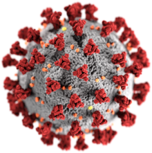
Illustration of a SARS-CoV-2 virion

A virus is a tiny infectious agent that reproduces inside the cells of living hosts. When infected, the host cell is forced to rapidly produce thousands of identical copies of the original virus. Unlike most living things, viruses do not have cells that divide; new viruses assemble in the infected host cell. But unlike simpler infectious agents like prions, they contain genes, which allow them to mutate and evolve. Over 4,800 species of viruses have been described in detail out of the millions in the environment. Their origin is unclear: some may have evolved from plasmids—pieces of DNA that can move between cells—while others may have evolved from bacteria.

Viruses are made of either two or three parts. All include genes. These genes contain the encoded biological information of the virus and are built from either DNA or RNA. All viruses are also covered with a protein coat to protect the genes. Some viruses may also have an envelope of fat-like substance that covers the protein coat, and makes them vulnerable to soap. A virus with this "viral envelope" uses it—along with specific receptors—to enter a new host cell. Viruses vary in shape from the simple helical and icosahedral to more complex structures. Viruses range in size from 20 to 300 nanometres; it would take 33,000 to 500,000 of them, side by side, to stretch to 1 centimetre (0.4 in).

Viruses spread in many ways. Although many are very specific about which host species or tissue they attack, each species of virus relies on a particular method to copy itself. Plant viruses are often spread from plant to plant by insects and other organisms, known as vectors. Some viruses of humans and other animals are spread by exposure to infected bodily fluids. Viruses such as influenza are spread through the air by droplets of moisture when people cough or sneeze. Viruses such as norovirus are transmitted by the faecal–oral route, which involves the contamination of hands, food and water. Rotavirus is often spread by direct contact with infected children. The human immunodeficiency virus, HIV, is transmitted by bodily fluids transferred during sex. Others, such as the dengue virus, are spread by blood-sucking insects.

Viruses, especially those made of RNA, can mutate rapidly to give rise to new types. Hosts may have little protection against such new forms. Influenza virus, for example, changes often, so a new vaccine is needed each year. Major changes can cause pandemics, as in the 2009 swine influenza that spread to most countries. Often, these mutations take place when the virus has first infected other animal hosts. Some examples of such "zoonotic" diseases include coronavirus in bats, and influenza in pigs and birds, before those viruses were transferred to humans.

Viral infections can cause disease in humans, animals and plants. In healthy humans and animals, infections are usually eliminated by the immune system, which can provide lifetime immunity to the host for that virus. Antibiotics, which work against bacteria, have no impact, but antiviral drugs can treat life-threatening infections. Those vaccines that produce lifelong immunity can prevent some infections.

Discovery

Scanning electron micrograph of HIV-1 viruses, coloured green, budding from a lymphocyte

In 1884, French microbiologist Charles Chamberland invented the Chamberland filter (or Chamberland–Pasteur filter), that contains pores smaller than bacteria. He could then pass a solution containing bacteria through the filter, and completely remove them. In the early 1890s, Russian biologist Dmitri Ivanovsky used this method to study what became known as the tobacco mosaic virus. His experiments showed that extracts from the crushed leaves of infected tobacco plants remain infectious after filtration.

At the same time, several other scientists showed that, although these agents (later called viruses) were different from bacteria and about one hundred times smaller, they could still cause disease. In 1899, Dutch microbiologist Martinus Beijerinck observed that the agent only multiplied when in dividing cells. He called it a "contagious living fluid" (Latin: contagium vivum fluidum)—or a "soluble living germ" because he could not find any germ-like particles. In the early 20th century, English bacteriologist Frederick Twort discovered viruses that infect bacteria, and French-Canadian microbiologist Félix d'Herelle described viruses that, when added to bacteria growing on agar, would lead to the formation of whole areas of dead bacteria. Counting these dead areas allowed him to calculate the number of viruses in the suspension.

The invention of the electron microscope in 1931 brought the first images of viruses. In 1935, American biochemist and virologist Wendell Meredith Stanley examined the tobacco mosaic virus and found it to be mainly made from protein. A short time later, this virus was shown to be made from protein and RNA. A problem for early scientists was that they did not know how to grow viruses without using live animals. The breakthrough came in 1931, when American pathologists Ernest William Goodpasture and Alice Miles Woodruff grew influenza, and several other viruses, in fertilised chickens' eggs. Some viruses could not be grown in chickens' eggs. This problem was solved in 1949, when John Franklin Enders, Thomas Huckle Weller, and Frederick Chapman Robbins grew polio virus in cultures of living animal cells. Over 4,800 species of viruses have been described in detail.

Origins

Viruses co-exist with life wherever it occurs. They have probably existed since living cells first evolved. Their origin remains unclear because they do not fossilize, so molecular techniques have been the best way to hypothesise about how they arose. These techniques rely on the availability of ancient viral DNA or RNA, but most viruses that have been preserved and stored in laboratories are less than 90 years old. Molecular methods have only been successful in tracing the ancestry of viruses that evolved in the 20th century. New groups of viruses might have repeatedly emerged at all stages of the evolution of life. There are three major theories about the origins of viruses:

Regressive theory
Viruses may have once been small cells that parasitised larger cells. Eventually, the genes they no longer needed for a parasitic way of life were lost. The bacteria Rickettsia and Chlamydia are living cells that, like viruses, can reproduce only inside host cells. This lends credence to this theory, as their dependence on being parasites may have led to the loss of the genes that once allowed them to live on their own.
Cellular origin theory
Some viruses may have evolved from bits of DNA or RNA that "escaped" from the genes of a larger organism. The escaped DNA could have come from plasmids—pieces of DNA that can move between cells—while others may have evolved from bacteria.
Coevolution theory
Viruses may have evolved from complex molecules of protein and DNA at the same time as cells first appeared on earth, and would have depended on cellular life for many millions of years.

There are problems with all of these theories. The regressive hypothesis does not explain why even the smallest of cellular parasites do not resemble viruses in any way. The escape or the cellular origin hypothesis does not explain the presence of unique structures in viruses that do not appear in cells. The coevolution, or "virus-first" hypothesis, conflicts with the definition of viruses, because viruses depend on host cells. Also, viruses are recognised as ancient, and to have origins that pre-date the divergence of life into the three domains. This discovery has led modern virologists to reconsider and re-evaluate these three classical hypotheses.

Structure

Simplified diagram of the structure of a virus

A virus particle, also called a virion, consists of genes made from DNA or RNA which are surrounded by a protective coat of protein called a capsid. The capsid is made of many smaller, identical protein molecules called capsomers. The arrangement of the capsomers can either be icosahedral (20-sided), helical, or more complex. There is an inner shell around the DNA or RNA called the nucleocapsid, made out of proteins. Some viruses are surrounded by a bubble of lipid (fat) called an envelope, which makes them vulnerable to soap and alcohol.

Size

Viruses are among the smallest infectious agents, and are too small to be seen by light microscopy; most of them can only be seen by electron microscopy. Their sizes range from 20 to 300 nanometres; it would take 30,000 to 500,000 of them, side by side, to stretch to one centimetre (0.4 in). In comparison, bacteria are typically around 1000 nanometres (1 micrometer) in diameter, and host cells of higher organisms are typically a few tens of micrometers. Some viruses such as megaviruses and pandoraviruses are relatively large viruses. At around 1000 nanometres, these viruses, which infect amoebae, were discovered in 2003 and 2013. They are around ten times wider (and thus a thousand times larger in volume) than influenza viruses, and the discovery of these "giant" viruses astonished scientists.

Genes

The genes of viruses are made from DNA (deoxyribonucleic acid) and, in many viruses, RNA (ribonucleic acid). The biological information contained in an organism is encoded in its DNA or RNA. Most organisms use DNA, but many viruses have RNA as their genetic material. The DNA or RNA of viruses consists of either a single strand or a double helix.

Viruses can reproduce rapidly because they have relatively few genes. For example, influenza virus has only eight genes and rotavirus has eleven. In comparison, humans have 20,000–25,000. Some viral genes contain the code to make the structural proteins that form the virus particle. Other genes make non-structural proteins found only in the cells the virus infects.

All cells, and many viruses, produce proteins that are enzymes that drive chemical reactions. Some of these enzymes, called DNA polymerase and RNA polymerase, make new copies of DNA and RNA. A virus's polymerase enzymes are often much more efficient at making DNA and RNA than the equivalent enzymes of the host cells, but viral RNA polymerase enzymes are error-prone, causing RNA viruses to mutate and form new strains.

In some species of RNA virus, the genes are not on a continuous molecule of RNA, but are separated. The influenza virus, for example, has eight separate genes made of RNA. When two different strains of influenza virus infect the same cell, these genes can mix and produce new strains of the virus in a process called reassortment.

Protein synthesis

Diagram of a typical eukaryotic cell, showing subcellular components. Organelles: (1) nucleolus (2) nucleus (3) ribosome (4) vesicle (5) rough endoplasmic reticulum (ER) (6) Golgi apparatus (7) cytoskeleton (8) smooth ER (9) mitochondria (10) vacuole (11) cytoplasm (12) lysosome (13) centrioles within centrosome (14) a virus shown to approximate scale

Proteins are essential to life. Cells produce new protein molecules from amino acid building blocks based on information coded in DNA. Each type of protein is a specialist that usually only performs one function, so if a cell needs to do something new, it must make a new protein. Viruses force the cell to make new proteins that the cell does not need, but are needed for the virus to reproduce. Protein synthesis consists of two major steps: transcription and translation.

Transcription is the process where information in DNA, called the genetic code, is used to produce RNA copies called messenger RNA (mRNA). These migrate through the cell and carry the code to ribosomes where it is used to make proteins. This is called translation because the protein's amino acid structure is determined by the mRNA's code. Information is hence translated from the language of nucleic acids to the language of amino acids.

Some nucleic acids of RNA viruses function directly as mRNA without further modification. For this reason, these viruses are called positive-sense RNA viruses. In other RNA viruses, the RNA is a complementary copy of mRNA and these viruses rely on the cell's or their own enzyme to make mRNA. These are called negative-sense RNA viruses. In viruses made from DNA, the method of mRNA production is similar to that of the cell. The species of viruses called retroviruses behave completely differently: they have RNA, but inside the host cell a DNA copy of their RNA is made with the help of the enzyme reverse transcriptase. This DNA is then incorporated into the host's own DNA, and copied into mRNA by the cell's normal pathways.

Life-cycle

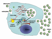
Life-cycle of a typical virus (left to right); following infection of a cell by a single virus, hundreds of offspring are released.

When a virus infects a cell, the virus forces it to make thousands more viruses. It does this by making the cell copy the virus's DNA or RNA, making viral proteins, which all assemble to form new virus particles.

There are six basic, overlapping stages in the life cycle of viruses in living cells:

  • Attachment is the binding of the virus to specific molecules on the surface of the cell. This specificity restricts the virus to a very limited type of cell. For example, the human immunodeficiency virus (HIV) infects only human T cells, because its surface protein, gp120, can only react with CD4 and other molecules on the T cell's surface. Plant viruses can only attach to plant cells and cannot infect animals. This mechanism has evolved to favour those viruses that only infect cells in which they are capable of reproducing.
  • Penetration follows attachment; viruses penetrate the host cell by endocytosis or by fusion with the cell.
  • Uncoating happens inside the cell when the viral capsid is removed and destroyed by viral enzymes or host enzymes, thereby exposing the viral nucleic acid.
  • Replication of virus particles is the stage where a cell uses viral messenger RNA in its protein synthesis systems to produce viral proteins. The RNA or DNA synthesis abilities of the cell produce the virus's DNA or RNA.
  • Assembly takes place in the cell when the newly created viral proteins and nucleic acid combine to form hundreds of new virus particles.
  • Release occurs when the new viruses escape or are released from the cell. Most viruses achieve this by making the cells burst, a process called lysis. Other viruses such as HIV are released more gently by a process called budding.

Effects on the host cell

Viruses have an extensive range of structural and biochemical effects on the host cell. These are called cytopathic effects. Most virus infections eventually result in the death of the host cell. The causes of death include cell lysis (bursting), alterations to the cell's surface membrane and apoptosis (cell "suicide"). Often cell death is caused by cessation of its normal activity due to proteins produced by the virus, not all of which are components of the virus particle.

Some viruses cause no apparent changes to the infected cell. Cells in which the virus is latent (inactive) show few signs of infection and often function normally. This causes persistent infections and the virus is often dormant for many months or years. This is often the case with herpes viruses.

Some viruses, such as Epstein-Barr virus, often cause cells to proliferate without causing malignancy; but some other viruses, such as papillomavirus, are an established cause of cancer. When a cell's DNA is damaged by a virus such that the cell cannot repair itself, this often triggers apoptosis. One of the results of apoptosis is destruction of the damaged DNA by the cell itself. Some viruses have mechanisms to limit apoptosis so that the host cell does not die before progeny viruses have been produced; HIV, for example, does this.

Viruses and diseases

There are many ways in which viruses spread from host to host but each species of virus uses only one or two. Many viruses that infect plants are carried by organisms; such organisms are called vectors. Some viruses that infect animals, including humans, are also spread by vectors, usually blood-sucking insects, but direct transmission is more common. Some virus infections, such as norovirus and rotavirus, are spread by contaminated food and water, by hands and communal objects, and by intimate contact with another infected person, while others are airborne (influenza virus). Viruses such as HIV, hepatitis B and hepatitis C are often transmitted by unprotected sex or contaminated hypodermic needles. To prevent infections and epidemics, it is important to know how each different kind of virus is spread.

In humans

Common human diseases caused by viruses include the common cold, influenza, chickenpox and cold sores. Serious diseases such as Ebola and AIDS are also caused by viruses. Many viruses cause little or no disease and are said to be "benign". The more harmful viruses are described as virulent. Viruses cause different diseases depending on the types of cell that they infect. Some viruses can cause lifelong or chronic infections where the viruses continue to reproduce in the body despite the host's defence mechanisms. This is common in hepatitis B virus and hepatitis C virus infections. People chronically infected with a virus are known as carriers. They serve as important reservoirs of the virus.

Endemic

If the proportion of carriers in a given population reaches a given threshold, a disease is said to be endemic. Before the advent of vaccination, infections with viruses were common and outbreaks occurred regularly. In countries with a temperate climate, viral diseases are usually seasonal. Poliomyelitis, caused by poliovirus often occurred in the summer months. By contrast colds, influenza and rotavirus infections are usually a problem during the winter months. Other viruses, such as measles virus, caused outbreaks regularly every third year. In developing countries, viruses that cause respiratory and enteric infections are common throughout the year. Viruses carried by insects are a common cause of diseases in these settings. Zika and dengue viruses for example are transmitted by the female Aedes mosquitoes, which bite humans particularly during the mosquitoes' breeding season.

Pandemic and emergent

Left to right: the African green monkey, source of SIV; the sooty mangabey, source of HIV-2; and the chimpanzee, source of HIV-1
 
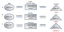
Origin and evolution of (A) SARS-CoV (B) MERS-CoV, and (C) SARS-CoV-2 in different hosts. All the viruses came from bats as coronavirus-related viruses before mutating and adapting to intermediate hosts and then to humans and causing the diseases SARS, MERS and COVID-19.

Although viral pandemics are rare events, HIV—which evolved from viruses found in monkeys and chimpanzees—has been pandemic since at least the 1980s. During the 20th century there were four pandemics caused by influenza virus and those that occurred in 1918, 1957 and 1968 were severe. Before its eradication, smallpox was a cause of pandemics for more than 3,000 years. Throughout history, human migration has aided the spread of pandemic infections; first by sea and in modern times also by air.

With the exception of smallpox, most pandemics are caused by newly evolved viruses. These "emergent" viruses are usually mutants of less harmful viruses that have circulated previously either in humans or in other animals.

Severe acute respiratory syndrome (SARS) and Middle East respiratory syndrome (MERS) are caused by new types of coronaviruses. Other coronaviruses are known to cause mild infections in humans, so the virulence and rapid spread of SARS infections—that by July 2003 had caused around 8,000 cases and 800 deaths—was unexpected and most countries were not prepared.

A related coronavirus emerged in Wuhan, China in November 2019 and spread rapidly around the world. Thought to have originated in bats and subsequently named severe acute respiratory syndrome coronavirus 2, infections with the virus cause a disease called COVID-19, that varies in severity from mild to deadly, and led to a pandemic in 2020. Restrictions unprecedented in peacetime have been placed on international travel, and curfews imposed in several major cities worldwide.

In plants

Peppers infected by mild mottle virus

There are many types of plant virus, but often they only cause a decrease in yield, and it is not economically viable to try to control them. Plant viruses are frequently spread from plant to plant by organisms called "vectors". These are normally insects, but some fungi, nematode worms and single-celled organisms have also been shown to be vectors. When control of plant virus infections is considered economical (perennial fruits, for example) efforts are concentrated on killing the vectors and removing alternate hosts such as weeds. Plant viruses are harmless to humans and other animals because they can only reproduce in living plant cells.

Bacteriophages

The structure of a typical bacteriophage

Bacteriophages are viruses that infect bacteria and archaea. They are important in marine ecology: as the infected bacteria burst, carbon compounds are released back into the environment, which stimulates fresh organic growth. Bacteriophages are useful in scientific research because they are harmless to humans and can be studied easily. These viruses can be a problem in industries that produce food and drugs by fermentation and depend on healthy bacteria. Some bacterial infections are becoming difficult to control with antibiotics, so there is a growing interest in the use of bacteriophages to treat infections in humans.

Host resistance

Innate immunity of animals

Animals, including humans, have many natural defences against viruses. Some are non-specific and protect against many viruses regardless of the type. This innate immunity is not improved by repeated exposure to viruses and does not retain a "memory" of the infection. The skin of animals, particularly its surface, which is made from dead cells, prevents many types of viruses from infecting the host. The acidity of the contents of the stomach destroys many viruses that have been swallowed. When a virus overcomes these barriers and enters the host, other innate defences prevent the spread of infection in the body. A special hormone called interferon is produced by the body when viruses are present, and this stops the viruses from reproducing by killing the infected cells and their close neighbours. Inside cells, there are enzymes that destroy the RNA of viruses. This is called RNA interference. Some blood cells engulf and destroy other virus-infected cells.

Adaptive immunity of animals

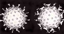
Two rotavirus particles: the one on the right is coated with antibodies which stop its attaching to cells and infecting them

Specific immunity to viruses develops over time and white blood cells called lymphocytes play a central role. Lymphocytes retain a "memory" of virus infections and produce many special molecules called antibodies. These antibodies attach to viruses and stop the virus from infecting cells. Antibodies are highly selective and attack only one type of virus. The body makes many different antibodies, especially during the initial infection. After the infection subsides, some antibodies remain and continue to be produced, usually giving the host lifelong immunity to the virus.

Plant resistance

Plants have elaborate and effective defence mechanisms against viruses. One of the most effective is the presence of so-called resistance (R) genes. Each R gene confers resistance to a particular virus by triggering localised areas of cell death around the infected cell, which can often be seen with the unaided eye as large spots. This stops the infection from spreading. RNA interference is also an effective defence in plants. When they are infected, plants often produce natural disinfectants that destroy viruses, such as salicylic acid, nitric oxide and reactive oxygen molecules.

Resistance to bacteriophages

The major way bacteria defend themselves from bacteriophages is by producing enzymes which destroy foreign DNA. These enzymes, called restriction endonucleases, cut up the viral DNA that bacteriophages inject into bacterial cells.

Prevention and treatment of viral disease

Vaccines

The structure of DNA showing the position of the nucleosides and the phosphorus atoms that form the "backbone" of the molecule

Vaccines simulate a natural infection and its associated immune response, but do not cause the disease. Their use has resulted in the eradication of smallpox and a dramatic decline in illness and death caused by infections such as polio, measles, mumps and rubella. Vaccines are available to prevent over fourteen viral infections of humans and more are used to prevent viral infections of animals. Vaccines may consist of either live or killed viruses. Live vaccines contain weakened forms of the virus, but these vaccines can be dangerous when given to people with weak immunity. In these people, the weakened virus can cause the original disease. Biotechnology and genetic engineering techniques are used to produce "designer" vaccines that only have the capsid proteins of the virus. Hepatitis B vaccine is an example of this type of vaccine. These vaccines are safer because they can never cause the disease.

Antiviral drugs

The structure of the DNA base guanosine and the antiviral drug aciclovir which functions by mimicking it

Since the mid-1980s, the development of antiviral drugs has increased rapidly, mainly driven by the AIDS pandemic. Antiviral drugs are often nucleoside analogues, which masquerade as DNA building blocks (nucleosides). When the replication of virus DNA begins, some of the fake building blocks are used. This prevents DNA replication because the drugs lack the essential features that allow the formation of a DNA chain. When DNA production stops the virus can no longer reproduce. Examples of nucleoside analogues are aciclovir for herpes virus infections and lamivudine for HIV and hepatitis B virus infections. Aciclovir is one of the oldest and most frequently prescribed antiviral drugs.

Other antiviral drugs target different stages of the viral life cycle. HIV is dependent on an enzyme called the HIV-1 protease for the virus to become infectious. There is a class of drugs called protease inhibitors, which bind to this enzyme and stop it from functioning.

Hepatitis C is caused by an RNA virus. In 80% of those infected, the disease becomes chronic, and they remain infectious for the rest of their lives unless they are treated. There is an effective treatment that uses the nucleoside analogue drug ribavirin. Treatments for chronic carriers of the hepatitis B virus have been developed by a similar strategy, using lamivudine and other anti-viral drugs. In both diseases, the drugs stop the virus from reproducing and the interferon kills any remaining infected cells.

HIV infections are usually treated with a combination of antiviral drugs, each targeting a different stage in the virus's life-cycle. There are drugs that prevent the virus from attaching to cells, others that are nucleoside analogues and some poison the virus's enzymes that it needs to reproduce. The success of these drugs is proof of the importance of knowing how viruses reproduce.

Role in ecology

Viruses are the most abundant biological entity in aquatic environments; one teaspoon of seawater contains about ten million viruses, and they are essential to the regulation of saltwater and freshwater ecosystems. Most are bacteriophages, which are harmless to plants and animals. They infect and destroy the bacteria in aquatic microbial communities and this is the most important mechanism of recycling carbon in the marine environment. The organic molecules released from the bacterial cells by the viruses stimulate fresh bacterial and algal growth.

Microorganisms constitute more than 90% of the biomass in the sea. It is estimated that viruses kill approximately 20% of this biomass each day and that there are fifteen times as many viruses in the oceans as there are bacteria and archaea. They are mainly responsible for the rapid destruction of harmful algal blooms, which often kill other marine life. The number of viruses in the oceans decreases further offshore and deeper into the water, where there are fewer host organisms.

Their effects are far-reaching; by increasing the amount of respiration in the oceans, viruses are indirectly responsible for reducing the amount of carbon dioxide in the atmosphere by approximately 3 gigatonnes of carbon per year.

Marine mammals are also susceptible to viral infections. In 1988 and 2002, thousands of harbour seals were killed in Europe by phocine distemper virus. Many other viruses, including caliciviruses, herpesviruses, adenoviruses and parvoviruses, circulate in marine mammal populations.

Personality theories of addiction

From Wikipedia, the free encyclopedia https://en.wikipedia.org/wiki/Personality_theories_of_addiction ...