Search This Blog

Friday, May 10, 2019

Childhood leukemia

From Wikipedia, the free encyclopedia

Childhood leukemia
Pediatric patients receiving chemotherapy.jpg
Two girls with acute lymphocytic leukemia demonstrating intravenous access for chemotherapy.

Childhood leukemia is leukemia that occurs in a child and is a type of childhood cancer. Childhood leukemia is the most common childhood cancer, accounting for 29% of cancers in children aged 0–14 in 2018. There are multiple forms of leukemia that occur in children, the most common being acute lymphoblastic leukemia (ALL) followed by acute myeloid leukemia (AML). Survival rates vary depending on the type of leukemia, but may be as high as 90% in ALL.

Leukemia is a hematological malignancy or a cancer of the blood. It develops in the bone marrow, the soft inner part of bones where new blood cells are made. When a child has leukemia, the bone marrow produces white blood cells that do not mature correctly. Normal healthy cells only reproduce when there is enough space for them. The body will regulate the production of cells by sending signals of when to stop production. When a child has leukemia, the cells do not respond to the signals telling them when to stop and when to produce cells. The bone marrow becomes crowded resulting in problems producing other blood cells.

Common childhood leukemia signs and symptoms include excessive tiredness, easy bruising or bleeding, bone pain and paleness.

Types

Leukemia is usually described either as "acute", which grows quickly, or "chronic", which grows slowly. The vast majority of childhood leukemia is acute, and chronic leukemias are more common in adults than in children. Acute leukemias typically develop and worsen quickly (over periods of days to weeks). Chronic leukemias develop over a slower period of time (months), but are more difficult to treat than acute leukemias. The following are some of the main types of leukemia that occur in children.

Acute lymphoblastic

The most common form childhood leukemia is acute lymphocytic (or lymphoblastic) leukemia (ALL), which makes up 75-80% of childhood leukemia diagnoses. ALL is a form of leukemia that affects lymphocytes, a type of white blood cells which fights infection. When a patient has ALL, the bone marrow makes too many immature white blood cells and they do not mature correctly. These white blood cells also do not work correctly to fight infection. The white blood cells over-produce, crowding the other blood cells in the bone marrow.

Acute myelogenous

Another type of acute leukemia is acute myelogenous leukemia (AML). AML accounts for most of the remaining cases of leukemia in children, comprising about 20% of childhood leukemia. AML is cancer of the blood in which too many myeloblasts (immature white blood cells) are produced in the bone marrow. The marrow continues to produce abnormal cells that crowd the other blood cells and do not work properly to fight infection.

Acute promyelocytic

Acute promyelocytic leukemia (APL) is a specific type of AML. In this leukemia promyelocytes are produced and build up in the bone marrow. A specific chromosome translocation (a type of genetic change) is found in patients with APL. Genes on chromosome 15 change places with genes on chromosome 17. This genetic change prevents the promyelocytes from maturing properly.

Chronic myelogenous

Chronic myelogenous leukemia (CML) is a chronic leukemia that develops slowly, over months to years. CML is rare in children, but does occur. CML patients have too many immature white blood cells being produced, and the cells crowd the other healthy blood cells. A chromosome translocation occurs in patients with CML. Part of chromosome 9 breaks off and attaches itself to chromosome 22, facilitating exchange of genetic material between chromosomes 9 and 22. The rearrangement of the chromosomes changes the positions and functions of certain genes, which causes uncontrolled cell growth.

Chronic lymphocytic leukemia (CLL) is another form of chronic leukemia, but is extremely rare in children.

Juvenile myelomonocytic

Juvenile myelomonocytic leukemia (JMML) is a form of leukemia in which myelomonocytic cells are overproduced. It is sometimes considered a myeloproliferative neoplasm. It is rare and most commonly occurs in children under the age of four. In JMML, the myelomonocytic cells produced by the bone marrow and invade the spleen, lungs, and intestines.

Signs and symptoms

Most initial symptoms of leukemia are related to problems with the bone-marrow function. There are a variety of symptoms that children may experience. The symptoms tend to appear quickly in acute leukemia and slowly over time in chronic leukemia. Symptoms in the different types of childhood leukemia include:
  • feelings of fatigue or weakness
  • repetitive infections or fever
  • bone and joint pain
  • refusing to walk, which likely results from bone pain or fatigue
  • easy bleeding or bruising (including petechiae)
  • increased paleness of skin
  • abdominal pain or fullness, which may cause shortness of breath or loss of appetite
  • swollen lymph nodes under the arms, in the groin, chest and neck.
  • enlarged spleen or liver
  • weight loss
  • rash

Causes

The exact cause of most cases of childhood leukemia is not known. Most children with leukemia do not have any known risk factors.

One hypothesis is that childhood acute lymphoblastic leukemia (ALL) is caused by a two-step process, starting with a prenatal genetic mutation and then exposure to infections While this theory is possible, there is not enough evidence in patients currently to either support or refute the relationship between infection and developing ALL.

There is evidence linking maternal alcohol consumption to AML development in children. Indoor insecticide exposure has also been linked to the development of childhood leukemias. High levels of coffee consumption during pregnancy (2-3 cups/day or greater) have been linked to childhood leukemia as well.

It has also been suggested that allergies are linked to the development of childhood leukemia but this is not supported by current evidence.

Diagnosis

Childhood leukemia is diagnosed in a variety of ways. The diagnostic procedures confirm if there is leukemia present, the extent of the leukemia (how far it has spread), and the type of leukemia. The diagnostic procedures are similar for the different types of leukemias:
  • A bone-marrow aspiration and biopsy to look for and collect leukemia cells. In aspiration, a fluid sample is removed from the marrow. In biopsy, bone marrow cells are removed. Usually both procedures are performed at the same time and used together to help with diagnosis.
  • Tests called immunophenotyping and cytogenetic analysis are performed on the cells to further determine the type and subtype of leukemia.
  • A complete blood count, which is a measurement of size, number, and maturity of different blood cells in blood.
  • Blood tests may include blood chemistry, evaluation of liver and kidney functions, and genetic studies.
  • A spinal tap: a special needle is placed into the lower back into the spinal canal, which is the area around the spinal cord. Cerebral spinal fluid is fluid that bathes the child's brain and spinal cord. A small amount of cerebral spinal fluid is sent for testing to determine if leukemia cells are present.

Treatment

Treatment for childhood leukemia is based on a number of factors, including the type of leukemia, characteristics of the leukemia, prognostic characteristics (children with worse prognostic characteristics receive more aggressive therapy, see Prognosis section), response to therapy, and extent of the disease at diagnosis. Treatment is typically managed by a team of health care professionals, consisting of pediatric oncologists, social workers, pediatric nurse specialists, and pediatricians among others.

Types

Young girl receiving chemotherapy treatment
 
While the exact treatment plan is determined by the type of leukemia and factors listed above, there are five types of therapies that are generally used to treat all childhood leukemias. Four of these are standard treatment and one is in clinical trials. The four specific types of treatments that are traditionally used are Chemotherapy, Stem cell transplant, Radiation therapy and Targeted therapy. Immunotherapy is another type of therapy that is currently in clinical trials.

Chemotherapy is a treatment that uses chemicals to interfere with the cancer cells ability to grow and reproduce. Chemotherapy can be used alone or in combination with other therapies. Chemotherapy can be given either as a pill to swallow orally, an injection into the fat or muscle, through an IV directly into the bloodstream or directly into the spinal column.

Stem cell transplant is a process in which the blood-forming cells that are abnormal (like leukemia cells) or that were destroyed by chemotherapy are replaced with healthy new blood-forming cells. A stem-cell transplant can help the human body produce more healthy white blood cells, red blood cells, or platelets. It also reduces the risk of life-threatening conditions such as anemia, or hemorrhage. Stem cell transplants can be done by obtaining cells from the bone-marrow, blood or umbilical-cord blood. Stem cell transplants can use the cells from one's self, called an autologous stem cell transplant or they can use cells from another person, known as an allogenic stem cell transplant. The type used in childhood leukemia is typically allogenic. The donors used must be a match to the child getting the transplant by a marker called HLA.
 
Radiation therapy uses various types of radiation to kill cancer cells. 

Targeted therapy is the use of medication to specifically kill the cancerous cells. The medication is able to leave healthy normal cells alone while it targets the cancer. These include tyrosine kinase inhibitors (TKIs), monoclonal antibodies, and proteasome inhibitors.

Immunotherapy is a type of therapy that uses the child's own immune system to fight the cancer. This therapy is currently in clinical trials.

ALL

Treatment for childhood ALL consists of three phases: Induction, Consolidation/Intensification, and Maintenance.
  • Induction is intended to kill the large majority of the cancer cells. It typically lasts for 4–6 weeks and uses chemotherapy and glucocorticoids. After induction, the goal is to put the cancer into remission. Remission means that cancer is no longer detected in the bone marrow or blood and that normal cells have returned to the bone marrow. However, remission does not mean that the cancer is cured. It is thought there are still cancer cells that are hiding in the body, so more treatment is needed to kill them.
  • Consolidation/Intensification is used to kill any remaining cells that have the potential to become cancerous. It consists of more chemotherapy and lasts for a few months.
  • Maintenance is a lower intensity chemotherapy regimen that used to kill any more remaining cells in the bone marrow that could regrow into cancer cells and cause the leukemia to come back. It lasts for 18–30 months.
Immunotherapy, radiation therapy, stem cell transplant, and targeted therapies may also be used in the treatment of ALL. This will depend on the extent of ALL, the characteristics of the ALL and if it has recurred (come back after initial treatment).

AML

Childhood AML is a more challenging cancer to treat than childhood ALL. Childhood AML treatment usually consists higher dose chemotherapy given over a shorter period of time compared to ALL treatment. Due to this shorter and more intense treatment, side effects are also more intense. These children are therefore treated in treatment centers or hospitals where they will stay for longer period of their treatment. Treatment for AML consists of 2 phases: Induction and Consolidation. There is no Maintenance phase of therapy in AML as it was not shown to lower chances of the cancer coming back.
  • Induction is aimed at killing leukemia in the blood and bone marrow. Its goal is to put the cancer into remission. Treatments used in induction therapy for childhood AML may include chemotherapy, targeted therapy, radiation therapy, stem cell transplant, or other treatments as part of a clinical trial. The exact treatment will vary depending on characteristics of the child and the cancer.
  • Consolidation begins after remission is obtained and is aimed at killing any remaining cancer cells. It will again vary depending on specifics about the patient and cancer. It typically will consist of chemotherapy followed by a stem cell transplant.
In addition to these treatments, there are also clinical trials of immunotherapy and targeted therapy for AML. The APL type of AML is also treated with all-trans retinoic acid or arsenic trioxide therapy in addition to what is listed above.

Other childhood leukemias

JMML is typically treated by chemotherapy followed by a stem cell transplant. CML is typically treated with targeted therapy and possibly a stem cell transplant if it comes back or does not respond to the targeted therapy at first.

Prognosis

The 5-year survival rate for children with leukemia is 83.6% in the USA. This means that 83.6% of children diagnosed with leukemia live for 5 years or more after their diagnosis. This is greatly improved from a 5-year survival rate of 36.5% in 1975. The improvement is largely attributed to advances in therapy, particularly therapy for ALL. The outlook or prognosis for an individual child is affected by the type of leukemia. In addition, there are certain characteristics of the patients and cancers that help doctors predict the prognosis (and determine treatment). These are referred to as prognostic factors. Generally prognostic factors are more meaningful in ALL than in AML.

ALL

The 5-year survival rate for children and adolescents under the age of 15 years diagnosed with ALL was 91.8% in the USA between 2007 and 2013. The survival rate for children under the age of 5 years with ALL was 94% during the same time period.

Prognostic factors in ALL:
  • Age at diagnosis: Children between the ages of 1–9 years with B-cell ALL (a specific type of ALL) have better cure rates than children less than 1 year old or over 10 years old. This does not seem to matter in T-cell ALL (another specific type of ALL).
  • White blood cell count at diagnosis: Children with very high white blood cell counts at diagnosis are higher risk patients than those with lower counts.
  • Specific type of ALL
  • Spread to other organs (such as the brain, spinal cord, and testicles) signifies worse prognosis
  • Chromosome changes: Patients whose leukemia cells have more chromosomes are more likely to be cured. Different chromosome translocations are also associated with different prognoses.
  • Initial treatment response: Children who respond to treatment quickly initially have a better prognosis.

AML

The survival rate for children under the age of 15 years with AML was 66.4% in the USA between 2007 and 2013. This is lower than the rates for ALL.

Prognostic factors for AML:
  • Age at diagnosis: Children under 2 years old may have a better prognosis than older children. However, how strong this link is is unclear.
  • White blood cell count at diagnosis: Children with lower white blood cell counts tend to have a better prognosis.
  • Children with Down Syndrome and AML typically have a good prognosis.
  • Specific type of AML: APL generally is a good prognosis.
  • Specific chromosome changes affect prognosis.
  • AML that started because of treatment for a different cancer usually has poorer prognosis.
  • Response to treatment: As with ALL, patients whose disease responds faster to treatment tend to have a better prognosis.
  • Children who are a normal weight usually have a better prognosis than those who are overweight or underweight.

After effects

As treatments for childhood leukemias have gotten better, there are more children surviving and living into adulthood. These survivors are at risk for long term after effects of treatment. The specific risks depend on the type of therapy that was given and the type of cancer the child had.

The older aggressive treatment regimens with cranial irradiation and higher doses of anthracyclines (such as doxorubicin) caused increased risk of solid tumors, heart failure, growth retardation, and cognitive defects. In types of childhood leukemias with good cure rates (mainly ALL), efforts are continually made to decrease the amount of toxicity caused by chemotherapy and other treatments.
Secondary cancers
Survivors who received treatment for childhood leukemia are at risk for developing a secondary cancer later in life. The risk of acquiring a second cancer is weighed against the benefit of receiving therapy for life-threatening leukemia.
 
Neurological
Survivors of ALL are at risk for various neurocognitive and neuropsychological issues that effect their quality of life. These include issues with attention span, vision, processing speed, memory, growth failure, malnutrition, obesity, reduced fertility, psychiatric problems and early death. All of the latent effects listed impact patients and create a low quality of life. Lower quality of life is directly related to depression and other psychiatric problems.
 
Growth and development
Some childhood leukemia treatments, notably stem cell transplants, can stunt growth. Growth hormone is sometimes given to help with this.
 
Fertility
Fertility may be affected in both boys and girls who receive leukemia treatment.
 
Bone problems
Bone problems or damage may result from glucocorticoids.
 
Emotional
Childhood leukemia is a very taxing disease, on the caregiver and the child. Some emotional issues that survivors have reported include: depression, anxiety, post-traumatic stress disorder, difficulties with interpersonal relationships, poor body image, and schizophrenia among other issues. However, it is unclear if the rates of mental and emotional problems are higher in childhood leukemia survivors than the general population. Regardless, some children may have emotional or psychological issues that may be addressed by doctors, other care team members, parents, and friends.

Epidemiology

Leukemia is the most common cancer in children, accounting for 25-30% of all cancers in children and adolescents. It most commonly is diagnosed in children when they are 1–4 years old. The median age of diagnosis is 6 years old. Childhood leukemia is more common in boys than girls. It is also more frequently diagnosed in white and Hispanic children. The incidence of childhood leukemia has been increasing over time. However, this may be because of increased ability to detect, diagnose, and report the disease, rather than an actual increase in children who are affected.

ALL is the most common type of childhood leukemia, accounting for 75-80% of diagnoses. AML is most commonly is diagnosed in 3-5-year-old children. As with childhood leukemia in general, it is more common in boys than girls and more common in white and Hispanic children.

AML is the second most common type of childhood leukemia, making up most of the remaining diagnoses. It is most commonly diagnosed in children less than 1 year old. Unlike ALL, it occurs equally in boys and girls and occurs equally across racial/ethnic groups.

There are a number of risk factors that have been studied for childhood leukemia. Genetic risk factors include: Down's Syndrome, Fanconi Anemia, damilial monosomy 7, Shwachman–Diamond syndrome, Bloom Syndrome, as well as mutations in specific gene mutations. Besides genetic risk factors, exposure to ionizing radiation is a known risk factor for childhood leukemia. Other factors that may be linked to development of childhood leukemia include: family history of blood cancers, maternal alcohol abuse, parental cigarette use, prior loss of pregnancy in the mother, older age of the mother, high birth weight, low birth weight, exposure to benzene, exposure to pesticides, and infections. However, whether or how much these factors actually contribute to the development of leukemia has yet to be determined and is unclear.

Pharmacoepigenetics

From Wikipedia, the free encyclopedia

Pharmacoepigenetics is an emerging field that studies the underlying epigenetic marking patterns that lead to variation in an individual's response to medical treatment.

Background

Due to genetic heterogeneity, environmental factors, and pathophysiological causes, individuals that exhibit similar disease expression may respond differently to identical drug treatments. Selecting treatments based on factors such as age, body-surface area, weight, gender, or disease stage has been shown to incompletely address this problem, so medical professionals are shifting toward using patient genomic data to select optimal treatments. Now, an increasing amount of evidence shows that epigenetics also plays an important role in determining the safety and efficacy of drug treatment in patients. Epigenetics is a bridge that connects individual genetics and environmental factors to explain some aspects of gene expression. Specifically, environmental factors have the potential to alter one’s epigenetic mechanisms in order to influence the expression of genes. For example, smoking cigarettes can alter the DNA methylation state of genes and thereby expression of genes through different mechanisms.

Epigenetic changes in genes caused by factors such as environment can result in abnormal gene expression and the initiation of diseases. The progression of diseases further alters the epigenetic patterns of the whole genome. While epigenetic changes are generally long lasting, and in some cases permanent, there is still the potential to change the epigenetic state of a gene. Thus, drugs have been developed to target aberrant epigenetic patterns in cells to either activate or suppress the epigenetically modified gene expression gene expression. This is known as epigenetic therapy. Besides being drug targets, epigenetic changes are also used as diagnostic and prognostic indicators to predict disease risk and progression, and this could be beneficial for the improvement of personalized medicine.

The development of the Human Epigenome Project and advances in epigenomics has given rise to a burgeoning field known as pharmacoepigenetics. Pharmacoepigenetics was initially developed to study how epigenetic patterns of drug transporters, drug-metabolizing enzymes, and nuclear receptors affect individuals’ response to the drug. Now, pharmacoepigenetics has an additional focus: the development of therapeutic epidrugs that can make changes to the epigenome in order to lessen the cause or symptoms of a disease in an individual. Even though a large gap still remains between the knowledge of epigenetic modifications on drug metabolism mechanisms and clinical applications, pharmacoepigenetics has become a rapidly growing field that has the potential to play an important role in personalized medicine. 

In order to develop effective epigenetic therapies, it is important to understand the underlying epigenetic mechanisms and the proteins that are involved. Various mechanisms and modifications play a role in epigenetic remodeling and signaling, including DNA methylation, histone modification, covalent modifications, RNA transcripts, microRNAs, mRNA, siRNA, and nucleosome positioning. In particular, scientists have extensively studied the associations of DNA methylation, histone modifications, regulatory microRNA with the development of diseases.

DNA methylation is the most widely studied epigenetic mechanism. Most of them occur at CpG sites. DNA methyltransferase is recruited to the site and adds methyl groups to the cytosine of the CpG dinucleotides. This allows the methyl-CpG binding proteins to bind to the methylated site and cause downregulation of genes. Histone modification is mainly achieved by modifying the N-terminal tails of histones. The mechanisms include acetylation, methylation, phosphorylation, unbiquitination, etc. They affect the compaction of chromatin structure, the accessibility of the DNA, and therefore the transcriptional level of specific genes. 

Additionally, microRNA is a type of noncoding RNA that is responsible for altering gene expression by targeting and marking mRNA transcripts for degradation. Since this process is a posttranscriptional modification, it does not involve changes in DNA sequence. The expression of microRNA is also regulated by other epigenetic mechanisms. Aberrant expression of microRNA facilitates disease development, making them good targets for epigenetic therapies. Epigenetic proteins involved in the regulation of gene transcription fall into three categories-writers, erasers, and readers. Both writers and erasers have enzymatic activity that allows them to covalently modify DNA or histone proteins. Readers have the ability to recognize and bind to specific sites on chromatin to alter epigenetic signatures.

Once the underlying epigenetic mechanisms are understood, it becomes possible to develop new ways to alter epigenetic marks such as "epidrugs", or epigenome editing, which is the overwriting of epigenetic patterns using man-made signals to direct epigenetic proteins to target loci. Furthermore, based on patients' unique epigenetic patterns, medical professionals can more accurately assign a safe and effective treatment including appropriate epigenetic drugs tailored to the patient.

Drug response and metabolism

Individual differences in drug metabolism and response can be partially explained by epigenetic changes. Epigenetic changes in genes that encode drug targets, enzymes, or transport proteins that affect the body's ability to absorb, metabolize, distribute and excrete substances that are foreign to the body (Xenobiotics) can result in changes in one's toxicity levels and drug response. One of the main effects of drug exposure early in life is altered ADME (Absorption, Distribution, Metabolism, and Excretion) gene expression. There is evidence that these genes are controlled by DNA methylation, histone acetylation, and miRNAs.

More needs to be understood about these mechanisms, but the hope is that it can lead to proper drug selection and dosage. Additionally, drug resistance can be acquired through epigenetic mechanisms. This is particularly common in chemotherapy, where cells that develop resistance to treatment continue to divide and survive. Pharmacoepigenetic treatment plans can consist of a single epidrug class or combine several in a unique therapy. The following are the examples of how drug response or metabolism related proteins are regulated by epigenetic mechanisms.

CXCR4 and DNA methylation

CXCR4 is a protein that acts as a coreceptor for the entry of HIV. It has been developed as a drug target for anti-HIV therapy. A study has shown that its expression is dysregulated by abnormal methylation patterns in some cancers. Thus, this could affect the efficiency and drug response to the anti-HIV therapy.

CYP1A1 methylation and histone modification

CYP1A1 is a protein that is well known for its role in chemical compounds and drug metabolism. A study in prostate cancer demonstrated that the protein's regulatory region was under the control of the histone modification H3K4me3, which typically indicates active gene expression in non-cancerous cells. This abnormal methylation typically causes histone modification and changes in chromatin structure at a local level, thus effecting gene expression.

ABCG2 and miRNA

ABCG2 is a protein that is responsible for multidrug resistance in cancer chemotherapy. Increased expression of ABCG2 is found in different drug resistant cancer cell lines and tumor tissues. One of the microRNA modifications changes its gene and protein expression by destabilizing its mRNA.

Epigenetics and human diseases

Epigenetics in cancer

While there is still a lot of work that needs to be done regarding the epigenetic modifications of specific cancers at various steps in tumor development, there is a general understanding of epigenetic modifications in genes that lead to abnormal expression and various types of cancer. These epigenetic biomarkers are being considered in clinical use as a tool to detect disease, classify tumors, and understand drug response to treatments such as target compounds, traditional chemotherapy agents, and epigenetic drugs. Human cancer is generally characterized by hypermethylation of specific promoters, which typically prevents the expression of DNA repair and tumor-suppressing genes, and the loss of DNA methylation on a global scale, which can allow for expression of oncogenes or result in a loss of imprinting. Histone modifications play an important role in the regulation of cellular processes, thus epigenetic changes resulting in changed structure can lead to abnormal transcription, DNA repair and replication. Below are some examples and then an overview of the ways these epigenetic modifications are being targeted.

Targeting epigenetic modifications in cancer

Epigenetic changes are highly present in cancer, therefore it is a good model to assess different ways in which epigenetic drugs can be used to make changes that turn up and turn down gene expression.

Targeting gain-of-function epigenetic mutations

DNA methyltransferase inhibitors are being pursued due to the hypermethylation of tumor suppressor genes and increased DNMTs that have been observed in cancer cells. Introduction of these inhibitors can result in reduced promoter methylation and expression of previously silenced tumor suppressor genes. Azacitidine and decitabine, which incorporate into the DNA and covalently trap the methyltransferases, have been approved by the FDA for myelodysplastic syndrome (a group of cancers where blood cells from the bone marrow do not mature properly into healthy blood cells) treatment and are currently being investigated for other cancers like leukemia. Other types of drugs are being developed like non-nucleoside analogues, which can covalently bind to DNMTs.

Some examples include procaine, hydralazine, and procainimide, but they lack specificity and potency making it hard to test them in clinical trials. DNA methyltranferase inhibitors are usually used at a low level due to their lack of specificity and toxic effects on normal cells. HDAC inhibitors are also being used, due to the changes in histone acetylation and the increased HDACs observed. While the mechanism is still under investigation, it is believed that adding the HDAC inhibitors results in increased histone acetylation and therefore the reactivation of transcription of tumor suppressor genes.

More so, HDACs can also remove acetyl groups from proteins that are not the histone, so it is thought that adding HDAC inhibitors may result in changes in transcription factor activity. There are around 14 different HDAC inhibitors being investigated in clinical trials for haematological and solid tumors, but more research needs to be done on the specificity and mechanisms by which they are inhibiting. Another way to alter epigenetic modifications is through the use of histone methyltransferase inhibitors.

Targeting loss-of-Function epigenetic mutations

Loss of function in genes encoding DNA demethylases or the overexpression of DNA methyltransferases can result in the hypermethylation of DNA promoters. Loss of function of DNA methyltransferases can lead to hypomethylation. Loss of function in chromosome remodeling, DNA repair, and cell cycle regulation genes can lead to uncontrolled growth of cells giving rise to cancer. Histone modification patterns can also lead to changes in genomes that can negatively affect these and other systems, making cancer more likely.

Cells that carry loss-of-function mutations can be targeted by drugs that induce synthetic lethality, a genetic/protein interaction where the loss of one component induces little change, but the loss of both components results in cell death. In cancer cells where one part of the interaction experiences a loss-of-function mutation, the other part can be interrupted by drug treatment to induce cell death in cancerous cells. Synthetic lethality is an attractive treatment option in patients with cancer since it there should be minimal / no effect on healthy cells. 

For example, with SWI/SNF loss of function mutations, DNA replication and repair is negatively affected and can give rise to tumors if cell growth goes unchecked. Mutations of these genes are common causes of cancers. These mutations are not directly targetable, but several synthetic lethal interactions can be exploited by cancer drugs to kill early cancer growth.

Additionally, loss-of-function mutations can be targeted by using the dynamic states of histone modifications. Loss of function mutations in demethylases, such as KDMK6A are common in cancer. By inducing upregulation of methyltransferase inhibitors, the effects of the loss-of-function mutation can be mitigated.

Development of drugs that target or modify epigenetic signatures of target genes is growing, especially as bioinformatic analysis increases our knowledge of the human genome and speeds up the search for synthetic lethal interactions. Most widely used to assess potential synthetic lethal interactions is using siRNA and CRISPR-Cas9 to modify target genes. CRISPRi and CRISPRa technology allows researchers to activate or inactivate target genes.

Lung cancer

In lung cancer the activation of both dominant and recessive oncogenes and inactivation of tumor suppressor genes has been observed. Frequently observed in lung cancer is the methylation of gene promoters that are involved in critical functions like cell-cycle control, repairing DNA, cell adhesion, proliferation, apoptosis, and motility. A few of the common genes frequently observed are APC, CDH1, CDKN2A, MGMT, and RASSF1A (a tumor suppressor). In the cases of CDKN2A and RASSF1A DNA these genes are methylated, resulting in the loss of tumor suppressor genes.

Various strategies such as using drugs like entinostat and azacitidine have been observed in clinical trials of non-small-cell lung carcinoma. The idea being that etinostat, a histone deacetylase inhibitor, can prevent the silencing of genes by allowing them to be accessible to transcription machinery. Azacitidine can be metabolized and incorporated into DNA and then recognized as a substrate for DNA methyltransferases, but since the enzyme is bound the methyltransferase cannot add methylation marks and thus silence crucial genes.

Heart failure

Histone modifications, DNA methylation, and microRNAs have been found to play an important role in heart disease. Previously, histone tail acetylation has been linked to cardiac hypertrophy or abnormal heart muscle thickening that is usually due to an increase in cardiomyocyte size or other cardiac muscle changes. The hypertrophic changes that occur in cardiac muscles cells result from the required acetylation of histone tails via acetyltransferases. In addition to acetyltransferases, histone deacetylases (HDACs) also aid in the regulation of muscle cells. Class II HDACs 5 and 9 inhibit the activity of a factor known as myocyte enhancer factor 2 (MEF2), which unable to bind prevents the expression of genes that produce hypertrophic effects.

Additionally, loci such as PECAM1, AMOTL2 and ARHGAP24 have been seen with different methylation patterns that are correlated with altered gene expression in cardiac tissue.

There are an increasing number of scientific publications that are finding that miRNA plays a key role in various aspects of heart failure. Examples of functions for miRNA include the regulation of the cardiomyocyte cell cycle and regulation of cardiomyocyte cell growth. Knowing the epigenetic modifications allows for the potential use of drugs to modify the epigenetic status of a target sequence. One could possibly target the miRNAs using antagomirs. Antagomirs are single strand RNAs that are complementary, which have been chemically engineered oligonucleotides that silence miRNAs so that they cannot degrade the mRNA that is needed for normal levels of expression. 

DNA methylation of CpGs can lead to a reduction of gene expression, and in some cases this decrease in gene product can contribute to disease. Therefore, in those instances it is important to have potential drugs that can alter the methylation status of the gene and increase expression levels. To increase gene expression, one may try to decrease CpG methylation by using a drug that works as DNA methytransferase inhibitor such as decitabine or 5-aza-2'-deoxycytidine.

On the other hand, some diseases result from a decrease in acetylase activity, which results in a decrease in gene expression. Some studies have shown that inhibiting HDAC activity can attenuate cardiac hypertrophy. trichostatin A and sodium butyrate are two HDAC inhinitors. Trichostatin A is known for its ability to inhibit class I and II HDACs from removing acetylases and decreasing gene expression. Sodium butyrate is another chemical that inhibits class I HDACs, thus resulting in the ability for transcription factors to easily access and express the gene.

Challenges in development of epigenetic therapies

There are a number of challenges with the developing epigenetic therapies for widespread medical use. While laboratory results indicate relationships between genes and potential drug interactions that could mitigate the effects of mutations, the complexity of the human genome and epigenome makes it difficult to develop therapies that are safe, efficient, and consistent. Epigenetic alteration may affect more systems than the target genes, which gives potential for deleterious effects to rise out of treatment. Additionally, epigenetic mutations can be a result of lineage.

As tissue gene expression is largely regulated by epigenetic interactions, certain tissue-specific cancers are difficult to target with epigenetic therapies. Additionally, genes that encode for elements that prevent one type of cancer in a cell, may have altered function in another and lead to another type of cancer. Trying to modify these proteins, such as EZH2, may give rise to other types of cancer. Selectivity is another hurdle in the development of therapies. Since many proteins are structurally similar, especially within the same protein family, Broad-spectrum inhibitors can't always be used since modifying the regulation of one protein may do the same to others in the family.

Based on the differences in these epigenetic patterns, scientists and physicians can further predict the drug response of each patient. One of the most compelling examples is methylation of the tumor suppressor gene at promoter sequence that codes for MGMT. MGMT is a DNA repair protein responsible for transferring methyl groups from O(6)-alkylguanine in DNA to itself to fight against mutagenesis and the buildup of toxic compounds that result from alkylating agents.

Therefore, MGMT is responsible for the repair of areas that have been damaged by toxins. This MGMT promoter region has been found to be highly methylated, and thereby repressed, in patients with various types of cancer. Several drugs such as procarbazine, streptozotocin, BCNU (carmustine), and temozolamide are designed to remodel DNA to reverse this abnormal methylation modification so that MGMT may be normally expressed and repair DNA. The methylation status of the promoter become the best predictor of responses to BCNU and temozolamide in patients with brain cancer.

Epigenetic inhibitors and therapies

Bromodomain and inhibitors (BET inhibitor)

Proteins containing bromodomains recognize and bind acetylated lysine residues in histones, causing chromatin structure modification and a subsequent shift in levels of gene expression. Bromodomain and extra-terminal (BET) proteins bind acetyl groups and work with RNAPII to help with transcription and elongation of chromatin. BET inhibitors have been able to prevent successful interaction between BET proteins and acetylated histones. Using a BET inhibitor can reduce the over expression of bromodomain proteins, which can cause aberrant chromatin remodeling, transcription regulation, and histone acetylation.

Histone acetylase inhibitors

Several studies have shown that histone acetyltransferase (HAT) inhibitors are useful in re-inducing expression of tumor suppression genes by stopping histone acetyltransferase activity to prevent chromatin condensation.

Protein methyltransferase (PMT) inhibitors: PMT's play a key role in methylating lysine and arginine residues to affect transcription levels of genes. It has been suggested that their enzymatic activity plays a role in cancer, as well as neurodegenerative and inflammatory diseases.

Histone deacetylase inhibitors

Using Histone deacetylase (HDAC) inhibitors allows for genes to remain transcriptionally active. HDACi's have been used in various Autoimmune Disorders, such as systemic lupus erythematosus, rheumatoid arthritis, and systemic onset juvenile idiopathic arthritis. They have also proven useful for treating cancer, since they are structurally diverse and only effect 2-10% of expressed genes. Using HDAC Inhibitors for the treatment of psychiatric and neurodegenerative diseases has shown promising results in early studies. Additionally, studies have demonstrated that HDACi are useful in minimizing damage after a stroke, and encouraging angiogenesis and myogenesis in embryonic cells.

DNA methyltransferase inhibitors

One of the common characteristics of various types of cancer is hypermethylation of a tumor suppressing gene. Repression of this methyltransferase action at targeted loci can prevent recurring transfer of methyl groups to these sites and keep them open to transcriptional machinery, allowing more tumor-suppression genes to be made. These drugs are typically cytidine derivatives. These drugs tether DNMT to the DNA and prevent their continued action. Treatments that inhibit DNMT function without attachment to DNA (which can cause toxic effects) show they could be effective treatment options but they are not developed enough to see widespread use.

Cancer epigenetics

From Wikipedia, the free encyclopedia

Cancer epigenetics is the study of epigenetic modifications to the DNA of cancer cells that do not involve a change in the nucleotide sequence. Epigenetic alterations may be just as important, or even more important, than genetic mutations in a cell's transformation to cancer. In cancers, loss of expression of genes occurs about 10 times more frequently by transcription silencing (caused by epigenetic promoter hypermethylation of CpG islands) than by mutations. As Vogelstein et al. point out, in a colorectal cancer there are usually about 3 to 6 driver mutations and 33 to 66 hitchhiker or passenger mutations. However, in colon tumors compared to adjacent normal-appearing colonic mucosa, there are about 600 to 800 heavily methylated CpG islands in promoters of genes in the tumors while these CpG islands are not methylated in the adjacent mucosa. Manipulation of epigenetic alterations holds great promise for cancer prevention, detection, and therapy. In different types of cancer, a variety of epigenetic mechanisms can be perturbed, such as silencing of tumor suppressor genes and activation of oncogenes by altered CpG island methylation patterns, histone modifications, and dysregulation of DNA binding proteins. Several medications which have epigenetic impact are now used in several of these diseases.
 
Epigenetics patterns in a normal and cancer cells
 
Epigenetic alterations in tumour progression

Mechanisms

DNA methylation

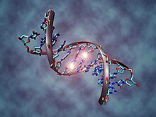
A DNA molecule fragment that is methylated at two cytosines
 
In somatic cells, patterns of DNA methylation are in general transmitted to daughter cells with high fidelity. However, epigenetic DNA methylation differs between normal cells and tumor cells in humans. The "normal" CpG methylation profile is often inverted in cells that become tumorigenic. In normal cells, CpG islands preceding gene promoters are generally unmethylated, and tend to be transcriptionally active, while other individual CpG dinucleotides throughout the genome tend to be methylated. However, in cancer cells, CpG islands preceding tumor suppressor gene promoters are often hypermethylated, while CpG methylation of oncogene promoter regions and parasitic repeat sequences is often decreased.

Hypermethylation of tumor suppressor gene promoter regions can result in silencing of those genes. This type of epigenetic mutation allows cells to grow and reproduce uncontrollably, leading to tumorigenesis. Genes commonly found to be transcriptionally silenced due to promoter hypermethylation include: Cyclin-dependent kinase inhibitor p16, a cell-cycle inhibitor; MGMT, a DNA repair gene; APC, a cell cycle regulator; MLH1, a DNA-repair gene; and BRCA1, another DNA-repair gene. Indeed, cancer cells can become addicted to the transcriptional silencing, due to promoter hypermethylation, of some key tumor suppressor genes, a process known as epigenetic addiction.

Hypomethylation of CpG dinucleotides in other parts of the genome leads to chromosome instability due to mechanisms such as loss of imprinting and reactivation of transposable elements. Loss of imprinting of insulin-like growth factor gene (IGF2) increases risk of colorectal cancer and is associated with Beckwith-Wiedemann syndrome which significantly increases the risk of cancer for newborns. In healthy cells, CpG dinucleotides of lower densities are found within coding and non-coding intergenic regions. Expression of some repetitive sequences and meiotic recombination at centromeres are repressed through methylation.

The entire genome of a cancerous cell contains significantly less methylcytosine than the genome of a healthy cell. In fact, cancer cell genomes have 20-50% less methylation at individual CpG dinucleotides across the genome. CpG islands found in promoter regions are usually protected from DNA methylation. In cancer cells CpG islands are hypomethylated  The regions flanking CpG islands called CpG island shores are where most DNA methylation occurs in the CpG dinucleotide context. Cancer cells are deferentially methylated at CpG island shores. In cancer cells, hypermethylation in the CpG island shores move into CpG islands, or hypomethylation of CpG islands move into CpG island shores eliminating sharp epigenetic boundaries between these genetic elements. In cancer cells "global hypomethylation" due to disruption in DNA methyltransferases (DNMTs) may promote mitotic recombination and chromosome rearrangement, ultimately resulting in aneuploidy when the chromosomes fail to separate properly during mitosis.

CpG island methylation is important in regulation of gene expression, yet cytosine methylation can lead directly to destabilizing genetic mutations and a precancerous cellular state. Methylated cytosines make hydrolysis of the amine group and spontaneous conversion to thymine more favorable. They can cause aberrant recruitment of chromatin proteins. Cytosine methylations change the amount of UV light absorption of the nucleotide base, creating pyrimidine dimers. When mutation results in loss of heterozygosity at tumor suppressor gene sites, these genes may become inactive. Single base pair mutations during replication can also have detrimental effects.

Histone modification

Eukaryotic DNA has a complex structure. It is generally wrapped around special proteins called histones to form a structure called a nucleosome. A nucleosome consists of 2 sets of 4 histones: H2A, H2B, H3, and H4. Additionally, histone H1 contributes to DNA packaging outside of the nucleosome. Certain histone modifying enzymes can add or remove functional groups to the histones, and these modifications influence the level of transcription of the genes wrapped around those histones and the level of DNA replication. Histone modification profiles of healthy and cancerous cells tend to differ. 

In comparison to healthy cells, cancerous cells exhibit decreased monoacetylated and trimethylated forms of histone H4 (decreased H4ac and H4me3). Additionally, mouse models have shown that a decrease in histone H4R3 asymmetric dimethylation (H4R3me2a) of the p19ARF promoter is correlated with more advanced cases of tumorigenesis and metastasis. In mouse models, the loss of histone H4 acetylation and trimethylation increases as tumor growth continues. Loss of histone H4 Lysine 16 acetylation (H4K16ac), which is a mark of aging at the telomeres, specifically loses its acetylation. Some scientists hope this particular loss of histone acetylation might be battled with a histone deacetylase (HDAC) inhibitor specific for SIRT1, an HDAC specific for H4K16.

Other histone marks associated with tumorigenesis include increased deacetylation (decreased acetylation) of histones H3 and H4, decreased trimethylation of histone H3 Lysine 4 (H3K4me3), and increased monomethylation of histone H3 Lysine 9 (H3K9me) and trimethylation of histone H3 Lysine 27 (H3K27me3). These histone modifications can silence tumor suppressor genes despite the drop in methylation of the gene's CpG island (an event that normally activates genes).

Some research has focused on blocking the action of BRD4 on acetylated histones, which has been shown to increase the expression of the Myc protein, implicated in several cancers. The development process of the drug to bind to BRD4 is noteworthy for the collaborative, open approach the team is taking.

The tumor suppressor gene p53 regulates DNA repair and can induce apoptosis in dysregulated cells. E Soto-Reyes and F Recillas-Targa elucidated the importance of the CTCF protein in regulating p53 expression.[26] CTCF, or CCCTC binding factor, is a zinc finger protein that insulates the p53 promoter from accumulating repressive histone marks. In certain types of cancer cells, the CTCF protein does not bind normally, and the p53 promoter accumulates repressive histone marks, causing p53 expression to decrease.

Mutations in the epigenetic machinery itself may occur as well, potentially responsible for the changing epigenetic profiles of cancerous cells. The histone variants of the H2A family are highly conserved in mammals, playing critical roles in regulating many nuclear processes by altering chromatin structure. One of the key H2A variants, H2A.X, marks DNA damage, facilitating the recruitment of DNA repair proteins to restore genomic integrity. Another variant, H2A.Z, plays an important role in both gene activation and repression. A high level of H2A.Z expression is detected in many cancers and is significantly associated with cellular proliferation and genomic instability. Histone variant macroH2A1 is important in the pathogenesis of many types of cancers, for instance in hepatocellular carcinoma. Other mechanisms include a decrease in H4K16ac may be caused by either a decrease in activity of a histone acetyltransferases (HATs) or an increase in deacetylation by SIRT1. Likewise, an inactivating frameshift mutation in HDAC2, a histone deacetylase that acts on many histone-tail lysines, has been associated with cancers showing altered histone acetylation patterns. These findings indicate a promising mechanism for altering epigenetic profiles through enzymatic inhibition or enhancement. 

DNA damage, caused by UV light, ionizing radiation, environmental toxins, and metabolic chemicals, can also lead to genomic instability and cancer. The DNA damage response to double strand DNA breaks (DSB) is mediated in part by histone modifications. At a DSB, MRE11-RAD50-NBS1 (MRN) protein complex recruits ataxia telangiectasia mutated (ATM) kinase which phosphorylates Serine 129 of Histone 2A. MDC1, mediator of DNA damage checkpoint 1, binds to the phosphopeptide, and phosphorylation of H2AX may spread by a positive feedback loop of MRN-ATM recruitment and phosphorylation. TIP60 acetylates the γH2AX, which is then polyubiquitylated. RAP80, a subunit of the DNA repair breast cancer type 1 susceptibility protein complex (BRCA1-A), binds ubiquitin attached to histones. BRCA1-A activity arrests the cell cycle at the G2/M checkpoint, allowing time for DNA repair, or apoptosis may be initiated.

MicroRNA gene silencing

In mammals, microRNAs (miRNAs) regulate about 60% of the transcriptional activity of protein-encoding genes. Some miRNAs also undergo methylation-associated silencing in cancer cells. Let-7 and miR15/16 play important roles in down-regulating RAS and BCL2 oncogenes, and their silencing occurs in cancer cells. Decreased expression of miR-125b1, a miRNA that functions as a tumor suppressor, was observed in prostate, ovarian, breast and glial cell cancers. In vitro experiments have shown that miR-125b1 targets two genes, HER2/neu and ESR1, that are linked to breast cancer. DNA methylation, specifically hypermethylation, is one of the main ways that the miR-125b1 is epigenetically silenced. In patients with breast cancer, hypermethylation of CpG islands located proximal to the transcription start site was observed. Loss of CTCF binding and an increase in repressive histone marks, H3K9me3 and H3K27me3, correlates with DNA methylation and miR-125b1 silencing. Mechanistically, CTCF may function as a boundary element to stop the spread of DNA methylation. Results from experiments conducted by Soto-Reyes et al. indicate a negative effect of methylation on the function and expression of miR-125b1. Therefore, they concluded that DNA methylation has a part in silencing the gene. Furthermore, some miRNA's are epigenetically silenced early on in breast cancer, and therefore these miRNA's could potentially be useful as tumor markers. The epigenetic silencing of miRNA genes by aberrant DNA methylation is a frequent event in cancer cells; almost one third of miRNA promoters active in normal mammary cells were found hypermethylated in breast cancer cells - that is a several fold greater proportion than is usually observed for protein coding genes.

Metabolic recoding of epigenetics in cancer

Dysregulation of metabolism allows tumor cells to generate needed building blocks as well as to modulate epigenetic marks to support cancer initiation and progression. Cancer-induced metabolic changes alter the epigenetic landscape, especially modifications on histones and DNA, thereby promoting malignant transformation, adaptation to inadequate nutrition, and metastasis. The accumulation of certain metabolites in cancer can target epigenetic enzymes to globally alter the epigenetic landscape. Cancer-related metabolic changes lead to locus-specific recoding of epigenetic marks. Cancer epigenetics can be precisely reprogramed by cellular metabolism through 1) dose-responsive modulation of cancer epigenetics by metabolites; 2) sequence-specific recruitment of metabolic enzymes; and 3) targeting of epigenetic enzymes by nutritional signals.

MicroRNA and DNA repair

DNA damage appears to be the primary underlying cause of cancer. If DNA repair is deficient, DNA damage tends to accumulate. Such excess DNA damage can increase mutational errors during DNA replication due to error-prone translesion synthesis. Excess DNA damage can also increase epigenetic alterations due to errors during DNA repair. Such mutations and epigenetic alterations can give rise to cancer.

Germ line mutations in DNA repair genes cause only 2–5% of colon cancer cases. However, altered expression of microRNAs, causing DNA repair deficiencies, are frequently associated with cancers and may be an important causal factor for these cancers.

Over-expression of certain miRNAs may directly reduce expression of specific DNA repair proteins. Wan et al. referred to 6 DNA repair genes that are directly targeted by the miRNAs indicated in parentheses: ATM (miR-421), RAD52 (miR-210, miR-373), RAD23B (miR-373), MSH2 (miR-21), BRCA1 (miR-182) and P53 (miR-504, miR-125b). More recently, Tessitore et al. listed further DNA repair genes that are directly targeted by additional miRNAs, including ATM (miR-18a, miR-101), DNA-PK (miR-101), ATR (miR-185), Wip1 (miR-16), MLH1, MSH2 and MSH6 (miR-155), ERCC3 and ERCC4 (miR-192) and UNG2 (mir-16, miR-34c and miR-199a). Of these miRNAs, miR-16, miR-18a, miR-21, miR-34c, miR-125b, miR-101, miR-155, miR-182, miR-185 and miR-192 are among those identified by Schnekenburger and Diederich as over-expressed in colon cancer through epigenetic hypomethylation. Over expression of any one of these miRNAs can cause reduced expression of its target DNA repair gene.

Up to 15% of the MLH1-deficiencies in sporadic colon cancers appeared to be due to over-expression of the microRNA miR-155, which represses MLH1 expression. However, the majority of 68 sporadic colon cancers with reduced expression of the DNA mismatch repair protein MLH1 were found to be deficient due to epigenetic methylation of the CpG island of the MLH1 gene.

In 28% of glioblastomas, the MGMT DNA repair protein is deficient but the MGMT promoter is not methylated. In the glioblastomas without methylated MGMT promoters, the level of microRNA miR-181d is inversely correlated with protein expression of MGMT and the direct target of miR-181d is the MGMT mRNA 3’UTR (the three prime untranslated region of MGMT mRNA). Thus, in 28% of glioblastomas, increased expression of miR-181d and reduced expression of DNA repair enzyme MGMT may be a causal factor. In 29–66% of glioblastomas, DNA repair is deficient due to epigenetic methylation of the MGMT gene, which reduces protein expression of MGMT. 

High mobility group A (HMGA) proteins, characterized by an AT-hook, are small, nonhistone, chromatin-associated proteins that can modulate transcription. MicroRNAs control the expression of HMGA proteins, and these proteins (HMGA1 and HMGA2) are architectural chromatin transcription-controlling elements. Palmieri et al showed that, in normal tissues, HGMA1 and HMGA2 genes are targeted (and thus strongly reduced in expression) by miR-15, miR-16, miR-26a, miR-196a2 and Let-7a

HMGA expression is almost undetectable in differentiated adult tissues but is elevated in many cancers. HGMA proteins are polypeptides of ~100 amino acid residues characterized by a modular sequence organization. These proteins have three highly positively charged regions, termed AT hooks, that bind the minor groove of AT-rich DNA stretches in specific regions of DNA. Human neoplasias, including thyroid, prostatic, cervical, colorectal, pancreatic and ovarian carcinoma, show a strong increase of HMGA1a and HMGA1b proteins. Transgenic mice with HMGA1 targeted to lymphoid cells develop aggressive lymphoma, showing that high HMGA1 expression is not only associated with cancers, but that the HMGA1 gene can act as an oncogene to cause cancer. Baldassarre et al., showed that HMGA1 protein binds to the promoter region of DNA repair gene BRCA1 and inhibits BRCA1 promoter activity. They also showed that while only 11% of breast tumors had hypermethylation of the BRCA1 gene, 82% of aggressive breast cancers have low BRCA1 protein expression, and most of these reductions were due to chromatin remodeling by high levels of HMGA1 protein.

HMGA2 protein specifically targets the promoter of ERCC1, thus reducing expression of this DNA repair gene. ERCC1 protein expression was deficient in 100% of 47 evaluated colon cancers (though the extent to which HGMA2 was involved is unknown).

Palmieri et al. showed that each of the miRNAs that target HMGA genes are drastically reduced in almost all human pituitary adenomas studied, when compared with the normal pituitary gland. Consistent with the down-regulation of these HMGA-targeting miRNAs, an increase in the HMGA1 and HMGA2-specific mRNAs was observed. Three of these microRNAs (miR-16, miR-196a and Let-7a) have methylated promoters and therefore low expression in colon cancer. For two of these, miR-15 and miR-16, the coding regions are epigenetically silenced in cancer due to histone deacetylase activity. When these microRNAs are expressed at a low level, then HMGA1 and HMGA2 proteins are expressed at a high level. HMGA1 and HMGA2 target (reduce expression of) BRCA1 and ERCC1 DNA repair genes. Thus DNA repair can be reduced, likely contributing to cancer progression.

DNA repair pathways

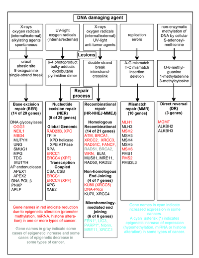
A chart of common DNA damaging agents, examples of lesions they cause in DNA, and pathways used to repair these lesions. Also shown are many of the genes in these pathways, an indication of which genes are epigenetically regulated to have reduced (or increased) expression in various cancers. It also shows genes in the error prone microhomology-mediated end joining pathway with increased expression in various cancers.

The chart in this section shows some frequent DNA damaging agents, examples of DNA lesions they cause, and the pathways that deal with these DNA damages. At least 169 enzymes are either directly employed in DNA repair or influence DNA repair processes. Of these, 83 are directly employed in repairing the 5 types of DNA damages illustrated in the chart. 

Some of the more well studied genes central to these repair processes are shown in the chart. The gene designations shown in red, gray or cyan indicate genes frequently epigenetically altered in various types of cancers. Wikipedia articles on each of the genes highlighted by red, gray or cyan describe the epigenetic alteration(s) and the cancer(s) in which these epimutations are found. Three review articles, and two broad experimental survey articles also document most of these epigenetic DNA repair deficiencies in cancers. 

Red-highlighted genes are frequently reduced or silenced by epigenetic mechanisms in various cancers. When these genes have low or absent expression, DNA damages can accumulate. Replication errors past these damages can lead to increased mutations and, ultimately, cancer. Epigenetic repression of DNA repair genes in accurate DNA repair pathways appear to be central to carcinogenesis.

The two gray-highlighted genes RAD51 and BRCA2, are required for homologous recombinational repair. They are sometimes epigenetically over-expressed and sometimes under-expressed in certain cancers. As indicated in the Wikipedia articles on RAD51 and BRCA2, such cancers ordinarily have epigenetic deficiencies in other DNA repair genes. These repair deficiencies would likely cause increased unrepaired DNA damages. The over-expression of RAD51 and BRCA2 seen in these cancers may reflect selective pressures for compensatory RAD51 or BRCA2 over-expression and increased homologous recombinational repair to at least partially deal with such excess DNA damages. In those cases where RAD51 or BRCA2 are under-expressed, this would itself lead to increased unrepaired DNA damages. Replication errors past these damages could cause increased mutations and cancer, so that under-expression of RAD51 or BRCA2 would be carcinogenic in itself. 

Cyan-highlighted genes are in the microhomology-mediated end joining (MMEJ) pathway and are up-regulated in cancer. MMEJ is an additional error-prone inaccurate repair pathway for double-strand breaks. In MMEJ repair of a double-strand break, an homology of 5-25 complementary base pairs between both paired strands is sufficient to align the strands, but mismatched ends (flaps) are usually present. MMEJ removes the extra nucleotides (flaps) where strands are joined, and then ligates the strands to create an intact DNA double helix. MMEJ almost always involves at least a small deletion, so that it is a mutagenic pathway. FEN1, the flap endonuclease in MMEJ, is epigenetically increased by promoter hypomethylation and is over-expressed in the majority of cancers of the breast, prostate, stomach, neuroblastomas, pancreas, and lung. PARP1 is also over-expressed when its promoter region ETS site is epigenetically hypomethylated, and this contributes to progression to endometrial cancer, BRCA-mutated ovarian cancer, and BRCA-mutated serous ovarian cancer. Other genes in the MMEJ pathway are also over-expressed in a number of cancers (see MMEJ for summary), and are also shown in blue.

Frequencies of epimutations in DNA repair genes

Deficiencies in DNA repair proteins that function in accurate DNA repair pathways increase the risk of mutation. Mutation rates are strongly increased in cells with mutations in DNA mismatch repair or in homologous recombinational repair (HRR). Individuals with inherited mutations in any of 34 DNA repair genes are at increased risk of cancer.

In sporadic cancers, a deficiency in DNA repair is occasionally found to be due to a mutation in a DNA repair gene, but much more frequently reduced or absent expression of DNA repair genes is due to epigenetic alterations that reduce or silence gene expression. For example, for 113 colorectal cancers examined in sequence, only four had a missense mutation in the DNA repair gene MGMT, while the majority had reduced MGMT expression due to methylation of the MGMT promoter region (an epigenetic alteration). Similarly, out of 119 cases of mismatch repair-deficient colorectal cancers that lacked DNA repair gene PMS2 expression, PMS2 protein was deficient in 6 due to mutations in the PMS2 gene, while in 103 cases PMS2 expression was deficient because its pairing partner MLH1 was repressed due to promoter methylation (PMS2 protein is unstable in the absence of MLH1). In the other 10 cases, loss of PMS2 expression was likely due to epigenetic overexpression of the microRNA, miR-155, which down-regulates MLH1.

Epigenetic defects in DNA repair genes are frequent in cancers. In the table, multiple cancers were evaluated for reduced or absent expression of the DNA repair gene of interest, and the frequency shown is the frequency with which the cancers had an epigenetic deficiency of gene expression. Such epigenetic deficiencies likely arise early in carcinogenesis, since they are also frequently found (though at somewhat lower frequency) in the field defect surrounding the cancer from which the cancer likely arose (see Table). 

Frequency of epigenetic reduction in DNA repair gene expression in sporadic cancers and in adjacent field defects
Cancer Gene Frequency in Cancer Frequency in Field Defect
Colorectal MGMT 46% 34%
Colorectal MGMT 47% 11%
Colorectal MGMT 70% 60%
Colorectal MSH2 13% 5%
Colorectal ERCC1 100% 40%
Colorectal PMS2 88% 50%
Colorectal XPF 55% 40%
Head and Neck MGMT 54% 38%
Head and Neck MLH1 33% 25%
Head and Neck MLH1 31% 20%
Stomach MGMT 88% 78%
Stomach MLH1 73% 20%
Esophagus MLH1 77%–100% 23%–79%

It appears that cancers may frequently be initiated by an epigenetic reduction in expression of one or more DNA repair enzymes. Reduced DNA repair likely allows accumulation of DNA damages. Error prone translesion synthesis past some of these DNA damages may give rise to a mutation with a selective advantage. A clonal patch with a selective advantage may grow and out-compete neighboring cells, forming a field defect. While there is no obvious selective advantage for a cell to have reduced DNA repair, the epimutation of the DNA repair gene may be carried along as a passenger when the cells with the selectively advantageous mutation are replicated. In the cells carrying both the epimutation of the DNA repair gene and the mutation with the selective advantage, further DNA damages will accumulate, and these could, in turn, give rise to further mutations with still greater selective advantages. Epigenetic defects in DNA repair may thus contribute to the characteristic high frequency of mutations in the genomes of cancers, and cause their carcinogenic progression. 

Cancers have high levels of genome instability, associated with a high frequency of mutations. A high frequency of genomic mutations increases the likelihood of particular mutations occurring that activate oncogenes and inactivate tumor suppressor genes, leading to carcinogenesis. On the basis of whole genome sequencing, cancers are found to have thousands to hundreds of thousands of mutations in their whole genomes. By comparison, the mutation frequency in the whole genome between generations for humans (parent to child) is about 70 new mutations per generation. In the protein coding regions of the genome, there are only about 0.35 mutations between parent/child generations (less than one mutated protein per generation). Whole genome sequencing in blood cells for a pair of identical twin 100-year-old centenarians only found 8 somatic differences, though somatic variation occurring in less than 20% of blood cells would be undetected.

While DNA damages may give rise to mutations through error prone translesion synthesis, DNA damages can also give rise to epigenetic alterations during faulty DNA repair processes. The DNA damages that accumulate due to epigenetic DNA repair defects can be a source of the increased epigenetic alterations found in many genes in cancers. In an early study, looking at a limited set of transcriptional promoters, Fernandez et al. examined the DNA methylation profiles of 855 primary tumors. Comparing each tumor type with its corresponding normal tissue, 729 CpG island sites (55% of the 1322 CpG sites evaluated) showed differential DNA methylation. Of these sites, 496 were hypermethylated (repressed) and 233 were hypomethylated (activated). Thus, there is a high level of epigenetic promoter methylation alterations in tumors. Some of these epigenetic alterations may contribute to cancer progression.

Epigenetic carcinogens

A variety of compounds are considered as epigenetic carcinogens—they result in an increased incidence of tumors, but they do not show mutagen activity (toxic compounds or pathogens that cause tumors incident to increased regeneration should also be excluded). Examples include diethylstilbestrol, arsenite, hexachlorobenzene, and nickel compounds.

Many teratogens exert specific effects on the fetus by epigenetic mechanisms. While epigenetic effects may preserve the effect of a teratogen such as diethylstilbestrol throughout the life of an affected child, the possibility of birth defects resulting from exposure of fathers or in second and succeeding generations of offspring has generally been rejected on theoretical grounds and for lack of evidence. However, a range of male-mediated abnormalities have been demonstrated, and more are likely to exist. FDA label information for Vidaza, a formulation of 5-azacitidine (an unmethylatable analog of cytidine that causes hypomethylation when incorporated into DNA) states that "men should be advised not to father a child" while using the drug, citing evidence in treated male mice of reduced fertility, increased embryo loss, and abnormal embryo development. In rats, endocrine differences were observed in offspring of males exposed to morphine. In mice, second generation effects of diethylstilbesterol have been described occurring by epigenetic mechanisms.

Cancer subtypes

Skin cancer

Melanoma is a deadly skin cancer that originates from melanocytes. Several epigenetic alterations are known to play a role in the transition of melanocytes to melanoma cells. These alterations are the consequence of deregulation of their corresponding enzymes. Several histone methyltransferases and demethylases are among these enzymes.

Prostate cancer

Prostate cancer kills around 35,000 men yearly, and about 220,000 men are diagnosed with prostate cancer per year, in North America alone. Prostate cancer is the second leading cause of cancer-caused fatalities in men, and within a man's lifetime, one in six men will have the disease. Alterations in histone acetylation and DNA methylation occur in various genes influencing prostate cancer. More than 90% of prostate cancers show gene silencing by CpG island hypermethylation of the GSTP1 gene promoter, which protects prostate cells from genomic damage that is caused by different oxidants or carcinogens. Real-time methylation-specific polymerase chain reaction (PCR) suggests that many other genes are also hypermethylated. Gene expression in the prostate may be modulated by nutrition and lifestyle changes.

Cervical cancer

The second most common malignant tumor in women is invasive cervical cancer (ICC) and more than 50% of all invasive cervical cancer (ICC) is caused by oncongenic human papillomavirus 16 (HPV16). Furthermore, cervix intraepithelial neoplasia (CIN) is primarily caused by oncogenic HPV16. As in many cases, the causative factor for cancer does not always take a direct route from infection to the development of cancer. Genomic methylation patterns have been associated with invasive cervical cancer. Within the HPV16L1 region, 14 tested CpG sites have significantly higher methylation in CIN3+ than in HPV16 genomes of women without CIN3. Only 2/16 CpG sites tested in HPV16 upstream regulatory region were found to have association with increased methylation in CIN3+. This suggests that the direct route from infection to cancer is sometimes detoured to a precancerous state in cervix intraepithelial neoplasia. Additionally, increased CpG site methylation was found in low levels in most of the five host nuclear genes studied, including 5/5 TERT, 1/4 DAPK1, 2/5 RARB, MAL, and CADM1. Furthermore, 1/3 of CpG sites in mitochondrial DNA were associated with increased methylation in CIN3+. Thus, a correlation exists between CIN3+ and increased methylation of CpG sites in the HPV16 L1 open reading frame. This could be a potential biomarker for future screens of cancerous and precancerous cervical disease.

Leukemia

Recent studies have shown that the mixed-lineage leukemia (MLL) gene causes leukemia by rearranging and fusing with other genes in different chromosomes, which is a process under epigenetic control.

Sarcoma

There are about 15,000 new cases of sarcoma in the US each year, and about 6,200 people were projected to die of sarcoma in the US in 2014. Sarcomas comprise a large number of rare, histogenetically heterogeneous mesenchymal tumors that, for example, include chondrosarcoma, Ewing's sarcoma, leiomyosarcoma, liposarcoma, osteosarcoma, synovial sarcoma, and (alveolar and embryonal) rhabdomyosarcoma. Several oncogenes and tumor suppressor genes are epigenetically altered in sarcomas. These include APC, CDKN1A, CDKN2A, CDKN2B, Ezrin, FGFR1, GADD45A, MGMT, STK3, STK4, PTEN, RASSF1A, WIF1, as well as several miRNAs. Expression of epigenetic modifiers such as that of the BMI1 component of the PRC1 complex is deregulated in chondrosarcoma, Ewing's sarcoma, and osteosarcoma, and expression of the EZH2 component of the PRC2 complex is altered in Ewing's sarcoma and rhabdomyosarcoma. Similarly, expression of another epigenetic modifier, the LSD1 histone demethylase, is increased in chondrosarcoma, Ewing's sarcoma, osteosarcoma, and rhabdomyosarcoma. Drug targeting and inhibition of EZH2 in Ewing's sarcoma, or of LSD1 in several sarcomas, inhibits tumor cell growth in these sarcomas.

Identification methods

Previously, epigenetic profiles were limited to individual genes under scrutiny by a particular research team. Recently, however, scientists have been moving toward a more genomic approach to determine an entire genomic profile for cancerous versus healthy cells.

Popular approaches for measuring CpG methylation in cells include:
Since bisulfite sequencing is considered the gold standard for measuring CpG methylation, when one of the other methods is used, results are usually confirmed using bisulfite sequencing. Popular approaches for determining histone modification profiles in cancerous versus healthy cells include:

Diagnosis and prognosis

Researchers are hoping to identify specific epigenetic profiles of various types and subtypes of cancer with the goal of using these profiles as tools to diagnose individuals more specifically and accurately. Since epigenetic profiles change, scientists would like to use the different epigenomic profiles to determine the stage of development or level of aggressiveness of a particular cancer in patients. For example, hypermethylation of the genes coding for Death-Associated Protein Kinase (DAPK), p16, and Epithelial Membrane Protein 3 (EMP3) have been linked to more aggressive forms of lung, colorectal, and brain cancers. This type of knowledge can affect the way that doctors will diagnose and choose to treat their patients.

Another factor that will influence the treatment of patients is knowing how well they will respond to certain treatments. Personalized epigenomic profiles of cancerous cells can provide insight into this field. For example, MGMT is an enzyme that reverses the addition of alkyl groups to the nucleotide guanine. Alkylating guanine, however, is the mechanism by which several chemotherapeutic drugs act in order to disrupt DNA and cause cell death. Therefore, if the gene encoding MGMT in cancer cells is hypermethylated and in effect silenced or repressed, the chemotherapeutic drugs that act by methylating guanine will be more effective than in cancer cells that have a functional MGMT enzyme.

Epigenetic biomarkers can also be utilized as tools for molecular prognosis. In primary tumor and mediastinal lymph node biopsy samples, hypermethylation of both CDKN2A and CDH13 serves as the marker for increased risk of faster cancer relapse and higher death rate of patients.

Treatment

Epigenetic control of the proto-onco regions and the tumor suppressor sequences by conformational changes in histones plays a role in the formation and progression of cancer. Pharmaceuticals that reverse epigenetic changes might have a role in a variety of cancers.

Recently, it is evidently known that associations between specific cancer histotypes and epigenetic changes can facilitate the development of novel epi-drugs. Drug development has focused mainly on modifying DNA methyltransferase, histone acetyltransferase (HAT) and histone deacetylase (HDAC).

Drugs that specifically target the inverted methylation pattern of cancerous cells include the DNA methyltransferase inhibitors azacitidine and decitabine. These hypomethylating agents are used to treat myelodysplastic syndrome, a blood cancer produced by abnormal bone marrow stem cells. These agents inhibit all three types of active DNA methyltransferases, and had been thought to be highly toxic, but proved to be effective when used in low dosage, reducing progression of myelodysplastic syndrome to leukemia.

Histone deacetylase (HDAC) inhibitors show efficacy in treatment of T cell lymphoma. two HDAC inhibitors, vorinostat and romidepsin, have been approved by the Food and Drug Administration. However, since these HDAC inhibitors alter the acetylation state of many proteins in addition to the histone of interest, knowledge of the underlying mechanism at the molecular level of patient response is required to enhance the efficiency of using such inhibitors as treatment. Treatment with HDAC inhibitors has been found to promote gene reactivation after DNA methyl-transferases inhibitors have repressed transcription. Panobinostat is approved for certain situations in myeloma.

Other pharmaceutical targets in research are histone lysine methyltransferases (KMT) and protein arginine methyltransferases (PRMT). Preclinical study has suggested that lunasin may have potentially beneficial epigenetic effects.

Delayed-choice quantum eraser

From Wikipedia, the free encyclopedia https://en.wikipedia.org/wiki/Delayed-choice_quantum_eraser A delayed-cho...