Search This Blog

Friday, May 26, 2023

Embryology

From Wikipedia, the free encyclopedia
1 - morula, 2 - blastula
 
1 - blastula, 2 - gastrula with blastopore; orange - ectoderm, red - endoderm

Embryology (from Greek ἔμβρυον, embryon, "the unborn, embryo"; and -λογία, -logia) is the branch of animal biology that studies the prenatal development of gametes (sex cells), fertilization, and development of embryos and fetuses. Additionally, embryology encompasses the study of congenital disorders that occur before birth, known as teratology.

Early embryology was proposed by Marcello Malpighi, and known as preformationism, the theory that organisms develop from pre-existing miniature versions of themselves. Aristotle proposed the theory that is now accepted, epigenesis. Epigenesis is the idea that organisms develop from seed or egg in a sequence of steps. Modern embryology developed from the work of Karl Ernst von Baer, though accurate observations had been made in Italy by anatomists such as Aldrovandi and Leonardo da Vinci in the Renaissance.

Comparative embryology

Preformationism and epigenesis

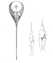
A tiny person (a homunculus) inside a sperm, as drawn by Nicolaas Hartsoeker in 1695

As recently as the 18th century, the prevailing notion in western human embryology was preformation: the idea that semen contains an embryo – a preformed, miniature infant, or homunculus – that simply becomes larger during development.

The competing explanation of embryonic development was epigenesis, originally proposed 2,000 years earlier by Aristotle. Much early embryology came from the work of the Italian anatomists Aldrovandi, Aranzio, Leonardo da Vinci, Marcello Malpighi, Gabriele Falloppio, Girolamo Cardano, Emilio Parisano, Fortunio Liceti, Stefano Lorenzini, Spallanzani, Enrico Sertoli, and Mauro Ruscóni. According to epigenesis, the form of an animal emerges gradually from a relatively formless egg. As microscopy improved during the 19th century, biologists could see that embryos took shape in a series of progressive steps, and epigenesis displaced preformation as the favored explanation among embryologists.

'CLEVAGE' Cleavage is the very beginning steps of a developing embryo. Cleavage refers to the many mitotic divisions that occur after the egg is fertilized by the sperm. The ways in which the cells divide is specific to certain types of animals and may have many forms.

Holoblastic

Holoblastic cleavage is the complete division of cells. Holoblastic cleavage can be radial (see: Radial cleavage), spiral (see: Spiral cleavage), bilateral (see: Bilateral cleavage), or rotational (see: Rotational cleavage). In holoblastic cleavage the entire egg will divide and become the embryo, whereas in meroblastic cleavage some cells will become the embryo and others will be the yolk sac.

Meroblastic

Meroblastic cleavage is the incomplete division of cells. The division furrow does not protrude into the yolky region as those cells impede membrane formation and this causes the incomplete separation of cells. Meroblastic cleavage can be bilateral (see: Bilateral cleavage), discoidal (see: Discoidal cleavage), or centrolecithal (see: Centrolecithal).

Basal phyla

Animals that belong to the basal phyla have holoblastic radial cleavage which results in radial symmetry (see: Symmetry in biology). During cleavage there is a central axis that all divisions rotate about. The basal phyla also has only one to two embryonic cell layers, compared to the three in bilateral animals (endoderm, mesoderm, and ectoderm).

Bilaterians

In bilateral animals cleavage can be either holoblastic or meroblastic depending on the species. During gastrulation the blastula develops in one of two ways that divide the whole animal kingdom into two-halves (see: Embryological origins of the mouth and anus). If in the blastula the first pore, or blastopore, becomes the mouth of the animal, it is a protostome; if the blastopore becomes the anus then it is a deuterostome. The protostomes include most invertebrate animals, such as insects, worms and molluscs, while the deuterostomes include the vertebrates. In due course, the blastula changes into a more differentiated structure called the gastrula. Soon after the gastrula is formed, three distinct layers of cells (the germ layers) from which all the bodily organs and tissues then develop.

Germ layers

  • The innermost layer, or endoderm, give rise to the digestive organs, the gills, lungs or swim bladder if present, and kidneys or nephrites.
  • The middle layer, or mesoderm, gives rise to the muscles, skeleton if any, and blood system.
  • The outer layer of cells, or ectoderm, gives rise to the nervous system, including the brain, and skin or carapace and hair, bristles, or scales.

Drosophila melanogaster (fruit fly)

Drosophila have been used as a developmental model for many years. The studies that have been conducted have discovered many useful aspects of development that not only apply to fruit flies but other species as well.

Outlined below is the process that leads to cell and tissue differentiation.

  1. Maternal-effect genes help to define the anterior-posterior axis using Bicoid (gene) and Nanos (gene).
  2. Gap genes establish 3 broad segments of the embryo.
  3. Pair-rule genes define 7 segments of the embryo within the confines of the second broad segment that was defined by the gap genes.
  4. Segment-polarity genes define another 7 segments by dividing each of the pre-existing 7 segments into anterior and posterior halves using a gradient of Hedgehog and Wnt.
  5. Homeotic (Hox) genes use the 14 segments as pinpoints for specific types of cell differentiation and the histological developments that correspond to each cell type.

Humans

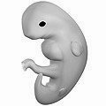
Humans are bilateral animals that have holoblastic rotational cleavage. Humans are also deuterostomes. In regard to humans, the term embryo refers to the ball of dividing cells from the moment the zygote implants itself in the uterus wall until the end of the eighth week after conception. Beyond the eighth week after conception (tenth week of pregnancy), the developing human is then called a fetus.

Evolutionary embryology

Evolutionary embryology is the expansion of comparative embryology by the ideas of Charles Darwin. Similarly to Karl Ernst von Baer's principles that explained why many species often appear similar to one another in early developmental stages, Darwin argued that the relationship between groups can be determined based upon common embryonic and larval structures.

Von Baer's principles

  1. The general features appear earlier in development than do the specialized features.
  2. More specialized characters develop from the more general ones.
  3. The embryo of a given species never resembles the adult form of a lower one.
  4. The embryo of a given species does resemble the embryonic form of a lower one.

Using Darwin's theory evolutionary embryologists have since been able to distinguish between homologous and analogous structures between varying species. Homologous structures are those that the similarities between them are derived from a common ancestor, such as the human arm and bat wings. Analogous structures are those that appear to be similar but have no common ancestral derivation.

Origins of modern embryology

Until the birth of modern embryology through observation of the mammalian ovum by Karl Ernst von Baer in 1827, there was no clear scientific understanding of embryology, although later discussions in this article show that some cultures had a fairly refined understanding of some of the principles. Only in the late 1950s when ultrasound was first used for uterine scanning, was the true developmental chronology of human fetus available. Karl Ernst von Baer along with Heinz Christian Pander, also proposed the germ layer theory of development which helped to explain how the embryo developed in progressive steps. Part of this explanation explored why embryos in many species often appear similar to one another in early developmental stages using his four principles.

Modern embryology research

Embryology is central to evolutionary developmental biology ("evo-devo"), which studies the genetic control of the development process (e.g. morphogens), its link to cell signalling, its roles in certain diseases and mutations, and its links to stem cell research. Embryology is the key to Gestational Surrogacy, which is when the sperm of the intended father and egg of intended mother are fused in a lab forming an embryo. This embryo is then put into the surrogate who carries the child to term.

Medical embryology

Medical embryology is used widely to detect abnormalities before birth. 2-5% of babies are born with an observable abnormality and medical embryology explores the different ways and stages that these abnormalities appear in. Genetically derived abnormalities are referred to as malformations. When there are multiple malformations, this is considered a syndrome. When abnormalities appear due to outside contributors, these are disruptions. The outside contributors causing disruptions are known as teratogens. Common teratogens are alcohol, retinoic acid, ionizing radiation or hyperthermic stress.

Vertebrate and invertebrate embryology

Many principles of embryology apply to invertebrates as well as to vertebrates. Therefore, the study of invertebrate embryology has advanced the study of vertebrate embryology. However, there are many differences as well. For example, numerous invertebrate species release a larva before development is complete; at the end of the larval period, an animal for the first time comes to resemble an adult similar to its parent or parents. Although invertebrate embryology is similar in some ways for different invertebrate animals, there are also countless variations. For instance, while spiders proceed directly from egg to adult form, many insects develop through at least one larval stage. For decades, a number of so-called normal staging tables were produced for the embryology of particular species, mainly focussing on external developmental characters. As variation in developmental progress makes comparison among species difficult, a character-based Standard Event System was developed, which documents these differences and allows for phylogenetic comparisons among species.

Birth of developmental biology

After the 1950s, with the DNA helical structure being unraveled and the increasing knowledge in the field of molecular biology, developmental biology emerged as a field of study which attempts to correlate the genes with morphological change, and so tries to determine which genes are responsible for each morphological change that takes place in an embryo, and how these genes are regulated.

As of today, human embryology is taught as a cornerstone subject in medical schools, as well as in biology and zoology programs at both an undergraduate and graduate level.

History

Ancient Egypt

Knowledge of the placenta goes back at least to ancient Egypt, where it was viewed as the seat of the soul. There was an Egyptian official with the title Opener of the Kings Placenta. An Egyptian text from the time of Akhenaten said that a human originates from the egg that grows in women.

Ancient India

A variety of conceptions on embryology appeared in ancient Asia. Descriptions of the amniotic sac appear in the Bhagavad Gita, Bhagavata Purana, and the Sushruta Samhita. . One of the Upanishads known as the Garbhopanisaḍ states that the embryo is "like water in the first night, in seven nights it is like a bubble, at the end of half a month it becomes a ball. At the end of a month it is hardened, in two months the head is formed". The Indian medical tradition in the Ayurveda also has conceptions of embryology from antiquity.

Ancient Greece

Pre-Socratic philosophers

Many pre-Socratic philosophers are recorded as having opinions on different aspects of embryology, although there is some bias in the description of their views in later authors such as Aristotle. According to Empedocles (whose views are described by Plutarch in the 1st century AD), who lived in the 5th century BC, the embryo derives and receives its blood from four vessels in all; two arteries and two veins. He also held sinews as originating from equal mixtures of earth and air. He further said men begin to form within the first month and are finished within fifty days. Asclepiades agreed that men are formed within fifty days, but he believed that women took a full two months to be fully knit. One observation, variously attributed to either Anaxagoras of Clazomenae or Alcmaeon of Croton, says that the milk produced by mammals is analogous to the white of fowl egg. Diogenes of Apollonia said that a mass of flesh forms first, only then followed by the development of bone and nerves. Diogenes recognized that the placenta was a nutritional source for the growing fetus. He also said that the development of males took four months, but that the development of females took five months. He did not think the embryo was alive. Alcmaeon also made some contributions, and he is the first person reported to have practiced dissection. One idea, first stated by Parmenides, was that there was a connection between the right side of the body and the male embryo, and between the left side of the body and the female embryo. According to Democritus and Epicurus, the fetus is nourished at the mouth inside the mother and there are comparable teats that supply this nourishment within the mother's body to the fetus. Discussion on various views regarding how long it takes for specific parts of the embryo to form appear in an anonymous document known as the Nutriment.

Ancient Greeks discussed whether only the male had a seed which developed into the embryo within the female womb, or both the male and the female each had a seed that made a contribution to the developing embryo. The difficulty that one-seed theorists confronted was to explain the maternal resemblance of the progeny. One issue that two-seed theorists confronted was why the female seed was needed if the male already had a seed. One common solution to this problem was to assert that the female seed was either inferior or inactive. Another question was the origin of the seed. The encephalomyelogenic theory stated that the seed originated from the brain or and/or bone marrow. Later came pangenesis, which asserted the seed was drawn from the whole body in order to explain the general resemblance in the body of the offspring. Later on hematogenous theory developed which asserted that the seed was drawn from the blood. A third question was how or in what form the progeny existed in the seed prior to developing into an embryo and a fetus. According to preformationists, the body of the progeny already existed in a pre-existing but undeveloped form in the seed. Three variants of preformationism were homoiomerous preformationism, anhomoiomerous preformationism, and homuncular preformationism. According to the first, the homoiomerous parts of the body (e.g. humors, bone) already exist pre-formed in the seed. The second held that it was the anhomoiomerous parts that were pre-formed. Finally, the third view held that the whole was already a unified organic thing. Preformationism was not the only view. According to epigenesists, parts of the embryo successively form after conception takes place.

Hippocrates

Some of the most well-known early ideas on embryology come from Hippocrates and the Hippocratic Corpus, where discussion on the embryo is usually given in the context of discussing obstetrics (pregnancy and childbirth). Some of the most relevant Hippocratic texts on embryology include the Regimen on Acute Diseases, On Semen, and On the Development of the Child. Hippocrates claimed that the development of the embryo is put into motion by fire and that nourishment comes from food and breath introduced into the mother. An outer layer of the embryo solidifies, and the fire within consumes humidity which makes way for development of bone and nerve. The fire in the innermost part becomes the belly and air channels are developed in order to route nourishment to it. The enclosed fire also helps form veins and allows for circulation. In this description, Hippocrates aims at describing the causes of development rather than describing what develops. Hippocrates also develops views similar to preformationism, where he claims that all parts of the embryo simultaneously develop. Hippocrates also believed that maternal blood nourishes the embryo. This blood flows and coagulates to help form the flesh of the fetus. This idea was derived from the observation that menstrual blood ceases during pregnancy, which Hippocrates took to imply that it was being redirected to fetal development. Hippocrates also claimed that the flesh differentiates into different organs of the body, and Hippocrates saw as analogous an experiment where a mixture of substances placed into water will differentiate into different layers. Comparing the seed to the embryo, Hippocrates further compared the stalk to the umbilical cord.

Aristotle

Some embryological discussion appears in the writings of Aristotle's predecessor Plato, especially in his Timaeus. One of his views were that the bone marrow acted as the seedbed, and that the soul itself was the seed out of which the embryo developed, though he did not explain how this development proceeded. Scholars also continue to debate the views he held on various other aspects of embryology. However, a much more voluminous discussion on the subject comes from the writings of Aristotle, especially as appears in his On the Generation of Animals. Some ideas related to embryology also appear in his History of Animals, On the Parts of Animals, On Respiration, and On the Motion of Animals. Means by which we know Aristotle studied embryology, and most likely his predecessors as well, was through studying developing embryos taken out from animals as well as aborted and miscarried human embryos. Aristotle believed that the female supplied the matter for the development of the embryo, formed from the menstrual blood whereas the semen that comes from the male shapes that matter. Aristotle's belief that both the male and female made a contribution to the actual fetus goes against some prior beliefs. According to Aeschylus and some Egyptian traditions, the fetus solely develops from the male contribution and that the female womb simply nourishes this growing fetus. On the other hand, the Melanesians held that the fetus is solely a product of the female contribution. Aristotle did not believe there were any external influences on the development of the embryo. Against Hippocrates, Aristotle believed that new parts of the body developed over time rather than all forming immediately and developing from then on. He also considered whether each new part derives from a previously formed part or develops independently of any previously formed part. On the basis that different parts of the body do not resemble each other, he decided in favor of the latter view. He also described development of fetal parts in terms of mechanical and automatic processes. In terms of the development of the embryo, he says it begins in a liquid-like state as the material secreted by the female combines with the semen of the male, and then the surface begins to solidify as it interacts with processes of heating and cooling. The first part of the body to differentiate is the heart, which Aristotle and many of his contemporaries believed was the location of reason and thinking. Aristotle claimed that vessels join to the uterus in order to supply nourishment to the developing fetus. Some of the most solid parts of the fetus cool and, as they lose moisture to heat, turn into nails, horns, hoofs, beaks, etc. Internal heat dries away moisture and forms sinews and bones and the skin results from drying of the flesh. Aristotle also describes the development of birds in eggs at length. He further described embryonic development in dolphins, some sharks, and many other animals. Aristotle singularly wrote more on embryology than any other pre-modern author, and his influence on the subsequent discussion on the subject for many centuries was immense, introducing into the subject forms of classification, a comparative method from various animals, discussion of the development of sexual characteristics, compared the development of the embryo to mechanistic processes, and so forth.

Later Greek embryology

Reportedly, some Stoics claimed that most parts of the body formed at once during embryological development. Some Epicureans claimed that the fetus is nourished by either the amniotic fluid or the blood, and that both male and female supply material to the development of the fetus. According to the writings of Tertullian, Herophilus in the 4th century BC described the ovaries and Fallopian tubes (but not past what was already described by Aristotle) and also dissected some embryos. One advance Herophilus made, against the conceptions of other individuals such as Aristotle, was that the brain was the center of intellect rather than the heart. Though not a part of Greek tradition, in Job 10, the formation of the embryo is likened to the curdling of milk into cheese, as described by Aristotle. Whereas Needham sees this statement in Job as part of the Aristotelian tradition, others see it as evidence that the milk analogy predates the Aristotelian Greek tradition and originates in Jewish circles. In addition, the Wisdom of Solomon (7:2) also has the embryo formed from menstrual blood. Soranus of Ephesus also wrote texts on embryology which went into use for a long time. Some rabbinic texts discuss the embryology of a female Greek writer named Cleopatra, a contemporary of Galen and Soranus, who was said to have claimed that the male fetus is complete in 41 days whereas the female fetus is complete in 81 days. Various other texts of less importance also appear and describe various aspects of embryology, though without making much progress from Aristotle. Plutarch has a chapter in one of his works titled "Whether was before, the hen or egg?" Discussion on embryological tradition also appears in many Neoplatonic traditions.

Next to Aristotle, the most impactful and important Greek writer on biology was Galen of Pergamum, and his works were transmitted throughout the Middle Ages. Galen discusses his understanding of embryology in two of his texts, those being his On the Natural Faculties and his On the Formation of the Foetus. There is an additional text spuriously attributed to Galen known as On the Question of whether the Embryo is an Animal. Galen described embryological development in four stages. In the first stage, the semen predominates. In the second stage, the embryo is filled with blood. In the third stage, the main outlines of the organs have developed but various other parts remain undeveloped. In the fourth stage, formation is complete and has reached a stage where we can call it a child. Galen described processes that played a role in furthering development of the embryo such as warming, drying, cooling, and combinations thereof. As this development plays out, the form of life of the embryo also moves from that like a plant to that of an animal (where the analogy between the root and umbilical cord is made). Galen claimed that the embryo forms from menstrual blood, by which his experimental analogy was that when you cut the vein of an animal and allow blood to flow out and into some mildly heated water, a sort of coagulation can be observed. He gave detailed descriptions of the position of the umbilical cord relative to other veins.

Patristics

The question of embryology is discussed among a number of patristic authors, largely in terms of theological questions such as whether the fetus has value and/or when it begins to have value. (Although a number of Christian authors continued the classical discussions on the description of the development of the embryo, such as Jacob of Serugh. Passing reference to the embryo also appears in the eighth hymn of Ephrem the Syrian's Paradise Hymns.) Many patristic treatments of embryology continued in the stream of Greek tradition. The earlier Greek and Roman view that it was not was reversed and all pre-natal infanticide was condemned. Tertullian held that the soul was present from the moment of conception. The Quinisext Council concluded that "we pay no attention to the subtle division as to whether the foetus is formed or unformed". In this time, then, the Roman practice of child exposure came to an end, where unwanted yet birthed children, usually females, were discarded by the parents to die. Other more liberal traditions followed Augustine, who instead viewed that the animation of life began on the 40th day in males and the 80th day in females but not prior. Before the 40th day for men and 80th day for women, the embryo was referred to as the embryo informatus, and after this period was reached, it was referred to as the embryo formatus. The notion originating from the Greeks that the male embryo developed faster remained in various authors until it was experimentally disproven by Andreas Ottomar Goelicke in 1723.

Various patristic literature from backgrounds ranging from Nestorian, Monophysite and Chalcedonian discuss and choose between three different conceptions on the relation between the soul and the embryo. According to one view, the soul pre-exists and enters the embryo at the moment of conception (prohyparxis). According to a second view, the soul enters into existence at the moment of conception (synhyparxis). In a third view, the soul enters into the body after it has been formed (methyparxis). The first option was proposed by Origen, but was increasingly rejected after the fourth century. On the other hand, the other two options were equally accepted after this point. The second position appears to have been proposed as a response to Origen's notion of a pre-existing soul. After the sixth century, the second position was also increasingly seen as Origenist and so rejected on those grounds. The writings of Origen were condemned during the Second Origenist Crises in 553. Those defending prohyparxis usually appealed to the Platonic notion of an eternally moving soul. Those defending the second position also appealed to Plato but rejected his notion on the eternality of the soul. Finally, those appealing to the third position appealed both to Aristotle and scripture. Aristotelian notions included the progression of the development of the soul, from an initial plant-like soul, to a sensitive soul found in animals and allows for movement and perception, and finally the formation of a rational soul which can only be found in the fully-formed human. Furthermore, some scriptural texts were seen as implying the formation of the soul temporally after the formation of the body (namely Genesis 2:7; Exodus 21:22-23; Zachariah 12:1). In the De hominis opificio of Gregory of Nyssa, Aristotle's triparitate notion of the soul was accepted. Gregory also held that the rational soul was present at conception. Theodoret argued based on Genesis 2:7 and Exodus 21:22 that the embryo is only ensouled after the body is fully formed. Based on Exodus 21:22 and Zachariah 12:1, the Monophysite Philoxenus of Mabbug claimed that the soul was created in the body forty days after conception. In his De opificio mundi, the Christian philosopher John Philoponus claimed that the soul is formed after the body. Later still, the author Leontius held that the body and soul were created simultaneously, though it is also possible he held that the soul pre-existed the body.

Some Monophysites and Chalcedonians seemed to have been compelled into accepting synhyparxis in the case of Jesus because of their view that the incarnation of Christ resulted in both one hypostasis and one nature, whereas some Nestorians claimed that Christ, like us, must have had his soul formed after the formation of his body because, per Hebrews 4:15, Christ was like us in all ways but sin. (On the other hand, Leontinus dismissed the relevance of Hebrews 4:15 on the basis that Christ differed from us not only in sinfulness but also conception without semen, making synhyparxis another of Christ's supernatural feats.) They felt comfortable holding this view, under their belief that the human nature of Jesus was separate from the divine hypostasis. Some Nestorians still wondered, however, if the body united with the soul in the moment the soul was created or whether it came with it only later. The Syriac author Babai argued for the former on the basis that the latter was hardly better than adoptionism. Maximus the Confessor ridiculed the Aristotelian notion of the development of the soul on the basis that it would make humans parents of both plants and animals. He held to synhyparxis and regarded the other two positions both as incorrect extremes. After the 7th century, Chalcedonian discussion on embryology is slight and the few works that touch on the topic support synhyparxis. But debate among other groups remains lively, still divided on similar sectarian grounds. The patriarch Timothy I argued that the Word first united with the body, and only later with the soul. He cited John 1:1, claiming on its basis that the Word became flesh first, not a human being first. Then, Jacob of Edessa rejected prohyparxis because Origen had defended it and methyparxis because he believed that it made the soul ontologically inferior and as only being made for the body. Then, Moses Bar Kepha claimed, for Christological reasons as a Monophysite, that only synhyparxis was acceptable. He claimed that Genesis 2:7 has no temporal sequence and that Exodus 21:22 regards the formation of the body and not the soul and so is not relevant. To argue against methyparxis, he reasoned that body and soul are both present at death and, because what is at the end must correspond to what is also at the beginning, conception must also have body and soul together.


The development of Embryology was cleary mentioned in ancient Buddhist text of Garbhāvakrāntisūtra (1st-4th century CE). It cleary mentions about human gestation period for 38 days.The text also talks about embryonic development in first three weeks as a liquid part of yogurt and in the third month, differentiation of body parts such as arms, leg,feeth and head.

Embryology in Jewish tradition

Many Jewish authors also discussed notions of embryology, especially as they appear in the Talmud. Much of the embryological data in the Talmud is part of discussions related to the impurity of the mother after childbirth. The embryo was described as the peri habbetten (fruit of the body) and it developed through various stages: (1) golem (formless and rolled-up) (2) shefir meruqqam (embroidered foetus) (3) ubbar (something carried) (4) walad (child) (5) walad shel qayama (viable child) (6) ben she-kallu khadashaw (child whose months have been completed). Some mystical notions regarding embryology appear in the Sefer Yetzirah. The text in the Book of Job relating to the fetus forming by analogy to the curdling of milk into cheese was cited in the Babylonian Talmud and in even greater detail in the Midrash: "When the womb of the woman is full of retained blood which then comes forth to the area of her menstruation, by the will of the Lord comes a drop of white-matter which falls into it: at once the embryo is created. [This can be] compared to milk being put in a vessel: if you add to it some lab-ferment [drug or herb], it coagulates and stands still; if not, the milk remains liquid." The Talmud sages held that there were two seeds that participated in the formation of the embryo, one from the male and one from the female, and that their relative proportions determine whether that develops into a male or a female. In the Tractate Nidda, the mother was said to provide a "red-seed" which allows for the development of skin, flesh, hair, and the black part of the eye (pupil), whereas the father provides the "white-seed" which forms the bones, nerves, brain, and the white part of the eye. And finally, God himself was thought to provide the spirit and soul, facial expressions, capacity for hearing and vision, movement, comprehension, and intelligence. Not all strands of Jewish tradition accepted that both the male and female contributed parts to the formation of the fetus. The 13th century medieval commentator Nachmanides, for example, rejected the female contribution. In Tractate Hullin in the Talmud, whether the organs of the child resemble more closely those of the mother or father is said to depend on which one contribute more matter to the embryo depending on the child. Rabbi Ishmael and other sages are said to have disagreed on one matter: they agreed that the male embryo developed on the 41st day, but disagreed on whether this was the case for the female embryo. Some believed that the female embryo was complete later, whereas others held that they were finished at the same time. The only ancient Jewish authors who associated abortion with homicide were Josephus and Philo of Alexandria in the 1st century. Some Talmudic texts discuss magical influences on the development of the embryo, such as one text which claims that if one sleeps on a bed that is pointed to the north–south will have a male child. According to Nachmanides, a child born of a cold drop of semen will be foolish, one born from a warm drop of semen will be passionate and irascible, and one born from a semen drop of medium temperature will be clever and level-headed. Some Talmudic discussions follow from Hippocratic claims that a child born on the eighth month could not survive, whereas others follow Aristotle in claiming that they sometimes could survive. One text even says that survival is possible on the seventh month, but not the eighth. Talmudic embryology, in various aspects, follows Greek discourses especially from Hippocrates and Aristotle, but in other areas, makes novel statements on the subject.

Embryology in the Islamic tradition

Passing reference to embryological notions also appear in the Qur'an (22:5), where the development of the embryo proceeds in four stages from drop, to a blood clot, to a partially developed stage, to a fully developed child. The notion of clay turning into flesh is seen by some as analogous to a text by Theodoret that describes the same process. The four stages of development in the Qur'an are similar to the four stages of embryological development as described by Galen. In the early 6th century, Sergius of Reshaina devoted himself to the translation of Greek medical texts into Syriac and became the most important figure in this process. Included in his translations were the relevant embryological texts of Galen. Anurshirvan founded a medical school in the southern Mesopotamian city of Gundeshapur, known as the Academy of Gondishapur, which also acted as a medium for the transmission, reception, and development of notions from Greek medicine. These factors helped the transmission of Greek notions on embryology, such as found in Galen, to enter into the Arabian milieu. Very similar embryonic descriptions also appear in the Syriac Jacob of Serugh's letter to the Archdeacon Mar Julian.

Embryological discussions also appear in the Islamic legal tradition.

Mammography

From Wikipedia, the free encyclopedia
https://en.wikipedia.org/wiki/Mammography
 
Mammography
Mammogram.jpg
Other namesMastography
ICD-10-PCSBH0
ICD-9-CM87.37
MeSHD008327
OPS-301 code3–10
MedlinePlus003380

Mammography (also called mastography) is the process of using low-energy X-rays (usually around 30 kVp) to examine the human breast for diagnosis and screening. The goal of mammography is the early detection of breast cancer, typically through detection of characteristic masses or microcalcifications.

As with all X-rays, mammograms use doses of ionizing radiation to create images. These images are then analyzed for abnormal findings. It is usual to employ lower-energy X-rays, typically Mo (K-shell X-ray energies of 17.5 and 19.6 keV) and Rh (20.2 and 22.7 keV) than those used for radiography of bones. Mammography may be 2D or 3D (tomosynthesis), depending on the available equipment and/or purpose of the examination. Ultrasound, ductography, positron emission mammography (PEM), and magnetic resonance imaging (MRI) are adjuncts to mammography. Ultrasound is typically used for further evaluation of masses found on mammography or palpable masses that may or may not be seen on mammograms. Ductograms are still used in some institutions for evaluation of bloody nipple discharge when the mammogram is non-diagnostic. MRI can be useful for the screening of high risk patients, for further evaluation of questionable findings or symptoms, as well as for pre-surgical evaluation of patients with known breast cancer, in order to detect additional lesions that might change the surgical approach (for example, from breast-conserving lumpectomy to mastectomy).

For the average woman, the U.S. Preventive Services Task Force recommends (2016) mammography every two years between the ages of 50 and 74, concluding that "the benefit of screening mammography outweighs the harms by at least a moderate amount from age 50 to 74 years and is greatest for women in their 60s". The American College of Radiology and American Cancer Society recommend yearly screening mammography starting at age 40. The Canadian Task Force on Preventive Health Care (2012) and the European Cancer Observatory (2011) recommend mammography every 2 to 3 years between ages 50 and 69. These task force reports point out that in addition to unnecessary surgery and anxiety, the risks of more frequent mammograms include a small but significant increase in breast cancer induced by radiation. Additionally, mammograms should not be performed with increased frequency in patients undergoing breast surgery, including breast enlargement, mastopexy, and breast reduction. The Cochrane Collaboration (2013) concluded after ten years that trials with adequate randomization did not find an effect of mammography screening on total cancer mortality, including breast cancer. The authors of this Cochrane review write: "If we assume that screening reduces breast cancer mortality by 15% and that overdiagnosis and over-treatment is at 30%, it means that for every 2,000 women invited for screening throughout 10 years, one will avoid dying of breast cancer and 10 healthy women, who would not have been diagnosed if there had not been screening, will be treated unnecessarily. Furthermore, more than 200 women will experience important psychological distress including anxiety and uncertainty for years because of false positive findings." The authors conclude that the time has come to re-assess whether universal mammography screening should be recommended for any age group. They state that universal screening may not be reasonable. The Nordic Cochrane Collection updated research in 2012 and stated that advances in diagnosis and treatment make mammography screening less effective today, rendering it "no longer effective". They conclude that "it therefore no longer seems reasonable to attend" for breast cancer screening at any age, and warn of misleading information on the internet. On the contrary, a report in the New England Journal of Medicine attributes the poor effectiveness of national mammography screening programs at reducing breast cancer mortality to radiation-induced cancers.

Mammography has a false-negative (missed cancer) rate of at least ten percent. This is partly due to dense tissue obscuring the cancer and the appearance of cancer on mammograms having a large overlap with the appearance of normal tissue. A meta-analysis review of programs in countries with organized screening found a 52% over-diagnosis rate.

History

As a medical procedure that induces ionizing radiation, the origin of mammography can be traced to the discovery of X-rays by Wilhelm Röntgen in 1895.

In 1913, German surgeon Albert Salomon performed a mammography study on 3,000 mastectomies, comparing X-rays of the breasts to the actual removed tissue, observing specifically microcalcifications. By doing so, he was able to establish the difference as seen on an X-ray image between cancerous and non-cancerous tumors in the breast. Salomon's mammographs provided substantial information about the spread of tumors and their borders.

In 1930, American physician and radiologist Stafford L. Warren published "A Roentgenologic Study of the Breast", a study where he produced stereoscopic X-rays images to track changes in breast tissue as a result of pregnancy and mastitis. In 119 women who subsequently underwent surgery, he correctly found breast cancer in 54 out of 58 cases.

As early as 1937, Jacob Gershon-Cohen developed a form a mammography for a diagnostic of breast cancer at earlier stages to improve survival rates. In the early 1950s, Uruguayan radiologist Raul Leborgne developed the breast compression technique to produce better quality images, and described the differences between benign and malign microcalcifications. In 1956, Gershon-Cohen conducted clinical trails on over 1,000 asymptomatic women at the Albert Einstein Medical Center on his screening technique, and the same year, Robert Egan at the University of Texas M.D. Anderson Cancer Center combined a technique of low kVp with high mA and single emulsion films to devise a method of screening mammography. He published these results in 1959 in a paper, subsequently vulgarized in a 1964 book called Mammography. The "Egan technique", as it became known, enabled physicians to detect calcification in breast tissue; of the 245 breast cancers that were confirmed by biopsy among 1,000 patients, Egan and his colleagues at M.D. Anderson were able to identify 238 cases by using his method, 19 of which were in patients whose physical examinations had revealed no breast pathology.

Use of mammography as a screening technique spread clinically after a 1966 study demonstrating the impact of mammograms on mortality and treatment led by Philip Strax. This study, based in New York, was the first large-scale randomized controlled trial of mammography screening.

Procedure

Illustration of a mammogram
 
A mobile mammography unit in New Zealand

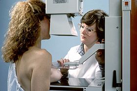
During the procedure, the breast is compressed using a dedicated mammography unit. Parallel-plate compression evens out the thickness of breast tissue to increase image quality by reducing the thickness of tissue that X-rays must penetrate, decreasing the amount of scattered radiation (scatter degrades image quality), reducing the required radiation dose, and holding the breast still (preventing motion blur). In screening mammography, both head-to-foot (craniocaudal, CC) view and angled side-view (mediolateral oblique, MLO) images of the breast are taken. Diagnostic mammography may include these and other views, including geometrically magnified and spot-compressed views of the particular area of concern. Deodorant, talcum powder or lotion may show up on the X-ray as calcium spots, so women are discouraged from applying them on the day of their exam. There are two types of mammogram studies: screening mammograms and diagnostic mammograms. Screening mammograms, consisting of four standard X-ray images, are performed yearly on patients who present with no symptoms. Diagnostic mammograms are reserved for patients with breast symptoms (such as palpable lumps, breast pain, skin changes, nipple changes, or nipple discharge), as follow-up for probably benign findings (coded BI-RADS 3), or for further evaluation of abnormal findings seen on their screening mammograms. Diagnostic mammograms may also performed on patients with personal and/or family histories of breast cancer. Patients with breast implants and other stable benign surgical histories generally do not require diagnostic mammograms.

Until some years ago, mammography was typically performed with screen-film cassettes. Today, mammography is undergoing transition to digital detectors, known as digital mammography or Full Field Digital Mammography (FFDM). The first FFDM system was approved by the FDA in the U.S. in 2000. This progress is occurring some years later than in general radiology. This is due to several factors:

  1. The higher spatial resolution demands of mammography
  2. Significantly increased expense of the equipment
  3. Concern by the FDA that digital mammography equipment demonstrate that it is at least as good as screen-film mammography at detecting breast cancers without increasing dose or the number of women recalled for further evaluation.

As of March 1, 2010, 62% of facilities in the United States and its territories have at least one FFDM unit. (The FDA includes computed radiography units in this figure.)

Tomosynthesis, otherwise known as 3D mammography, was first introduced in clinical trials in 2008 and has been Medicare-approved in the United States since 2015. As of 2023, 3D mammography has become widely available in the US and has been shown to have improved sensitivity and specificity over 2D mammography.

Mammograms are either looked at by one (single reading) or two (double reading) trained professionals: these film readers are generally radiologists, but may also be radiographers, radiotherapists, or breast clinicians (non-radiologist physicians specializing in breast disease). Double reading, which is standard practice in the UK, but less common in the US, significantly improves the sensitivity and specificity of the procedure. Clinical decision support systems may be used with digital mammography (or digitized images from analogue mammography), but studies suggest these approaches do not significantly improve performance or provide only a small improvement.

Digital

Digital mammography is a specialized form of mammography that uses digital receptors and computers instead of X-ray film to help examine breast tissue for breast cancer. The electrical signals can be read on computer screens, permitting more manipulation of images to allow radiologists to view the results more clearly. Digital mammography may be "spot view", for breast biopsy, or "full field" (FFDM) for screening.

Digital mammography is also utilized in stereotactic biopsy. Breast biopsy may also be performed using a different modality, such as ultrasound or magnetic resonance imaging (MRI).

While radiologists had hoped for more marked improvement, the effectiveness of digital mammography was found comparable to traditional X-ray methods in 2004, though there may be reduced radiation with the technique and it may lead to fewer retests. Specifically, it performs no better than film for post-menopausal women, who represent more than three-quarters of women with breast cancer. The U.S. Preventive Services Task Force concluded that there was insufficient evidence to recommend for or against digital mammography.

Digital mammography is a NASA spin-off, utilizing technology developed for the Hubble Space Telescope. As of 2007, about 8% of American screening centers used digital mammography. Around the globe, systems by Fujifilm Corporation are the most widely used. In the United States, GE's digital imaging units typically cost US$300,000 to $500,000, far more than film-based imaging systems. Costs may decline as GE begins to compete with the less expensive Fuji systems.

3D mammography

Three-dimensional mammography, also known as digital breast tomosynthesis (DBT), tomosynthesis, and 3D breast imaging, is a mammogram technology that creates a 3D image of the breast using X-rays. When used in addition to usual mammography, it results in more positive tests. Cost effectiveness is unclear as of 2016. Another concern is that it more than doubles the radiation exposure.

Photon counting

Photon-counting mammography was introduced commercially in 2003 and was shown to reduce the X-ray dose to the patient by approximately 40% compared to conventional methods while maintaining image quality at an equal or higher level. The technology was subsequently developed to enable spectral imaging with the possibility to further improve image quality, to distinguish between different tissue types, and to measure breast density.

Galactography

A galactography (or breast ductography) is a now infrequently used type of mammography used to visualize the milk ducts. Prior to the mammography itself, a radiopaque substance is injected into the duct system. This test is indicated when nipple discharge exists.

Scoring

Mammogram results are often expressed in terms of the BI-RADS Assessment Category, often called a "BI-RADS score". The categories range from 0 (Incomplete) to 6 (Known biopsy – proven malignancy). In the UK mammograms are scored on a scale from 1–5 (1 = normal, 2 = benign, 3 = indeterminate, 4 = suspicious of malignancy, 5 = malignant). Evidence suggests that accounting for genetic risk, factors improve breast cancer risk prediction.

"Work-up" process

In the past several years, the "work-up" process has become highly formalized. It generally consists of screening mammography, diagnostic mammography, and biopsy when necessary, often performed via stereotactic core biopsy or ultrasound-guided core biopsy. After a screening mammogram, some women may have areas of concern which cannot be resolved with only the information available from the screening mammogram. They would then be called back for a "diagnostic mammogram". This phrase essentially means a problem-solving mammogram. During this session, the radiologist will be monitoring each of the additional films as they are taken by a radiographer. Depending on the nature of the finding, ultrasound may often be used as well.

Generally, the cause of the unusual appearance is found to be benign. If the cause cannot be determined to be benign with sufficient certainty, a biopsy may be recommended. The biopsy procedure will be used to obtain actual tissue from the site for the pathologist to examine microscopically to determine the precise cause of the abnormality. In the past, biopsies were most frequently done in surgery, under local or general anesthesia. The majority are now done with needles in conjunction with either ultrasound or mammographic guidance to be sure that the area of concern is the area that is biopsied. These core biopsies require only local anesthesia, similar to what would be given during a minor dental procedure.

Benefits

Mammography can detect cancer early when it’s most treatable and can be treated less invasively (thereby helping to preserve quality of life).

According to National Cancer Institute data, since mammography screening became widespread in the mid-1980s, the U.S. breast cancer death rate, unchanged for the previous 50 years, has dropped well over 30 percent. In European countries like Denmark and Sweden, where mammography screening programs are more organized, the breast cancer death rate has been cut almost in half over the last 20 years.

A study published in Cancer Epidemiology, Biomarkers & Prevention shows mammography screening cuts the risk of dying from breast cancer nearly in half. A recent study published in Cancer showed that more than 70 percent of the women who died from breast cancer in their 40s at major Harvard teaching hospitals were among the 20 percent of women who were not being screened. Some scientific studies have shown that the most lives are saved by screening beginning at age 40.

A recent study in the British Medical Journal shows that early detection of breast cancer – as with mammography – significantly improves breast cancer survival.

The benefits of mammography screening at decreasing breast cancer mortality in randomized trials are not found in observational studies performed long after implementation of breast cancer screening programs (for instance, Bleyer et al.) These discrepancies can be explained by cancers caused by mammograms.

When to start screening

In 2014, the Surveillance, Epidemiology, and End Results Program of the National Institutes of Health reported the occurrence rates of breast cancer based on 1000 women in different age groups. In the 40–44 age group, the incidence was 1.5 and in the 45–49 age group, the incidence was 2.3. In the older age groups, the incidence was 2.7 in the 50–54 age group and 3.2 in the 55–59 age group. While screening between ages 40 and 50 is somewhat controversial, the preponderance of the evidence indicates that there is a benefit in terms of early detection. Currently, the American Cancer Society, the American Congress of Obstetricians and Gynecologists (ACOG), the American College of Radiology, and the Society of Breast Imaging encourage annual mammograms beginning at age 40.

The National Cancer Institute encourages mammograms every one to two years for women ages 40 to 49. In contrast, the American College of Physicians, a large internal medicine group, has recently encouraged individualized screening plans as opposed to wholesale biannual screening of women aged 40 to 49. In 2009, the U.S. Preventive Services Task Force recommended that screening of women ages 40 to 49 be based on individual risk factors, and that screening should not be routine in this age group. Their report says that the benefits of screenings before the age of 50 do not outweigh the risks.

Starting screening at age 40

One in six breast cancers occur in women in their 40s. The ten year risk for breast cancer in a 40-year-old woman is 1 in 69 and only increases with age; 40 percent of all the years of life saved by mammography are for women in their 40s.

Screening mammography shows greatest benefit—a 39.6 percent mortality reduction—from annual screening of women 40–84 years old. This screening regimen saves 71 percent more lives than (the USPSTF-recommended regimen of) biennial screening of women 50–74 years old, which had a 23.2 percent mortality reduction. By not getting a yearly mammogram after age 40, women increase their odds of dying from breast cancer and that treatment for any advanced cancers ultimately found will be more extensive and more expensive.

Note that women at elevated risk for breast cancer due to family history or other factors should speak with their doctor about starting screening earlier than age 40.

Arguments against the USPTF recommendations

Approximately 75 percent of women diagnosed with breast cancer have no family history of breast cancer or other factors that put them at high risk for developing the disease (so screening only high-risk women misses majority of cancers). An analysis by Hendrick and Helvie, published in the American Journal of Roentgenology, showed that if USPSTF breast cancer screening guidelines were followed, approximately 6,500 additional women each year in the U.S. would die from breast cancer.

The largest (Hellquist et al) and longest running (Tabar et al) breast cancer screening studies in history, re-confirmed that regular mammography screening cut breast cancer deaths by roughly a third in all women ages 40 and over (including women ages 40–49). This renders the USPSTF calculations off by half. They used a 15% mortality reduction to calculate how many women needed to be invited to be screened to save a life. With the now re-confirmed 29% (or up) figure, the number to be screened using the USPSTF formula is half of their estimate and well within what they considered acceptable by their formula.

According to the USPSTF report, even for women 50+, skipping a mammogram every other year would miss up to 30 percent of cancers. A recent study published in Cancer showed that more than 70 percent of the women who died from breast cancer in their 40s at major Harvard teaching hospitals were among the 20 percent of women who were not being screened.

There is a concern about bias and lack of experience regarding the panel that made the recommendations. The USPSTF did not contain or involve a single breast cancer expert (oncologist, radiologist, breast surgeon or radiation oncologist), but did have current or former members of the insurance industry (which some would argue has a vested interest in not paying for mammograms).

Arguments against mammography

Normal (left) versus cancerous (right) mammography image

The use of mammography as a screening tool for the detection of early breast cancer in otherwise healthy women without symptoms is seen by some as controversial.

Keen and Keen indicated that repeated mammography starting at age fifty saves about 1.8 lives over 15 years for every 1,000 women screened. This result has to be seen against the adverse effects of errors in diagnosis, over-treatment, and radiation exposure.

The Cochrane analysis of screening indicates that it is "not clear whether screening does more good than harm". According to their analysis, 1 in 2,000 women will have her life prolonged by 10 years of screening, while 10 healthy women will undergo unnecessary breast cancer treatment. Additionally, 200 women will experience significant psychological stress due to false positive results.

Newman posits that screening mammography does not reduce death overall, but causes significant harm by inflicting cancer scare and unnecessary surgical interventions. The Nordic Cochrane Collection notes that advances in diagnosis and treatment of breast cancer may make breast cancer screening no longer effective in decreasing death from breast cancer, and therefore no longer recommend routine screening for healthy women as the risks might outweigh the benefits.

Of every 1,000 U.S. women who are screened, about 7% will be called back for a diagnostic session (although some studies estimate the number to be closer to 10% to 15%). About 10% of those who are called back will be referred for a biopsy. Of the 10% referred for biopsy, about 3.5% will have cancer and 6.5% will not. Of the 3.5% who have cancer, about 2 will have an early stage cancer that will be cured after treatment.

Mammography may also produce false negatives. Estimates of the numbers of cancers missed by mammography are usually around 20%. Reasons for not seeing the cancer include observer error, but more frequently it is because the cancer is hidden by other dense tissue in the breast, and even after retrospective review of the mammogram the cancer cannot be seen. Furthermore, one form of breast cancer, lobular cancer, has a growth pattern that produces shadows on the mammogram that are indistinguishable from normal breast tissue.

Mortality

The Cochrane Collaboration states that the best quality evidence does not demonstrate a reduction in mortality or a reduction in mortality from all types of cancer from screening mammography.

The Canadian Task Force found that for women ages 50 to 69, screening 720 women once every 2 to 3 years for 11 years would prevent one death from breast cancer. For women ages 40 to 49, 2,100 women would need to be screened at the same frequency and period to prevent a single death from breast cancer.

Women whose breast cancer was detected by screening mammography before the appearance of a lump or other symptoms commonly assume that the mammogram "saved their lives". In practice, the vast majority of these women received no practical benefit from the mammogram. There are four categories of cancers found by mammography:

  1. Cancers that are so easily treated that a later detection would have produced the same rate of cure (women would have lived even without mammography).
  2. Cancers so aggressive that even early detection is too late to benefit the patient (women who die despite detection by mammography).
  3. Cancers that would have receded on their own or are so slow-growing that the woman would die of other causes before the cancer produced symptoms (mammography results in over-diagnosis and over-treatment of this class).
  4. A small number of breast cancers that are detected by screening mammography and whose treatment outcome improves as a result of earlier detection.

Only 3% to 13% of breast cancers detected by screening mammography will fall into this last category. Clinical trial data suggests that 1 woman per 1,000 healthy women screened over 10 years falls into this category. Screening mammography produces no benefit to any of the remaining 87% to 97% of women. The probability of a woman falling into any of the above four categories varies with age.

A 2016 review for the United States Preventive Services Task Force found that mammography was associated with an 8%-33% decrease in breast cancer mortality in different age groups, but that this decrease was not statistically significant at the age groups of 39–49 and 70–74. The same review found that mammography significantly decreased the risk of advanced cancer among women aged 50 and older by 38%, but among those aged 39 to 49 the risk reduction was a non-significant 2%. The USPSTF made their review based on data from randomized controlled trials (RCT) studying breast cancer in women between the ages of 40-49.

The lack of effectiveness of mammography screening in reducing mortality may be explained by cancers caused by mammograms.

False positives

The goal of any screening procedure is to examine a large population of patients and find the small number most likely to have a serious condition. These patients are then referred for further, usually more invasive, testing. Thus a screening exam is not intended to be definitive; rather it is intended to have sufficient sensitivity to detect a useful proportion of cancers. The cost of higher sensitivity is a larger number of results that would be regarded as suspicious in patients without disease. This is true of mammography. The patients without disease who are called back for further testing from a screening session (about 7%) are sometimes referred to as "false positives". There is a trade-off between the number of patients with disease found and the much larger number of patients without disease that must be re-screened.

Research shows that false-positive mammograms may affect women's well-being and behavior. Some women who receive false-positive results may be more likely to return for routine screening or perform breast self-examinations more frequently. However, some women who receive false-positive results become anxious, worried, and distressed about the possibility of having breast cancer, feelings that can last for many years.

False positives also mean greater expense, both for the individual and for the screening program. Since follow-up screening is typically much more expensive than initial screening, more false positives (that must receive follow-up) means that fewer women may be screened for a given amount of money. Thus as sensitivity increases, a screening program will cost more or be confined to screening a smaller number of women.

Overdiagnosis

The central harm of mammographic breast cancer screening is overdiagnosis: the detection of abnormalities that meet the pathologic definition of cancer but will never progress to cause symptoms or death. Dr. H. Gilbert Welch, a researcher at Dartmouth College, states that "screen-detected breast and prostate cancer survivors are more likely to have been over-diagnosed than actually helped by the test." Estimates of overdiagnosis associated with mammography have ranged from 1% to 54%. In 2009, Peter C. Gotzsche and Karsten Juhl Jørgensen reviewed the literature and found that 1 in 3 cases of breast cancer detected in a population offered mammographic screening is over-diagnosed. In contrast, a 2012 panel convened by the national cancer director for England and Cancer Research UK concluded that 1 in 5 cases of breast cancer diagnosed among women who have undergone breast cancer screening are over-diagnosed. This means an over-diagnosis rate of 129 women per 10,000 invited to screening.

False negatives

Mammograms also have a rate of missed tumors, or "false negatives". Accurate data regarding the number of false negatives are very difficult to obtain because mastectomies cannot be performed on every woman who has had a mammogram to determine the false negative rate. Estimates of the false negative rate depend on close follow-up of a large number of patients for many years. This is difficult in practice because many women do not return for regular mammography making it impossible to know if they ever developed a cancer. In his book The Politics of Cancer, Dr. Samuel S. Epstein claims that in women ages 40 to 49, one in four cancers are missed at each mammography. Researchers have found that breast tissue is denser among younger women, making it difficult to detect tumors. For this reason, false negatives are twice as likely to occur in pre-menopausal mammograms (Prate). This is why the screening program in the UK does not start calling women for screening mammograms until age 50.

The importance of these missed cancers is not clear, particularly if the woman is getting yearly mammograms. Research on a closely related situation has shown that small cancers that are not acted upon immediately, but are observed over periods of several years, will have good outcomes. A group of 3,184 women had mammograms that were formally classified as "probably benign". This classification is for patients who are not clearly normal but have some area of minor concern. This results not in the patient being biopsied, but rather in having early follow up mammography every six months for three years to determine whether there has been any change in status. Of these 3,184 women, 17 (0.5%) did have cancers. Most importantly, when the diagnosis was finally made, they were all still stage 0 or 1, the earliest stages. Five years after treatment, none of these 17 women had evidence of re-occurrence. Thus, small early cancers, even though not acted on immediately, were still reliably curable.

Radiation

The radiation exposure associated with mammography is a potential risk of screening, which appears to be greater in younger women. In scans where women receive 0.25–20 Gray (Gy) of radiation, they have more of an elevated risk of developing breast cancer. A study of radiation risk from mammography concluded that for women 40 years of age and older, the risk of radiation-induced breast cancer was minuscule, particularly compared with the potential benefit of mammographic screening, with a benefit-to-risk ratio of 48.5 lives saved for each life lost due to radiation exposure. This also correlates to a decrease in breast cancer mortality rates by 24%. However, this estimate is based on modelling, not observations. In contrast epidemiologic studies show a high incidence of breast cancer following mammography screening. Organizations such as the National Cancer Institute and United States Preventive Task Force do not take such risks into account when formulating screening guidelines.

Other risks

The majority of health experts agree that the risk of breast cancer for asymptomatic women under 35 is not high enough to warrant the risk of radiation exposure. For this reason, and because the radiation sensitivity of the breast in women under 35 is possibly greater than in older women, most radiologists do not recommend screening mammography on women under 40. However, if there is a significant risk of cancer in a particular patient (due to genetic tests, positive family history, etc), mammography prior to age may still be important. Often, the radiologist will try to avoid mammography by using ultrasound or MRI imaging.

Pain

The mammography procedure can be painful. Reported pain rates range from 6–76%, with 23–95% experiencing pain or discomfort. Experiencing pain is a significant predictor in women not re-attending screening. There are few proven interventions to reduce pain in mammography, but evidence suggests that giving women information about the mammography procedure prior to it taking place may reduce the pain and discomfort experienced. Furthermore, research has found that standardised compression levels can help to reduce patients' pain while still allowing for optimal diagnostic images to be produced.

Attendance

Many factors affect how many people attend breast cancer screenings. For example, people from minority ethnic communities are also less likely to attend cancer screening. In the UK, women of South Asian heritage are the least likely to attend breast cancer screening. Research is still needed to identify specific barriers for the different South Asian communities. For example, a study showed that British-Pakistani women faced cultural and language barriers and were not aware that breast screening takes place in a female-only environment.

People with mental illnesses are also less likely to attend cancer screening appointments. In Northern Ireland women with mental health problems were shown to be less likely to attend screening for breast cancer, than women without. The lower attendance numbers remained the same even when marital status and social deprivation were taken into account.

Regulation

Mammography discover facilities in the United States and its territories (including military bases) are subject to the Mammography Quality Standards Act (MQSA). The act requires annual inspections and accreditation every three years through an FDA-approved body. Facilities found deficient during the inspection or accreditation process can be barred from performing mammograms until corrective action has been verified or, in extreme cases, can be required to notify past patients that their exams were sub-standard and should not be relied upon.

At this time, MQSA applies only to traditional mammography and not to related scans, such as breast ultrasound, stereotactic breast biopsy, or breast MRI.

Many states in the US require a notification to be given to women with dense breasts to inform them that mammography is less accurate if breast tissue density is high. In 2019, the Food and Drug Administration proposed a rule that would require doctors inform these women that they may need other imaging tests in addition to mammograms.

Alternative examination methods

For patients who do not want to undergo mammography, MRI and also breast computed tomography (also called breast CT) offer a painless alternative. Whether the respective method is suitable depends on the clinical picture and it is decided by the physician.

Personality theories of addiction

From Wikipedia, the free encyclopedia https://en.wikipedia.org/wiki/Personality_theories_of_addiction ...