Search This Blog

Sunday, May 19, 2019

Addison's disease

From Wikipedia, the free encyclopedia

Addison's disease
Other namesAddison disease, chronic adrenal insufficiency, hypocortisolism, hypoadrenalism, primary adrenal insufficiency
Addisons hyperpigmentation.jpg
Classic darkening of the skin due to increased pigment as seen in Addison's disease
SpecialtyEndocrinology
SymptomsAbdominal pain, weakness, weight loss, darkening of the skin
ComplicationsAdrenal crisis
Usual onsetMiddle-aged females
CausesProblems with the adrenal gland
Diagnostic methodBlood tests, urine tests, medical imaging
TreatmentCorticosteroid such as hydrocortisone and fludrocortisone
Frequency0.9–1.4 per 10,000 people (developed world)

Addison's disease, also known as primary adrenal insufficiency and hypocortisolism, is a long-term endocrine disorder in which the adrenal glands do not produce enough steroid hormones. Symptoms generally come on slowly and may include abdominal pain, weakness, and weight loss. Darkening of the skin in certain areas may also occur. Under certain circumstances, an adrenal crisis may occur with low blood pressure, vomiting, lower back pain, and loss of consciousness. An adrenal crisis can be triggered by stress, such as from an injury, surgery, or infection.

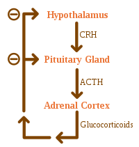
Addison's disease arises from problems with the adrenal gland such that not enough of the steroid hormone cortisol and possibly aldosterone are produced, most often due to damage by the body's own immune system in the developed world and tuberculosis in the developing world. Other causes include certain medications, sepsis, and bleeding into both adrenal glands. Secondary adrenal insufficiency is caused by not enough adrenocorticotropic hormone (ACTH) (produced by the pituitary gland) or CRH (produced by the hypothalamus). Despite this distinction, adrenal crises can happen in all forms of adrenal insufficiency. Addison's disease is generally diagnosed by blood tests, urine tests, and medical imaging.

Treatment involves replacing the absent hormones. This involves taking a corticosteroid such as hydrocortisone and fludrocortisone. These medications are usually taken by mouth. Lifelong, continuous steroid replacement therapy is required, with regular follow-up treatment and monitoring for other health problems. A high-salt diet may also be useful in some people. If symptoms worsen, an injection of corticosteroid is recommended and people should carry a dose with them. Often, large amounts of intravenous fluids with the sugar dextrose are also required. Without treatment, an adrenal crisis can result in death.

Addison's disease affects about 0.9 to 1.4 per 10,000 people in the developed world. It occurs most frequently in middle-aged females. Secondary adrenal insufficiency is more common. Long-term outcomes with treatment are typically good. It is named after Thomas Addison, a graduate of the University of Edinburgh Medical School, who first described the condition in 1855. The adjective "addisonian" is used to describe features of the condition, as well as people with Addison's disease.

Signs and symptoms

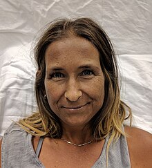
A Caucasian woman with Addison's disease
 
Legs of a Caucasian woman with Addison's disease
 
The symptoms of Addison's disease develop gradually and may become established before they are recognized. They can be nonspecific and are potentially attributable to other medical conditions. 

The signs and symptoms include fatigue; lightheadedness upon standing or difficulty standing, muscle weakness, fever, weight loss, anxiety, nausea, vomiting, diarrhea, headache, sweating, changes in mood or personality, and joint and muscle pains. Some people have cravings for salt or salty foods due to the loss of sodium through their urine. Hyperpigmentation of the skin may be seen, particularly when the person lives in a sunny area, as well as darkening of the palmar crease, sites of friction, recent scars, the vermilion border of the lips, and genital skin. These skin changes are not encountered in secondary and tertiary hypoadrenalism.

On physical examination, these clinical signs may be noticed:
  • Low blood pressure with or without orthostatic hypotension (blood pressure that decreases with standing)
  • Darkening (hyperpigmentation) of the skin, including areas not exposed to the sun. Characteristic sites of darkening are skin creases (e.g., of the hands), nipple, and the inside of the cheek (buccal mucosa); also, old scars may darken. This occurs because melanocyte-stimulating hormone (MSH) and ACTH share the same precursor molecule, pro-opiomelanocortin (POMC). After production in the anterior pituitary gland, POMC gets cleaved into gamma-MSH, ACTH, and beta-lipotropin. The subunit ACTH undergoes further cleavage to produce alpha-MSH, the most important MSH for skin pigmentation. In secondary and tertiary forms of adrenal insufficiency, skin darkening does not occur, as ACTH is not overproduced.
Addison's disease is associated with the development of other autoimmune diseases, such as type I diabetes, thyroid disease (Hashimoto's thyroiditis), celiac disease, or vitiligo. Addison's disease may be the only manifestation of undiagnosed celiac disease. Both diseases share the same genetic risk factors (HLA-DQ2 and HLA-DQ8 haplotypes).

The presence of Addison's in addition to mucocutaneous candidiasis, hypoparathyroidism, or both, is called autoimmune polyendocrine syndrome type 1. The presence of Addison's in addition to autoimmune thyroid disease, type 1 diabetes, or both, is called autoimmune polyendocrine syndrome type 2.

Addisonian crisis

An "Addisonian crisis" or "adrenal crisis" is a constellation of symptoms that indicates severe adrenal insufficiency. This may be the result of either previously undiagnosed Addison's disease, a disease process suddenly affecting adrenal function (such as adrenal hemorrhage), or an intercurrent problem (e.g., infection, trauma) in someone known to have Addison's disease. It is a medical emergency and potentially life-threatening situation requiring immediate emergency treatment. 

Characteristic symptoms are:

Causes

The negative feedback loop for glucocorticoids
 
Causes of adrenal insufficiency can be categorized by the mechanism through which they cause the adrenal glands to produce insufficient cortisol. These are adrenal dysgenesis (the gland has not formed adequately during development), impaired steroidogenesis (the gland is present but is biochemically unable to produce cortisol) or adrenal destruction (disease processes leading to glandular damage).

Adrenal destruction

Autoimmune adrenalitis is the most common cause of Addison's disease in the industrialized world. Autoimmune destruction of the adrenal cortex is caused by an immune reaction against the enzyme 21-hydroxylase (a phenomenon first described in 1992). This may be isolated or in the context of autoimmune polyendocrine syndrome (APS type 1 or 2), in which other hormone-producing organs, such as the thyroid and pancreas, may also be affected.

Adrenal destruction is also a feature of adrenoleukodystrophy, and when the adrenal glands are involved in metastasis (seeding of cancer cells from elsewhere in the body, especially lung), hemorrhage (e.g., in Waterhouse–Friderichsen syndrome or antiphospholipid syndrome), particular infections (tuberculosis, histoplasmosis, coccidioidomycosis), or the deposition of abnormal protein in amyloidosis.

Adrenal dysgenesis

All causes in this category are genetic, and generally very rare. These include mutations to the SF1 transcription factor, congenital adrenal hypoplasia due to DAX-1 gene mutations and mutations to the ACTH receptor gene (or related genes, such as in the Triple-A or Allgrove syndrome). DAX-1 mutations may cluster in a syndrome with glycerol kinase deficiency with a number of other symptoms when DAX-1 is deleted together with a number of other genes.

Impaired steroidogenesis

To form cortisol, the adrenal gland requires cholesterol, which is then converted biochemically into steroid hormones. Interruptions in the delivery of cholesterol include Smith–Lemli–Opitz syndrome and abetalipoproteinemia

Of the synthesis problems, congenital adrenal hyperplasia is the most common (in various forms: 21-hydroxylase, 17α-hydroxylase, 11β-hydroxylase and 3β-hydroxysteroid dehydrogenase), lipoid CAH due to deficiency of StAR and mitochondrial DNA mutations. Some medications interfere with steroid synthesis enzymes (e.g., ketoconazole), while others accelerate the normal breakdown of hormones by the liver (e.g., rifampicin, phenytoin).

Diagnosis

Suggestive features

Routine laboratory investigations may show:

Testing

In suspected cases of Addison's disease, demonstration of low adrenal hormone levels even after appropriate stimulation (called the ACTH stimulation test or synacthen test) with synthetic pituitary ACTH hormone tetracosactide is needed for the diagnosis. Two tests are performed, the short and the long test. Dexamethasone does not cross-react with the assay and can be administered concomitantly during testing. 

The short test compares blood cortisol levels before and after 250 micrograms of tetracosactide (intramuscular or intravenous) is given. If, one hour later, plasma cortisol exceeds 170 nmol/l and has risen by at least 330 nmol/l to at least 690 nmol/l, adrenal failure is excluded. If the short test is abnormal, the long test is used to differentiate between primary adrenal insufficiency and secondary adrenocortical insufficiency. 

The long test uses 1 mg tetracosactide (intramuscular). Blood is taken 1, 4, 8, and 24 hr later. Normal plasma cortisol level should reach 1000 nmol/l by 4 hr. In primary Addison's disease, the cortisol level is reduced at all stages, whereas in secondary corticoadrenal insufficiency, a delayed but normal response is seen. 

Other tests may be performed to distinguish between various causes of hypoadrenalism, including renin and adrenocorticotropic hormone levels, as well as medical imaging - usually in the form of ultrasound, computed tomography or magnetic resonance imaging

Adrenoleukodystrophy, and the milder form, adrenomyeloneuropathy, cause adrenal insufficiency combined with neurological symptoms. These diseases are estimated to be the cause of adrenal insufficiency in about 35% of diagnosed male with idiopathic Addison’s disease, and should be considered in the differential diagnosis of any male with adrenal insufficiency. Diagnosis is made by a blood test to detect very long chain fatty acids.

Treatment

Maintenance

Treatment for Addison's disease involves replacing the missing cortisol, sometimes in the form of hydrocortisone tablets, or prednisone tablets in a dosing regimen that mimics the physiological concentrations of cortisol. Alternatively, one-quarter as much prednisolone may be used for equal glucocorticoid effect as hydrocortisone. Treatment is usually lifelong. In addition, many people require fludrocortisone as replacement for the missing aldosterone. 

People with Addison's are often advised to carry information on them (e.g., in the form of a MedicAlert bracelet or information card) for the attention of emergency medical services personnel who might need to attend to their needs. It is also recommended that a needle, syringe, and injectable form of cortisol be carried for emergencies. People with Addison's disease are advised to increase their medication during periods of illness or when undergoing surgery or dental treatment. Immediate medical attention is needed when severe infections, vomiting, or diarrhea occur, as these conditions can precipitate an Addisonian crisis. A person who is vomiting may require injections of hydrocortisone instead.

Crisis

Standard therapy involves intravenous injections of glucocorticoids and large volumes of intravenous saline solution with dextrose (glucose). This treatment usually brings rapid improvement. If intravenous access is not immediately available, intramuscular injection of glucocorticoids can be used. When the person can take fluids and medications by mouth, the amount of glucocorticoids is decreased until a maintenance dose is reached. If aldosterone is deficient, maintenance therapy also includes oral doses of fludrocortisone acetate.

Prognosis

Outcomes are typically good when treated. Most can expect to live relatively normal lives. Someone with the disease should be observant of symptoms of an "Addison's crisis" while the body is strained, as in rigorous exercise or being sick, the latter often needing emergency treatment with intravenous injections to treat the crisis.

Individuals with Addison's disease have more than a doubled mortality rate. Furthermore, individuals with Addison's disease and diabetes mellitus have an almost 4 time increase in mortality compared to individuals with only diabetes.

Epidemiology

The frequency rate of Addison's disease in the human population is sometimes estimated at roughly one in 100,000. Some put the number closer to 40–144 cases per million population (1/25,000–1/7,000). Addison's can affect persons of any age, sex, or ethnicity, but it typically presents in adults between 30 and 50 years of age. Research has shown no significant predispositions based on ethnicity.

History

Discovery

Addison’s disease is named after Thomas Addison, the British physician who first described the condition in On the Constitutional and Local Effects of Disease of the Suprarenal Capsules (1855). He originally described it as "melasma suprarenale," but later physicians gave it the medical eponym "Addison's disease" in recognition of Addison's discovery.

All six of the original people under Addison's care had tuberculosis of the adrenal glands. While the six under Addison in 1855 all had adrenal tuberculosis, the term "Addison's disease" does not imply an underlying disease process. 

The condition was initially considered a form of anemia associated with the adrenal glands. Because little was known at the time about the adrenal glands (then called "Supra-Renal Capsules"), Addison’s monograph describing the condition was an isolated insight. As the adrenal function became better known, Addison’s monograph became known as an important medical contribution and a classic example of careful medical observation.

Other animals

Hypoadrenocorticism is uncommon in dogs, and rare in cats. Individual cases have been reported in a grey seal, a red panda, a flying fox, and a sloth.

In dogs, hypoadrenocorticism has been diagnosed in many breeds. Vague symptoms which wax and wane can cause delay in recognition of the presence of the disease. Female dogs appear more affected than male dogs, though this may not be the case in all breeds. The disease is most often diagnosed in dogs which are young to middle aged, but it can occur at any age from 4 months to 14 years. Treatment of hypoadrenocorticism must replace the hormones (cortisol and aldosterone) which the dog cannot produce itself. This is achieved either by daily treatment with fludrocortisone, or monthly injections with desoxycorticosterone pivalate (DOCP) and daily treatment with a glucocorticoid, such as prednisone. Several follow-up blood tests are required so that the dose can be adjusted until the dog is receiving the correct amount of treatment, because the medications used in the therapy of hypoadrenocorticism can cause excessive thirst and urination if not prescribed at the lowest effective dose. In anticipation of stressful situations, such as staying in a boarding kennel, dogs require an increased dose of prednisone. Lifelong treatment is required, but the prognosis for dogs with hypoadrenocorticism is very good.
Vagus nerve

From Wikipedia, the free encyclopedia

Vagus nerve
Gray791.png
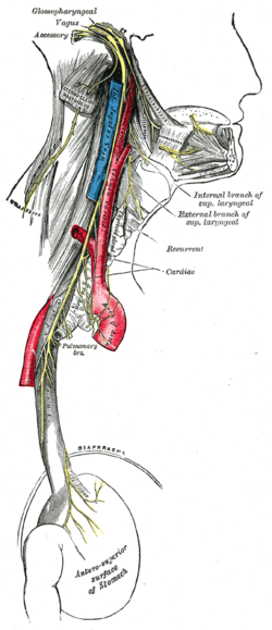
Plan of the upper portions of the glossopharyngeal, vagus, and accessory nerves.
Gray793.png
Course and distribution of the glossopharyngeal, vagus, and accessory nerves.
Details
InnervatesLevator veli palatini, Salpingopharyngeus, Palatoglossus, Palatopharyngeus, Superior pharyngeal constrictor, Middle pharyngeal constrictor, visceratyppi, Inferior pharyngeal constrictor, viscera rass
Identifiers
Latinnervus vagus
MeSHD014630
NeuroNames702
TAA14.2.01.153
FMA5731

The vagus nerve, historically cited as the pneumogastric nerve, is the tenth cranial nerve or CN X, and interfaces with the parasympathetic control of the heart, lungs, and digestive tract. The vagus nerves are paired but are normally referred to in the singular. It is the longest nerve of the autonomic nervous system in the human body.

Structure

Upon leaving the medulla oblongata between the pyramid and the inferior cerebellar peduncle, the vagus nerve extends through the jugular foramen, then passes into the carotid sheath between the internal carotid artery and the internal jugular vein down to the neck, chest, and abdomen, where it contributes to the innervation of the viscera, reaching all the way to the colon. Besides giving some output to various organs, the vagus nerve comprises between 80% and 90% of afferent nerves mostly conveying sensory information about the state of the body's organs to the central nervous system. The right and left vagus nerves descend from the cranial vault through the jugular foramina, penetrating the carotid sheath between the internal and external carotid arteries, then passing posterolateral to the common carotid artery. The cell bodies of visceral afferent fibers of the vagus nerve are located bilaterally in the inferior ganglion of the vagus nerve (nodose ganglia). 

The right vagus nerve gives rise to the right recurrent laryngeal nerve, which hooks around the right subclavian artery and ascends into the neck between the trachea and esophagus. The right vagus then crosses anterior to the right subclavian artery, runs posterior to the superior vena cava, descends posterior to the right main bronchus, and contributes to cardiac, pulmonary, and esophageal plexuses. It forms the posterior vagal trunk at the lower part of the esophagus and enters the diaphragm through the esophageal hiatus

The left vagus nerve enters the thorax between left common carotid artery and left subclavian artery and descends on the aortic arch. It gives rise to the left recurrent laryngeal nerve, which hooks around the aortic arch to the left of the ligamentum arteriosum and ascends between the trachea and esophagus. The left vagus further gives off thoracic cardiac branches, breaks up into the pulmonary plexus, continues into the esophageal plexus, and enters the abdomen as the anterior vagal trunk in the esophageal hiatus of the diaphragm.
The vagus runs parallel to the common carotid artery and internal jugular vein inside the carotid sheath.

Nuclei

The vagus nerve includes axons which emerge from or converge onto four nuclei of the medulla:
  1. The dorsal nucleus of vagus nerve – which sends parasympathetic output to the viscera, especially the intestines
  2. The nucleus ambiguus – which gives rise to the branchial efferent motor fibers of the vagus nerve and preganglionic parasympathetic neurons that innervate the heart
  3. The solitary nucleus – which receives afferent taste information and primary afferents from visceral organs
  4. The spinal trigeminal nucleus – which receives information about deep/crude touch, pain, and temperature of the outer ear, the dura of the posterior cranial fossa and the mucosa of the larynx

Development

The motor division of the vagus nerve is derived from the basal plate of the embryonic medulla oblongata, while the sensory division originates from the cranial neural crest.

Function

The vagus nerve supplies motor parasympathetic fibers to all the organs (except the adrenal glands), from the neck down to the second segment of the transverse colon. The vagus also controls a few skeletal muscles, including:
This means that the vagus nerve is responsible for such varied tasks as heart rate, gastrointestinal peristalsis, sweating, and quite a few muscle movements in the mouth, including speech (via the recurrent laryngeal nerve). It also has some afferent fibers that innervate the inner (canal) portion of the outer ear (via the auricular branch, also known as Alderman's nerve) and part of the meninges.

Efferent vagus nerve fibers innervating the pharynx and back of the throat are responsible for the gag reflex. In addition, 5-HT3 receptor-mediated afferent vagus stimulation in the gut due to gastroenteritis is a cause of vomiting. Stimulation of the vagus nerve in the cervix uteri (as in some medical procedures) can lead to a vasovagal response

The vagus nerve also plays a role in satiation following food consumption. Knocking out vagal nerve receptors has been shown to cause hyperphagia (greatly increased food intake).

Vagus nerve and the heart

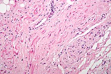
H&E stained fibers of the vagus nerve (bottom right) innervate the sinoatrial node tissue (middle left)
 
Parasympathetic innervation of the heart is partially controlled by the vagus nerve and is shared by the thoracic ganglia. Vagal and spinal ganglionic nerves mediate the lowering of the heart rate. The right vagus branch innervates the sinoatrial node. In healthy people, parasympathetic tone from these sources are well-matched to sympathetic tone. Hyperstimulation of parasympathetic influence promotes bradyarrhythmias. When hyperstimulated, the left vagal branch predisposes the heart to conduction block at the atrioventricular node

At this location, neuroscientist Otto Loewi first demonstrated that nerves secrete substances called neurotransmitters, which have effects on receptors in target tissues. In his experiment, Loewi electrically stimulated the vagus nerve of a frog heart, which slowed the heart. Then he took the fluid from the heart and transferred it to a second frog heart without a vagus nerve. The second heart slowed down without an electrical stimulation. Loewi described the substance released by the vagus nerve as vagusstoff, which was later found to be acetylcholine. Drugs that inhibit the muscarinic receptors (anticholinergics) such as atropine and scopolamine, are called vagolytic because they inhibit the action of the vagus nerve on the heart, gastrointestinal tract, and other organs. Anticholinergic drugs increase heart rate and are used to treat bradycardia.

Physical and emotional effects

Excessive activation of the vagal nerve during emotional stress, which is a parasympathetic overcompensation for a strong sympathetic nervous system response associated with stress, can also cause vasovagal syncope due to a sudden drop in cardiac output, causing cerebral hypoperfusion. Vasovagal syncope affects young children and women more than other groups. It can also lead to temporary loss of bladder control under moments of extreme fear.

Research has shown that women having had complete spinal cord injury can experience orgasms through the vagus nerve, which can go from the uterus, cervix, and, it is presumed, the vagina to the brain.

Insulin signaling activates the adenosine triphosphate (ATP)-sensitive potassium (KATP) channels in the arcuate nucleus, decreases AgRP release, and through the vagus nerve, leads to decreased glucose production by the liver by decreasing gluconeogenic enzymes: Phosphoenolpyruvate carboxykinase, Glucose 6-phosphatase.

Clinical significance

Vagus nerve stimulation

Vagus nerve stimulation (VNS) therapy using a neurostimulator implanted in the chest is a treatment used since 1997 to control seizures in epilepsy patients and has been approved for treating drug-resistant cases of clinical depression. A non-invasive VNS device that stimulates an afferent branch of the vagus nerve is also being developed and will soon undergo trials.

Clinical trials have started in Antwerp, Belgium, using VNS for the treatment of tonal tinnitus after a breakthrough study published in early 2011 by researchers at the University of Texas - Dallas showed successful tinnitus-suppression in rats when tones were paired with brief pulses of stimulation of the vagus nerve.

VNS may also be achieved by one of the vagal maneuvers: holding the breath for a few seconds, dipping the face in cold water, coughing, or tensing the stomach muscles as if to bear down to have a bowel movement. Patients with supraventricular tachycardia, atrial fibrillation, and other illnesses may be trained to perform vagal maneuvers (or find one or more on their own).

Vagus nerve blocking (VBLOC) therapy is similar to VNS but used only during the day. In a six-month open-label trial involving three medical centers in Australia, Mexico, and Norway, vagus nerve blocking helped 31 obese participants lose an average of nearly 15 percent of their excess weight. As of 2008 a year-long 300-participant double-blind, phase II trial had begun.

Vagotomy

Vagotomy (cutting of the vagus nerve) is a now-obsolete therapy that was performed for peptic ulcer disease. Vagotomy is currently being researched as a less invasive alternative weight-loss procedure to gastric bypass surgery. The procedure curbs the feeling of hunger and is sometimes performed in conjunction with putting bands on patients' stomachs, resulting in an average of 43% of excess weight lost at six months with diet and exercise.

One serious side effect of a vagotomy is a vitamin B12 deficiency later in life – perhaps after about 10 years – that is similar to pernicious anemia. The vagus normally stimulates the stomach's parietal cells to secrete acid and intrinsic factor. Intrinsic factor is needed to absorb vitamin B12 from food. The vagotomy reduces this secretion and ultimately leads to the deficiency, which, if left untreated, causes nerve damage, tiredness, dementia, paranoia, and ultimately death.

Researchers from Aarhus University and Aarhus University Hospital have demonstrated that vagotomy prevents (halves the risk of) the development of Parkinson's disease, suggesting that Parkinson's disease begins in the gastrointestinal tract and spreads via the vagus nerve to the brain. Or giving further evidence to the theory that dysregulated environmental stimuli, such as that received by the vagus nerve from the gut, may have a negative effect on the dopamine reward system of the substantia nigra, thereby causing Parkinsons disease.

Chagas disease

The neuropathy in Chagas disease spreads in part via the major parasympathetic branches of the vagus nerve.

History

Etymology

The Latin word vagus means literally "wandering" (the words vagrant, vagabond, and vague come from the same root). Sometimes the right and left branches together are spoken of in the plural and are thus called vagi (/ˈv/VAY-jy). The vagus was also historically called the pneumogastric nerve since it innervates both the lungs and the stomach.

National Academies of Sciences, Engineering, and Medicine

From Wikipedia, the free encyclopedia

The National Academies of Sciences, Engineering, and Medicine
The Keck Center of the National Academies by Matthew Bisanz.JPG
The Keck Center of the National Academies in Washington, D.C.
Formation1863 (as National Academy of Sciences), 1916 (as National Research Council), 2015 (as National Academies of Sciences, Engineering, and Medicine)
HeadquartersWashington, D.C., USA
Membership
Scientists, engineers, and health professionals
Official language
English

The National Academies of Sciences, Engineering, and Medicine (also known as NASEM or the National Academies) is the collective scientific national academy of the United States. The name is used interchangeably in two senses: (1) as an umbrella term for its three quasi-independent honorific member organizations (the National Academy of Sciences (NAS), the National Academy of Engineering (NAE), and the National Academy of Medicine (NAM)). And (2) as the brand for studies and reports issued by the operating arm of the three academies, the National Research Council (NRC). The NRC was first formed in 1916 as an activity of the NAS. Now jointly governed by all three academies, it produces some 200 publications annually which are published by the National Academies Press.

History

The US National Academy of Sciences was created by an Act of Incorporation dated March 3, 1863, which was signed by then President of the United States Abraham Lincoln The Act stated that "... the Academy shall, whenever called upon by any department of the Government, investigate, examine, experiment, and report upon any subject of science or art . ... " With the American civil war raging, the new Academy was presented with few problems to solve, but it did address matters of "... coinage, weights and measures, iron ship hulls, and the purity of whiskey ..." All subsequently affiliated organizations have been created under this same overall congressional charter, including the two younger academies, National Academy of Engineering (NAE) (created in 1964) and NAM (created as the Institute of Medicine in 1970 and rechartered as NAM in 2015).

Under this same charter, the National Research Council was created in 1916. On June 19 of that year, then US President Woodrow Wilson requested that the National Academy of Sciences organize a "National Research Council". The purpose of the Council (at first called the National Research Foundation) was in part to foster and encourage "the increased use of scientific research in the development of American industries ... the employment of scientific methods in strengthening the national defense ... and such other applications of science as will promote the national security and welfare."

At the time, the Academy's effort to support national defense readiness, the Committee on Nitric Acid Supply, was approved by Secretary of War Newton D. Baker. Nitric acid was the substance basic in the making of propellants such as cordite, high explosives, dyes, fertilizers, and other products but availability was limited due to World War I. The NRC, through its committee, recommended importing Chilean saltpeter and the construction of four new ordinance plants. These recommendations were accepted by the War Department in June 1917, although the plants were not completed prior to the end of the war.

In 1918, Wilson formalized the NRC's existence under Executive Order 2859. Wilson's order declared the function of the NRC to be in general:
"(T)o stimulate research in the mathematical. physical, and biological sciences. and in the application of these sciences to engineering, agriculture. medicine. and other useful arts. with the object of increasing knowledge, of strengthening the national defense, and of contributing in other ways to the public welfare."
During World War I, the United States was at war, the NRC operated as the Department of Science and Research of the Council of National Defense as well as the Science and Research Division of the United States Army Signal Corps. When war was first declared, the Council had organized committees on antisubmarine and gas warfare.

On June 1, 1917, the council convened a meeting of scientific representatives of the United Kingdom and France with interested parties from the U.S. on the subject of submarine detection. Another meeting with the British and French was held in Paris in October 1918, at which more details of their work was disclosed. As a result of these meetings, the NRC recommended that scientists be brought together to work on the problems associated with submarine detection. Due to the success of council-directed research in producing a sound-based method of detecting submarines, as well as other military innovations, the NRC was retained at the end of the war, though it was gradually decoupled from the military. 

NRC's Articles of Organization have been changed only three times: in 1956, January 1993, and July 2015.

Honorific societies

The National Academy of Sciences, National Academy of Engineering and National Academy of Medicine are honorary membership organizations, each of which has its own governing Council, and each of which elects its own new members. The membership of the three academies totals more than 6,300 scientists, engineers, and health professionals. New members for each organization are elected annually by current members, based on their distinguished and continuing achievements in original research. By the terms of the original 1863 Congressional charter, the three academies serve pro bono as "advisers to the nation on science, engineering, and medicine."

Program units

The program units, formerly known as the National Research Council, are collectively the operating arm of the three academies for the purpose of providing objective policy advice. Although separately chartered (see above), it falls legally under the overall charter of the National Academy of Sciences, whose ultimate fiduciary body is the NAS Council. In actual practice, the NAS Council delegates governing authority to a Governing Board of the National Research Council that is chaired jointly by the presidents of the three academies, with additional members chosen by them or specified in the charters of the academies. 

Under this three-academy umbrella, the program units produce reports that shape policies, inform public opinion, and advance the pursuit of science, engineering, and medicine.

There are seven major divisions: Division of Behavioral and Social Sciences and Education, Division of Earth and Life Studies, Division of Engineering and Physical Sciences, Health and Medicine Division, Policy and Global Affairs Division, Transportation Research Board, and the Gulf Research Program.

Division on Engineering and Physical Sciences

The Division on Engineering and Physical Sciences has activities organized around:

The study process

The National Academies attempt to obtain authoritative, objective, and scientifically balanced answers to difficult questions of national importance. Top scientists, engineers, health professionals, and other experts (not limited to those in academies membership) are enlisted to address the scientific and technical aspects of some of society's problems. These experts are volunteers who serve on study committees that are convened to answer specific sets of questions. All committee members serve without pay. NASEM itself does not perform original research; rather it provides independent advice. Federal agencies are the primary financial sponsors of the Academies' work; additional studies are funded by state agencies, foundations, other private sponsors, and the National Academies endowment. The external sponsors have no control over the conduct or results of a study, once the statement of task and budget are finalized. Study committees gather information from many sources in public meetings but deliberate in private in order to avoid political, special interest, and sponsor influence. 

All reports go through an extensive external review facilitated by the internal Report Review Committee (also consisting of members from the NAS, NAE, and NAM).

Through this study process, the National Academies produce around 200 reports each year. Recent reports cover such topics as addressing the obesity epidemic, the use of forensics in the courtroom, invasive plants, pollinator collapse, underage drinking, the Hubble Telescope, vaccine safety, the hydrogen economy, transportation safety, climate change, and homeland security. Many reports influence policy decisions; some are instrumental in enabling new research programs; others provide independent program reviews. The National Academies Press is the publisher for the National Academies of Sciences, Engineering, and Medicine, and makes its publications available for free online reading, and the full book PDFs have been available for free download since 2011.

Notable reports

Reports on climate change

In 2001, the NRC published the report Climate Change Science: An Analysis of Some Key Questions, which emphasized the fact that national policy decisions made now and in the long-term future will influence the extent of any damage suffered by vulnerable human populations and ecosystems later in this century. The report endorsed findings of the Intergovernmental Panel on Climate Change (IPCC) as representing the views of the scientific community:
The changes observed over the last several decades are likely mostly due to human activities, but we cannot rule out that some significant part of these changes is also a reflection of natural variability. Human-induced warming and associated sea level rise are expected to continue through the 21st century ... The IPCC's conclusion that most of the observed warming of the last 50 years is likely to have been due to the increase in greenhouse gas concentrations accurately reflects the current thinking of the scientific community on this issue.
In 2013, the NRC published the report Abrupt Impacts of Climate Change: Anticipating Surprises, which provided an updated look at the issue of abrupt climate change and its potential impacts. This study differed from previous treatments of abrupt changes by focusing on abrupt climate changes and also abrupt climate impacts that have the potential to severely affect the physical climate system, natural systems, or human systems, often affecting multiple interconnected areas of concern.

Report on sexual assault

In 2013, the NRC published the report Estimating the Incidence of Rape and Sexual Assault, which pointed out that approximately 80 percent of sexual assaults go unreported to law enforcement. The report recommends that the National Crime Victimization Survey adopt new approaches to interviews of rape victims, including changing the wording of questions. 

In an article about the report, Amber Stevenson, clinical supervisor and therapist at the Nashville Sexual Assault Center, said that victim-blaming was the main issue preventing victims from coming forward:
As long as we as a community continue to make victim-blaming statements, such as, "She put herself in this situation,"..."She didn't fight back, she must have wanted it," we will continue to see rapes go unreported ... We have to stop blaming the victim. The conversation needs to shift to the person who chose to rape.

Report on integrity in research

The 1992 report, Responsible Science: Ensuring the Integrity of the Research Process was updated in 2017 by the report, Fostering Integrity in Research:
... as experience has accumulated with various forms of research misconduct, detrimental research practices, and other forms of misconduct, as subsequent empirical research has revealed more about the nature of scientific misconduct, and because technological and social changes have altered the environment in which science is conducted, it is clear that the framework established more than two decades ago needs to be updated.
One of the report's main concerns is that a growing percentage of recently published research turns out to be not reproducible due in part to inadequate support of standards of transparency in many fields as well as to various other detrimental research practices.

Other programs

The Christine Mirzayan Science and Technology Policy Fellowship is an annual program for current or recent graduate students to spend three months working in the National Academies. The Academies also administered the Marian Koshland Science Museum in downtown Washington until its closing in 2017; the Museum has since been replaced by LabX, a program of online resources and nationwide public events that aims to increase awareness of scientific and evidence-based solutions to community problems.

Surgery

From Wikipedia, the free encyclopedia https://en.wikipedia.org/w...