Search This Blog

Tuesday, May 17, 2022

Neuroepigenetics

From Wikipedia, the free encyclopedia

Neuroepigenetics is the study of how epigenetic changes to genes affect the nervous system. These changes may effect underlying conditions such as addiction, cognition, and neurological development.

Mechanisms

Neuroepigenetic mechanisms regulate gene expression in the neuron. Often, these changes take place due to recurring stimuli. Neuroepigenetic mechanisms involve proteins or protein pathways that regulate gene expression by adding, editing or reading epigenetic marks such as methylation or acetylation. Some of these mechanisms include ATP-dependent chromatin remodeling, LINE1, and prion protein-based modifications. Other silencing mechanisms include the recruitment of specialized proteins that methylate DNA such that the core promoter element is inaccessible to transcription factors and RNA polymerase. As a result, transcription is no longer possible. One such protein pathway is the REST co-repressor complex pathway. There are also several non-coding RNAs that regulate neural function at the epigenetic level. These mechanisms, along with neural histone methylation, affect arrangement of synapses, neuroplasticity, and play a key role in learning and memory.

Methylation

DNA methyltransferases (DNMTs) are involved in regulation of the electrophysiological landscape of the brain through methylation of CpGs. Several studies have shown that inhibition or depletion of DNMT1 activity during neural maturation leads to hypomethylation of the neurons by removing the cell's ability to maintain methylation marks in the chromatin. This gradual loss of methylation marks leads to changes in the expression of crucial developmental genes that may be dosage sensitive, leading to neural degeneration. This was observed in the mature neurons in the dorsal portion of the mouse prosencephalon, where there was significantly greater amounts of neural degeneration and poor neural signaling in the absence of DNMT1. Despite poor survival rates amongst the DNMT1-depleted neurons, some of the cells persisted throughout the lifespan of the organism. The surviving cells reaffirmed that the loss of DNMT1 led to hypomethylation in the neural cell genome. These cells also exhibited poor neural functioning. In fact, a global loss of neural functioning was also observed in these model organisms, with the greatest amounts neural degeneration occurring in the prosencephalon.

Other studies showed a trend for DNMT3a and DNMT3b. However, these DNMT's add new methyl marks on unmethylated DNA, unlike DNMT1. Like DNMT1, the loss of DNMT3a and 3b resulted in neuromuscular degeneration two months after birth, as well as poor survival rates amongst the progeny of the mutant cells, even though DNMT3a does not regularly function to maintain methylation marks. This conundrum was addressed by other studies which recorded rare loci in mature neurons where DNMT3a acted as a maintenance DNMT. The Gfap locus, which codes for the formation and regulation of the cytoskeleton of astrocytes, is one such locus where this activity is observed. The gene is regularly methylated to downregulate glioma related cancers. DNMT inhibition leads to decreased methylation and increased synaptic activity. Several studies show that the methylation-related increase or decrease in synaptic activity occurs due to the upregulation or downregulation of receptors at the neurological synapse. Such receptor regulation plays a major role in many important mechanisms, such as the 'fight or flight' response. The glucocorticoid receptor (GR) is the most studied of these receptors. During stressful circumstances, there is a signaling cascade that begins from the pituitary gland and terminates due to a negative feedback loop from the adrenal gland. In this loop, the increase in the levels of the stress response hormone results in the increase of GR. Increase in GR results in the decrease of cellular response to the hormone levels. It has been shown that methylation of the I7 exon within the GR locus leads to a lower level of basal GR expression in mice. These mice were more susceptible to high levels of stress as opposed to mice with lower levels of methylation at the I7 exon. Up-regulation or down-regulation of receptors through methylation leads to change in synaptic activity of the neuron.

Hypermethylation, CpG islands, and tumor suppressing genes

CpG Islands (CGIs) are regulatory elements that can influence gene expression by allowing or interfering with transcription initiation or enhancer activity. CGIs are generally interspersed with the promoter regions of the genes they affect and may also affect more than one promoter region. In addition they may also include enhancer elements and be separate from the transcription start site. Hypermethylation at key CGIs can effectively silence expression of tumor suppressing genes and is common in gliomas. Tumor suppressing genes are those which inhibit a cell's progression towards cancer. These genes are commonly associated with important functions which regulate cell-cycle events. For example, PI3K and p53 pathways are affected by CGI promoter hypermethylation, this includes the promoters of the genes CDKN2/p16, RB, PTEN, TP53 and p14ARF. Importantly, glioblastomas are known to have high frequency of methylation at CGIs/promoter sites. For example, Epithelial Membrane Protein 3 (EMP3) is a gene which is involved in cell proliferation as well as cellular interactions. It is also thought to function as a tumor suppressor, and in glioblastomas is shown to be silenced via hypermethylation. Furthermore, introduction of the gene into EMP3-silenced neuroblasts results in reduced colony formation as well as suppressed tumor growth. In contrast, hypermethylation of promoter sites can also inhibit activity of oncogenes and prevent tumorigenesis. Such oncogenic pathways as the transformation growth factor (TGF)-beta signaling pathway stimulate cells to proliferate. In glioblastomas the overactivity of this pathway is associated with aggressive forms of tumor growth. Hypermethylation of PDGF-B, the TGF-beta target, inhibits uncontrolled proliferation.

Hypomethylation and aberrant histone modification

Global reduction in methylation is implicated in tumorigenesis. More specifically, wide spread CpG demethylation, contributing to global hypomethylation, is known to cause genomic instability leading to development of tumors. An important effect of this DNA modification is its transcriptional activation of oncogenes. For example, expression of MAGEA1 enhanced by hypomethylation interferes with p53 function.

Aberrant patterns of histone modifications can also take place at specific loci and ultimately manipulate gene activity. In terms of CGI promoter sites, methylation and loss of acetylation occurs frequently at H3K9. Furthermore, H3K9 dimethylation and trimethylation are repressive marks which, along with bivalent differentially methylated domains, are hypothesized to make tumor suppressing genes more susceptible to silencing. Abnormal presence or lack of methylation in glioblastomas are strongly linked to genes which regulate apoptosis, DNA repair, cell proliferation, and tumor suppression. One of the best known examples of genes affected by aberrant methylation that contributes to formation of glioblastomas is MGMT, a gene involved in DNA repair which encodes the protein O6-methylguanine-DNA methyltransferase. Methylation of the MGMT promoter is an important predictor of the effectiveness of alkylating agents to target glioblastomas. Hypermethylation of the MGMT promoter causes transcriptional silencing and is found in several cancer types including glioma, lymphoma, breast cancer, prostate cancer, and retinoblastoma.

Neuroplasticity

Neuroplasticity refers to the ability of the brain to undergo synaptic rearrangement as a response to recurring stimuli. Neurotrophin proteins play a major role in synaptic rearrangement, amongst other factors. Depletion of neurotrophin BDNF or BDNF signaling is one of the main factors in developing diseases such as Alzheimer's disease, Huntington's disease, and depression. Neuroplasticity can also occur as a consequence of targeted epigenetic modifications such as methylation and acetylation. Exposure to certain recurring stimuli leads to demethylation of particular loci and remethylation in a pattern that leads to a response to that particular stimulus. Like the histone readers, erasers and writers also modify histones by removing and adding modifying marks respectively. An eraser, neuroLSD1, is a modified version of the original Lysine Demethylase 1(LSD1) that exists only in neurons and assists with neuronal maturation. Although both versions of LSD1 share the same target, their expression patterns are vastly different and neuroLSD1 is a truncated version of LSD1. NeuroLSD1 increases the expression of immediate early genes (IEGs) involved in cell maturation. Recurring stimuli lead to differential expression of neuroLSD1, leading to rearrangement of loci. The eraser is also thought to play a major role in the learning of many complex behaviors and is way through which genes interact with the environment.

Neurodegenerative diseases

Alzheimer's disease

Alzheimer's disease (AD) is a neurodegenerative disease known to progressively affect memory and incite cognitive degradation. Epigenetic modifications both globally and on specific candidate genes are thought to contribute to the etiology of this disease. Immunohistochemical analysis of post-mortem brain tissues across several studies have revealed global decreases in both 5-methylcytosine (5mC) and 5-hydroxymethylcytosine (5hmC) in AD patients compared with controls. However, conflicting evidence has shown elevated levels of these epigenetic markers in the same tissues. Furthermore, these modifications appear to be affected early on in tissues associated with the pathophysiology of AD. The presence of 5mC at the promoters of genes is generally associated with gene silencing. 5hmC, which is the oxidized product of 5mC, via ten-eleven-translocase (TET), is thought to be associated with activation of gene expression, though the mechanisms underlying this activation are not fully understood.

Regardless of variations in results of methylomic analysis across studies, it is known that the presence of 5hmC increases with differentiation and aging of cells in the brain. Furthermore, genes which have a high prevalence of 5hmC are also implicated in the pathology of other age related neurodegenerative diseases, and are key regulators of ion transport, neuronal development, and cell death. For example, over-expression of 5-Lipoxygenase (5-LOX), an enzyme which generates pro-inflammatory mediators from arachidonic acid, in AD brains is associated with high prevalence of 5hmC at the 5-LOX gene promoter region.

Amyotrophic Lateral Sclerosis

DNA modifications at different transcriptional sites have been shown to contribute to neurodegenerative diseases. These include harmful transcriptional alterations such as those found in motor neuron functionality associated with Amyotrophic Lateral Sclerosis (ALS). Degeneration of upper and lower motor neurons, which contributes to muscle atrophy in ALS patients, is linked to chromatin modifications among a group of key genes. One important site that is regulated by epigenetic events is the hexanucleotide repeat expansion in C9orf72 within the chromosome 9p21. Hypermethylation of the C9orf72 related CpG Islands is shown to be associated with repeat expansion in ALS affected tissues. Overall, silencing of the C9orf72 gene may result in haploinsufficiency, and may therefore influence the presentation of disease. The activity of chromatin modifiers is also linked to prevalence of ALS. DNMT3A is an important methylating agent and has been shown to be present throughout the central nervous systems of those with ALS. Furthermore, over-expression of this de novo methyl transferase is also implicated in cell death of motor-neuron analogs.

Mutations in the FUS gene, that encodes an RNA/DNA binding protein, are causally linked to ALS. ALS patients with such mutations have increased levels of DNA damage. The protein encoded by the FUS gene is employed in the DNA damage response. It is recruited to DNA double-strand breaks and catalyzes recombinational repair of such breaks. In response to DNA damage, the FUS protein also interacts with histone deacetylase I, a protein employed in epigenetic alteration of histones. This interaction is necessary for efficient DNA repair. These findings suggest that defects in epigenetic signalling and DNA repair contribute to the pathogenesis of ALS.

Neuro-oncology

A multitude of genetic and epigenetic changes in DNA profiles in brain cells are thought to be linked to tumorgenesis. These alterations, along with changes in protein functions, are shown to induce uncontrolled cell proliferation, expansion, and metastasis. While genetic events such as deletions, translocations, and amplification give rise to activation of oncogenes and deactivation of tumor suppressing genes, epigenetic changes silence or up-regulate these same genes through key chromatin modifications.

Neurotoxicity

Neurotoxicity refers to damage made to the central or peripheral nervous systems due to chemical, biological, or physical exposure to toxins. Neurotoxicity can occur at any age and its effects may be short-term or long-term, depending on the mechanism of action of the neurotoxin and degree of exposure.

Certain metals are considered essential due to their role in key biochemical and physiological pathways, while the remaining metals are characterized as being nonessential. Nonessential metals do not serve a purpose in any biological pathway and the presence and accumulation in the brain of most can lead to neurotoxicity. These nonessential metals, when found inside the body compete with essential metals for binding sites, upset antioxidant balance, and their accumulation in the brain can lead to harmful side effects, such as depression and intellectual disability. An increase in nonessential heavy metal concentrations in air, water and food sources, and household products has increased the risk of chronic exposure.

Acetylation, methylation and histone modification are some of the most common epigenetic markers. While these changes do not directly affect the DNA sequence, they are able to alter the accessibility to genetic components, such as the promoter or enhancer regions, necessary for gene expression. Studies have shown that long-term maternal exposure to lead (Pb) contributes to decreased methylation in areas of the fetal epigenome, for example the interspaced repetitive sequences (IRSs) Alu1 and LINE-1. The hypomethylation of these IRSs has been linked to increased risk for cancers and autoimmune diseases later in life. Additionally, studies have found a relationship between chronic prenatal Pb exposure and neurological diseases, such as Alzheimer's and schizophrenia, as well as developmental issues. Furthermore, the acetylation and methylation changes induced by overexposure to lead result in decreased neurogenesis and neuron differentiation ability, and consequently interfere with early brain development.

Overexposure to essential metals can also have detrimental consequences on the epigenome. For example, when manganese, a metal normally used by the body as a cofactor, is present at high concentrations in the blood it can negatively affect the central nervous system. Studies have shown that accumulation of manganese leads to dopaminergic cell death and consequently plays a role in the onset of Parkinson's disease (PD). A hallmark of Parkinson's disease is the accumulation of α-Synuclein in the brain. Increased exposure to manganese leads to the downregulation of protein kinase C delta (PKCδ) through decreased acetylation and results in the misfolding of the α-Synuclein protein that allows aggregation and triggers apoptosis of dopaminergic cells.

Research

The field has only recently seen a growth in interest, as well as in research, due to technological advancements that facilitate better resolution of the minute modifications made to DNA. However, even with the significant advances in technology, studying the biology of neurological phenomena, such as cognition and addiction, comes with its own set of challenges. Biological study of cognitive processes, especially with humans, has many ethical caveats. Some procedures, such as brain biopsies of Rett Syndrome patients, usually call for a fresh tissue sample that can only be extricated from the brain of deceased individual. In such cases, the researchers have no control over the age of brain tissue sample, thereby limiting research options. In case of addiction to substances such as alcohol, researchers utilize mouse models to mirror the human version of the disease. However, the mouse models are administered greater volumes of ethanol than humans normally consume in order to obtain more prominent phenotypes. Therefore, while the model organism and the tissue samples provide an accurate approximation of the biology of neurological phenomena, these approaches do not provide a complete and precise picture of the exact processes underlying a phenotype or a disease.

Neuroepigenetics had also remained underdeveloped due to the controversy surrounding the classification of genetic modifications in matured neurons as epigenetic phenomena. This discussion arises due to the fact that neurons do not undergo mitosis after maturation, yet the conventional definition of epigenetic phenomena emphasizes heritable changes passed on from parent to offspring. However, various histone modifications are placed by epigenetic modifiers such as DNA methyltransferases (DNMT) in neurons and these marks regulate gene expression throughout the life span of the neuron. The modifications heavily influence gene expression and arrangement of synapses within the brain. Finally, although not inherited, most of these marks are maintained throughout the life of the cell once they are placed on chromatin.

Hypokinesia

From Wikipedia, the free encyclopedia
 
Hypokinesia
SpecialtyPsychiatry, neurology

Hypokinesia is one of the classifications of movement disorders, and refers to decreased bodily movement. Hypokinesia is characterized by a partial or complete loss of muscle movement due to a disruption in the basal ganglia. Hypokinesia is a symptom of Parkinson's disease shown as muscle rigidity and an inability to produce movement. It is also associated with mental health disorders and prolonged inactivity due to illness, amongst other diseases.

The other category of movement disorder is hyperkinesia that features an exaggeration of unwanted movement, such as twitching or writhing in Huntington's disease or Tourette syndrome.

Spectrum of disorders

Hypokinesia describes a variety of more specific disorders:

Hypokinetic disorder Characteristics
Akinesia (α- a-, "without", κίνησις kinēsis, "motion") Inability to initiate voluntary movement.
Bradykinesia (βραδύς bradys, "slow", κίνησις kinēsis, "motion") Slowness of initiation of voluntary movement with a progressive reduction in speed and range of repetitive actions, such as voluntary finger-tapping. It occurs in Parkinson's disease and other disorders of the basal ganglia. It is one of the four key symptoms of parkinsonism, which are bradykinesia, tremor, rigidity, and postural instability.
Dysarthria A condition which affects the muscles necessary for speech, it causes difficulty in speech production despite a continued cognitive understanding of language. Often caused by Parkinson's disease, patients experience weakness, paralysis, or lack of coordination in the motor-speech system, causing respiration, phonation, prosody, and articulation to be affected. Problems including tone, speed of communication, breath control, volume, and timing are displayed. Hypokinetic dysarthria particularly affects the volume of speech, prompting treatment with a speech language pathologist.
Dyskinesia This is characterized by a diminished ability for voluntary movements, as well as the presence of involuntary movements. The hands and upper body are the areas most likely to be affected by tremors and tics. In some cases, Parkinson's patients experience dyskinesia as a negative side effect of dopamine medications.
Dystonia A movement disorder characterised by sustained muscle contractions, frequently causing twisted and repetitive movements, or abnormal postures.
Freezing This is characterized by an inability to move muscles in any desired direction.
Neuroleptic malignant syndrome Resulting from heavy exposure to drugs that block dopamine receptors, victims can experience fever, rigidity, mental status change, dysautonomia, tremors, dystonia, and myoclonus. While this disorder is extremely rare, immediate attention is necessary because of the high risk of death.
Rigidity Resistance to externally imposed ("passive") joint movements, such as when a doctor flexes a patient's arm at the elbow joint. It does not depend on imposed speed and can be elicited at very low speeds of passive movement in both directions. Cogwheel rigidity and leadpipe rigidity are two types identified with Parkinson's disease:
  • Leadpipe rigidity is sustained resistance to passive movement throughout the whole range of motion, with no fluctuations.
  • Cogwheel rigidity is jerky resistance to passive movement as muscles tense and relax.

Spasticity, a special form of rigidity, is present only at the start of passive movement. It is rate-dependent and only elicited upon a high-speed movement. These various forms of rigidity can be seen in different forms of movement disorders, such as Parkinson's disease.

Postural instability A disturbance in balance that impairs the ability to maintain an upright posture when standing and walking. In Parkinsons disease it is correlated with greater disability and more depression, as well as with frequency of falls and fear of falls (which, itself, can be significantly disabling).

A person with medication-induced dystonia

Causes

The most common cause of Hypokinesia is Parkinson's disease, and conditions related to Parkinson's disease.

Other conditions may also cause slowness of movements. These include hypothyroidism and severe depression. These conditions need to be carefully ruled out, before a diagnosis of Parkinsonism is made.

The remainder of this article describes Hypokinesia associated with Parkinson's disease, and conditions related to Parkinson's disease.

Pathophysiology

Associated neurotransmitters

Dopamine

The main neurotransmitter thought to be involved in hypokinesia is dopamine. Essential to the basal ganglionic-thalamocortical loop, which processes motor function, dopamine depletion is common in these areas of hypokinesic patients. Bradykinesia is correlated with lateralized dopaminergic depletion in the substantia nigra. The dopamine pathway in the substantia nigra is essential to motor function, and commonly a lesion in this area correlates with displayed hypokinesia. Tremor and rigidity, however, seem to be only partially due to dopamine deficits in the substantia nigra, suggesting other processes are involved in motor control. Treatments for hypokinesia often either attempt to prevent dopamine degradation by MAO-B or increase the amount of neurotransmitter present in the system.

GABA and glutamate

The inhibitory neurotransmitter GABA and the excitatory glutamate are found in many parts of the central nervous system, including in the motor pathways that involve hypokinesia. In one pathway, glutamate in the substantia nigra excites the release of GABA into the thalamus, which then inhibits the release of glutamate in the cortex and thereby reduces motor activity. If too much glutamate is initially in the substantia nigra, then through interaction with GABA in the thalamus and glutamate in the cortex, movements will be reduced or will not occur at all.

Another direct pathway from the basal ganglia sends GABA inhibitory messages to the globus pallidus and substantia nigra, which then send GABA to the thalamus. In the indirect pathway, the basal ganglia send GABA to the globus pallidus which then sends it to the subthalamic nucleus, which then disinhibited sends glutamate to the output structures of the basal ganglia. Inhibition of GABA release could disrupt the feedback loop to the basal ganglia and produce hypokinesic movements.

GABA and glutamate often interact with each other and with dopamine directly. In the basal ganglia, the nigrostriatal pathway is where GABA and dopamine are housed in the same neurons and released together.

Neurobiology

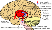
Hypokinetic symptoms arise from damage to the basal ganglia, which plays a role in producing force and computing the effort necessary to make a movement. Two possible neural pathways enable the basal ganglia to produce movement. When activated, the direct pathway sends sensory and motor information from the cerebral cortex to the first structure of the basal ganglia, the putamen. That information directly inhibits the globus pallidus internal and allows free movement. The indirect pathway, traveling through the putamen, globus pallidus external, and subthalamic nucleus, activates the globus pallidus internal threshold and inhibits the thalamus from communicating with the motor cortex, producing hypokinetic symptoms.

Basal ganglia (red) and related structures (blue)

When levels of dopamine decrease, the normal wave-firing pattern of basal ganglia neural oscillations changes and the tendency for oscillations increases, particularly in the beta wave of the basal ganglia. Recent research indicates, when oscillations fire simultaneously, processing is disrupted at the thalamus and cortex, affecting activities such as motor planning and sequence learning, as well as causing hypokinetic tremors.

Treatments

Dopaminergic drugs

Dopaminergic drugs are commonly used in the early stages of the hypokinesia to treat patients. With increased intake, though, they can become ineffective because of the development of noradrenergic lesions. While initially the dopaminergic drugs may be effective, these noradrenergic lesions are associated with hypokinesic gait disorder development later on.

Some Parkinson's patients are unable to move during sleep, prompting the diagnosis of "nocturnal hypokinesia". Physicians have experienced success treating this sleep disorder with slow-release or night-time dopaminergic drugs, and in some cases, continuous stimulation by the dopamine agonist rotigotine. Despite improved mobility during sleep, many Parkinson's patients report an extremely uncomfortable sleeping experience even after dopaminergic treatments.

Deep brain stimulation

Once the reaction to dopaminergic drugs begins to fluctuate in Parkinson's patients, deep brain stimulation (DBS) of the subthalamic nucleus and internal globus pallidus is often used to treat hypokinesia. DBS, like dopaminergic drugs, initially provides relief, but chronic use causes worse hypokinesia and freezing of gait. Lower-frequency DBS in irregular patterns has been shown to be more effective and less detrimental in treatment.

Parkinson surgery

Posteroventral pallidotomy (PVP) is a specific kind of DBS that destroys a small part of the globus pallidus by scarring the neural tissue, reducing brain activity and therefore tremors and rigidity. PVP is suspected to recalibrate basal ganglia activity in the thalamocortical pathway. PVP in the dominant hemisphere has been reported to disrupt executive function verbal processing abilities, and bilateral PVP may disturb processes of focused attention.

Many akinesia patients also form a linguistic akinesia in which their ability to produce verbal movements mirrors their physical akinesia symptoms, especially after unsuccessful PVP. Patients are usually able to maintain normal levels of fluency, but often stop midsentence, unable to remember or produce a desired word. According to a study of Parkinson's patients with articulatory hypokinesia, subjects with faster rates of speech experienced more problems trying to produce conversational language than those who normally spoke at slower rates.

Methylphenidate

Methylphenidate, commonly used to treat ADHD, has been used in conjunction with levodopa to treat hypokinesia in the short term. The two work together to increase dopamine levels in the striatum and prefrontal cortex. Methylphenidate mainly inhibits dopamine and noradrenaline reuptake by blocking presynaptic transporters, and levodopa increases the amount of dopamine, generally improving hypokinesic gait. Some patients, however, have adverse reactions of nausea and headache to the treatment and the long-term effects of the drug treatment still need to be assessed.

Stem cells

New treatments include increasing the number of dopamine cells by transplanting stem cells into the basal ganglia or stimulating endogenous stem cell production and movement to the basal ganglia. The successful integration of stem cells can relieve hypokinetic symptoms and decrease the necessary dose of dopaminergic drugs. However, a variety of complications, including possible tumor formation, inappropriate cell migration, rejection of cells by the immune system, and cerebral hemorrhage are possible, causing many physicians to believe the risks outweigh the possible benefits.

NOP receptor antagonists

Another treatment, still in an experimental stage, is the administration of nociception FQ peptide (NOP) receptor antagonists. This treatment has been shown to reduce hypokinesia in animal studies when increasing nociception FQ in the substantia nigra and subthalamic nucleus. Low doses can be taken with dopaminergic treatment to decrease the amount of L-dopa needed, which can reduce its long-term side effects and improve motor performance.

Dance therapy

Dance therapy has also been shown to reduce hypokinesic movements and rigidity, though targeted more at the muscular aspects of the disorder than the neural activity.

Associations

Cognitive impairment

Bradykinesia has been shown to precede impairment of executive functions, working memory, and attention. These cognitive deficiencies can be tied to nonfunction of the basal ganglia and prefrontal cortex, which is also linked to the motor-dysfunction of hypokinesia. Tremor and rigidity have not had observable connections to cognitive impairments, supporting the idea that they are not as involved in the dopamine pathway in the basal ganglionic-thalamocortical loop. Dopaminergic treatments have shown improvement in cognitive functions associated with hypokinesia, suggesting they are also dependent on dopamine levels in the system.

Motor motivation

Often debated is whether the efficiency, vigor, and speed of movements in patients with hypokinesia are tied to motivation for rewarding and against punishing stimuli. The basal ganglia have been tied to the incentives behind movement, therefore suggesting a cost/benefit analysis of planned movement could be affected in hypokinesia. Rewards have not been shown to change the aspects of a hypokinesic individual's movement. In fact, the motor planning and control of a patient with hypokinesia is already as efficient as possible (as shown by slightly faster, but generally the same movement after deep brain stimulation of the subthalamic nucleus). This suggests that hypokinetic individuals simply have a narrower range of movement that does not increase relative to motivation.

Other studies have come to the same conclusion about rewards and hypokinesia, but have shown that aversive stimuli can, in fact, reduce hypokinesic movement. Dopamine is either less involved or has a more complex role in the response to punishment than it does to rewards, as the hypodopaminergic striatum allows more movement in response to aversive stimuli.

Demographic differentiation

Gender

More men than women typically develop hypokinesia, which is reflected in young and middle-aged populations where females have displayed higher levels of nigrostriatal dopamine than males. In the elderly, however, this differentiation is not present. Typically, women exhibit more tremor in the beginning development of hypokinesia. In the disorder, men tend to display more rigidity and women more bradykinesic motor behavior.

Age of onset

Hypokinesia is displayed in the brain and outwardly slightly different depending on when an individual is first affected. In young-onset hypokinesia (younger than 45 years of age), typically slightly more cell loss occurs in the substantia nigra with more displayed dystonia and muscle stiffness. In old-onset hypokinesia (older than 70 years of age), typically more of a hypokinesic gait and difficulty walking and no dystonia are seen. Both onsets can display resting tremor, although more generally found in old-onset cases.

Symptoms

Stress causes alterations of cerebral circulation, increasing blood flow in the supramarginal gyrus and angular gyrus of the parietal lobe, the frontal lobe, and the superior temporal gyrus of the left hemisphere. Also, an increase in cardiac activity and change in the tonus of the heart vessels occurs, which is an elementary indication of stress development. In patients with normal stress, an adaptive fight-or-flight response is usually triggered by sympathetic nervous system activation. Hypokinesia patients experience these typical stress symptoms on a regular basis because of damage to the basal ganglia system. Therefore, when a hypokinesia victim is under stress, he or she does not display a typical fight-or-flight response, placing the patient under greater danger from potentially harmful stimuli. Low-impact exercise, elimination of drug and alcohol use, and regular meditation can help to restore normal stress responses in hypokinesia patients.

Connections to other medical conditions

Though it is often most associated with Parkinson's disease, hypokinesia can be present in a wide variety of other conditions.

Condition Connection to hypokinesia
Stroke Damage to certain areas of the brain due to lack of oxygenation has been found to cause hypokinetic symptoms. Frontal and subcortical lesions caused by stroke are more likely to cause hypokinesia than posterior lesions.
Schizophrenia The lack of connections between the right supplementary motor area to the pallidum and the left primary motor cortex to the thalamus shown in patients with schizophrenia is thought to lead to hypokinesia.
Hyperammonemia Chronic hyperammonemia and liver disease can alter neurotransmission of GABA and glutamate by increasing the amount of glutamate in the substantia nigra and inhibiting movement.
Progressive supranuclear palsy Very similar to Parkinson's disease, supranuclear palsy does not actually display the hypokinetic characteristic of progressive loss of movement, despite small amplitude. Diagnosis of hypokinesia can help to distinguish this disorder from Parkinson's.

Alcohol and pregnancy

From Wikipedia, the free encyclopedia
 
Alcohol and pregnancy
Photo of baby with FAS.jpg
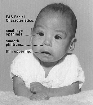
Baby with fetal alcohol syndrome, showing some of the characteristic facial features
SpecialtyGynaecology Neonatology Pediatrics Psychiatry Obstetrics Toxicology
ComplicationsMiscarriage Stillbirth

Alcohol use in pregnancy includes use of alcohol at any time during gestation, including the time before a mother-to-be is aware that she is pregnant. Alcohol use at some point during pregnancy is common and appears to be rising in prevalence.

Alcohol use during pregnancy has been associated with spontaneous abortion, stillbirth, low birthweight, and prematurity, along with a variety of birth defects and developmental abnormalities with ranging severity. Defects caused by gestational exposure to alcohol are collectively referred to as Fetal alcohol spectrum disorders (FASDs), with the most severe form termed fetal alcohol syndrome (FAS). However, not all pregnancies complicated by alcohol use will result in spontaneous abortion, stillbirth, low birthweight, and prematurity, and not all infants exposed to alcohol in utero will have FASDs or FAS.

The variance seen in outcomes of alcohol consumption during pregnancy is poorly understood, however genetic and social risk factors for more severe outcomes have both been suggested. The effect of quantity and gestational timing of alcohol consumption is also poorly understood. However, there is no amount of alcohol that is known to be safe to drink while pregnant, and there is no safe time point or trimester of pregnancy during which alcohol consumption has been proven to be safe. Therefore, medical consensus is to recommend complete abstinence from alcohol during pregnancy.

Some evidence suggests that the likelihood of FASD, FAS, miscarriage and stillbirth increases with higher quantity and longer duration of alcohol consumption during pregnancy. Therefore, it is never too late to reduce the likelihood of FASDs, FAS, and alcohol related pregnancy complications by avoiding or limiting alcohol use.

Embryology

Different body systems in the infant grow, mature and develop at specific times during gestation. The consumption of alcohol during one or more of these developmental stages may only result in one or few conditions.

During the first weeks of pregnancy babies grow at a rapid pace, even before the mothers know they are pregnant. From conception and to the third week, the most susceptible systems and organs are the brain, spinal cord, and heart. These crucial organs start forming in early stages of pregnancy, which are very sensitive and critical periods in human development. Though these body systems complete their development later in the pregnancy, the effects of alcohol consumption early in the pregnancy can result in defects to these systems and organs. During the fourth week of gestation, the limbs are being formed and it is at this point that alcohol can effect the development of arms, legs, fingers and toes. The eyes and ears also form during the fourth week and are more susceptible to the effects of alcohol. By the sixth week of gestation, the teeth and palate are forming and alcohol consumption at this time will affect these structures. Alcohol use in this window is responsible for many of the facial characteristics of fetal alcohol syndrome. By the 20th week of gestation the formation of organs and organ systems is well-developed. The infant is still susceptible to the damaging effects of alcohol. Therefore, it would be safer for women to stop drinking prior to trying to conceive.

The baby's brain, body, and organs are developing throughout pregnancy and can be affected by exposure to alcohol at any time. Because every pregnancy is different, drinking alcohol may hurt one baby more than another. A child that has been affected by alcohol before birth may appear 'normal' at birth. Intellectual disabilities may not appear until the child begins school.

According to a study done by the University of Houston, through the use of Speckle variance optical cohearance tomography (SVOCT), it was discovered that 45 minutes after pregnant mice were exposed to a binge like bolus of ethanol, a dramatic decrease in the size and number of blood vessels in the fetal brains of the mice was observed. Thus, demonstrating the magnitude of potential damage caused by a single prenatal alcohol exposure.

Alcohol during pregnancy

The developing fetus is exposed to the alcohol through the placenta and umbilical cord. Alcohol metabolizes slowly in the fetus and remains for a long time when compared to an adult because of re-uptake of alcohol-containing amniotic fluid. Alcohol exposure has serious implications on the developing fetus as well as the mother. When a woman is planning for pregnancy, she should keep in mind that there is no safe limit for alcohol consumption. It can lead to premature birth and problems may manifest later as the child continues to grow. One of the main problematic outcomes in the developing baby is fetal alcohol syndrome (FAS), which is characterized by: cleft palate and/or cleft lip, disproportionate physical development of the body, and various disabilities like attention deficiency, low memory and coordination ability, as well as improper functioning of various body organs like the kidneys, heart and bones. A large range of other developmental abnormalities are also associated with alcohol consumption during pregnancy, including an abnormal appearance, short height, low body weight, small head size, poor coordination, low intelligence, behavioral problems, hearing loss, and vision problems. These less severe outcomes are collectively termed Fetal alcohol spectrum disorders (FASDs). Those affected are more likely to have trouble in school, legal problems, participate in high-risk behaviors, and have trouble with alcohol and recreational drug use. Spontaneous abortion, stillbirth, low birthweight, and prematurity are other common outcomes, along with increased likelihood of domestic violence and potential harm to the infant.

These effects can be magnified especially during the first and third trimester of pregnancy when the baby is growing rapidly. Alcohol consumption in the first trimester of pregnancy, which is a crucial developmental stage of fetal growth, can have serious consequences. The developing fetus can be exposed to alcohol in the earliest weeks of pregnancy. During the third week, alcohol can affect the heart and central nervous system of the fetus. If the mother continues to drink, the eyes, legs and arms of the fetus can be adversely affected. Continuous exposure further through the sixth week can have negative impact on ear and teeth development. palate and external genitalia can be affected if the mother persists drinking. During the twelfth week, frequent alcohol exposure can negatively impact the brain development which affects cognitive, learning and behavioral skills before birth. Consumption of excessive alcohol can lead to Fetal Alcohol Syndrome which can produce irreversible lifetime changes in physical, mental and neurobehavioral development of the fetus. Alcohol during pregnancy not only affects the developing fetus, but it also has adverse health outcomes on the mother as well. It can harm the fertility of women who are planning for pregnancy. Adverse effects of alcohol can lead to malnutrition, seizures, vomiting and dehydration. The mother can suffer from anxiety and depression which can result in child abuse/neglect. It has also been observed that when the pregnant mother withdraws from alcohol, its effects are visible on the infant as well. The baby remains in an irritated mood, cries frequently, doesn't sleep properly, has weakening of sucking ability and increased hunger.

Alcohol consumption during pregnancy may increase the risk that the child will develop acute myeloid leukemia at a young age.

In the past, alcohol was used as a common tocolytic agent. Tocolytic agents are drugs that are used to prevent preterm labor (born at less than 37 weeks gestation) by suppressing uterine contraction. However, alcohol is no longer used in current practice due to safety concerns for the mother and her baby. A Cochrane Systematic Review has also shown that ethanol is no better than placebo (sugar water) to suppress preterm birth and neonatal mortality. Not only is ethanol worse than other beta-mimetic drugs (type of tocolytic agents) at postponing birth, it also leads to a higher rate of low birthweight babies, babies with breathing problems at birth and neonatal death.

Signs and symptoms

When an infant is born and appears to be healthy, they may still have non-visible disorders and organ defects due to exposure to alcohol during gestation. Social problems in children have been found to be associated with their mothers' alcohol use during pregnancy. Alcohol is a cause of microcephaly. Alcohol use during pregnancy does not effect the ability to breastfeed the infant – in addition, an infant may breastfeed even if their mother continues to consume alcohol after giving birth. An infant born to a mother with an alcohol dependency may go through alcohol withdrawal after the birth.

One of the major effects of alcohol consumption during pregnancy is Fetal Alcohol Spectrum Disorders (FASDs), of which Fetal Alcohol Syndrome (FAS) is the most severe form. It is shown that small amounts of alcohol consumed during pregnancy does not show any health related issues in the face, but behavioral issues can be seen. There is a wide range of symptoms seen in persons suffering from FASDs which include:

  • Abnormal facial features, such as a smooth ridge between the nose and upper lip (this ridge is called the philtrum)
  • Small head size
  • Shorter-than-average height
  • Low birth weight
  • Poor coordination
  • Hyperactive behavior
  • Difficulty with attention
  • Poor memory
  • Difficulty in school (especially with math)
  • Learning disabilities
  • Speech and language delays
  • Intellectual disability or low IQ
  • Poor reasoning and judgment skills
  • Sleep and sucking problems as a baby
  • Vision or hearing problems
  • Problems with the heart, kidneys or bones.

There are five types of FASDs depending on the symptoms:

(1) Fetal Alcohol Syndrome;

(2) Alcohol-Related Neurodevelopmental Disorder;

(3) Alcohol-Related Birth Defects;

(4) Static Encephalopathy;

(5) Neurobehavioral Disorder Associated with Prenatal Alcohol Exposure.

There are three approaches to treatment of FAS:

(1) At Home – A stable and loving home, along with a regular routine, simple rules to follow and where rewards are given for positive behavior, is a good environment for children with FAS.

(2) Medications – Medications are used to specifically treat symptoms of FASDs and not FAS entirely. Some of the medications used are antidepressants, stimulants, neuroleptics and anti-anxiety drugs.

(3) Counseling – Children with FAS benefit from behavioral and functional training, social skill training and tutoring. Support groups and talk therapy not only help the children suffering from FAS, but also help the parents and siblings of these children.

Treatment

A woman may elect to discontinue alcohol once she discovers that she is pregnant. However, women can experience serious symptoms that accompany alcohol withdrawal during pregnancy. According to the World Health Organization, these symptoms can be treated during pregnancy with brief use of benzodiazepine tranquilizers.

Medications

Currently, the FDA has approved three medications—naltrexone, acamprosate, and disulfiram—for the treatment of alcohol use disorder (AUD). However, there is insufficient data regarding the safety of these medications for pregnant women.

  • Naltrexone is a nonselective opioid antagonist that is used to treat AUD and opioid use disorder. The long-term effects of naltrexone on the fetus are currently unknown. Animal studies show that naltrexone administered during pregnancy increases the incidence of early fetal loss; however, there are insufficient data available to identify the extent to which this is a risk in pregnant women.
  • Acamprosate functions as both an antagonist of NMDA and glutamate and an agonist at GABAA receptors, although its molecular mechanism is not completely understood. Acamprosate has been shown to be effective at preventing alcohol relapse during abstinence. Animal data, however, suggests that acamprosate can have possible teratogenic effects on fetuses.
  • Disulfiram prevents relapse by blocking the metabolism of acetaldehyde after consumption of alcohol which leads to headache, nausea, and vomiting. Some evidence suggests that disulfiram use during the first trimester is associated with an increased risk of congenital malformations such as reduction defects and cleft palate. Additionally, the effects of disulfiram can involve hypertension which can be harmful to both the pregnant woman and the fetus.

American Psychiatric Association guidelines recommend that medications not be used to treat alcohol use disorder in pregnant women except in cases of acute alcohol withdrawals or other co-existing conditions. Instead, behavioral interventions are usually preferred as treatments for pregnant women with AUD.  Medications should only be used for pregnant women after carefully considering potential risks and harms of the medications versus the benefits of alcohol cessation.

Epidemiology

Alcohol consumption during pregnancy is relatively common, and its prevalence has been on the rise. An estimated 7.6% of pregnant women use alcohol, while 1.4% of pregnant women report binge drinking during their pregnancy. The highest prevalence estimates of reported alcohol use during pregnancy were among women who are aged 35–44 years (14.3%), white (8.3%), college graduates (10.0%), or employed (9.6%). Furthermore, alcohol-related congenital abnormalities occur at an incidence of roughly one out of 67 women who drink alcohol during pregnancy. The use of alcohol during pregnancy occurs at different rates across the world, potentially due to various cultural differences and legislation. The five countries with the highest prevalence of alcohol use during pregnancy are Ireland (60%), Belarus (47%), Denmark (46%), the UK (41%), and the Russian Federation (37%).

One of the biggest challenges in uncovering the true prevalence of FAS and the associated disorders is understanding how to recognize the syndrome, which largely depends on the age and physical features of the individual being diagnosed. Using medical and other records, CDC studies have identified 0.2 to 1.5 infants with FAS for every 1,000 live births in certain areas of the United States. A more recent CDC study analyzed medical and other records and found FAS in 0.3 out of 1,000 children from 7 to 9 years of age.

Public health recommendations

Starting in 1981, the Surgeon General of the United States started releasing a warning asking pregnant women to abstain from alcohol for the remainder of gestation. The American Academy of Pediatrics issued a set of recommendations in 2015: "During pregnancy no amount of alcohol intake should be considered safe; there is no safe trimester to drink alcohol; all forms of alcohol, such as beer, wine, and liquor, pose similar risk; and binge drinking poses dose-related risk to the developing fetus." The World Health Organization recommends that alcohol should be avoided entirely during pregnancy, given the relatively unknown effects of even small amounts of alcohol during pregnancy. The United Kingdom's National Institute for Health and Clinical Excellence recommends "that if you're pregnant or planning to become pregnant, the safest approach is not to drink alcohol at all to keep risks to your baby to a minimum."

There has been some controversy surrounding the zero-tolerance approach taken by many countries toward alcohol consumption during pregnancy. A 2000 article wrote that the concern about the risk of FAS may be inflated beyond the level warranted by existing evidence of its prevalence and impact and argued that equating a low level of drinking with unavoidable harm to the fetus may have negative social, legal and health impacts.

Optimal tax

From Wikipedia, the free encyclopedia https://en.wikipedia.org/wiki/Optimal_tax   Optimal tax theory or the theory of optimal taxation is ...