Search This Blog

Saturday, August 15, 2020

Phagocytosis

From Wikipedia, the free encyclopedia
 
Overview of phagocytosis
 
Phagocytosis versus exocytosis
 
Phagocytosis (from Ancient Greek φαγεῖν (phagein) , meaning 'to eat', and κύτος, (kytos) , meaning 'cell') is the process by which a cell uses its plasma membrane to engulf a large particle (≥ 0.5 μm), giving rise to an internal compartment called the phagosome. It is one type of endocytosis.

The engulfing of a pathogen by a phagocyte

In a multicellular organism's immune system, phagocytosis is a major mechanism used to remove pathogens and cell debris. The ingested material is then digested in the phagosome. Bacteria, dead tissue cells, and small mineral particles are all examples of objects that may be phagocytized. Some protozoa use phagocytosis as means to obtain nutrients.

History

Phagocytosis was first noted by Canadian physician William Osler (1876), and later studied and named by Élie Metchnikoff (1880, 1883).

In immune system

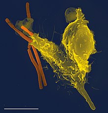
Scanning electron micrograph of a phagocyte (yellow, right) phagocytosing anthrax bacilli (orange, left)

Phagocytosis is one of the main mechanisms of the innate immune defense. It is one of the first processes responding to infection, and is also one of the initiating branches of an adaptive immune response. Although most cells are capable of phagocytosis, some cell types perform it as part of their main function. These are called 'professional phagocytes.' Phagocytosis is old in evolutionary terms, being present even in invertebrates.

Professional phagocytic cells

Light microscopic video sequence of a neutrophil from human blood phagocytosing a bacterium

Neutrophils, macrophages, monocytes, dendritic cells, osteoclasts and eosinophils can be classified as professional phagocytes. The first three have the greatest role in immune response to most infections.
The role of neutrophils is patrolling the bloodstream and rapid migration to the tissues in large numbers only in case of infection. There they have direct microbicidal effect by phagocytosis. After ingestion, neutrophils are efficient in intracellular killing of pathogens. Neutrophils phagocytose mainly via the Fcγ receptors and complement receptors 1 and 3. The microbicidal effect of neutrophils is due to a large repertoire of molecules present in pre-formed granules. Enzymes and other molecules prepared in these granules are proteases, such as collagenase, gelatinase or serine proteases, myeloperoxidase, lactoferrin and antibiotic proteins. Degranulation of these into the phagosome, accompanied by high reactive oxygen species production (oxidative burst) is highly microbicidal.

Monocytes, and the macrophages that mature from them, leave blood circulation to migrate through tissues. There they are resident cells and form a resting barrier. Macrophages initiate phagocytosis by mannose receptors, scavenger receptors, Fcγ receptors and complement receptors 1, 3 and 4. Macrophages are long-lived and can continue phagocytosis by forming new lysosomes.

Dendritic cells also reside in tissues and ingest pathogens by phagocytosis. Their role is not killing or clearance of microbes, but rather breaking them down for antigen presentation to the cells of the adaptive immune system.

Initiating receptors

Receptors for phagocytosis can be divided into two categories by recognised molecules. The first, opsonic receptors, are dependent on opsonins. Among these are receptors that recognise the Fc part of bound IgG antibodies, deposited complement or receptors, that recognise other opsonins of cell or plasma origin. Non-opsonic receptors include lectin-type receptors, Dectin receptor, or scavenger receptors. Some phagocytic pathways require a second signal from pattern recognition receptors (PRRs) activated by attachment to pathogen-associated molecular patterns (PAMPS), which leads to NF-κB activation.

Fcγ receptors

Fcγ receptors recognise IgG coated targets. The main recognised part is the Fc fragment. The molecule of the receptor contain an intracellular ITAM domain or associates with an ITAM-containing adaptor molecule. ITAM domains transduce the signal from the surface of the phagocyte to the nucleus. For example, activating receptors of human macrophages are FcγRI, FcγRIIA, and FcγRIII. Fcγ receptor mediated phagocytosis includes formation of protrusions of the cell called a 'phagocytic cup' and activates an oxidative burst in neutrophils.

Complement receptors

These receptors recognise targets coated in C3b, C4b and C3bi from plasma complement. The extracellular domain of the receptors contains a lectin-like complement-binding domain. Recognition by complement receptors is not enough to cause internalisation without additional signals. In macrophages, the CR1, CR3 and CR4 are responsible for recognition of targets. Complement coated targets are internalised by 'sinking' into the phagocyte membrane, without any protrusions.

Mannose receptors

Mannose and other pathogen-associated sugars, such as fucose, are recognised by the mannose receptor. Eight lectin-like domains form the extracellular part of the receptor. The ingestion mediated by the mannose receptor is distinct in molecular mechanisms from Fcγ receptor or complement receptor mediated phagocytosis.

Phagosome

Engulfment of material is facilitated by the actin-myosin contractile system. The phagosome is the organelle formed by phagocytosis of material. It then moves toward the centrosome of the phagocyte and is fused with lysosomes, forming a phagolysosome and leading to degradation. Progressively, the phagolysosome is acidified, activating degradative enzymes.

Degradation can be oxygen-dependent or oxygen-independent.
  • Oxygen-dependent degradation depends on NADPH and the production of reactive oxygen species. Hydrogen peroxide and myeloperoxidase activate a halogenating system, which leads to the creation of hypochlorite and the destruction of bacteria.
  • Oxygen-independent degradation depends on the release of granules, containing enzymes such as lysozymes, and cationic proteins such as defensins. Other antimicrobial peptides are present in these granules, including lactoferrin, which sequesters iron to provide unfavourable growth conditions for bacteria. Other enzymes like hyaluronidase, lipase, collagenase, elastase, ribonuclease, deoxyribonuclease also play an important role in preventing the spread of infection and degradation of essential microbial biomolecules leading to cell death.
Leukocytes generate hydrogen cyanide during phagocytosis, and can kill bacteria, fungi, and other pathogens by generating several other toxic chemicals.

Some bacteria, for example Treponema pallidum, Escheria coli and Staphylococcus aureus, are able to avoid phagocytosis by several mechanisms.

In apoptosis

Following apoptosis, the dying cells need to be taken up into the surrounding tissues by macrophages in a process called efferocytosis. One of the features of an apoptotic cell is the presentation of a variety of intracellular molecules on the cell surface, such as calreticulin, phosphatidylserine (from the inner layer of the plasma membrane), annexin A1, oxidised LDL and altered glycans. These molecules are recognised by receptors on the cell surface of the macrophage such as the phosphatidylserine receptor or by soluble (free-floating) receptors such as thrombospondin 1, GAS6, and MFGE8, which themselves then bind to other receptors on the macrophage such as CD36 and alpha-v beta-3 integrin. Defects in apoptotic cell clearance is usually associated with impaired phagocytosis of macrophages. Accumulation of apoptotic cell remnants often causes autoimmune disorders; thus pharmacological potentiation of phagocytosis has a medical potential in treatment of certain forms of autoimmune disorders.

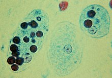
Trophozoites of Entamoeba histolytica with ingested erythrocytes

In protists

In many protists, phagocytosis is used as a means of feeding, providing part or all of their nourishment. This is called phagotrophic nutrition, distinguished from osmotrophic nutrition which takes place by absorption.
  • In some, such as amoeba, phagocytosis takes place by surrounding the target object with pseudopods, as in animal phagocytes. In humans, the amoebozoan Entamoeba histolytica can phagocytose red blood cells.
  • Ciliates also engage in phagocytosis. In ciliates there is a specialized groove or chamber in the cell where phagocytosis takes place, called the cytostome or mouth.
As in phagocytic immune cells, the resulting phagosome may be merged with lysosomes containing digestive enzymes, forming a phagolysosome. The food particles will then be digested, and the released nutrients are diffused or transported into the cytosol for use in other metabolic processes.
Mixotrophy can involve phagotrophic nutrition and phototrophic nutrition.

Macrophage

From Wikipedia, the free encyclopedia

Macrophage
Macrophage.jpg
A macrophage stretching its "arms" (filopodia) to engulf two particles, possibly pathogens, in a mouse. Trypan blue exclusion staining.
Details
Pronunciation/ˈmakrə(ʊ)feɪdʒ/
SystemImmune system
FunctionPhagocytosis
Identifiers
LatinMacrophagocytus
Acronym(s)Mφ, MΦ
MeSHD008264
THH2.00.03.0.01007
FMA63261

Macrophages (abbreviated as Mφ, or MP) (Greek: large eaters, from Greek μακρός (makrós) = large, φαγεῖν (phagein) = to eat) are a type of white blood cell of the immune system that engulfs and digests cellular debris, foreign substances, microbes, cancer cells, and anything else that does not have the type of proteins specific to healthy body cells on its surface in a process called phagocytosis.
These large phagocytes are found in essentially all tissues, where they patrol for potential pathogens by amoeboid movement. They take various forms (with various names) throughout the body (e.g., histiocytes, Kupffer cells, alveolar macrophages, microglia, and others), but all are part of the mononuclear phagocyte system. Besides phagocytosis, they play a critical role in nonspecific defense (innate immunity) and also help initiate specific defense mechanisms (adaptive immunity) by recruiting other immune cells such as lymphocytes. For example, they are important as antigen presenters to T cells. In humans, dysfunctional macrophages cause severe diseases such as chronic granulomatous disease that result in frequent infections.

Beyond increasing inflammation and stimulating the immune system, macrophages also play an important anti-inflammatory role and can decrease immune reactions through the release of cytokines. Macrophages that encourage inflammation are called M1 macrophages, whereas those that decrease inflammation and encourage tissue repair are called M2 macrophages. This difference is reflected in their metabolism; M1 macrophages have the unique ability to metabolize arginine to the "killer" molecule nitric oxide, whereas rodent M2 macrophages have the unique ability to metabolize arginine to the "repair" molecule ornithine. However, this dichotomy has been recently questioned as further complexity has been discovered.

Human macrophages are about 21 micrometres (0.00083 in) in diameter and are produced by the differentiation of monocytes in tissues. They can be identified using flow cytometry or immunohistochemical staining by their specific expression of proteins such as CD14, CD40, CD11b, CD64, F4/80 (mice)/EMR1 (human), lysozyme M, MAC-1/MAC-3 and CD68.

Macrophages were first discovered by Élie Metchnikoff, a Russian zoologist, in 1884.

Structure

Types

Drawing of a macrophage when fixed and stained by giemsa dye

A majority of macrophages are stationed at strategic points where microbial invasion or accumulation of foreign particles is likely to occur. These cells together as a group are known as the mononuclear phagocyte system and were previously known as the reticuloendothelial system. Each type of macrophage, determined by its location, has a specific name:

Cell Name Anatomical Location
Adipose tissue macrophages Adipose tissue (fat)
Monocytes Bone marrow / blood
Kupffer cells Liver
Sinus histiocytes Lymph nodes
Alveolar macrophages (dust cells) Pulmonary alveoli
Tissue macrophages (histiocytes) leading to giant cells Connective tissue
Microglia Central nervous system
Hofbauer cells Placenta
Intraglomerular mesangial cells Kidney
Osteoclasts Bone
Epithelioid cells Granulomas
Red pulp macrophages (sinusoidal lining cells) Red pulp of spleen
Peritoneal macrophages Peritoneal cavity
LysoMac Peyer's patch

Investigations concerning Kupffer cells are hampered because in humans, Kupffer cells are only accessible for immunohistochemical analysis from biopsies or autopsies. From rats and mice, they are difficult to isolate, and after purification, only approximately 5 million cells can be obtained from one mouse. 

Macrophages can express paracrine functions within organs that are specific to the function of that organ. In the testis, for example, macrophages have been shown to be able to interact with Leydig cells by secreting 25-hydroxycholesterol, an oxysterol that can be converted to testosterone by neighbouring Leydig cells. Also, testicular macrophages may participate in creating an immune privileged environment in the testis, and in mediating infertility during inflammation of the testis.

Cardiac resident macrophages participate in electrical conduction via gap junction communication with cardiac myocytes.

Macrophages can be classified on basis of the fundamental function and activation. According to this grouping there are classically-activated (M1) macrophages, wound-healing macrophages (also known as alternatively-activated (M2) macrophages), and regulatory macrophages (Mregs).

Development

Macrophages that reside in adult healthy tissues either derive from circulating monocytes or are established before birth and then maintained during adult life independently of monocytes. By contrast, most of the macrophages that accumulate at diseased sites typically derive from circulating monocytes. When a monocyte enters damaged tissue through the endothelium of a blood vessel, a process known as leukocyte extravasation, it undergoes a series of changes to become a macrophage. Monocytes are attracted to a damaged site by chemical substances through chemotaxis, triggered by a range of stimuli including damaged cells, pathogens and cytokines released by macrophages already at the site. At some sites such as the testis, macrophages have been shown to populate the organ through proliferation. Unlike short-lived neutrophils, macrophages survive longer in the body, up to several months.

Function

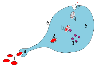
Steps of a macrophage ingesting a pathogen:
a. Ingestion through phagocytosis, a phagosome is formed
b. The fusion of lysosomes with the phagosome creates a phagolysosome; the pathogen is broken down by enzymes
c. Waste material is expelled or assimilated (the latter not pictured)
Parts:
1. Pathogens
2. Phagosome
3. Lysosomes
4. Waste material
5. Cytoplasm
6. Cell membrane

Phagocytosis

Macrophages are professional phagocytes and are highly specialized in removal of dying or dead cells and cellular debris. This role is important in chronic inflammation, as the early stages of inflammation are dominated by neutrophils, which are ingested by macrophages if they come of age (see CD31 for a description of this process).

The neutrophils are at first attracted to a site, where they perform their function and die, before they are phagocytized by the macrophages. When at the site, the first wave of neutrophils, after the process of aging and after the first 48 hours, stimulate the appearance of the macrophages whereby these macrophages will then ingest the aged neutrophils.

The removal of dying cells is, to a greater extent, handled by fixed macrophages, which will stay at strategic locations such as the lungs, liver, neural tissue, bone, spleen and connective tissue, ingesting foreign materials such as pathogens and recruiting additional macrophages if needed. 

When a macrophage ingests a pathogen, the pathogen becomes trapped in a phagosome, which then fuses with a lysosome. Within the phagolysosome, enzymes and toxic peroxides digest the pathogen. However, some bacteria, such as Mycobacterium tuberculosis, have become resistant to these methods of digestion. Typhoidal Salmonellae induce their own phagocytosis by host macrophages in vivo, and inhibit digestion by lysosomal action, thereby using macrophages for their own replication and causing macrophage apoptosis. Macrophages can digest more than 100 bacteria before they finally die due to their own digestive compounds.

Role in adaptive immunity

Macrophages are versatile cells that play many roles. As scavengers, they rid the body of worn-out cells and other debris. Along with dendritic cells, they are foremost among the cells that present antigens, a crucial role in initiating an immune response. As secretory cells, monocytes and macrophages are vital to the regulation of immune responses and the development of inflammation; they produce a wide array of powerful chemical substances (monokines) including enzymes, complement proteins, and regulatory factors such as interleukin-1. At the same time, they carry receptors for lymphokines that allow them to be "activated" into single-minded pursuit of microbes and tumour cells.

After digesting a pathogen, a macrophage will present the antigen (a molecule, most often a protein found on the surface of the pathogen and used by the immune system for identification) of the pathogen to the corresponding helper T cell. The presentation is done by integrating it into the cell membrane and displaying it attached to an MHC class II molecule (MHCII), indicating to other white blood cells that the macrophage is not a pathogen, despite having antigens on its surface.

Eventually, the antigen presentation results in the production of antibodies that attach to the antigens of pathogens, making them easier for macrophages to adhere to with their cell membrane and phagocytose. In some cases, pathogens are very resistant to adhesion by the macrophages.

The antigen presentation on the surface of infected macrophages (in the context of MHC class II) in a lymph node stimulates TH1 (type 1 helper T cells) to proliferate (mainly due to IL-12 secretion from the macrophage). When a B-cell in the lymph node recognizes the same unprocessed surface antigen on the bacterium with its surface bound antibody, the antigen is endocytosed and processed. The processed antigen is then presented in MHCII on the surface of the B-cell. T cells that express the T cell receptor which recognizes the antigen-MHCII complex (with co-stimulatory factors- CD40 and CD40L) cause the B-cell to produce antibodies that help opsonisation of the antigen so that the bacteria can be better cleared by phagocytes.

Macrophages provide yet another line of defense against tumor cells and somatic cells infected with fungus or parasites. Once a T cell has recognized its particular antigen on the surface of an aberrant cell, the T cell becomes an activated effector cell, producing chemical mediators known as lymphokines that stimulate macrophages into a more aggressive form.

Macrophage subtypes

There are several activated forms of macrophages. In spite of a spectrum of ways to activate macrophages, there are two main groups designated M1 and M2. M1 macrophages: as mentioned earlier (previously referred to as classically activated macrophages), M1 "killer" macrophages are activated by LPS and IFN-gamma, and secrete high levels of IL-12 and low levels of IL-10. M1 macrophages have pro-inflammatory, bactericidal, and phagocytic functions. In contrast, the M2 "repair" designation (also referred to as alternatively activated macrophages) broadly refers to macrophages that function in constructive processes like wound healing and tissue repair, and those that turn off damaging immune system activation by producing anti-inflammatory cytokines like IL-10. M2 is the phenotype of resident tissue macrophages, and can be further elevated by IL-4. M2 macrophages produce high levels of IL-10, TGF-beta and low levels of IL-12. Tumor-associated macrophages are mainly of the M2 phenotype, and seem to actively promote tumor growth.

Macrophages exist in a variety of phenotypes which are determined by the role they play in wound maturation. Phenotypes can be predominantly separated into two major categories; M1 and M2. M1 macrophages are the dominating phenotype observed in the early stages of inflammation and are activated by four key mediators: interferon-γ (IFN-γ), tumor necrosis factor (TNF), and damage associated molecular patterns (DAMPs). These mediator molecules create a pro-inflammatory response that in return produce pro-inflammatory cytokines like Interleukin-6 and TNF. Unlike M1 macrophages, M2 macrophages secrete an anti-inflammatory response via the addition of Interleukin-4 or Interleukin-13. They also play a role in wound healing and are needed for revascularization and reepithelialization. M2 macrophages are divided into four major types based on their roles: M2a, M2b, M2c, and M2d. How M2 phenotypes are determined is still up for discussion but studies have shown that their environment allows them to adjust to whichever phenotype is most appropriate to efficiently heal the wound.

M2 macrophages are needed for vascular stability. They produce vascular epithelial growth factor-A and TGF-β1. There is a phenotype shift from M1 to M2 macrophages in acute wounds, however this shift is impaired for chronic wounds. This dysregulation results in insufficient M2 macrophages and its corresponding growth factors that aid in wound repair. With a lack of these growth factors/anti-inflammatory cytokines and an overabundance of pro-inflammatory cytokines from M1 macrophages chronic wounds are unable to heal in a timely manner. Normally, after neutrophils eat debris/pathogens they perform apoptosis and are removed. At this point, inflammation is not needed and M1 undergoes a switch to M2 (anti-inflammatory). However, dysregulation occurs as the M1 macrophages are unable/do not phagocytose neutrophils that have undergone apoptosis leading to increased macrophage migration and inflammation.

Both M1 and M2 macrophages play a role in promotion of atherosclerosis. M1 macrophages promote atherosclerosis by inflammation. M2 macrophages can remove cholesterol from blood vessels, but when the cholesterol is oxidized, the M2 macrophages become apoptotic foam cells contributing to the atheromatous plaque of atherosclerosis.

Role in muscle regeneration

The first step to understanding the importance of macrophages in muscle repair, growth, and regeneration is that there are two "waves" of macrophages with the onset of damageable muscle use – subpopulations that do and do not directly have an influence on repairing muscle. The initial wave is a phagocytic population that comes along during periods of increased muscle use that are sufficient to cause muscle membrane lysis and membrane inflammation, which can enter and degrade the contents of injured muscle fibers. These early-invading, phagocytic macrophages reach their highest concentration about 24 hours following the onset of some form of muscle cell injury or reloading. Their concentration rapidly declines after 48 hours. The second group is the non-phagocytic types that are distributed near regenerative fibers. These peak between two and four days and remain elevated for several days during the hopeful muscle rebuilding. The first subpopulation has no direct benefit to repairing muscle, while the second non-phagocytic group does.

It is thought that macrophages release soluble substances that influence the proliferation, differentiation, growth, repair, and regeneration of muscle, but at this time the factor that is produced to mediate these effects is unknown. It is known that macrophages' involvement in promoting tissue repair is not muscle specific; they accumulate in numerous tissues during the healing process phase following injury.

Role in wound healing

Macrophages are essential for wound healing. They replace polymorphonuclear neutrophils as the predominant cells in the wound by day two after injury. Attracted to the wound site by growth factors released by platelets and other cells, monocytes from the bloodstream enter the area through blood vessel walls. Numbers of monocytes in the wound peak one to one and a half days after the injury occurs. Once they are in the wound site, monocytes mature into macrophages. The spleen contains half the body's monocytes in reserve ready to be deployed to injured tissue.

The macrophage's main role is to phagocytize bacteria and damaged tissue, and they also debride damaged tissue by releasing proteases. Macrophages also secrete a number of factors such as growth factors and other cytokines, especially during the third and fourth post-wound days. These factors attract cells involved in the proliferation stage of healing to the area. Macrophages may also restrain the contraction phase. Macrophages are stimulated by the low oxygen content of their surroundings to produce factors that induce and speed angiogenesis and they also stimulate cells that re-epithelialize the wound, create granulation tissue, and lay down a new extracellular matrix. By secreting these factors, macrophages contribute to pushing the wound healing process into the next phase.

Role in limb regeneration

Scientists have elucidated that as well as eating up material debris, macrophages are involved in the typical limb regeneration in the salamander. They found that removing the macrophages from a salamander resulted in failure of limb regeneration and a scarring response.

Role in iron homeostasis

As described above, macrophages play a key role in removing dying or dead cells and cellular debris. Erythrocytes have a lifespan on average of 120 days and so are constantly being destroyed by macrophages in the spleen and liver. Macrophages will also engulf macromolecules, and so play a key role in the pharmacokinetics of parenteral irons.

The iron that is released from the haemoglobin is either stored internally in ferritin or is released into the circulation via ferroportin. In cases where systemic iron levels are raised, or where inflammation is present, raised levels of hepcidin act on macrophage ferroportin channels, leading to iron remaining within the macrophages.

Role in pigment retainment

Melanophage

Melanophages are a subset of tissue-resident macrophages able to absorb pigment, either native to the organism or exogenous (such as tattoos), from extracellular space. In contrast to dendritic juncional melanocytes, which synthesize melanosomes and contain various stages of their development, the melanophages only accumulate phagocytosed melanin in lysosome-like phagosomes. This occurs repeatedly as the pigment from dead dermal macrophages is phagocytosed by their successors, preserving the tattoo in the same place.

Role in tissue homeostasis

Every tissue harbors its own specialized population of resident macrophages, which entertain reciprocal interconnections with the stroma and functional tissue. These resident macrophages are sessile (non-migratory), provide essential growth factors to support the physiological function of the tissue (e.g. macrophage-neuronal crosstalk in the guts), and can actively protect the tissue from inflammatory damage.

Clinical significance

Due to their role in phagocytosis, macrophages are involved in many diseases of the immune system. For example, they participate in the formation of granulomas, inflammatory lesions that may be caused by a large number of diseases. Some disorders, mostly rare, of ineffective phagocytosis and macrophage function have been described, for example.

As a host for intracellular pathogens

In their role as a phagocytic immune cell macrophages are responsible for engulfing pathogens to destroy them. Some pathogens subvert this process and instead live inside the macrophage. This provides an environment in which the pathogen is hidden from the immune system and allows it to replicate.

Diseases with this type of behaviour include tuberculosis (caused by Mycobacterium tuberculosis) and leishmaniasis (caused by Leishmania species).

In order to minimize the possibility of becoming the host of an intracellular bacteria, macrophages have evolved defense mechanisms such as induction of nitric oxide and reactive oxygen intermediates, which are toxic to microbes. Macrophages have also evolved the ability to restrict the microbe's nutrient supply and induce autophagy.

Tuberculosis

Once engulfed by a macrophage, the causative agent of tuberculosis, Mycobacterium tuberculosis, avoids cellular defenses and uses the cell to replicate.

Leishmaniasis

Upon phagocytosis by a macrophage, the Leishmania parasite finds itself in a phagocytic vacuole. Under normal circumstances, this phagocytic vacuole would develop into a lysosome and its contents would be digested. Leishmania alter this process and avoid being destroyed; instead, they make a home inside the vacuole.

Chikungunya

Infection of macrophages in joints is associated with local inflammation during and after the acute phase of Chikungunya (caused by CHIKV or Chikungunya virus).

Others

Adenovirus (most common cause of pink eye) can remain latent in a host macrophage, with continued viral shedding 6–18 months after initial infection.

Brucella spp. can remain latent in a macrophage via inhibition of phagosomelysosome fusion; causes brucellosis (undulant fever).

Legionella pneumophila, the causative agent of Legionnaires' disease, also establishes residence within macrophages.

Heart disease

Macrophages are the predominant cells involved in creating the progressive plaque lesions of atherosclerosis.

Focal recruitment of macrophages occurs after the onset of acute myocardial infarction. These macrophages function to remove debris, apoptotic cells and to prepare for tissue regeneration.

HIV infection

Macrophages also play a role in human immunodeficiency virus (HIV) infection. Like T cells, macrophages can be infected with HIV, and even become a reservoir of ongoing virus replication throughout the body. HIV can enter the macrophage through binding of gp120 to CD4 and second membrane receptor, CCR5 (a chemokine receptor). Both circulating monocytes and macrophages serve as a reservoir for the virus. Macrophages are better able to resist infection by HIV-1 than CD4+ T cells, although susceptibility to HIV infection differs among macrophage subtypes.

Cancer

Macrophages can contribute to tumor growth and progression by promoting tumor cell proliferation and invasion, fostering tumor angiogenesis and suppressing antitumor immune cells. Attracted to oxygen-starved (hypoxic) and necrotic tumor cells they promote chronic inflammation. Inflammatory compounds such as tumor necrosis factor (TNF)-alpha released by the macrophages activate the gene switch nuclear factor-kappa B. NF-κB then enters the nucleus of a tumor cell and turns on production of proteins that stop apoptosis and promote cell proliferation and inflammation. Moreover, macrophages serve as a source for many pro-angiogenic factors including vascular endothelial factor (VEGF), tumor necrosis factor-alpha (TNF-alpha), Macrophage colony-stimulating factor (M-CSF/CSF1) and IL-1 and IL-6 contributing further to the tumor growth. Macrophages have been shown to infiltrate a number of tumors. Their number correlates with poor prognosis in certain cancers including cancers of breast, cervix, bladder, brain and prostate. Tumor-associated macrophages (TAMs) are thought to acquire an M2 phenotype, contributing to tumor growth and progression. Some tumors can also produce factors, including M-CSF/CSF1, MCP-1/CCL2 and Angiotensin II, that trigger the amplification and mobilization of macrophages in tumors. Research in various study models suggests that macrophages can sometimes acquire anti-tumor functions. For example, macrophages may have cytotoxic activity to kill tumor cells directly; also the co-operation of T-cells and macrophages is important to suppress tumors. This co-operation involves not only the direct contact of T-cell and macrophage, with antigen presentation, but also includes the secretion of adequate combinations of cytokines, which enhance T-cell antitumor activity. Recent study findings suggest that by forcing IFN-α expression in tumor-infiltrating macrophages, it is possible to blunt their innate protumoral activity and reprogram the tumor microenvironment toward more effective dendritic cell activation and immune effector cell cytotoxicity. Additionally, subcapsular sinus macrophages in tumor-draining lymph nodes can suppress cancer progression by containing the spread of tumor-derived materials.

Cancer therapy

Experimental studies indicate that macrophages can affect all therapeutic modalities, including surgery, chemotherapy, radiotherapy, immunotherapy and targeted therapy. Macrophages can influence treatment outcomes both positively and negatively. Macrophages can be protective in different ways: they can remove dead tumor cells (in a process called phagocytosis) following treatments that kill these cells; they can serve as drug depots for some anticancer drugs; they can also be activated by some therapies to promote antitumor immunity. Macrophages can also be deleterious in several ways: for example they can suppress various chemotherapies, radiotherapies and immunotherapies. Because macrophages can regulate tumor progression, therapeutic strategies to reduce the number of these cells, or to manipulate their phenotypes, are currently being tested in cancer patients. However, macrophages are also involved in antibody mediated cytotoxicity (ADCC)and this mechanism has been proposed to be important for certain cancer immunotherapy antibodies.

Obesity

It has been observed that increased number of pro-inflammatory macrophages within obese adipose tissue contributes to obesity complications including insulin resistance and diabetes type 2.

Within the fat (adipose) tissue of CCR2 deficient mice, there is an increased number of eosinophils, greater alternative macrophage activation, and a propensity towards type 2 cytokine expression. Furthermore, this effect was exaggerated when the mice became obese from a high fat diet. This is partially caused by a phenotype switch of macrophages induced by necrosis of fat cells (adipocytes). In an obese individual some adipocytes burst and undergo necrotic death, which causes the residential M2 macrophages to switch to M1 phenotype. This is one of the causes of a low-grade systemic chronic inflammatory state associated with obesity.

Intestinal macrophages

Though very similar in structure to tissue macrophages, intestinal macrophages have evolved specific characteristics and functions given their natural environment, which is in the digestive tract. Macrophages and intestinal macrophages have high plasticity causing their phenotype to be altered by their environments. Like macrophages, intestinal macrophages are differentiated monocytes, though intestinal macrophages have to coexist with the microbiome in the intestines. This is a challenge considering the bacteria found in the gut are not recognized as "self" and could be potential targets for phagocytosis by the macrophage.

To prevent the destruction of the gut bacteria, intestinal macrophages have developed key differences compared to other macrophages. Primarily, intestinal macrophages do not induce inflammatory responses. Whereas tissue macrophages release various inflammatory cytokines, such as IL-1, IL-6 and TNF-α, intestinal macrophages do not produce or secrete inflammatory cytokines. This change is directly caused by the intestinal macrophages environment. Surrounding intestinal epithelial cells release TGF-β, which induces the change from proinflammatory macrophage to noninflammatory macrophage.

Even though the inflammatory response is downregulated in intestinal macrophages, phagocytosis is still carried out. There is no drop off in phagocytosis efficiency as intestinal macrophages are able to effectively phagocytize the bacteria,S. typhimurium and E. coli, but intestinal macrophages still do not release cytokines, even after phagocytosis. Also, intestinal macrophages do not express lipoplysaccharide (LPS), IgA, or IgG receptors. The lack of LPS receptors is important for the gut as the intestinal macrophages do not detect the microbe-associated molecular patterns (MAMPS/PAMPS) of the intestinal microbiome. Nor do they express IL-2 and IL-3 growth factor receptors.

Role in disease

Intestinal macrophages have been shown to play a role in inflammatory bowel disease (IBD), such as Crohn's disease (CD) and ulcerative colitis (UC). In a healthy gut, intestinal macrophages limit the inflammatory response in the gut, but in a disease-state, intestinal macrophage numbers and diversity are altered. This leads to inflammation of the gut and disease symptoms of IBD. Intestinal macrophages are critical in maintaining gut homeostasis. The presence of inflammation or pathogen alters this homeostasis, and concurrently alters the intestinal macrophages. There has yet to be a determined mechanism for the alteration of the intestinal macrophages by recruitment of new monocytes or changes in the already present intestinal macrophages.

Multi-drug-resistant tuberculosis

From Wikipedia, the free encyclopedia
 
Multi-drug-resistance tuberculosis
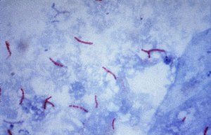
Mycobacterium tuberculosis Ziehl-Neelsen stain 02.jpg
Mycobacterium tuberculosis bacteria seen by microscope
SpecialtyInfectious disease

Multi-drug-resistant tuberculosis (MDR-TB) is a form of tuberculosis (TB) infection caused by bacteria that are resistant to treatment with at least two of the most powerful first-line anti-TB medications (drugs), isoniazid and rifampin. Some forms of TB are also resistant to second-line medications, and are called extensively drug-resistant TB (XDR-TB).

Tuberculosis is caused by infection with the bacteria Mycobacterium tuberculosis. Almost one in four people in the world are infected with TB bacteria. Only when the bacteria become active do people become ill with TB. Bacteria become active as a result of anything that can reduce the person's immunity, such as HIV, advancing age, diabetes or other immunocompromising illnesses. TB can usually be treated with a course of four standard, or first-line, anti-TB drugs (i.e., isoniazid, rifampin and any fluoroquinolone).

However, beginning with the first antibiotic treatment for TB in 1943, some strains of the TB bacteria developed resistance to the standard drugs through genetic changes (see mechanisms.) Currently the majority of multidrug-resistant cases of TB are due to one strain of TB bacteria called the Beijing lineage. This process accelerates if incorrect or inadequate treatments are used, leading to the development and spread of multidrug-resistant TB (MDR-TB). Incorrect or inadequate treatment may be due to use of the wrong medications, use of only one medication (standard treatment is at least two drugs), not taking medication consistently or for the full treatment period (treatment is required for several months). Treatment of MDR-TB requires second-line drugs (i.e., fluoroquinolones, aminoglycosides, and others), which in general are less effective, more toxic and much more expensive than first-line drugs. Treatment schedules for MDR-TB involving fluoroquinolones and aminoglycosides can run for 2 years, compared to the 6 months of first-line drug treatment, and cost over US$100,000. If these second-line drugs are prescribed or taken incorrectly, further resistance can develop leading to XDR-TB.

Resistant strains of TB are already present in the population, so MDR-TB can be directly transmitted from an infected person to an uninfected person. In this case a previously untreated person develops a new case of MDR-TB. This is known as primary MDR-TB, and is responsible for up to 75% of cases. Acquired MDR-TB develops when a person with a non-resistant strain of TB is treated inadequately, resulting in the development of antibiotic resistance in the TB bacteria infecting them. These people can in turn infect other people with MDR-TB.

MDR-TB caused an estimated 600,000 new TB cases and 240,000 deaths in 2016 and MDR-TB accounts for 4.1% of all new TB cases and 19% of previously treated cases worldwide.[12] Globally, most MDR-TB cases occur in South America, Southern Africa, India, China, and the former Soviet Union.

Treatment of MDR-TB requires treatment with second-line drugs, usually four or more anti-TB drugs for a minimum of 6 months, and possibly extending for 18–24 months if rifampin resistance has been identified in the specific strain of TB with which the patient has been infected. Under ideal program conditions, MDR-TB cure rates can approach 70%.

Mechanism of drug resistance

The TB bacteria has natural defenses against some drugs, and can acquire drug resistance through genetic mutations. The bacteria does not have the ability to transfer genes for resistance between organisms through plasmids (see horizontal transfer). Some mechanisms of drug resistance include:
  1. Cell wall: The cell wall of M. tuberculosis (TB) contains complex lipid molecules which act as a barrier to stop drugs from entering the cell.
  2. Drug modifying & inactivating enzymes: The TB genome codes for enzymes (proteins) that inactivate drug molecules. These enzymes are usually phosphorylate, acetylate, or adenylate drug compounds.
  3. Drug efflux systems: The TB cell contains molecular systems that actively pump drug molecules out of the cell.
  4. Mutations: Spontaneous mutations in the TB genome can alter proteins which are the target of drugs, making the bacteria drug resistant.
One example is a mutation in the rpoB gene, which encodes the beta subunit of the bacteria's RNA polymerase. In non-resistant TB, rifampin binds the beta subunit of RNA polymerase and disrupt transcription elongation. Mutation in the rpoB gene changes the sequence of amino acids and eventual conformation of the beta subunit. In this case rifampin can no longer bind or prevent transcription, and the bacteria is resistant.

Other mutations make the bacterium resistant to other drugs. For example, there are many mutations that confer resistance to isoniazid (INH), including in the genes katG, inhA, ahpC and others. Amino acid replacements in the NADH binding site of InhA apparently result in INH resistance by preventing the inhibition of mycolic acid biosynthesis, which the bacterium uses in its cell wall. Mutations in the katG gene make the enzyme catalase peroxidase unable to convert INH to its biologically active form. Hence, INH is ineffective and the bacteria is resistant. The discovery of new molecular targets is essential to overcome drug resistant problems.

In some TB bacteria, the acquisition of these mutations can be explained other mutations in the DNA recombination, recognition and repair machinery. Mutations in these genes allow the bacteria to have a higher overall mutation rate and to accumulate mutations that cause drug resistance more quickly.

Extensively drug-resistant TB

MDR-TB can become resistant to the major second-line TB drug groups: fluoroquinolones (moxifloxacin, ofloxacin) and injectable aminoglycoside or polypeptide drugs (amikacin, capreomycin, kanamycin). When MDR-TB is resistant to at least one drug from each group, it is classified as extensively drug-resistant tuberculosis (XDR-TB).

In a study of MDR-TB patients from 2005 to 2008 in various countries, 43.7% had resistance to at least one second-line drug. About 9% of MDR-TB cases are resistant to a drug from both classes and classified as XDR-TB.

In the past 10 years TB strains have emerged in Italy, Iran, India, and South Africa which are resistant to all available first and second line TB drugs, classified as totally drug-resistant tuberculosis, though there is some controversy over this term. Increasing levels of resistance in TB strains threaten to complicate the current global public health approaches to TB control. New drugs are being developed to treat extensively resistant forms but major improvements in detection, diagnosis, and treatment will be needed.

Prevention

There are several ways that drug resistance to TB, and drug resistance in general, can be prevented:
  1. Rapid diagnosis & treatment of TB: One of the greatest risk factors for drug resistant TB is problems in treatment and diagnosis, especially in developing countries. If TB is identified and treated soon, drug resistance can be avoided.
  2. Completion of treatment: Previous treatment of TB is an indicator of MDR TB. If the patient does not complete his/her antibiotic treatment, or if the physician does not prescribe the proper antibiotic regimen, resistance can develop. Also, drugs that are of poor quality or less in quantity, especially in developing countries, contribute to MDR TB.
  3. Patients with HIV/AIDS should be identified and diagnosed as soon as possible. They lack the immunity to fight the TB infection and are at great risk of developing drug resistance.
  4. Identify contacts who could have contracted TB: i.e. family members, people in close contact, etc.
  5. Research: Much research and funding is needed in the diagnosis, prevention and treatment of TB and MDR TB.
"Opponents of a universal tuberculosis treatment, reasoning from misguided notions of cost-effectiveness, fail to acknowledge that MDRTB is not a disease of poor people in distant places. The disease is infectious and airborne. Treating only one group of patients looks inexpensive in the short run, but will prove disastrous for all in the long run."— Paul Farmer 

DOTS-Plus

Community-based treatment programs such as DOTS-Plus, a MDR-TB-specialized treatment using the popular Directly Observed Therapy – Short Course (DOTS) initiative, have shown considerable success in the world. In these locales, these programs have proven to be a good option for proper treatment of MDR-TB in poor, rural areas. A successful example has been in Lima, Peru, where the program has seen cure rates of over 80%.

However, TB clinicians have expressed concern in the DOTS program administered in the Republic of Georgia because it is anchored in a passive case finding. This means that the system depends on patients coming to health care providers, without conducting compulsory screenings. As medical anthropologists like Erin Koch have shown, this form of implementation does not suit all cultural structures. They urge that the DOTS protocol be constantly reformed in the context of local practices, forms of knowledge and everyday life.

Erin Koch has used Paul Farmer's concept of "structural" violence as a perspective for understanding how "institutions, environment, poverty, and power reproduce, solidify, and naturalize the uneven distribution of disease and access to resources". She has also studied the effectiveness of the DOTS protocol in the widespread disease of tuberculosis in the Georgian prison system. Unlike the DOTS passive case finding used for the general Georgian public, the multiple-level surveillance in the prison system has proven more successful in reducing the spread of tuberculosis while increasing rates of cure.

Koch critically notes that because the DOTS protocol aims to change the individual's behavior without addressing the need to change the institutional, political, and economic contexts, certain limitations arise, such as MDR tuberculosis.

Treatment

Usually, multidrug-resistant tuberculosis can be cured with long treatments of second-line drugs, but these are more expensive than first-line drugs and have more adverse effects. The treatment and prognosis of MDR-TB are much more akin to those for cancer than to those for infection. MDR-TB has a mortality rate of up to 80%, which depends on a number of factors, including:
  1. How many drugs the organism is resistant to (the fewer the better)
  2. How many drugs the patient is given (patients treated with five or more drugs do better)
  3. The expertise and experience of the physician responsible
  4. How co-operative the patient is with treatment (treatment is arduous and long, and requires persistence and determination on the part of the patient)
  5. Whether the patient is HIV-positive or not (HIV co-infection is associated with an increased mortality).
The majority of patients suffering from multi-drug-resistant tuberculosis do not receive treatment, as they are found in underdeveloped countries or in poverty. Denial of treatment remains a difficult human rights issue, as the high cost of second-line medications often precludes those who cannot afford therapy.

A study of cost-effective strategies for tuberculosis control supported three major policies. First, the treatment of smear-positive cases in DOTS programs must be the foundation of any tuberculosis control approach, and should be a basic practice for all control programs. Second, there is a powerful economic case for treating smear-negative and extra-pulmonary cases in DOTS programs along with treating smear-negative and extra-pulmonary cases in DOTS programs as a new WHO "STOP TB" approach and the second global plan for tuberculosis control. Last, but not least, the study shows that significant scaling up of all interventions is needed in the next 10 years if the millennium development goal and related goals for tuberculosis control are to be achieved. If the case detection rate can be improved, this will guarantee that people who gain access to treatment facilities are covered and that coverage is widely distributed to people who do not now have access.

In general, treatment courses are measured in months to years; MDR-TB may require surgery, and death rates remain high despite optimal treatment. However, good outcomes for patients are still possible.

The treatment of MDR-TB must be undertaken by physicians experienced in the treatment of MDR-TB. Mortality and morbidity in patients treated in non-specialist centers are significantly higher to those of patients treated in specialist centers. Treatment of MDR-TB must be done on the basis of sensitivity testing: it is impossible to treat such patients without this information. When treating a patient with suspected MDR-TB, pending the result of laboratory sensitivity testing, the patient could be started on SHREZ (Streptomycin+ isonicotinyl Hydrazine+ Rifampicin+Ethambutol+ pyraZinamide) and moxifloxacin with cycloserine. There is evidence that previous therapy with a drug for more than a month is associated with diminished efficacy of that drug regardless of in vitro tests indicating susceptibility. Hence, a detailed knowledge of the treatment history of each patient is essential. In addition to the obvious risks (i.e., known exposure to a patient with MDR-TB), risk factors for MDR-TB include HIV infection, previous incarceration, failed TB treatment, failure to respond to standard TB treatment, and relapse following standard TB treatment.

A gene probe for rpoB is available in some countries. This serves as a useful marker for MDR-TB, because isolated RMP resistance is rare (except when patients have a history of being treated with rifampicin alone). If the results of a gene probe (rpoB) are known to be positive, then it is reasonable to omit RMP and to use SHEZ+MXF+cycloserine. The reason for maintaining the patient on INH is that INH is so potent in treating TB that it is foolish to omit it until there is microbiological proof that it is ineffective (even though isoniazid resistance so commonly occurs with rifampicin resistance).

For treatment of RR- and MDT-TB, WHO treatment guidelines are as follows: "a regimen with at least five effective TB medicines during the intensive phase is recommended, including pyrazinamide and four core second-line TB medicines – one chosen from Group A, one from Group B, and at least two from Group C3 (conditional recommendation, very low certainty in the evidence). If the minimum number of effective TB medicines cannot be composed as given above, an agent from Group D2 and other agents from Group D3 may be added to bring the total to five. It is recommended that the regimen be further strengthened with high-dose isoniazid and/or ethambutol (conditional recommendation, very low certainty in the evidence)."  Medicines recommended are the following:
  • Group A: Fluoroquinolones (levofloxacinm moxifloxicin, gatifloxacin), linezolid, bedaquiline
  • Group B: clofazimine, cycloserine/terizidone
  • Group C: Other core second-line agents (ethambutol, delamanid, pyrazinamide, imipenem-cilastatin/meropenem, amikacin/streptomycin, ethionamide/prothionamide, p-aminosalicylic acid)
For patients with RR-TB or MDR-TB, "not previously treated with second-line drugs and in whom resistance to fluoroquinolones and second-line injectable agents was excluded or is considered highly unlikely, a shorter MDR-TB regimen of 9–12 months may be used instead of the longer regimens (conditional recommendation, very low certainty in the evidence)." 

In general, resistance to one drug within a class means resistance to all drugs within that class, but a notable exception is rifabutin: Rifampicin-resistance does not always mean rifabutin-resistance, and the laboratory should be asked to test for it. It is possible to use only one drug within each drug class. If it is difficult finding five drugs to treat then the clinician can request that high-level INH-resistance be looked for. If the strain has only low-level INH-resistance (resistance at 0.2 mg/l INH, but sensitive at 1.0 mg/l INH), then high dose INH can be used as part of the regimen. When counting drugs, PZA and interferon count as zero; that is to say, when adding PZA to a four-drug regimen, another drug must be chosen to make five. It is not possible to use more than one injectable (STM, capreomycin or amikacin), because the toxic effect of these drugs is additive: If possible, the aminoglycoside should be given daily for a minimum of three months (and perhaps thrice weekly thereafter). Ciprofloxacin should not be used in the treatment of tuberculosis if other fluoroquinolones are available. As of 2008, Cochrane reports that trials of other fluoroquinolones are ongoing.

There is no intermittent regimen validated for use in MDR-TB, but clinical experience is that giving injectable drugs for five days a week (because there is no-one available to give the drug at weekends) does not seem to result in inferior results. Directly observed therapy helps to improve outcomes in MDR-TB and should be considered an integral part of the treatment of MDR-TB.

Response to treatment must be obtained by repeated sputum cultures (monthly if possible). Treatment for MDR-TB must be given for a minimum of 18 months and cannot be stopped until the patient has been culture-negative for a minimum of nine months. It is not unusual for patients with MDR-TB to be on treatment for two years or more.

Patients with MDR-TB should be isolated in negative-pressure rooms, if possible. Patients with MDR-TB should not be accommodated on the same ward as immunosuppressed patients (HIV-infected patients, or patients on immunosuppressive drugs). Careful monitoring of compliance with treatment is crucial to the management of MDR-TB (and some physicians insist on hospitalisation if only for this reason). Some physicians will insist that these patients remain isolated until their sputum is smear-negative, or even culture-negative (which may take many months, or even years). Keeping these patients in hospital for weeks (or months) on end may be a practical or physical impossibility, and the final decision depends on the clinical judgement of the physician treating that patient. The attending physician should make full use of therapeutic drug monitoring (in particular, of the aminoglycosides) both to monitor compliance and to avoid toxic effects.

Some supplements may be useful as adjuncts in the treatment of tuberculosis, but, for the purposes of counting drugs for MDR-TB, they count as zero (if four drugs are already in the regimen, it may be beneficial to add arginine or vitamin D or both, but another drug will be needed to make five). Supplements are: arginine (peanuts are a good source), vitamin D, Dzherelo, V5 Immunitor.

The drugs listed below have been used in desperation, and it is uncertain as to whether they are effective at all. They are used when it is not possible to find five drugs from the list above. imipenem, co-amoxiclav clofazimine, prochlorperazine, metronidazole.

On 28 December 2012, the U.S. Food and Drug Administration (FDA) approved bedaquiline (marketed as Sirturo by Johnson & Johnson) to treat multi-drug resistant tuberculosis, the first new treatment in 40 years. Sirturo is to be used in a combination therapy for patients who have failed standard treatment and have no other options. Sirturo is an adenosine triphosphate synthase (ATP synthase) inhibitor.

The following drugs are experimental compounds that are not commercially available, but may be obtained from the manufacturer as part of a clinical trial or on a compassionate basis. Their efficacy and safety are unknown: pretomanid (manufactured by Novartis, developed in partnership with TB Alliance), and delamanid.

In cases of extremely resistant disease, surgery to remove infection portions of the lung is, in general, the final option. The center with the largest experience in this is the National Jewish Medical and Research Center in Denver, Colorado. In 17 years of experience, they have performed 180 operations; of these, 98 were lobectomies and 82 were pneumonectomies. There is a 3.3% operative mortality, with an additional 6.8% dying following the operation; 12% experienced significant morbidity (in particular, extreme breathlessness). Of 91 patients who were culture-positive before surgery, only 4 were culture-positive after surgery.

The resurgence of tuberculosis in the United States, the advent of HIV-related tuberculosis, and the development of strains of TB resistant to the first-line therapies developed in recent decades—serve to reinforce the thesis that Mycobacterium tuberculosis, the causative organism, makes its own preferential option for the poor. The simple truth is that almost all tuberculosis deaths result from a lack of access to existing effective therapy.

Treatment success rates remain unacceptably low globally with variation between regions. 2016 data published by the WHO reported treatment success rates of Multi-drug resistant TB globally. For those started on treatment for multi-drug resistant TB 56% successfully completed treatment, either treatment course completion or eradication of disease; 15% of those died while in treatment; 15% were lost to follow-up; 8% had treatment failure and there was no data on the remaining 6%. Treatment success rate was highest in the World Health Organization Mediterranean region at 65%. Treatment success rates were lower than 50% in the Ukraine, Mozambique, Indonesia and India. Areas with poor TB surveillance infrastructure had higher rates of loss to follow-up of treatment.

57 countries reported outcomes for patients started on extreme-drug resistant Tuberculosis, this included 9258 patients. 39% completed treatment successfully, 26% of patients died and treatment failed for 18%. 84% of the extreme Drug resistant Cohort was made up of only three countries; India, Russian Federation and Ukraine. Shorter treatment regimes for MDR-TB have been found to be beneficial having higher treatment success rates.

Epidemiology

Cases of MDR tuberculosis have been reported in every country surveyed. MDR-TB most commonly develops in the course of TB treatment, and is most commonly due to doctors giving inappropriate treatment, or patients missing doses or failing to complete their treatment. Because MDR tuberculosis is an airborne pathogen, persons with active, pulmonary tuberculosis caused by a multidrug-resistant strain can transmit the disease if they are alive and coughing. TB strains are often less fit and less transmissible, and outbreaks occur more readily in people with weakened immune systems (e.g., patients with HIV). Outbreaks among non immunocompromised healthy people do occur, but are less common.

As of 2013, 3.7% of new tuberculosis cases have MDR-TB. Levels are much higher in those previously treated for tuberculosis - about 20%. WHO estimates that there were about 0.5 million new MDR-TB cases in the world in 2011. About 60% of these cases occurred in Brazil, China, India, the Russian Federation and South Africa alone. In Moldova, the crumbling health system has led to the rise of MDR-TB. In 2013, the Mexico–United States border was noted to be "a very hot region for drug resistant TB", though the number of cases remained small.

It has been known for many years that INH-resistant TB is less virulent in guinea pigs, and the epidemiological evidence is that MDR strains of TB do not dominate naturally. A study in Los Angeles, California, found that only 6% of cases of MDR-TB were clustered. Likewise, the appearance of high rates of MDR-TB in New York City in the early 1990s was associated with the explosion of AIDS in that area. In New York City, a report issued by city health authorities states that fully 80 percent of all MDR-TB cases could be traced back to prisons and homeless shelters. When patients have MDR-TB, they require longer periods of treatment—about two years of multidrug regimen. Several of the less powerful second-line drugs, which are required to treat MDR-TB, are also more toxic, with side effects such as nausea, abdominal pain, and even psychosis. The Partners in Health team had treated patients in Peru who were sick with strains that were resistant to ten and even twelve drugs. Most such patients require adjuvant surgery for any hope of a cure.

Somalia

MDR-TB is widespread in Somalia, where 8.7% of newly discovered TB cases are restistant to Rifampicin and Isoniazid, in patients which were treated previously the share was 47%.

Refugees from Somalia brought an until then unknown variant of MDR tuberculosis with them to Europe. A few number of cases in four different countries were considered by the European Centre for Disease Prevention and Control to pose no risk to the native population.

Russian prisons

One of the so-called "hot-spots" of drug-resistant tuberculosis is within the Russian prison system. Infectious disease researchers Nachega & Chaisson report that 10% of the one million prisoners within the system have active TB. One of their studies found that 75% of newly diagnosed inmates with TB are resistant to at least one drug; 40% of new cases are multi-drug resistant. In 1997, TB accounted for almost half of all Russian prison deaths, and as Bobrik et al. point out in their public health study, the 90% reduction in TB incidence contributed to a consequential fall in the prisoner death rate in the years following 1997. Baussano et al. articulate that concerning statistics like these are especially worrisome because spikes in TB incidence in prisons are linked to corresponding outbreaks in surrounding communities. Additionally, rising rates of incarceration, especially in Central Asian and Eastern European countries like Russia, have been correlated with higher TB rates in civilian populations. Even as the DOTS program is expanded throughout Russian prisons, researchers such as Shin et al. have noted that wide-scale interventions have not had their desired effect, especially with regard to the spread of drug-resistant strains of TB.

Contributing factors

There are several elements of the Russian prison system that enable the spread of MDR-TB and heighten its severity. Overcrowding in prisons is especially conducive to the spread of tuberculosis; an inmate in a prison hospital has (on average) 3 meters of personal space, and an inmate in a correctional colony has 2 meters. Specialized hospitals and treatment facilities within the prison system, known as TB colonies, are intended to isolate infected prisoners to prevent transmission; however, as Ruddy et al. demonstrate, there are not enough of these colonies to sufficiently protect staff and other inmates. Additionally, many cells lack adequate ventilation, which increases likelihood of transmission. Bobrik et al. have also noted food shortages within prisons, which deprive inmates of the nutrition necessary for healthy functioning.

Comorbidity of HIV within prison populations has also been shown to worsen health outcomes. Nachega & Chaisson articulate that while HIV-infected prisoners are not more susceptible MDR-TB infection, they are more likely to progress to serious clinical illness if infected. According to Stern, HIV infection is 75 times more prevalent in Russian prison populations than in the civilian population. Therefore, prison inmates are both more likely to become infected with MDR-TB initially and to experience severe symptoms because of previous exposure to HIV.

Shin et al. emphasize another factor in MDR-TB prevalence in Russian prisons: alcohol and substance use. Ruddy et al. showed that risk for MDR-TB is three times higher among recreational drug users than non-users. Shin et al.'s study demonstrated that alcohol usage was linked to poorer outcomes in MDR-TB treatment; they also noted that a majority of subjects within their study (many of whom regularly used alcohol) were nevertheless cured by their aggressive treatment regimen.

Non-compliance with treatment plans is often cited as a contributor to MDR-TB transmission and mortality. Indeed, of the 80 newly-released TB-infected inmates in Fry et al.'s study, 73.8% did not report visiting a community dispensary for further treatment. Ruddy et al. cite release from facilities as one of the main causes of interruption in prisoner's TB treatment, in addition to non-compliance within the prison and upon reintegration into civilian life. Fry et al.'s study also listed side effects of TB treatment medications (especially in HIV positive individuals), financial worries, housing insecurities, family problems, and fear of arrest as factors that prevented some prisoners from properly adhering to TB treatment. They also note that some researchers have argued that the short-term gains TB-positive prisoners receive, such as better food or work exclusion, may dis-incentivize becoming cured. In their World Health Organization article, Gelmanova et al. posit that non-adherence to TB treatment indirectly contributes to bacterial resistance. Although ineffective or inconsistent treatment does not "create" resistant strains, mutations within the high bacterial load in non-adherent prisoners can cause resistance.

Nachega & Chaisson argue that inadequate TB control programs are the strongest driver of MDR-TB incidence. They note that prevalence of MDR-TB is 2.5 times higher in areas of poorly controlled TB. Russian-based therapy (i.e., not DOTS) has been criticized by Kimerling et al. as "inadequate" in properly controlling TB incidence and transmission. Bobrik et al. note that treatment for MDR-TB is equally inconsistent; the second-line drugs used to treat the prisoners lack specific treatment guidelines, infrastructure, training, or follow-up protocols for prisoners reentering civilian life.

Policy impacts

As Ruddy et al. note in their scholarly article, Russia's recent penal reforms will greatly reduce the number of inmates inside prison facilities and thus increase the number of ex-convicts integrated into civilian populations. Because the incidence of MDR-TB is strongly predicted by past imprisonment, the health of Russian society will be greatly impacted by this change. Formerly incarcerated Russians will re-enter civilian life and remain within that sphere; as they live as civilians, they will infect others with the contagions they were exposed to in prison. Researcher Vivian Stern argues that the risk of transmission from prison populations to the general public calls for an integration of prison healthcare and national health services to better control both TB and MDR-TB. While second-line drugs necessary for treating MDR-TB are arguably more expensive than a typical regimen of DOTS therapy, infectious disease specialist Paul Farmer posits that the outcome of leaving infected prisoners untreated could cause a massive outbreak of MDR-TB in civilian populations, thereby inflicting a heavy toll on society. Additionally, as MDR-TB spreads, the threat of the emergence of totally-drug-resistant TB becomes increasingly apparent.

Delayed-choice quantum eraser

From Wikipedia, the free encyclopedia https://en.wikipedia.org/wiki/Delayed-choice_quantum_eraser A delayed-cho...