Search This Blog

Sunday, July 22, 2018

Molten salt reactor

    From Wikipedia, the free encyclopedia
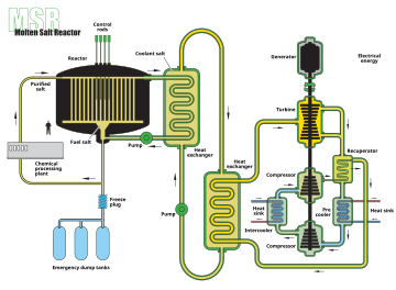
    Example of a molten salt reactor scheme

    A molten salt reactor (MSR) is a class of generation IV nuclear fission reactor in which the primary nuclear reactor coolant, or even the fuel itself, is a molten salt mixture. MSRs can run at higher temperatures than water-cooled reactors for a higher thermodynamic efficiency, while staying at low vapour pressure.

    The nuclear fuel may be solid or dissolved in the coolant. In many designs the nuclear fuel dissolved in the coolant is uranium tetrafluoride (UF4). The fluid becomes critical in a graphite core that serves as the moderator. Some solid-fuel designs propose ceramic fuel dispersed in a graphite matrix, with the molten salt providing low pressure, high temperature cooling. The salts are much more efficient than compressed helium (another potential coolant in Generation IV reactor designs) at removing heat from the core, reducing the need for pumping and piping and reducing the core size.

    The concept was established in the 1950s. The early Aircraft Reactor Experiment was primarily motivated by the small size that the design could provide, while the Molten-Salt Reactor Experiment was a prototype for a thorium fuel cycle breeder nuclear power plant. The increased research into Generation IV reactor designs included a renewed interest in the technology.[2]

    History

    Aircraft reactor experiment

    Aircraft Reactor Experiment building at ORNL. It was later retrofitted for the MSRE.

    Extensive research into molten salt reactors started with the U.S. aircraft reactor experiment (ARE) in support of the U.S. Aircraft Nuclear Propulsion program. The ARE was a 2.5 MWth nuclear reactor experiment designed to attain a high energy density for use as an engine in a nuclear-powered bomber.

    The project included experiments, including high temperature reactor and engine tests collectively called the Heat Transfer Reactor Experiments: HTRE-1, HTRE-2 and HTRE-3 at the National Reactor Test Station (now Idaho National Laboratory) as well as an experimental high-temperature molten salt reactor at Oak Ridge National Laboratory – the ARE.

    The ARE used molten fluoride salt NaF-ZrF4-UF4 (53-41-6 mol%) as fuel, moderated by beryllium oxide (BeO). Liquid sodium was a secondary coolant.

    The experiment had a peak temperature of 860 °C. It produced 100 MWh over nine days in 1954. This experiment used Inconel 600 alloy for the metal structure and piping.[3]

    After ARE, another reactor was operated at the Critical Experiments Facility of the Oak Ridge National Laboratory in 1957. It was part of the circulating-fuel reactor program of the Pratt & Whitney Aircraft Company (PWAC). This was called the PWAR-1, the Pratt and Whitney Aircraft Reactor-1. The experiment was run for only a few weeks and at essentially zero nuclear power, but it reached criticality. The operating temperature was held constant at approximately 675 °C (1,250 °F). The PWAR-1 used NaF-ZrF4-UF4 as the primary fuel and coolant, making it one of the three critical molten salt reactors ever built.[4]

    Molten-salt reactor experiment

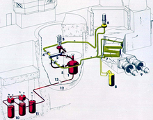
    MSRE plant diagram

    Oak Ridge National Laboratory (ORNL) took the lead in researching the MSR through the 1960s. Much of their work culminated with the Molten-Salt Reactor Experiment (MSRE). The MSRE was a 7.4 MWth test reactor simulating the neutronic "kernel" of a type of epithermal thorium molten salt breeder reactor called the liquid fluoride thorium reactor. The large (expensive) breeding blanket of thorium salt was omitted in favor of neutron measurements.  The MSRE was located at ORNL. Its piping, core vat and structural components were made from Hastelloy-N, moderated by pyrolytic graphite. It went critical in 1965 and ran for four years. The fuel for the MSRE was LiF-BeF2-ZrF4-UF4 (65-29-5-1). The graphite core moderated it. Its secondary coolant was FLiBe (2LiF-BeF2). It reached temperatures as high as 650 °C and operated for the equivalent of about 1.5 years of full power operation.

    Oak Ridge National Laboratory molten salt breeder reactor

    The culmination of the Oak Ridge National Laboratory research during the 1970–1976 timeframe resulted in a proposed molten salt breeder reactor (MSBR) design which would use LiF-BeF2-ThF4-UF4 (72-16-12-0.4) as fuel. It was to be moderated by graphite with a 4-year replacement schedule. The secondary coolant was to be NaF-NaBF4. Its peak operating temperature was to be 705 °C.[5] Despite the success, the MSR program closed down in the early 1970s in favor of the liquid metal fast-breeder reactor (LMFBR),[6] after which research stagnated in the United States.[7][8] As of 2011, the ARE and the MSRE remained the only molten-salt reactors ever operated.

    The MSBR project received funding until 1976. Inflation-adjusted to 1991 dollars, the project received $38.9 million from 1968 to 1976.[9]

    Officially, the program was cancelled because:

    • The political and technical support for the program in the United States was too thin geographically. Within the United States, only in Oak Ridge, Tennessee, was the technology well understood.[6]
    • The MSR program was in competition with the fast breeder program at the time, which got an early start and had copious government development funds allocated to many parts of the United States. When the MSR development program had progressed far enough to justify an expanded program leading to commercial development, the AEC could not justify the diversion of substantial funds from the LMFBR to a competing program.[6]

    Oak Ridge National Laboratory denatured molten salt reactor (DMSR)

    In 1980, the engineering technology division at Oak Ridge National Laboratory published a paper entitled "Conceptual Design Characteristics of a Denatured Molten-Salt Reactor with Once-Through Fueling." In it, the authors "examine the conceptual feasibility of a molten-salt power reactor fueled with denatured uranium-235 (i.e. with low-enriched uranium) and operated with a minimum of chemical processing." The main priority behind the design characteristics is proliferation resistance.[10] Lessons learned from past projects and research at ORNL were considered. Although the DMSR can theoretically be fueled partially by thorium or plutonium, fueling solely with low enriched uranium (LEU) helps maximize proliferation resistance.

    Another important goal of the DMSR was to minimize R&D and to maximize feasibility. The Generation IV international Forum (GIF) includes "salt processing" as a technology gap for molten salt reactors.[11] The DMSR requires minimal chemical processing because it is a burner rather than a breeder. Both reactors built at ORNL were burner designs. In addition, the choices to use graphite for neutron moderation and enhanced Hastelloy-N for piping simplify the design and reduce R&D.

    United Kingdom

    The UK's Atomic Energy Research Establishment (AERE) were developing an alternative MSR design across its National Laboratories at Harwell, Culham, Risley and Winfrith. AERE opted to focus on a lead-cooled 2.5 GWe Molten Salt Fast Reactor (MSFR) concept using a chloride.[12] They also researched the option of helium gas as an alternative coolant.

    The UK MSFR would be fuelled by plutonium, a fuel considered to be 'free' by the program's research scientists, because of the UK's plutonium stockpile.

    Despite their different designs, ORNL and AERE maintained contact during this period with information exchange and expert visits. Theoretical work on the concept was conducted between 1964 and 1966, while experimental work was ongoing between 1968 and 1973. The program received annual government funding of around £100,000-£200,000 (equivalent to £2m-£3m in 2005). This funding came to an end in 1974, partly due to the success of the Prototype Fast Reactor at Dounreay which was considered a priority for funding as it went critical in the same year.[12]

    AERE reports and findings from its MSR Program conducted in the 1960s and 1970s are available for public viewing at the UK National Archives in Kew, London.[12]

    Soviet Union

    In the USSR, a molten-salt reactor research program was started in the second half of the 1970s at the Kurchatov Institute. It included theoretical and experimental studies, particularly the investigation of mechanical, corrosion and radiation properties of the molten salt container materials. The main findings supported the conclusion that there were no physical nor technological obstacles to the practical implementation of MSRs.[15] A reduction in activity occurred after 1986 due to the Chernobyl accident, along with a general stagnation of nuclear power and the nuclear industry.[16](p381)

    Twenty-first century

    Canada

    Terrestrial Energy Inc. (TEI), a Canadian-based company, is developing a DMSR design called the Integral Molten Salt Reactor (IMSR). The IMSR is designed to be deployable as a small modular reactor (SMR). Their design currently undergoing licensing is 400MW thermal (190MW electrical). With high operating temperatures, the IMSR has applications in industrial heat markets as well as traditional power markets. The main design features include neutron moderation from graphite, fueling with low-enriched uranium and a compact and replaceable Core-unit. Decay heat is removed passively using nitrogen (with air as an emergency alternative). The latter feature permits the operational simplicity necessary for industrial deployment.[17]

    Terrestrial has completed the first phase of a prelicensing review by the Canadian Nuclear Safety Commission, which provides a regulatory opinion that the design features are generally safe enough to eventually obtain a license to construct the reactor.[18]

    China

    Under Jiang Mianheng's direction, China initiated a thorium molten-salt reactor research project. It was formally announced at the Chinese Academy of Sciences (CAS) annual conference in January 2011.[19] A 100-MW demonstrator of the solid fuel version (TMSR-SF), based on pebble bed technology, was to be ready by 2024. Initially, a 10-MW pilot and a larger demonstrator of the liquid fuel (TMSR-LF) variant were targeted for 2024 and 2035 respectively.[20][21] However, China has accelerated its program to build two 12 MW reactors underground at Wuwei research facilities in Gansu Province by 2020.[22] Heat from the reaction will be used to produce electricity, hydrogen, industrial chemicals, drinking water through desalination, and minerals.[22] The project also seeks to test new corrosion resistant materials.[22]

    In 2017, ANSTO/Shanghai Institute Of Applied Physics announced the creation of a NiMo-SiC alloy for use in molten salt reactors.[23][24]

    Denmark

    Seaborg Technologies, a company based in Denmark, is developing the core for a Molten Salt Waste-burner (MSW). The MSW is a high temperature, single salt, thermal MSR designed to go critical on a combination of thorium and nuclear waste from conventional nuclear reactors. The MSW design is modular. The reactor core is estimated to be replaced every 6–10 years. However, the fuel will not be replaced and will burn for the entire power plant lifetime. The first version of the Seaborg core is planned to produce 50 MWth power and could consume approximately 1 ton (not considering natural decays) of transuranic waste over its 60 years power plant lifetime. After 60 years the 233U concentration in the fuel salt is high enough to initiate a closed thorium fuel cycle in the next generation power plant.[25]

    France

    The CNRS project EVOL (Evaluation and viability of liquid fuel fast reactor system) project, with the objective of proposing a design of the MSFR (Molten Salt Fast Reactor),[26] released its final report in 2014.[27] The various molten salt reactor projects like FHR, MOSART, MSFR, and TMSR have common themes in basic R&D areas, according to a 2014 paper giving an overview of the MSR in a GenV context.[28] Another paper gives an overview of the MSFR.[29] More resources are available in the MSFR bibliography.[30]

    The EVOL project will be continued by the EU-funded SAMOFAR (Safety Assessment of the Molten Salt Fast Reactor) project, in which several European research institutes and universities collaborate[31].

    India

    Ratan Kumar Sinha, Chairman of Atomic Energy Commission of India, stated in 2013: "India is also investigating Molten Salt Reactor (MSR) technology. We have molten salt loops operational at BARC."[32]

    Japan

    The Fuji Molten Salt Reactor is a 100 to 200 MWe LFTR, using technology similar to the Oak Ridge project. A consortium including members from Japan, the U.S. and Russia are developing the project. The project would likely take 20 years to develop a full size reactor,[33] but the project seems to lack funding.[34]

    United Kingdom

    The Alvin Weinberg Foundation is a British non-profit organization founded in 2011, dedicated to raising awareness about the potential of thorium energy and LFTR. It was formally launched at the House of Lords on 8 September 2011.[35][36][37] It is named after American nuclear physicist Alvin M. Weinberg, who pioneered thorium molten salt reactor research.

    A study on MSRs completed in July 2015 by Energy Process Developments, funded by Innovate UK, summarizes MSR activity internationally. It looks at the feasibility of developing a pilot scale demonstration MSR in the UK. A review of potential UK sites is given along with an insight into the UK regulatory process for innovative reactor technology. The technical review of six MSR designs led to the selection of the Stable Salt Reactor, designed by Moltex Energy, as most suitable for UK implementation.[38] However, Moltex "failed to get engagement from the UK government quickly enough" and so will target Canada instead.[39]

    United States

    Idaho National Laboratory designed a molten-salt-cooled, molten-salt-fuelled reactor with a prospective output of 1000 MWe.[40]

    Kirk Sorensen, former NASA scientist and chief nuclear technologist at Teledyne Brown Engineering, is a long-time promoter of the thorium fuel cycle, coining the term liquid fluoride thorium reactor. In 2011, Sorensen founded Flibe Energy, a company aimed at developing 20–50 MW LFTR reactor designs to power military bases. (It is easier to approve novel military designs than civilian power station designs in today's US nuclear regulatory environment).

    Transatomic Power was created by Ph.D. students from MIT including CEO Leslie Dewan and Mark Massie, and Russ Wilcox of E Ink.[45] They are pursuing what they term a Waste-Annihilating Molten Salt Reactor (acronym WAMSR), intending to consume existing spent nuclear fuel.[46] Transatomic received venture capital funding in early 2015.[47]

    In January 2016, the United States Department of Energy announced a $80m award fund to develop Generation IV reactor designs.[48] One of the two beneficiaries, Southern Company will use the funding to develop a Molten Chloride Fast Reactor (MCFR), a type of MSR developed earlier by British scientists.[12]

    Variants

    Liquid-salt very-high-temperature reactor

    It is essentially a standard VHTR design that uses liquid salt as a coolant in the primary loop, rather than a single helium loop. It relies on "TRISO" fuel dispersed in graphite. Early AHTR research focused on graphite would be in the form of graphite rods that would be inserted in hexagonal moderating graphite blocks, but current studies focus primarily on pebble-type fuel. The LS-VHTR has many attractive features, including the ability to work at very high temperatures (the boiling point of most molten salt candidates is >1400 °C); low-pressure cooling that can be used to more easily match hydrogen production facility conditions (most thermochemical cycles require temperatures in excess of 750 °C); better electric conversion efficiency than a helium-cooled VHTR operating at similar conditions; passive safety systems and better retention of fission products in the event of an accident.[citation needed] This concept is now referred to as "fluoride salt-cooled high-temperature reactor" (FHR).[49]

    Liquid fluoride thorium reactor (LFTR)

    Advocates estimate that five hundred metric tons of thorium could supply all U.S. energy needs for one year.[50] The U.S. Geological Survey estimates that the largest known U.S. thorium deposit, the Lemhi Pass district on the Montana-Idaho border, contains thorium reserves of 64,000 metric tons.[51]

    Molten-salt fueling options

    The LFTR design was strongly supported by Alvin Weinberg, who patented the light-water reactor and was a director of the U.S.'s Oak Ridge National Laboratory. In 2016 Nobel prize winning physicist Carlo Rubbia, former Director General of CERN, claimed that one of the main reasons why research was cut is that thorium is difficult to turn into a nuclear weapon.[52]
    Thorium is not for tomorrow but unless you do any development, it will not get there. — Dr Carlo Rubbia, Nobel Laureate and former Director General of CERN, January 2016[52]
    Alternatives to thorium include enriched uranium-235 or fissile material from dismantled nuclear weapons.[53]

    Molten-salt-cooled reactors

    Molten-salt-cooled solid-fuel reactors are quite different from molten-salt-fueled reactors. They are called "molten salt reactor system" in the Generation IV proposal, also called Molten Salt Converter Reactor (MSCR). These reactors were additionally referred to as advanced high-temperature reactors (AHTRs), but since about 2010 the preferred DOE designation is fluoride high-temperature reactors (FHR).[54]

    The FHR concept cannot reprocess fuel easily and has fuel rods that need to be fabricated and validated, delaying deployment by up to twenty years[citation needed] from project inception. However, since it uses fabricated fuel, reactor manufacturers can still profit by selling fuel assemblies.

    The FHR retains the safety and cost advantages of a low-pressure, high-temperature coolant, also shared by liquid metal cooled reactors. Notably, steam is not created in the core (as is present in BWRs), and no large, expensive steel pressure vessel (as required for PWRs). Since it can operate at high temperatures, the conversion of the heat to electricity can use an efficient, lightweight Brayton cycle gas turbine.

    Much of the current research on FHRs is focused on small, compact heat exchangers that reduce molten salt volumes and associated costs.[55]

    Molten salts can be highly corrosive and corrosivity increases with temperature. For the primary cooling loop, a material is needed that can withstand corrosion at high temperatures and intense radiation. Experiments show that Hastelloy-N and similar alloys are suited to these tasks at operating temperatures up to about 700 °C. However, operating experience is limited. Still higher operating temperatures are desirable – at 850 °C thermochemical production of hydrogen becomes possible. Materials for this temperature range have not been validated, though carbon composites, molybdenum alloys (e.g. TZM), carbides, and refractory metal based or ODS alloys might be feasible.

    Dual-fluid molten salt reactors

    A prototypical example of a dual fluid reactor is the lead-cooled, salt-fueled reactor.

    Fused salt selection

    Molten FLiBe

    The salt mixtures are chosen to make the reactor safer and more practical. Fluoride salts are favored, because fluorine has only one stable isotope (F-19), and does not easily become radioactive under neutron bombardment. Both of these make fluorine better than chlorine, which has two stable isotopes (Cl-35 and Cl-37), as well as a slow-decaying isotope between them which facilitates neutron absorption by Cl-35. Compared to chlorine and other halides, fluorine also absorbs fewer neutrons and slows ("moderates") neutrons better. Low-valence fluorides boil at high temperatures, though many pentafluorides and hexafluorides boil at low temperatures. They also must be very hot before they break down into their constituent elements. Such molten salts are "chemically stable" when maintained well below their boiling points.

    On the other hand, some salts are so useful that isotope separation of the halide is worthwhile. Chlorides permit fast breeder reactors to be constructed using molten salts. Much less research has been done on reactor designs using chloride salts. Chlorine, unlike fluorine, must be purified to isolate the heavier stable isotope, chlorine-37, thus reducing production of sulfur tetrafluoride that occurs when chlorine-35 absorbs a neutron to become chlorine-36, then degrades by beta decay to sulfur-36.

    Similarly, any lithium present in a salt mixture must be in the form of purified lithium-7, because lithium-6 effectively captures neutrons and produces tritium. Even if pure 7Li is used, salts containing lithium will cause significant tritium production, comparable with heavy water reactors.

    Reactor salts are usually close to eutectic mixtures to reduce their melting point. A low melting point simplifies melting the salt at startup and reduces the risk of the salt freezing as it is cooled in the heat exchanger.

    Due to the high "redox window" of fused fluoride salts, the redox potential of the fused salt system can be changed. Fluorine-Lithium-Beryllium ("FLiBe") can be used with beryllium additions to lower the redox potential and almost eliminate corrosion. However, since beryllium is extremely toxic, special precautions must be engineered into the design to prevent its release into the environment. Many other salts can cause plumbing corrosion, especially if the reactor is hot enough to make highly reactive hydrogen.

    To date, most research has focused on FLiBe, because lithium and beryllium are reasonably effective moderators and form a eutectic salt mixture with a lower melting point than each of the constituent salts. Beryllium also performs neutron doubling, improving the neutron economy. This process occurs when the beryllium nucleus re-emits two neutrons after absorbing a single neutron. For the fuel carrying salts, generally 1% or 2% (by mole) of UF4 is added. Thorium and plutonium fluorides have also been used.

    Comparison of the neutron capture and moderating efficiency of several materials. Red are Be-bearing, blue are ZrF4-bearing and green are LiF-bearing salts.[56]
    Material Total neutron capture
    relative to graphite
    (per unit volume)
    Moderating ratio
    (Avg. 0.1 to 10 eV)
    Heavy water 0.2 11449
    ZrH[57][58][59] ~0.2 ~0 if <0 .14="" ev="" if="">0.14 eV
    Light water 75 246
    Graphite 1 863
    Sodium 47 2
    UCO 285 2
    UO2 3583 0.1
    2LiF–BeF2 8 60
    LiF–BeF2–ZrF4 (64.5–30.5–5) 8 54
    NaF–BeF2 (57–43) 28 15
    LiF–NaF–BeF2 (31–31–38) 20 22
    LiF–ZrF4 (51–49) 9 29
    NaF–ZrF4 (59.5–40.5) 24 10
    LiF-NaF–ZrF4 (26–37–37) 20 13
    KF–ZrF4 (58–42) 67 3
    RbF–ZrF4 (58–42) 14 13
    LiF–KF (50–50) 97 2
    LiF–RbF (44–56) 19 9
    LiF–NaF–KF (46.5–11.5–42) 90 2
    LiF–NaF–RbF (42–6–52) 20 8

    Fused salt purification

    Techniques for preparing and handling molten salt were first developed at Oak Ridge National Lab.[60] The purpose of salt purification was to eliminate oxides, sulfur and metal impurities. Oxides could result in the deposition of solid particles in reactor operation. Sulfur had to be removed because of its corrosive attack on nickel-based alloys at operational temperature. Structural metal such as chromium, nickel, and iron had to be removed for corrosion control.

    A water content reduction purification stage using HF and helium sweep gas was specified to run at 400 °C. Oxide and sulfur contamination in the salt mixtures were removed using gas sparging of HF – H2 mixture, with the salt heated to 600 °C.[60](p8) Structural metal contamination in the salt mixtures were removed using hydrogen gas sparging, at 700 °C.[60](p26) Solid ammonium hydrofluoride was proposed as a safer alternative for oxide removal.[61]

    Fused salt processing

    The possibility of online processing can be an MSR advantage. Continuous processing would reduce the inventory of fission products, control corrosion and improve neutron economy by removing fission products with high neutron absorption cross-section, especially xenon. This makes the MSR particularly suited to the neutron-poor thorium fuel cycle. Online fuel processing can introduce risks of fuel processing accidents,[62](p15) which can trigger release of radio isotopes.

    In some thorium breeding scenarios, the intermediate product protactinium-233 would be removed from the reactor and allowed to decay into highly pure uranium-233, an attractive bomb-making material. More modern designs propose to use a lower specific power or a separate large thorium breeding blanket. This dilutes the protactinium to such an extent that few protactinium atoms absorb a second neutron or, via a (n, 2n) reaction (in which an incident neutron is not absorbed but instead knocks a neutron out of the nucleus), generate uranium-232. Because U-232 has a short half-life and its decay chain contains hard gamma emitters, it makes the isotopic mix of uranium less attractive for bomb-making. This benefit would come with the added expense of a larger fissile inventory or a 2-fluid design with a large quantity of blanket salt.

    The necessary fuel salt reprocessing technology has been demonstrated, but only at laboratory scale. A prerequisite to full-scale commercial reactor design is the R&D to engineer an economically competitive fuel salt cleaning system.

    Fissile fuel reprocessing issues

    Changes in the composition of a MSR fast neutron (kg/GW)

    Reprocessing refers to the chemical separation of fissionable uranium and plutonium from spent nuclear fuel.[63] The recovery of uranium or plutonium could increase the risk of nuclear proliferation. In the United States the regulatory regime has varied dramatically in different administrations.[63]

    In the original 1971 Molten Salt Breeder Reactor proposal, uranium reprocessing was scheduled every ten days as part of reactor operation.[64](p181) Subsequently, a once-through fueling design was proposed that limited uranium reprocessing to every thirty years at the end of useful salt life.[65](p98) A mixture of uranium-238 was called for to make sure recovered uranium would not be weapons grade. This design is referred to as denatured molten salt reactor.[66] If reprocessing were to be prohibited then the uranium would be disposed with other fission products.

    Comparison to light water reactors

    MSRs, especially those with the fuel dissolved in the salt differ considerably from conventional reactors. Reactor core pressure can be low and the temperature much higher. In this respect an MSR is more similar to a liquid metal cooled reactor than to a conventional light water cooled reactor. MSRs are often planned as breeding reactors with a closed fuel cycle – as opposed to the once-through fuel currently used in U.S. nuclear reactors.

    Safety concepts rely on a negative temperature coefficient of reactivity and a large possible temperature rise to limit reactivity excursions. As an additional method for shutdown, a separate, passively cooled container below the reactor can be included. In case of problems and for regular maintenance the fuel is drained from the reactor. This stops the nuclear reaction and acts as another second cooling system. Neutron-producing accelerators have been proposed for some super-safe subcritical experimental designs.[67]

    Cost estimates from the 1970s were slightly lower than for conventional light-water reactors.[68]

    The temperatures of some proposed designs are high enough to produce process heat for hydrogen production or other chemical reactions. Because of this, they are included in the GEN-IV roadmap for further study.[69]
    Advantages

    MSR offers many potential advantages over current light water reactors:[5]
     
    • Inherently safe design (safety by passive components and the strong negative temperature coefficient of reactivity of some designs). In some designs, the fuel and the coolant are the same fluid, so a loss of coolant removes the reactor's fuel. Unlike steam, fluoride salts dissolve poorly in water, and do not form burnable hydrogen. Unlike steel and solid uranium oxide, molten salts are not damaged by the core's neutron bombardment.
    • A low-pressure MSR lacks a LWR's high-pressure radioactive steam and therefore do not experience leaks of radioactive steam and cooling water, and the expensive containment, steel core vessel, piping and safety equipment needed to contain radioactive steam.
    • MSRs make closed nuclear fuel cycles cheaper and more practical. If fully implemented, a closed nuclear fuel cycle reduces environmental impacts: The chemical separation makes long-lived actinides back into reactor fuel. The discharged wastes are mostly fission products (nuclear ashes) with short half-lives. This reduces the needed geologic containment to 300 years rather than the tens of thousands of years needed by a light-water reactor's spent nuclear fuel. It also permits society to use more-abundant nuclear fuels.
    • The fuel's liquid phase might be pyroprocessed to separate fission products (nuclear ashes) from actinide fuels. This may have advantages over conventional reprocessing, though much development is still needed.
    • Fuel rods are not required.
    • In new solid-fueled reactor designs, the longest-lead item is the safety testing of fuel element designs. Fuel tests usually must cover several three-year refueling cycles. However, several molten salt fuels have already been validated.
    • Some designs can "burn" problematic transuranic elements from traditional solid-fuel nuclear reactors.
    • An MSR can react to load changes in less than 60 seconds (unlike "traditional" solid-fuel nuclear power plants that suffer from xenon poisoning).
    • Molten salt reactors can run at high temperatures, yielding high production efficiency. This reduces the size, expense and environmental impacts of a power plant.
    • MSRs can offer a high "specific power," that is high power at a low mass as demonstrated by the ARE.[3] Simplified MSR power plants may be suitable for ships.
    • A possibly good neutron economy makes the MSR attractive for the neutron poor thorium fuel cycle.
    • LWR's (and most other solid-fuel reactors) have no fundamental "off switch", but once the initial criticality is overcome, an MSR is comparatively easy and fast to turn off by letting the freeze plug melt.

    Disadvantages

  • Little development compared to most Gen IV designs .
  • Required onsite chemical plant to manage core mixture and remove fission products.
  • Required regulatory changes to deal with radically different design features.
  • MSR designs rely on nickel-based alloys to hold the molten salt. Alloys based on nickel and iron are prone to embrittlement under high neutron flux.[65](p83)
  • Corrosion risk.[70]
  • As a breeder reactor, a modified MSR might be able to produce weapons-grade nuclear material.[71]
  • The MSRE and aircraft nuclear reactors used enrichment levels so high that they approach the levels of nuclear weapons. These levels would be illegal in most modern regulatory regimes for power plants. Some modern designs avoid this issue.[72]
  • Neutron damage to solid moderator materials can limit the core lifetime of an MSR that makes moderately fast neutrons. For example, the MSRE was designed so that its graphite moderator sticks had very low tolerances, so neutron damage could change their size without damage. "Two fluid" MSR designs are unable to use graphite piping because graphite changes size when it is bombarded with neutrons, and graphite pipes would crack and leak.

Optogenetics

From Wikipedia, the free encyclopedia
 
Optogenetics (from Greek optikós, meaning 'seen, visible') is a biological technique which involves the use of light to control cells in living tissue, typically neurons, that have been genetically modified to express light-sensitive ion channels. It is a neuromodulation method that uses a combination of techniques from optics and genetics to control and monitor the activities of individual neurons in living tissue—even within freely-moving animals—and to precisely measure these manipulation effects in real-time. The key reagents used in optogenetics are light-sensitive proteins. Neuronal control is achieved using optogenetic actuators like channelrhodopsin, halorhodopsin, and archaerhodopsin, while optical recording of neuronal activities can be made with the help of optogenetic sensors for calcium (GCaMP), vesicular release (synapto-pHluorin), neurotransmitter (GluSnFRs), or membrane voltage (arc lightning, ASAP1). Control (or recording) of activity is restricted to genetically defined neurons and performed in a spatiotemporal-specific manner by light.

In 2010, optogenetics was chosen as the "Method of the Year" across all fields of science and engineering by the interdisciplinary research journal Nature Methods. At the same time, optogenetics was highlighted in the article on "Breakthroughs of the Decade" in the academic research journal Science.[5] These journals also referenced recent public-access general-interest video Method of the year video and textual SciAm summaries of optogenetics.

History

The "far-fetched" possibility of using light for selectively controlling precise neural activity (action potential) patterns within subtypes of cells in the brain was thought of by Francis Crick in his Kuffler Lectures at the University of California in San Diego in 1999.[6] An earlier use of light to activate neurons was carried out by Richard Fork,[7] who demonstrated laser activation of neurons within intact tissue, although not in a genetically-targeted manner. The earliest genetically targeted method that used light to control rhodopsin-sensitized neurons was reported in January 2002, by Boris Zemelman (now at UT Austin) and Gero Miesenböck, who employed Drosophila rhodopsin cultured mammalian neurons.[8] In 2003, Zemelman and Miesenböck developed a second method for light-dependent activation of neurons in which single inotropic channels TRPV1, TRPM8 and P2X2 were gated by photocaged ligands in response to light.[9] Beginning in 2004, the Kramer and Isacoff groups developed organic photoswitches or "reversibly caged" compounds in collaboration with the Trauner group that could interact with genetically introduced ion channels.[10][11] TRPV1 methodology, albeit without the illumination trigger, was subsequently used by several laboratories to alter feeding, locomotion and behavioral resilience in laboratory animals.[12][13][14] However, light-based approaches for altering neuronal activity were not applied outside the original laboratories, likely because the easier to employ channelrhodopsin was cloned soon thereafter.[15]
Peter Hegemann, studying the light response of green algae at the University of Regensburg, had discovered photocurrents that were too fast to be explained by the classic g-protein-coupled animal rhodopsins.[16] Teaming up with the electrophysiologist Georg Nagel at the Max Planck Institute in Frankfurt, they could demonstrate that a single gene from the alga Chlamydomonas produced large photocurents when expressed in the oocyte of a frog.[17] To identify expressing cells, they replaced the cytoplasmic tail of the algal protein with the fluorescent protein YFP, generating the first generally applicable optogenetic tool.[15] Zhuo-Hua Pan of Wayne State University, researching on restore sight to blindness, thought about using channelrhodopsin when it came out in late 2003. By February 2004, he was trying channelrhodopsin out in ganglion cells—the neurons in our eyes that connect directly to the brain—that he had cultured in a dish. Indeed, the transfected neurons became electrically active in response to light.[18] In April 2005, Susana Lima and Miesenböck reported the first use of genetically-targeted P2X2 photostimulation to control the behaviour of an animal.[19] They showed that photostimulation of genetically circumscribed groups of neurons, such as those of the dopaminergic system, elicited characteristic behavioural changes in fruit flies. In August 2005, Karl Deisseroth's laboratory in the Bioengineering Department at Stanford including graduate students Ed Boyden and Feng Zhang (both now at MIT) published the first demonstration of a single-component optogenetic system in cultured mammalian neurons,[20][21] using the channelrhodopsin-2(H134R)-eYFP construct from Nagel and Hegemann.[15] The groups of Gottschalk and Nagel were first to use channelrhodopsin-2 for controlling neuronal activity in an intact animal, showing that motor patterns in the roundworm Caenorhabditis elegans could be evoked by light stimulation of genetically selected neural circuits (published in December 2005).[22] In mice, controlled expression of optogenetic tools is often achieved with cell-type-specific Cre/loxP methods developed for neuroscience by Joe Z. Tsien back in the 1990s[23] to activate or inhibit specific brain regions and cell-types in vivo.[24]

The primary tools for optogenetic recordings have been genetically encoded calcium indicators (GECIs). The first GECI to be used to image activity in an animal was cameleon, designed by Atsushi Miyawaki, Roger Tsien and coworkers.[25] Cameleon was first used successfully in an animal by Rex Kerr, William Schafer and coworkers to record from neurons and muscle cells of the nematode C. elegans.[26] Cameleon was subsequently used to record neural activity in flies[27] and zebrafish.[28] In mammals, the first GECI to be used in vivo was GCaMP,[29] first developed by Nakai and coworkers.[30] GCaMP has undergone numerous improvements, and GCaMP6[31] in particular has become widely used throughout neuroscience.

In 2010, Karl Deisseroth at Stanford University was awarded the inaugural HFSP Nakasone Award "for his pioneering work on the development of optogenetic methods for studying the function of neuronal networks underlying behavior". In 2012, Gero Miesenböck was awarded the InBev-Baillet Latour International Health Prize for "pioneering optogenetic approaches to manipulate neuronal activity and to control animal behaviour." In 2013, Ernst Bamberg, Ed Boyden, Karl Deisseroth, Peter Hegemann, Gero Miesenböck and Georg Nagel were awarded The Brain Prize for "their invention and refinement of optogenetics."[32][33] Karl Deisseroth was awarded the Else Kröner Fresenius Research Prize 2017 (4 million euro) for his "contributions to the understanding of the biological basis of psychiatric disorders".

Description

Fig 1. Channelrhodopsin-2 (ChR2) induces temporally precise blue light-driven activity in rat prelimbic prefrontal cortical neurons. a) In vitro schematic (left) showing blue light delivery and whole-cell patch-clamp recording of light-evoked activity from a fluorescent CaMKllα::ChR2-EYFP expressing pyramidal neuron (right) in an acute brain slice. b) In vivo schematic (left) showing blue light (473 nm) delivery and single-unit recording. (bottom left) Coronal brain slice showing expression of CaMKllα::ChR2-EYFP in the prelimbic region. Light blue arrow shows tip of the optical fiber; black arrow shows tip of the recording electrode (left). White bar, 100 µm. (bottom right) In vivo light recording of prefrontal cortical neuron in a transduced CaMKllα::ChR2-EYFP rat showing light-evoked spiking to 20 Hz delivery of blue light pulses (right). Inset, representative light-evoked single-unit response.[34]
 
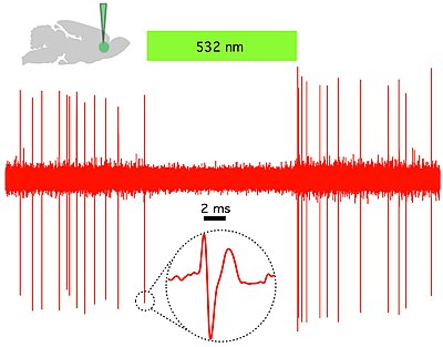
Fig 2. Halorhodopsin (NpHR) rapidly and reversibly silences spontaneous activity in vivo in rat prelimbic prefrontal cortex. (Top left) Schematic showing in vivo green (532 nm) light delivery and single- unit recording of a spontaneously active CaMKllα::eNpHR3.0- EYFP expressing pyramidal neuron. (Right) Example trace showing that continuous 532 nm illumination inhibits single-unit activity in vivo. Inset, representative single unit event; Green bar, 10 seconds.[34]
 
A nematode expressing the light-sensitive ion channel Mac. Mac is a proton pump originally isolated in the fungus Leptosphaeria maculans and now expressed in the muscle cells of C. elegans that opens in response to green light and causes hyperpolarizing inhibition. Of note is the extension in body length that the worm undergoes each time it is exposed to green light, which is presumably caused by Mac's muscle-relaxant effects.[35]
 
A nematode expressing ChR2 in its gubernacular-oblique muscle group responding to stimulation by blue light. Blue light stimulation causes the gubernacular-oblique muscles to repeatedly contract, causing repetitive thrusts of the spicule, as would be seen naturally during copulation.[36]
 
Optogenetics provides millisecond-scale temporal precision which allows the experimenter to keep pace with fast biological information processing (for example, in probing the causal role of specific action potential patterns in defined neurons). Indeed, to probe the neural code, optogenetics by definition must operate on the millisecond timescale to allow addition or deletion of precise activity patterns within specific cells in the brains of intact animals, including mammals (see Figure 1). By comparison, the temporal precision of traditional genetic manipulations (employed to probe the causal role of specific genes within cells, via "loss-of-function" or "gain of function" changes in these genes) is rather slow, from hours or days to months. It is important to also have fast readouts in optogenetics that can keep pace with the optical control. This can be done with electrical recordings ("optrodes") or with reporter proteins that are biosensors, where scientists have fused fluorescent proteins to detector proteins. An example of this is voltage-sensitive fluorescent protein (VSFP2).[37] Additionally, beyond its scientific impact optogenetics represents an important case study in the value of both ecological conservation (as many of the key tools of optogenetics arise from microbial organisms occupying specialized environmental niches), and in the importance of pure basic science as these opsins were studied over decades for their own sake by biophysicists and microbiologists, without involving consideration of their potential value in delivering insights into neuroscience and neuropsychiatric disease.[38]

Light-activated proteins: channels, pumps and enzymes

The hallmark of optogenetics therefore is introduction of fast light-activated channels, pumps, and enzymes that allow temporally precise manipulation of electrical and biochemical events while maintaining cell-type resolution through the use of specific targeting mechanisms. Among the microbial opsins which can be used to investigate the function of neural systems are the channelrhodopsins (ChR2, ChR1, VChR1, and SFOs) to excite neurons and anion-conducting channelrhodopsins for light-induced inhibition. Light-driven ion pumps are also used to inhibit neuronal activity, e.g. halorhodopsin (NpHR),[39] enhanced halorhodopsins (eNpHR2.0 and eNpHR3.0, see Figure 2),[40] archaerhodopsin (Arch), fungal opsins (Mac) and enhanced bacteriorhodopsin (eBR).[41]

Optogenetic control of well-defined biochemical events within behaving mammals is now also possible. Building on prior work fusing vertebrate opsins to specific G-protein coupled receptors[42] a family of chimeric single-component optogenetic tools was created that allowed researchers to manipulate within behaving mammals the concentration of defined intracellular messengers such as cAMP and IP3 in targeted cells.[43] Other biochemical approaches to optogenetics (crucially, with tools that displayed low activity in the dark) followed soon thereafter, when optical control over small GTPases and adenylyl cyclases was achieved in cultured cells using novel strategies from several different laboratories.[44][45][46][47][48] This emerging repertoire of optogenetic probes now allows cell-type-specific and temporally precise control of multiple axes of cellular function within intact animals.[49]

Hardware for light application

Another necessary factor is hardware (e.g. integrated fiberoptic and solid-state light sources) to allow specific cell types, even deep within the brain, to be controlled in freely behaving animals. Most commonly, the latter is now achieved using the fiberoptic-coupled diode technology introduced in 2007,[50][51][52] though to avoid use of implanted electrodes, researchers have engineered ways to inscribe a "window" made of zirconia that has been modified to be transparent and implanted in mice skulls, to allow optical waves to penetrate more deeply to stimulate or inhibit individual neurons.[53] To stimulate superficial brain areas such as the cerebral cortex, optical fibers or LEDs can be directly mounted to the skull of the animal. More deeply implanted optical fibers have been used to deliver light to deeper brain areas. Complementary to fiber-tethered approaches, completely wireless techniques have been developed utilizing wirelessly delivered power to headborne LEDs for unhindered study of complex behaviors in freely behaving organisms.[54]

Expression of optogenetic actuators

Optogenetics also necessarily includes the development of genetic targeting strategies such as cell-specific promoters or other customized conditionally-active viruses, to deliver the light-sensitive probes to specific populations of neurons in the brain of living animals (e.g. worms, fruit flies, mice, rats, and monkeys). In invertebrates such as worms and fruit flies some amount of all-trans-retinal (ATR) is supplemented with food. A key advantage of microbial opsins as noted above is that they are fully functional without the addition of exogenous co-factors in vertebrates.[52]

Technique

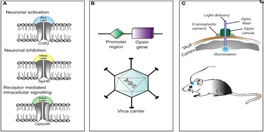
Three primary components in the application of optogenetics
are as follows (A) Identification or synthesis of a
light-sensitive protein (opsin) such as channelrhodopsin-2
(ChR2), halorhodopsin (NpHR), etc... (B) The design of a
system to introduce the genetic material containing the opsin
into cells for protein expression such as application of Cre
 recombinase or an adeno-associated-virus (C) application of
light emitting instruments.[55]

The technique of using optogenetics is flexible and adaptable to the experimenter's needs. For starters, experimenters genetically engineer a microbial opsin based on the gating properties (rate of excitability, refractory period, etc..) required for the experiment.

There is a challenge in introducing the microbial opsin, an optogenetic actuator, into a specific region of the organism in question. A rudimentary approach is to introduce an engineered viral vector that contains the optogenetic actuator gene attached to a recognizable promoter such as CAMKIIα. This allows for some level of specificity as cells that already contain and can translate the given promoter will be infected with the viral vector and hopefully express the optogenetic actuator gene.

Another approach is the creation of transgenic mice where the optogenetic actuator gene is introduced into mice zygotes with a given promoter, most commonly Thy1. Introduction of the optogenetic actuator at an early stage allows for a larger genetic code to be incorporated and as a result, increases the specificity of cells to be infected.

A third and rather novel approach that has been developed is creating transgenic mice with Cre recombinase, an enzyme that catalyzes recombination between two lox-P sites. Then by introducing an engineered viral vector containing the optogenetic actuator gene in between two lox-P sites, only the cells containing the Cre recombinase will express the microbial opsin. This last technique has allowed for multiple modified optogenetic actuators to be used without the need to create a whole line of transgenic animals every time a new microbial opsin is needed.

After the introduction and expression of the microbial opsin, depending on the type of analysis being performed, application of light can be placed at the terminal ends or the main region where the infected cells are situated. Light stimulation can be performed with a vast array of instruments from light emitting diodes (LEDs) or diode-pumped solid state (DPSS). These light sources are most commonly connected to a computer through a fiber optic cable. Recent advances include the advent of wireless head-mounted devices that also apply LED to targeted areas and as a result give the animal more freedom of mobility to reproduce in vivo results.[56][57]

Issues

Although already a powerful scientific tool, optogenetics, according to Doug Tischer & Orion D. Weiner of the University of California San Francisco, should be regarded as a "first-generation GFP" because of its immense potential for both utilization and optimization.[58] With that being said, the current approach to optogenetics is limited primarily by its versatility. Even within the field of Neuroscience where it is most potent, the technique is less robust on a subcellular level.[59]

Selective expression

One of the main problems of optogenetics is that not all the cells in question may express the microbial opsin gene at the same level. Thus, even illumination with a defined light intensity will have variable effects on individual cells. Optogenetic stimulation of neurons in the brain is even less controlled as the light intensity drops exponentially from the light source (e.g. implanted optical fiber).

Moreover, mathematical modelling shows that selective expression of opsin in specific cell types can dramatically alter the dynamical behavior of the neural circuitry. In particular, optogenetic stimulation that preferentially targets inhibitory cells can transform the excitability of the neural tissue from Type 1 — where neurons operate as integrators — to Type 2 where neurons operate as resonators.[60] Type 1 excitable media sustain propagating waves of activity whereas Type 2 excitable media do not. The transformation from one to the other explains how constant optical stimulation of primate motor cortex elicits gamma-band (40–80 Hz) oscillations in the manner of a Type 2 excitable medium. Yet those same oscillations propagate far into the surrounding tissue in the manner of a Type 1 excitable medium.[61]

Nonetheless, it remains difficult to target opsin to defined subcellular compartments, e.g. the plasma membrane, synaptic vesicles, or mitochondria.[59][62] Restricting the opsin to specific regions of the plasma membrane such as dendrites, somata or axon terminals would provide a more robust understanding of neuronal circuitry.[59]

Kinetics and synchronization

An issue with channelrhodopsin-2 is that its gating properties don't mimic in vivo cation channels of cortical neurons. A solution to this issue with a protein's kinetic property is introduction of variants of channelrhodopsin-2 with more favorable kinetics.[55][56]

Another one of the technique's limitations is that light stimulation produces a synchronous activation of infected cells and this removes any individual cell properties of activation among the population affected. Therefore, it is difficult to understand how the cells in the population affected communicate with one another or how their phasic properties of activation may relate to the circuitry being observed.

Optogenetic activation has been combined with functional magnetic resonance imaging (ofMRI) to elucidate the connectome, a thorough map of the brain’s neural connections. The results, however, are limited by the general properties of fMRI.[59][63] The readouts from this neuroimaging procedure lack the spatial and temporal resolution appropriate for studying the densely packed and rapid-firing neuronal circuits.[63]

Excitation spectrum

The opsin proteins currently in use have absorption peaks across the visual spectrum, but remain considerable sensitivity to blue light.[59] This spectral overlap makes it very difficult to combine opsin activation with genenetically encoded indictors (GEVIs, GECIs, GluSnFR, synapto-pHluorin), most of which need blue light excitation. Opsins with infrared activation would, at a standard irradiance value, increase light penetration and augment resolution through reduction of light scattering.

Applications

The field of optogenetics has furthered the fundamental scientific understanding of how specific cell types contribute to the function of biological tissues such as neural circuits in vivo (see references from the scientific literature below). Moreover, on the clinical side, optogenetics-driven research has led to insights into Parkinson's disease[64][65] and other neurological and psychiatric disorders. Indeed, optogenetics papers in 2009 have also provided insight into neural codes relevant to autism, Schizophrenia, drug abuse, anxiety, and depression.[41][66][67][68]

Identification of particular neurons and networks

Amygdala

Optogenetic approaches have been used to map neural circuits in the amygdala that contribute to fear conditioning.[69][70][71][72] One such example of a neural circuit is the connection made from the basolateral amygdala to the dorsal-medial prefrontal cortex where neuronal oscillations of 4 Hz have been observed in correlation to fear induced freezing behaviors in mice. Transgenic mice were introduced with channelrhodoposin-2 attached with a parvalbumin-Cre promoter that selectively infected interneurons located both in the basolateral amygdala and the dorsal-medial prefrontal cortex responsible for the 4 Hz oscillations. The interneurons were optically stimulated generating a freezing behavior and as a result provided evidence that these 4 Hz oscillations may be responsible for the basic fear response produced by the neuronal populations along the dorsal-medial prefrontal cortex and basolateral amygdala.[73]

Olfactory bulb

Optogenetic activation of olfactory sensory neurons was critical for demonstrating timing in odor processing[74] and for mechanism of neuromodulatory mediated olfactory guided behaviors (e.g. aggression, mating)[75] In addition, with the aid of optogenetics, evidence has been reproduced to show that the "afterimage" of odors is concentrated more centrally around the olfactory bulb rather than on the periphery where the olfactory receptor neurons would be located. Transgenic mice infected with channel-rhodopsin Thy1-ChR2, were stimulated with a 473 nm laser transcranially positioned over the dorsal section of the olfactory bulb. Longer photostimulation of mitral cells in the olfactory bulb led to observations of longer lasting neuronal activity in the region after the photostimulation had ceased, meaning the olfactory sensory system is able to undergo long term changes and recognize differences between old and new odors.[76]

Nucleus accumbens

Optogenetics, freely moving mammalian behavior, in vivo electrophysiology, and slice physiology have been integrated to probe the cholinergic interneurons of the nucleus accumbens by direct excitation or inhibition. Despite representing less than 1% of the total population of accumbal neurons, these cholinergic cells are able to control the activity of the dopaminergic terminals that innervate medium spiny neurons (MSNs) in the nucleus accumbens.[77] These accumbal MSNs are known to be involved in the neural pathway through which cocaine exerts its effects, because decreasing cocaine-induced changes in the activity of these neurons has been shown to inhibit cocaine conditioning. The few cholinergic neurons present in the nucleus accumbens may prove viable targets for pharmacotherapy in the treatment of cocaine dependence[41]

Cages for rat equipped of optogenetics leds commutators which permit in vivo to study animal behavior during optogenetics' stimulations.

Prefrontal cortex

In vivo and in vitro recordings (by the Cooper laboratory) of individual CAMKII AAV-ChR2 expressing pyramidal neurons within the prefrontal cortex demonstrated high fidelity action potential output with short pulses of blue light at 20 Hz (Figure 1).[34] The same group recorded complete green light-induced silencing of spontaneous activity in the same prefrontal cortical neuronal population expressing an AAV-NpHR vector (Figure 2).[34]

Heart

Optogenetics was applied on atrial cardiomyocytes to end spiral wave arrhythmias, found to occur in atrial fibrillation, with light.[78] This method is still in the development stage. A recent study explored the possibilities of optogenetics as a method to correct for arrythmias and resynchronize cardiac pacing. The study introduced channelrhodopsin-2 into cardiomyocytes in ventricular areas of hearts of transgenic mice and performed in vitro studies of photostimulation on both open-cavity and closed-cavity mice. Photostimulation led to increased activation of cells and thus increased ventricular contractions resulting in increasing heart rates. In addition, this approach has been applied in cardiac resynchronization therapy (CRT) as a new biological pacemaker as a substitute for electrode based-CRT.[79] Lately, optogenetics has been used in the heart to defibrillate ventricular arrhythmias with local epicardial illumination,[80] a generalized whole heart illumination[81] or with customized stimulation patterns based on arrhythmogenic mechanisms in order to lower defibrillation energy.[82]

Spiral ganglion

Optogenetic stimulation of the spiral ganglion in deaf mice restored auditory activity.[83][84] Optogenetic application onto the cochlear region allows for the stimulation or inhibition of the spiral ganglion cells (SGN). In addition, due to the characteristics of the resting potentials of SGN's, different variants of the protein channelrhodopsin-2 have been employed such as Chronos and CatCh. Chronos and CatCh variants are particularly useful in that they have less time spent in their deactivated states, which allow for more activity with less bursts of blue light emitted. The result being that the LED producing the light would require less energy and the idea of cochlear prosthetics in association with photo-stimulation, would be more feasible.[85]

Brainstem

Optogenetic stimulation of a modified red-light excitable channelrhodopsin (ReaChR) expressed in the facial motor nucleus enabled minimally invasive activation of motoneurons effective in driving whisker movements in mice.[86] One novel study employed optogenetics on the Dorsal Ralphe Nucleus to both activate and inhibit dopaminergic release onto the ventral tegmental area. To produce activation transgenic mice were infected with channelrhodopsin-2 with a TH-Cre promoter and to produce inhibition the hyperpolarizing opsin NpHR was added onto the TH-Cre promoter. Results showed that optically activating dopaminergic neurons led to an increase in social interactions, and their inhibition decreased the need to socialize only after a period of isolation.[87]

Precise temporal control of interventions

The currently available optogenetic actuators allow for the accurate temporal control of the required intervention (i.e. inhibition or excitation of the target neurons) with precision routinely going down to the millisecond level. Therefore, experiments can now be devised where the light used for the intervention is triggered by a particular element of behavior (to inhibit the behavior), a particular unconditioned stimulus (to associate something to that stimulus) or a particular oscillatory event in the brain (to inhibit the event). This kind of approach has already been used in several brain regions:

Hippocampus

Sharp waves and ripple complexes (SWRs) are distinct high frequency oscillatory events in the hippocampus thought to play a role in memory formation and consolidation. These events can be readily detected by following the oscillatory cycles of the on-line recorded local field potential. In this way the onset of the event can be used as a trigger signal for a light flash that is guided back into the hippocampus to inhibit neurons specifically during the SWRs and also to optogenetically inhibit the oscillation itself[88] These kinds of "closed-loop" experiments are useful to study SWR complexes and their role in memory.

Cellular biology/cell signaling pathways

 
Optogenetic control of cellular forces and induction of mechanotransduction. Pictured cells receive an hour of imaging concurrent with blue light that pulses every 60 seconds. This is also indicated when the blue point flashes onto the image. The cell relaxes for an hour without light activation and then this cycle repeats again. The square inset magnifies the cell's nucleus.

The optogenetic toolkit has proven pivotal for the field of neuroscience as it allows precise manipulation of neuronal excitability. Moreover, this technique has been shown to extend outside neurons to an increasing number of proteins and cellular functions.[58] Cellular scale modifications including manipulation of contractile forces relevant to cell migration, cell division and wound healing have been optogenetically manipulated.[89] The field has not developed to the point where processes crucial to cellular and developmental biology and cell signaling including protein localization, post-translational modification and GTP loading can be consistently controlled via optogenetics.[58]

Photosensitive proteins utilized in various cell signaling pathways

While this extension of optogenetics remains to be further investigated, there are various conceptual methodologies that may prove to immediately robust. There is a considerable body of literature outlining photosensitive proteins that have been utilized in cell signaling pathways.[58] CRY2, LOV, DRONPA and PHYB are photosynthetic proteins involved in inducible protein association whereby activation via light can induce/turn off a signaling cascade via recruitment of a signaling domain to its respective substrate.[90][91][92][93] LOV and PHYB are photosensitive proteins that engage in homodimerization and/or heterodimerization to recruit some DNA-modifying protein, translocate to the site of DNA and alter gene expression levels.[94][95][96] CRY2, a protein that inherently clusters when active, has been fused with signaling domains and subsequently photoactivated allowing for clustering-based activation.[97] Proteins LOV and Dronpa have also been adapted to cell signaling manipulation; exposure to light induces conformational changes in the photosensitive protein which can subsequently reveal a previously obscured signaling domain and/or activate a protein that was otherwise allosterically inhibited.[98][99] LOV has been fused to caspase 3 to produce a construct capable of inducing apoptosis upon light stimulation.[100]

Optogenetic temporal control of signals

A different set of signaling cascades respond to stimulus timing duration and dynamics.[101] Adaptive signaling pathways, for instance, adjust in accordance to the current level of the projected stimulus and display activity only when these levels change as opposed to responding to absolute levels of the input.[102] Stimulus dynamics also can trigger activity; treating PC12 cells with epidermal growth factor (inducing a transient profile of ERK activity) leads to cellular proliferation whereas introduction of nerve growth factor (inducing a sustained profile of ERK activity) is associated with a different cellular decision whereby the PC12 cells differentiate into neuron-like cells.[103] This discovery was guided pharmacologically but the finding was replicated utilizing optogenetic inputs instead.[104] This ability to optogenetically control signals for various time durations is being explored to elucidate various cell signaling pathways where there is not a strong enough understanding to utilize either drug/genetic manipulation.

Natural science

From Wikipedia, the free encyclopedia https://en.wikipedia.org/w...