Search This Blog

Friday, July 10, 2015

Symbiogenesis


From Wikipedia, the free encyclopedia


Electron micrograph of a mitochondrion showing its mitochondrial matrix and membranes

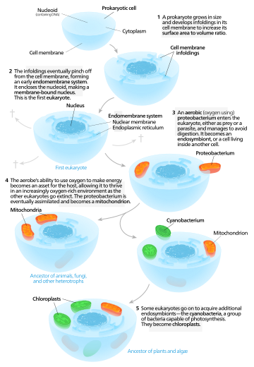
Symbiogenesis, or endosymbiotic theory, is an evolutionary theory that explains the origin of eukaryotic cells from prokaryotes. It states that several key organelles of eukaryotes originated as a symbiosis between separate single-celled organisms. According to this theory, mitochondria, plastids (for example chloroplasts), and possibly other organelles representing formerly free-living bacteria were taken inside another cell as an endosymbiont around 1.5 billion years ago. Molecular and biochemical evidence suggest that mitochondria developed from proteobacteria (in particular, Rickettsiales, the SAR11 clade,[1][2] or close relatives) and chloroplasts from cyanobacteria (in particular, nitrogen-fixing filamentous cyanobacteria[3][4]).

History


Endosymbiotic theory

The endosymbiotic (Greek: ἔνδον endon "within", σύν syn "together" and βίωσις biosis "living") theories were first articulated by the Russian botanist Konstantin Mereschkowski in 1910,[5] although the fundamental elements of the theory were described in a paper five years earlier.[6][7] Mereschkowski was familiar with work by botanist Andreas Schimper, who had observed in 1883 that the division of chloroplasts in green plants closely resembled that of free-living cyanobacteria, and who had himself tentatively proposed (in a footnote) that green plants had arisen from a symbiotic union of two organisms.[8] Ivan Wallin extended the idea of an endosymbiotic origin to mitochondria in the 1920s.[9][10] A Russian botanist Boris Kozo-Polyansky was the first to explain the theory in terms of Darwinian evolution.[11] In his 1924 book Symbiogenesis: A New Principle of Evolution he wrote, "The theory of symbiogenesis is a theory of selection relying on the phenomenon of symbiosis."[12] These theories were initially dismissed or ignored. More detailed electron microscopic comparisons between cyanobacteria and chloroplasts (for example studies by Hans Ris published in 1961[13]), combined with the discovery that plastids and mitochondria contain their own DNA[14] (which by that stage was recognized to be the hereditary material of organisms) led to a resurrection of the idea in the 1960s.

The endosymbiotic theory was advanced and substantiated with microbiological evidence by Lynn Margulis in a 1967 paper, On the origin of mitosing cells.[15] In her 1981 work Symbiosis in Cell Evolution she argued that eukaryotic cells originated as communities of interacting entities, including endosymbiotic spirochaetes that developed into eukaryotic flagella and cilia. This last idea has not received much acceptance, because flagella lack DNA and do not show ultrastructural similarities to bacteria or archaea (see also: Evolution of flagella and Prokaryotic cytoskeleton). According to Margulis and Dorion Sagan,[16] "Life did not take over the globe by combat, but by networking" (i.e., by cooperation). The possibility that peroxisomes may have an endosymbiotic origin has also been considered, although they lack DNA. Christian de Duve proposed that they may have been the first endosymbionts, allowing cells to withstand growing amounts of free molecular oxygen in the Earth's atmosphere. However, it now appears that they may be formed de novo, contradicting the idea that they have a symbiotic origin.[17]

It is thought that over millennia these endosymbionts transferred some of their own DNA to the host cell's nucleus (called "endosymbiotic gene transfer") during the evolutionary transition from a symbiotic community to an instituted eukaryotic cell. The endosymbiotic theory is considered to be a type of saltational evolution.[18]
One model for the origin of mitochondria and plastids.

From endosymbionts to organelles

According to Keeling and Archibald,[19] the usual way to distinguish organelles from endosymbionts is by their reduced genome sizes. As an endosymbiont evolves into an organelle, most of their genes are transferred to the host cell genome. The host cell and organelle need to develop a transport mechanism that enables transfer back of the protein products needed by the organelle but now manufactured by the cell. However, using the example of the freshwater amoeboid Paulinella chromatophora, which contains chromatophores found to be evolved from cyanobacteria, these authors argue that this is not the only possible criterion, another one being that the host cell has assumed control of the regulation of the former endosymbiont's division, bringing it in synchrony with the cell's own division.[19] Nowack and her colleagues[20] performed gene sequencing on the chromatophore (1.02Mb) and found that only 867 proteins were encoded by these photosynthetic cells. Comparisons with their closest free living cyanobacteria of the genus Synechococcus (having a genome size of 3Mb with 3300 genes) revealed that chromatophores underwent a drastic genome shrinkage. Chromatophores contained genes that were accountable for photosynthesis but were deficient in genes that could carry out other biosynthetic functions signifying that these endosymbiotic cells were highly dependent on their hosts for their survival and growth mechanisms. Thus, these chromatophores were found to be non-functional for organelle-specific purposes when compared to mitochondria and plastids. This distinction could have promoted the early evolution of photosynthetic organelles.

Evidence

Evidence that mitochondria and plastids arose from bacteria is as follows:[21][22][23]
  • New mitochondria and plastids are formed only through a process similar to binary fission.
  • If a cell's mitochondria or chloroplasts are removed, they do not have the means to create new ones.[24] For example, in some algae, such as Euglena, the plastids can be destroyed by certain chemicals or prolonged absence of light without otherwise affecting the cell. In such a case, the plastids will not regenerate.
  • Transport proteins called porins are found in the outer membranes of mitochondria and chloroplasts, are also found in bacterial cell membrane.[25][26][27]
  • A membrane lipid cardiolipin is exclusively found in the inner mitochondrial membrane and bacterial cell membrane.[28]
  • Both mitochondria and plastids contain single circular DNA that is different from that of the cell nucleus and that is similar to that of bacteria (both in their size and structure).
  • The genomes, including the specific genes, are basically similar between mitochondria and the Rickettsial bacteria.[29]
  • Genome comparisons indicate that cyanobacteria contributed to the genetic origin of plastids.[30]
  • DNA sequence analysis and phylogenetic estimates suggest that nuclear DNA contains genes that probably came from plastids.
  • These organelles' ribosomes are like those found in bacteria (70S).
  • Proteins of organelle origin, like those of bacteria, use N-formylmethionine as the initiating amino acid.
  • Much of the internal structure and biochemistry of plastids, for instance the presence of thylakoids and particular chlorophylls, is very similar to that of cyanobacteria. Phylogenetic estimates constructed with bacteria, plastids, and eukaryotic genomes also suggest that plastids are most closely related to cyanobacteria.
  • Mitochondria have several enzymes and transport systems similar to those of bacteria.
  • Some proteins encoded in the nucleus are transported to the organelle, and both mitochondria and plastids have small genomes compared to bacteria. This is consistent with an increased dependence on the eukaryotic host after forming an endosymbiosis. Most genes on the organellar genomes have been lost or moved to the nucleus. Most genes needed for mitochondrial and plastid function are located in the nucleus. Many originate from the bacterial endosymbiont.
  • Plastids are present in very different groups of protists, some of which are closely related to forms lacking plastids. This suggests that if chloroplasts originated de novo, they did so multiple times, in which case their close similarity to each other is difficult to explain.
  • Many of these protists contain "primary" plastids that have not yet been acquired from other plastid-containing eukaryotes.
  • Among eukaryotes that acquired their plastids directly from bacteria (known as Archaeplastida), the glaucophyte algae have chloroplasts that strongly resemble cyanobacteria. In particular, they have a peptidoglycan cell wall between the two membranes.

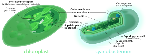
Comparison of chloroplasts and cyanobacteria showing their similarities.

Secondary endosymbiosis

Primary endosymbiosis involves the engulfment of a bacterium by another free living organism. Secondary endosymbiosis occurs when the product of primary endosymbiosis is itself engulfed and retained by another free living eukaryote. Secondary endosymbiosis has occurred several times and has given rise to extremely diverse groups of algae and other eukaryotes. Some organisms can take opportunistic advantage of a similar process, where they engulf an alga and use the products of its photosynthesis, but once the prey item dies (or is lost) the host returns to a free living state. Obligate secondary endosymbionts become dependent on their organelles and are unable to survive in their absence (for a review see McFadden 2001[31]). RedToL, the Red Algal Tree of Life Initiative funded by the National Science Foundation highlights the role red algae or Rhodophyta played in the evolution of our planet through secondary endosymbiosis.

One possible secondary endosymbiosis in process has been observed by Okamoto & Inouye (2005). The heterotrophic protist Hatena behaves like a predator until it ingests a green alga, which loses its flagella and cytoskeleton, while Hatena, now a host, switches to photosynthetic nutrition, gains the ability to move towards light and loses its feeding apparatus.[32]

The process of secondary endosymbiosis left its evolutionary signature within the unique topography of plastid membranes. Secondary plastids are surrounded by three (in euglenophytes and some dinoflagellates) or four membranes (in haptophytes, heterokonts, cryptophytes, and chlorarachniophytes). The two additional membranes are thought to correspond to the plasma membrane of the engulfed alga and the phagosomal membrane of the host cell. The endosymbiotic acquisition of a eukaryote cell is represented in the cryptophytes; where the remnant nucleus of the red algal symbiont (the nucleomorph) is present between the two inner and two outer plastid membranes.[citation needed]

Despite the diversity of organisms containing plastids, the morphology, biochemistry, genomic organisation, and molecular phylogeny of plastid RNAs and proteins suggest a single origin of all extant plastids – although this theory is still debated.[33][34]

Some species including Pediculus humanus have multiple chromosomes in the mitochondrion. This and the phylogenetics of the genes encoded within the mitochondrion suggest that mitochondria have multiple ancestors, that these were acquired by endosymbiosis on several occasions rather than just once, and that there have been extensive mergers and rearrangements of genes on the several original mitochondrial chromosomes.[35]

Prokaryote

From Wikipedia, the free encyclopedia

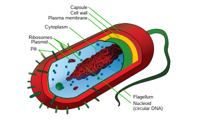
Cell structure of a bacterium, a member of one of the two domains of prokaryotic life

Comparison of Eukaryotes vs. Prokaryotes
3D animation of a prokaryotic cell that shows all the elements that compose it

A prokaryote is a single-celled organism that lacks a membrane-bound nucleus (karyon), mitochondria, or any other membrane-bound organelles.[1] The word prokaryote comes from the Greek πρό- (pro-) "before" and καρυόν (karyon) "nut or kernel".[2][3] Prokaryotes can be divided into two domains, Archaea and Bacteria, with the remainder of species, called eukaryotes, in the third domain Eukaryota.[4]

In the prokaryotes all the intracellular water-soluble components (proteins, DNA and metabolites) are located together in the cytoplasm enclosed by the cell membrane, rather than in separate cellular compartments. Bacteria, however, do possess protein-based bacterial microcompartments, which are thought to act as primitive organelles enclosed in protein shells.[5][6] Some prokaryotes also have multicellular stages in their life cycles, such as myxobacteria, or create large colonies, like cyanobacteria.[7]

Molecular studies have provided insight into the evolution and interrelationships of the three domains of biological species.[8] Eukaryotes are organisms, including humans, whose cells have a well defined membrane-bound nucleus (containing chromosomal DNA) and organelles. The division between prokaryotes and eukaryotes reflects the existence of two very different levels of cellular organization. Distinctive types of prokaryotes include extremophiles and methanogens; these are common in some extreme environments.[1]

Structure

Research indicates that all prokaryotes have a prokaryotic cytoskeleton, albeit more primitive than that of the eukaryotes. Besides homologues of actin and tubulin (MreB and FtsZ), the helically arranged building-block of the flagellum, flagellin, is one of the most significant cytoskeletal proteins of bacteria, as it provides structural backgrounds of chemotaxis, the basic cell physiological response of bacteria. At least some prokaryotes also contain intracellular structures that can be seen as primitive organelles. Membranous organelles (or intracellular membranes) are known in some groups of prokaryotes, such as vacuoles or membrane systems devoted to special metabolic properties, such as photosynthesis or chemolithotrophy. In addition, some species also contain protein-enclosed microcompartments, which have distinct physiological roles (e.g. carboxysomes or gas vacuoles).
Most prokaryotes are between 1 µm and 10 µm, but they can vary in size from 0.2 µm to 750 µm (Thiomargarita namibiensis).

Prokaryotic cell structure
Flagellum
Long, whip-like protrusion that aids in cellular locomotion.
Cell membrane
Surrounds the cell's cytoplasm and regulates the flow of substances in and out of the cell.
Cell wall (except genera Mycoplasma and Thermoplasma)
Outer covering of most cells that protects the bacterial cell and gives it shape.
Cytoplasm
A gel-like substance composed mainly of water that also contains enzymes, salts, cell components, and various organic molecules.
Ribosome
Cell structures responsible for protein production.
Nucleoid
Area of the cytoplasm that contains the single bacterial DNA molecule.
Glycocalyx
Inclusions

Morphology of prokaryotic cells

Prokaryotic cells have various shapes; the four basic shapes of bacteria are:[9]
The archaeon Haloquadratum has flat square-shaped cells.[10]

Reproduction

Bacteria and archaea reproduce through asexual reproduction, usually by binary fission. Genetic exchange and recombination still occur, but this is a form of horizontal gene transfer and is not a replicative process, simply involving the transference of DNA between two cells, as in bacterial conjugation.

DNA transfer

DNA transfer between prokaryotic cells occurs in bacteria and archaea, although it has been mainly studied in bacteria. In bacteria, gene transfer occurs by three processes. These are (1) bacterial virus (bacteriophage)-mediated transduction, (2) plasmid-mediated conjugation, and (3) natural transformation. Transduction of bacterial genes by bacteriophage appears to reflect an occasional error during intracellular assembly of virus particles, rather than an adaptation of the host bacteria. The transfer of bacterial DNA is under the control of the bacteriophage’s genes rather than bacterial genes. Conjugation in the well-studied E. coli system is controlled by plasmid genes, and is an adaptation for distributing copies of a plasmid from one bacterial host to another. Infrequently during this process, a plasmid may integrate into the host bacterial chromosome, and subsequently transfer part of the host bacterial DNA to another bacterium. Plasmid mediated transfer of host bacterial DNA (conjugation) also appears to be an accidental process rather than a bacterial adaptation.

Natural bacterial transformation involves the transfer of DNA from one bacterium to another through the intervening medium. Unlike transduction and conjugation, transformation is clearly a bacterial adaptation for DNA transfer, because it depends on numerous bacterial gene products that specifically interact to perform this complex process.[11] For a bacterium to bind, take up and recombine donor DNA into its own chromosome, it must first enter a special physiological state called competence. About 40 genes are required in Bacillus subtilis for the development of competence.[12] The length of DNA transferred during B. subtilis transformation can be as much as a third to the whole chromosome.[13][14] Transformation is a common mode of DNA transfer, and 67 prokaryotic species are thus far known to be naturally competent for transformation.[15] The development of competence in nature is usually associated with stressful environmental conditions, and appears to be an adaptation for promoting repair of DNA damage in recipient cells.[16]

Among archaea, Halobacterium volcanii forms cytoplasmic bridges between cells that appear to be used for transfer of DNA from one cell to another.[17] Another archaeon, Sulfolobus solfataricus, transfers DNA between cells by direct contact. Frols et al.[18] found that exposure of S. solfataricus to DNA damaging agents induces cellular aggregation, and suggested that cellular aggregation may enhance DNA transfer among cells to provide increased repair of damaged DNA via homologous recombination.

Sociality

While prokaryotes are considered strictly unicellular, most can form stable aggregate communities.[19] When such communities are encased in a stabilizing polymer matrix ("slime"), they may be called "biofilms".[20] Cells in biofilms often show distinct patterns of gene expression (phenotypic differentiation) in time and space. Also, as with multicellular eukaryotes, these changes in expression often appear to result from cell-to-cell signaling, a phenomenon known as quorum sensing.

Biofilms may be highly heterogeneous and structurally complex and may attach to solid surfaces, or exist at liquid-air interfaces, or potentially even liquid-liquid interfaces. Bacterial biofilms are often made up of microcolonies (approximately dome-shaped masses of bacteria and matrix) separated by "voids" through which the medium (e.g., water) may flow relatively uninhibited. The microcolonies may join together above the substratum to form a continuous layer, closing the network of channels separating microcolonies. This structural complexity—combined with observations that oxygen limitation (a ubiquitous challenge for anything growing in size beyond the scale of diffusion) is at least partially eased by movement of medium throughout the biofilm—has led some to speculate that this may constitute a circulatory system [21] and many researchers have started calling prokaryotic communities multicellular (for example [22]). Differential cell expression, collective behavior, signaling, programmed cell death, and (in some cases) discrete biological dispersal[23] events all seem to point in this direction. However, these colonies are seldom if ever founded by a single founder (in the way that animals and plants are founded by single cells), which presents a number of theoretical issues. Most explanations of co-operation and the evolution of multicellularity have focused on high relatedness between members of a group (or colony, or whole organism). If a copy of a gene is present in all members of a group, behaviors that promote cooperation between members may permit those members to have (on average) greater fitness than a similar group of selfish individuals[24] (see inclusive fitness and Hamilton's rule).

Should these instances of prokaryotic sociality prove to be the rule rather than the exception, it would have serious implications for the way we view prokaryotes in general, and the way we deal with them in medicine.[25] Bacterial biofilms may be 100 times more resistant to antibiotics than free-living unicells and may be nearly impossible to remove from surfaces once they have colonized them.[26] Other aspects of bacterial cooperation—such as bacterial conjugation and quorum-sensing-mediated pathogenicity, present additional challenges to researchers and medical professionals seeking to treat the associated diseases.

Environment

Prokaryotes have diversified greatly throughout their long existence. The metabolism of prokaryotes is far more varied than that of eukaryotes, leading to many highly distinct prokaryotic types. For example, in addition to using photosynthesis or organic compounds for energy, as eukaryotes do, prokaryotes may obtain energy from inorganic compounds such as hydrogen sulfide. This enables prokaryotes to thrive in harsh environments as cold as the snow surface of Antarctica, studied in cryobiology or as hot as undersea hydrothermal vents and land-based hot springs.

Prokaryotes live in nearly all environments on Earth. Some archaea and bacteria thrive in harsh conditions, such as high temperatures (thermophiles) or high salinity (halophiles). Organisms such as these are referred to as extremophiles.[27] Many archaea grow as plankton in the oceans. Symbiotic prokaryotes live in or on the bodies of other organisms, including humans.

Classification

In 1977, Carl Woese proposed dividing prokaryotes into the Bacteria and Archaea (originally Eubacteria and Archaebacteria) because of the major differences in the structure and genetics between the two groups of organisms. Archaea were originally thought to be extremophiles, living only in inhospitable conditions such as extremes of temperature, pH, and radiation but have since been found in all types of habitats. The resulting arrangement of Eukaryota (also called "Eukarya"), Bacteria, and Archaea is called the three-domain system, replacing the traditional two-empire system.[28]

One criticism of this classification points out that the word "prokaryote" is based on what these organisms are not (they are not eukaryotic), rather than what they are (either archaea or bacteria).[29]

Evolution of prokaryotes

Evolutionary tree showing the common ancestry of organisms from all three domains of life. Bacteria are colored blue, eukaryotes red, and archaea green.
Relative positions of some of the phyla included are shown around the tree.

Phylogenetic tree showing the diversity of prokaryotes, compared to eukaryotes

The current model of the evolution of the first living organisms is that these were some form of prokaryotes, which may have evolved out of protocells. In general, the eukaryotes are thought to have evolved later in the history of life.[30] However, some authors have questioned this conclusion, arguing that the current set of prokaryotic species may have evolved from more complex eukaryotic ancestors through a process of simplification.[31][32][33] Others have argued that the three domains of life arose simultaneously, from a set of varied cells that formed a single gene pool.[34] This controversy was summarized in 2005:[35]
There is no consensus among biologists concerning the position of the eukaryotes in the overall scheme of cell evolution. Current opinions on the origin and position of eukaryotes span a broad spectrum including the views that eukaryotes arose first in evolution and that prokaryotes descend from them, that eukaryotes arose contemporaneously with eubacteria and archeabacteria and hence represent a primary line of descent of equal age and rank as the prokaryotes, that eukaryotes arose through a symbiotic event entailing an endosymbiotic origin of the nucleus, that eukaryotes arose without endosymbiosis, and that eukaryotes arose through a symbiotic event entailing a simultaneous endosymbiotic origin of the flagellum and the nucleus, in addition to many other models, which have been reviewed and summarized elsewhere.
The oldest known fossilized prokaryotes were laid down approximately 3.5 billion years ago, only about 1 billion years after the formation of the Earth's crust. Eukaryotes only appear in the fossil record later, and may have formed from endosymbiosis of multiple prokaryote ancestors. The oldest known fossil eukaryotes are about 1.7 billion years old. However, some genetic evidence suggests eukaryotes appeared as early as 3 billion years ago.[36]

While Earth is the only place in the universe where life is known to exist, some have suggested that there is evidence on Mars of fossil or living prokaryotes;[37][38] but this possibility remains the subject of considerable debate and skepticism.[39][40]

Relationship to eukaryotes

The division between prokaryotes and eukaryotes is usually considered the most important distinction or difference among organisms. The distinction is that eukaryotic cells have a "true" nucleus containing their DNA, whereas prokaryotic cells do not have a nucleus. Both eukaryotes and prokaryotes contain large RNA/protein structures called ribosomes, which produce protein.

Another difference is that ribosomes in prokaryotes are smaller than in eukaryotes. However, two organelles found in many eukaryotic cells, mitochondria and chloroplasts, contain ribosomes similar in size and makeup to those found in prokaryotes.[41] This is one of many pieces of evidence that mitochondria and chloroplasts are themselves descended from free-living bacteria. This theory holds that early eukaryotic cells took in primitive prokaryotic cells by phagocytosis and adapted themselves to incorporate their structures, leading to the mitochondria we see today.

The genome in a prokaryote is held within a DNA/protein complex in the cytosol called the nucleoid, which lacks a nuclear envelope.[42] The complex contains a single, cyclic, double-stranded molecule of stable chromosomal DNA, in contrast to the multiple linear, compact, highly organized chromosomes found in eukaryotic cells. In addition, many important genes of prokaryotes are stored in separate circular DNA structures called plasmids.[2]

Prokaryotes lack mitochondria and chloroplasts. Instead, processes such as oxidative phosphorylation and photosynthesis take place across the prokaryotic cell membrane.[43] However, prokaryotes do possess some internal structures, such as prokaryotic cytoskeletons.[44][45] It has been suggested that the bacterial order Planctomycetes have a membrane around their nucleoid and contain other membrane-bound cellular structures.[46] However, further investigation revealed that Planctomycetes cells are not compartmentalized or nucleated and like the other bacterial membrane systems are all interconnected.[47]

Prokaryotic cells are usually much smaller than eukaryotic cells.[2] Therefore, prokaryotes have a larger surface-area-to-volume ratio, giving them a higher metabolic rate, a higher growth rate, and as a consequence, a shorter generation time than eukaryotes.[2]

Personality theories of addiction

From Wikipedia, the free encyclopedia https://en.wikipedia.org/wiki/Personality_theories_of_addiction ...