Search This Blog

Monday, April 26, 2021

Superspreading event

From Wikipedia, the free encyclopedia

9th floor layout of the Hotel Metropole in Hong Kong, showing where a superspreading event of severe acute respiratory syndrome (SARS) occurred in 2003

A superspreading event (SSEV) is an event in which an infectious disease is spread much more than usual, while an unusually contagious organism infected with a disease is known as a superspreader. In the context of a human-borne illness, a superspreader is an individual who is more likely to infect others, compared with a typical infected person. Such superspreaders are of particular concern in epidemiology.

Some cases of superspreading conform to the 80/20 rule, where approximately 20% of infected individuals are responsible for 80% of transmissions, although superspreading can still be said to occur when superspreaders account for a higher or lower percentage of transmissions. In epidemics with such superspreader events, the majority of individuals infect relatively few secondary contacts.

SSEVs are shaped by multiple factors including a decline in herd immunity, nosocomial infections, virulence, viral load, misdiagnosis, airflow dynamics, immune suppression, and co-infection with another pathogen.

Definition

Although loose definitions of superspreader events exist, some effort has been made at defining what qualifies as a superspreader event (SSEV). Lloyd-Smith et al. (2005) define a protocol to identify a superspreader event as follows:

  1. estimate the effective reproductive number, R, for the disease and population in question;
  2. construct a Poisson distribution with mean R, representing the expected range of Z due to stochasticity without individual variation;
  3. define an SSEV as any infected person who infects more than Z(n) others, where Z(n) is the nth percentile of the Poisson(R) distribution.

This protocol defines a 99th-percentile SSEV as a case which causes more infections than would occur in 99% of infectious histories in a homogeneous population.

During the SARS-CoV-1 2002–2004 SARS outbreak from China, epidemiologists defined a superspreader as an individual with at least eight transmissions of the disease.

Superspreaders may or may not show any symptoms of the disease.

On April 2020 Jonathan Kay reported in relation to the COVID-19 pandemic:

Putting aside hospitals, private residences and old-age homes, almost all of these superspreader events (SSEVs) took place in the context of (1) parties, (2) face-to-face professional networking events and meetings, (3) religious gatherings, (4) sports events, (5) meat-processing facilities, (6) ships at sea, (7) singing groups, and, yes, (8) funerals.

Factors in transmission

How an infection spreads in a community with immunized and non-immunized members.

Superspreaders have been identified who excrete a higher than normal number of pathogens during the time they are infectious. This causes their contacts to be exposed to higher viral/bacterial loads than would be seen in the contacts of non-superspreaders with the same duration of exposure.

Basic reproductive number

The basic reproduction number R0 is the average number of secondary infections caused by a typical infective person in a totally susceptible population. The basic reproductive number is found by multiplying the average number of contacts by the average probability that a susceptible individual will become infected, which is called the shedding potential.

R0 = Number of contacts × Shedding potential

Individual reproductive number

The individual reproductive number represents the number of secondary infections caused by a specific individual during the time that individual is infectious. Some individuals have significantly higher than average individual reproductive numbers and are known as superspreaders. Through contact tracing, epidemiologists have identified superspreaders in measles, tuberculosis, rubella, monkeypox, smallpox, Ebola hemorrhagic fever and SARS.

Co-infections with other pathogens

Studies have shown that men with HIV who are co-infected with at least one other sexually transmitted disease, such as gonorrhea, hepatitis C, and herpes simplex 2 virus, have a higher HIV shedding rate than men without co-infection. This shedding rate was calculated in men with similar HIV viral loads. Once treatment for the co-infection has been completed, the HIV shedding rate returns to levels comparable to men without co-infection.

Lack of herd immunity

Herd immunity, or herd effect, refers to the indirect protection that immunized community members provide to non-immunized members in preventing the spread of contagious disease. The greater the number of immunized individuals, the less likely an outbreak can occur because there are fewer susceptible contacts. In epidemiology, herd immunity is known as a dependent happening because it influences transmission over time. As a pathogen that confers immunity to the survivors moves through a susceptible population, the number of susceptible contacts declines. Even if susceptible individuals remain, their contacts are likely to be immunized, preventing any further spread of the infection. The proportion of immune individuals in a population above which a disease may no longer persist is the herd immunity threshold. Its value varies with the virulence of the disease, the efficacy of the vaccine, and the contact parameter for the population. That is not to say that an outbreak can't occur, but it will be limited.

Superspreaders during outbreaks or pandemics

COVID-19 pandemic: 2020–present

The South Korean spread of confirmed cases of SARS-CoV-2 infection jumped suddenly starting on 19–20 February 2020. On 19 February, the number of confirmed cases increased by 20. On 20 February, 58 or 70 new cases were confirmed, giving a total of 104 confirmed cases, according to the Centers for Disease Control and Prevention Korea (KCDC). According to Reuters, KCDC attributed the sudden jump to 70 cases linked to "Patient 31", who had participated in a gathering in Daegu at the Shincheonji Church of Jesus the Temple of the Tabernacle of the Testimony. On 20 February, the streets of Daegu were empty in reaction to the Shincheonji outbreak. A resident described the reaction, stating "It's like someone dropped a bomb in the middle of the city. It looks like a zombie apocalypse." On 21 February, the first death was reported. According to the mayor of Daegu, the number of suspected cases as of 21 February is 544 among 4,400 examined followers of the church. Later in the outbreak, in May, A 29-year-old man visited several Seoul nightclubs in one night and resulted in accumulated infections of at least 79 other people.

A business conference in Boston (MA) from February 26–28 was a superspreading event.

Between 27 February and 1 March, a Tablighi Jamaat event at Masjid Jamek, Seri Petaling in Kuala Lumpur, Malaysia attended by approximately 16,000 people resulted in a major outbreak across the country. By May 16, 3,348 COVID-19 cases - 48% of Malaysia's total at the time - were linked to the event, and with approximately 10% of attendees visiting from overseas, the event resulted in virus spreading across Southeast Asia. Cases in Cambodia, Indonesia, Vietnam, Brunei, the Philippines and Thailand were traced back to the mosque gathering.

In New York, a lawyer contracted the illness then spread it to at least twenty other individuals in his community in New Rochelle, creating a cluster of cases that quickly passed 100, accounting for more than half of SARS-CoV2 coronavirus cases in the state during early March 2020. For comparison, the basic reproduction number of the virus, which is the average number of additional people that a single case will infect without any preventative measures, is between 1.4 and 3.9.

On March 06, preacher Baldev Singh returned to India after being infected while traveling in Italy and Germany. He subsequently died, becoming the first coronavirus fatality in the State of Punjab. Testing revealed that he'd infected 26 locals, including 19 relatives, while tracing discovered that he'd had direct contact with more than 550 people. Fearing an outbreak, India's government instituted a quarantine in 27 March 2020 in the, affecting 40,000 residents from 20 villages. Initial reports claimed that Baldev Singh had ignored self-quarantine orders, and police collaborated with singer Sidhu Moose Wala to release a rap music video blaming the dead man for bringing the virus to Punjab. But Baldev Singh's fellow travelers insisted that no such order had been given, leading to accusations that local authorities had scapegoated him to avoid scrutiny of their own failures.

A Tablighi Jamaat religious congregation that took place in Delhi's Nizamuddin Markaz Mosque in early March 2020 was a coronavirus super-spreader event, with more than 4,000 confirmed cases and at least 27 deaths linked to the event reported across the country. Over 9,000 missionaries may have attended the congregation, with the majority being from various states of India, and 960 attendees from 40 foreign countries. On 18 April, 4,291 confirmed cases of COVID-19 linked to this event by the Union Health Ministry represented a third of all the confirmed cases of India. Around 40,000 people, including Tablighi Jamaat attendees and their contacts, were quarantined across the country.

On 11 May 2020, it came to light that a worker at a fish processing plant in Tema, Ghana is believed to have infected over 500 other people with COVID-19.

As of 18 July 2020, more than one thousand suspected superspreading events had been logged, for example a cluster of 187 people who were infected after eating at a Harper's Restaurant and Brew Pub in East Lansing, Michigan.

On Saturday, September 26, 2020, President Trump announced his Supreme Court Justice nominee, Amy Coney Barrett. The announcement took place at the White House Rose Garden, where around 30 people attentively watched. The event has since been dubbed a “superspreader” event. Less than a week after the event, President Trump himself was diagnosed with SARS-CoV-2, as well as others who attended the Rose Garden event. By October 7, the Federal Emergency Management Agency memo revealed that 34 White House staff members, housekeepers, and other contacts had contracted the virus. The superspreader event caused controversy throughout the United States, as President Trump was already under scrutiny for not taking the virus seriously. After the president’s recovery from COVID-19, the White House did not host any more superspreader events.

Public health experts have said that the storming of the Capitol on January 6, 2021 was a potential COVID-19 superspreading event. Few members of the crowd storming the Capitol wore face coverings, with many coming from out of town, and few of the rioters were immediately detained and identified.

Several factors are identified as contributing to superspreading events with COVID-19: closed spaces with poor ventilation, crowds, and close contact settings ("three Cs").

Statistical analyses of the frequency of coronavirus superspreading events, including SARS-CoV-2 and SARS, have shown that they correspond to fat-tailed events, indicating that they are extreme, but likely, occurrences.

A SARS-CoV-2 superspreading events database maintained by a group of researchers at the London School of Hygiene and Tropical Medicine includes more than 1,600 superspreading events from around the world.

SARS outbreak: 2003

Guangdong Province in southeastern China where the first outbreak of SARS occurred in 2003.

The first cases of SARS occurred in mid-November 2002 in the Guangdong Province of China. This was followed by an outbreak in Hong Kong in February 2003. A Guangdong Province doctor, Liu Jianlun, who had treated SARS cases there, had contracted the virus and was symptomatic. Despite his symptoms, he traveled to Hong Kong to attend a family wedding. He stayed on the ninth floor of the Metropole Hotel in Kowloon, infecting 16 other hotel guests also staying on that floor. The guests then traveled to Canada, Singapore, Taiwan, and Vietnam, spreading SARS to those locations and transmitting what became a global epidemic.

In another case during this same outbreak, a 54-year-old male was admitted to a hospital with coronary heart disease, chronic kidney failure and type II diabetes mellitus. He had been in contact with a patient known to have SARS. Shortly after his admission he developed fever, cough, myalgia and sore throat. The admitting physician suspected SARS. The patient was transferred to another hospital for treatment of his coronary artery disease. While there, his SARS symptoms became more pronounced. Later, it was discovered he had transmitted SARS to 33 other patients in just two days. He was transferred back to the original hospital where he died of SARS.

In his post-mortem reflection, Low remained puzzled as to the reason for this phenomenon and speculated that "possible explanations for (the superspreaders') enhanced infectivity include the lack of early implementation of infection control precautions, higher load of SCoV, or larger amounts of respiratory secretions."

The SARS outbreak was eventually contained, but not before it caused 8,273 cases and 775 deaths. Within two weeks of the original outbreak in Guangdong Province, SARS had spread to 29 countries.

Measles outbreak: 1989

Rates of measles vaccination worldwide in 2010

Measles is a highly contagious, air-borne virus that reappears even among vaccinated populations. In one Finnish town in 1989, an explosive school-based outbreak resulted in 51 cases, several of whom had been previously vaccinated. One child alone infected 22 others. It was noted during this outbreak that when vaccinated siblings shared a bedroom with an infected sibling, seven out of nine became infected as well.

Typhoid fever

Typhoid fever is a human-specific disease caused by the bacterium Salmonella typhi. It is highly contagious and becoming resistant to antibiotics. S. typhi is susceptible to creating asymptomatic carriers. The most famous carriers are Mary Mallon, known as Typhoid Mary, from New York City, and Mr. N. the Milker, from Folkstone, England. Both were active around the same time. Mallon infected 51 people from 1902 to 1909. Mr. N. infected more than 200 people over 14 years from 1901 to 1915. At the request of health officials, Mr. N. gave up working in food service. Mallon was at first also compliant, choosing other work – but eventually she returned to cooking and caused further outbreaks. She was involuntarily quarantined at Brothers Island in New York, where she stayed until she died in November 1938, aged 69.

It has been found that Salmonella typhi persists in infected mice macrophages that have cycled from an inflammatory state to a non-inflammatory state. The bacteria remain and reproduce without causing further symptoms in the mice, and this helps to explain why carriers are asymptomatic. Identifying superspreaders A method to detect superspreaders in complex networks has been suggested by Kitsak et al.

Orthornavirae

From Wikipedia, the free encyclopedia
 
Orthornavirae
Virus classification e
(unranked): Virus
Realm: Riboviria
Kingdom: Orthornavirae
Phyla and classes

Positive-strand RNA viruses

Negative-strand RNA viruses

Double-stranded RNA viruses

Orthornavirae is a kingdom of viruses that have genomes made of ribonucleic acid (RNA) and which encode an RNA-dependent RNA polymerase (RdRp). The RdRp is used to transcribe the viral RNA genome into messenger RNA (mRNA) and to replicate the genome. Viruses in this kingdom also share a number of characteristics involving evolution, including high rates of genetic mutations, recombinations, and reassortments.

Viruses in Orthornavirae belong to the realm Riboviria. They are descended from a common ancestor that may have been a non-viral molecule that encoded a reverse transcriptase instead of an RdRp for replication. The kingdom is subdivided into five phyla that separate member viruses based on their genome type, host range, and genetic similarity. Viruses with three genome types are included: positive-strand RNA viruses, negative-strand RNA viruses, and double-stranded RNA viruses.

Many of the most widely known viral diseases are caused by RNA viruses in the kingdom, which includes coronaviruses, the Ebola virus, influenza viruses, the measles virus, and the rabies virus. The first virus to be discovered, tobacco mosaic virus, belongs to the kingdom. In modern history, RdRp-encoding RNA viruses have caused numerous disease outbreaks, and they infect many economically important crops. Most eukaryotic viruses, including most human, animal, and plant viruses, are RdRp-encoding RNA viruses. In contrast, there are relatively few prokaryotic viruses in the kingdom.

Etymology

The first part of Orthornavirae comes from Greek ὀρθός [orthós], meaning straight, the middle part, rna, refers to RNA, and -virae is the suffix used for virus kingdoms.

Characteristics

Structure

Genome type and replication cycle of different RNA viruses

RNA viruses in Orthornavirae typically do not encode that many proteins, but most positive-sense, single-stranded (+ssRNA) viruses and some double-stranded RNA (dsRNA) viruses encode a major capsid protein that has a single jelly roll fold, so named because the folded structure of the protein contains a structure that resembles a jelly roll. Many also encode an envelope, a type of lipid membrane that typically surrounds the capsid. In particular, the viral envelope is near-universal among negative-sense, single-stranded (-ssRNA) viruses.

Genome

Viruses in Orthornavirae have three different types of genomes: dsRNA, +ssRNA, and -ssRNA. Single-stranded RNA viruses have either a positive or negative sense strand, and dsRNA viruses have both. This structure of the genome is important in terms of transcription to synthesize viral mRNA as well as replication of the genome, both of which are carried out by the viral enzyme RNA-dependent RNA polymerase (RdRp), also called RNA replicase.

Replication and transcription

Positive-strand RNA viruses

Positive-strand RNA viruses have genomes that can function as mRNA, so transcription is not necessary. However, +ssRNA will produce dsRNA forms as part of the process of replicating their genomes. From the dsRNA, additional positive strands are synthesized, which may be used as mRNA or for genomes for progeny. Because +ssRNA viruses create intermediate dsRNA forms, they have to avoid the host's immune system in order to replicate. +ssRNA viruses accomplish this by replicating in membrane-associated vesicles that are used as replication factories. For many +ssRNA viruses, subgenomic portions of the genome will be transcribed to translate specific proteins, whereas others will transcribe a polyprotein that is cleaved to produce separate proteins.

Negative-strand RNA viruses

Negative-strand RNA viruses have genomes that function as templates from which mRNA can be synthesized directly by RdRp. Replication is the same process but executed on the positive sense antigenome, during which RdRp ignores all transcription signals so that a complete -ssRNA genome can be synthesized. -ssRNA viruses vary between those that inititate transcription by the RdRp creating a cap on the 5'-end (usually pronounced "five prime end") of the genome or by snatching a cap from host mRNA and attaching it to the viral RNA. For many -ssRNA viruses, at the end of transcription, RdRp stutters on a uracil in the genome, synthesizing hundreds of adenines in a row as part of creating a polyadenylated tail for the mRNA. Some -ssRNA viruses are essentially ambisense, and have proteins encoded by both the positive and negative strand, so mRNA is synthesized directly from the genome and from a complementary strand.

Double-stranded RNA viruses

For dsRNA viruses, RdRp transcribes mRNA by using the negative strand as a template. Positive strands may also be used as templates to synthesize negative strands for the construction of genomic dsRNA. dsRNA is not a molecule produced by cells, so cellular life has evolved mechanisms to detect and inactivate viral dsRNA. To counter this, dsRNA viruses typically retain their genomes inside of viral capsid in order to evade the host's immune system.

Evolution

RNA viruses in Orthornavirae experience a high rate of genetic mutations because RdRp is prone to making errors in replication since it typically lacks proofreading mechanisms to repair errors. Mutations in RNA viruses are often influenced by host factors such as dsRNA-dependent adenosine deaminases, which edit viral genomes by changing adenosines to inosines. Mutations in genes that are essential for replication lead to a reduced number of progeny, so viral genomes typically contain sequences that are highly conserved over time with relatively few mutations.

Many RdRp-encoding RNA viruses also experience a high rate of genetic recombination, though rates of recombination vary significantly, with lower rates in -ssRNA viruses and higher rates in dsRNA and +ssRNA viruses. There are two types of recombination: copy choice recombination and ressortment. Copy choice recombination occurs when the RdRp switches templates during synthesis without releasing the prior, newly created RNA strand, which generates a genome of mixed ancestry. Reassortment, which is restricted to viruses with segmented genomes, has segments from different genomes packaged into a single virion, or virus particle, which also produces hybrid progeny.

For reassortment, some segmented viruses package their genomes into multiple virions, which produces genomes that are random mixtures of parents, whereas for those that are packaged into a single virion, typically individual segments are swapped. Both forms of recombination can only occur if more than one virus is present in a cell, and the more alleles are present, the more likely recombination is to occur. A key difference between copy choice recombination and reassortment is that copy choice recombination can occur anywhere in a genome, whereas reassortment swaps fully-replicated segments. Therefore, copy choice recombination can produce non-functional viral proteins whereas reassortment cannot.

The mutation rate of a virus is associated with the rate of genetic recombinations. Higher mutation rates increase both the number of advantageous and disadvantageous mutations, whereas higher rates of recombination allows for beneficial mutations to be separated from deleterious ones. Therefore, higher rates of mutations and recombinations, up to a certain point, improve viruses' ability to adapt. Notable examples of this include reassortments that enable cross-species transmission of influenza viruses, which have led to numerous pandemics, as well as the emergence of drug-resistance influenza strains via mutations that were reassorted.

Phylogenetics

Phylogenetic tree with phylum branches highlighted. Negarnaviricota (brown), Duplornaviricota (green), Kitrinoviricota (pink), Pisuviricota (blue), and Lenarviricota (yellow)

The exact origin of Orthornavirae is not well established, but the viral RdRp shows a relation to the reverse transcriptase (RT) enzymes of group II introns that encode RTs and retrotransposons, the latter of which are self-replicating DNA sequences that integrate themselves into other parts of the same DNA molecule. Within the kingdom, +ssRNA viruses are likely to be the oldest lineage, dsRNA viruses appear to have emerged on multiple occasions from +ssRNA viruses, and -ssRNA viruses in turn appear to be related to reoviruses, which are dsRNA viruses.

Classification

RNA viruses that encode RdRp are assigned to the kingdom Orthornavirae, which contains five phyla and several taxa that are unassigned to a phylum due to lack of information. The five phyla are separated based on the genome types, host ranges, and genetic similarity of member viruses.

  • Phylum: Duplornaviricota, which contains dsRNA viruses that infect prokaryotes and eukaryotes, which do not cluster with members of Pisuviricota, and which encode a capsid composed of a 60 homo- or heterodimers of capsid proteins organized on a lattice with pseudo T = 2 symmetry
  • Phylum: Kitrinoviricota, which contains +ssRNA viruses that infect eukaryotes and which do not cluster with members of Pisuviricota
  • Phylum: Lenarviricota, which contains +ssRNA viruses that infect prokaryotes and eukaryotes and which do not cluster with members of Kitrinoviricota
  • Phylum: Negarnaviricota, which contains all -ssRNA viruses
  • Phylum: Pisuviricota, which contains +ssRNA and dsRNA viruses that infect eukaryotes and which do not cluster with other phyla

The unassigned taxa are listed hereafter (-viridae denotes family and -virus denotes genus).

The kingdom contains three groups in the Baltimore classification system, which groups viruses together based on their manner of mRNA synthesis, and which is often used alongside standard virus taxonomy, which is based on evolutionary history. Those three groups are Group III: dsRNA viruses, Group IV: +ssRNA viruses, and Group V: -ssRNA viruses.

Disease

RNA viruses are associated with a wide range of disease, including many of the most widely known viral diseases. Notable disease-causing viruses in Orthornavirae include:

Animal viruses in Orthornavirae include orbiviruses, which cause various diseases in ruminants and horses, including Bluetongue virus, African horse sickness virus, Equine encephalosis virus, and epizootic hemorrhagic disease virus. The vesicular stomatitis virus causes disease in cattle, horses, and pigs. Bats harbor many viruses including ebolaviruses and henipaviruses, which also can cause disease in humans. Similarly, arthropod viruses in the Flavivirus and Phlebovirus genera are numerous and often transmitted to humans. Coronaviruses and influenza viruses cause disease in various vertebrates, including bats, birds, and pigs.

Plant viruses in the kingdom are numerous and infect many economically important crops. Tomato spotted wilt virus is estimated to cause more than 1 billion USD in damages annually, affecting more than 800 plant species including chrysanthemum, lettuce, peanut, pepper, and tomato. Cucumber mosaic virus infects more than 1,200 plant species and likewise causes significant crop losses. Potato virus Y causes significant reductions in yield and quality for pepper, potato, tobacco, and tomato, and Plum pox virus is the most important virus among stone fruit crops. Brome mosaic virus, while not causing significant economic losses, is found throughout much of the world and primarily infects grasses, including cereals.

History

Diseases caused by RNA viruses in Orthornavirae have been known throughout much of history, but their cause was only discovered in modern times. As a whole, RNA viruses were discovered during a time period of major advancements in molecular biology, including the discovery of mRNA as the immediate carrier of genetic information for protein synthesis. Tobacco mosaic virus was discovered in 1898 and was the first virus to be discovered. Viruses in the kingdom that are transmitted by arthropods have been a key target in the development of vector control, which often aims to prevent viral infections. In modern history, numerous disease outbreaks have been caused by RdRp-encoding RNA viruses, including outbreaks caused by coronaviruses, ebola, and influenza.

Orthornavirae was established in 2019 as a kingdom within the realm Riboviria, intended to accommodate all RdRp-encoding RNA viruses. Prior to 2019, Riboviria was established in 2018 and included only RdRp-encoding RNA viruses. In 2019, Riboviria was expanded to also include reverse transcribing viruses, placed under the kingdom Pararnavirae, so Orthornavirae was established to separate RdRp-encoding RNA viruses from reversing transcribing viruses.

Bacteriophage

From Wikipedia, the free encyclopedia
https://en.wikipedia.org/wiki/Bacteriophage

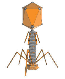
Atomic structural model of bacteriophage T4
 
The structure of a typical myovirus bacteriophage
 
Anatomy and infection cycle of phage T4.

A bacteriophage (/bækˈtɪərif/), also known informally as a phage (/ˈf/), is a virus that infects and replicates within bacteria and archaea. The term was derived from "bacteria" and the Greek φαγεῖν (phagein), meaning "to devour". Bacteriophages are composed of proteins that encapsulate a DNA or RNA genome, and may have structures that are either simple or elaborate. Their genomes may encode as few as four genes (e.g. MS2) and as many as hundreds of genes. Phages replicate within the bacterium following the injection of their genome into its cytoplasm.

Bacteriophages are among the most common and diverse entities in the biosphere. Bacteriophages are ubiquitous viruses, found wherever bacteria exist. It is estimated there are more than 1031 bacteriophages on the planet, more than every other organism on Earth, including bacteria, combined. Viruses are the most abundant biological entity in the water column of the world's oceans, and the second largest component of biomass after prokaryotes, where up to 9x10⁸ virions per millilitre have been found in microbial mats at the surface, and up to 70% of marine bacteria may be infected by phages.

Phages have been used since the late 20th century as an alternative to antibiotics in the former Soviet Union and Central Europe, as well as in France. They are seen as a possible therapy against multi-drug-resistant strains of many bacteria (see phage therapy). On the other hand, phages of Inoviridae have been shown to complicate biofilms involved in pneumonia and cystic fibrosis and to shelter the bacteria from drugs meant to eradicate disease, thus promoting persistent infection.

Classification

Bacteriophages occur abundantly in the biosphere, with different genomes, and lifestyles. Phages are classified by the International Committee on Taxonomy of Viruses (ICTV) according to morphology and nucleic acid.

Bacteriophage P22, a member of the Podoviridae by morphology due to its short, non-contractile tail

It has been suggested that members of Picobirnaviridae infect bacteria, but not mammals.

Another proposed family is "Autolykiviridae" (dsDNA).

History

In 1896, Ernest Hanbury Hankin reported that something in the waters of the Ganges and Yamuna rivers in India had a marked antibacterial action against cholera and it could pass through a very fine porcelain filter. In 1915, British bacteriologist Frederick Twort, superintendent of the Brown Institution of London, discovered a small agent that infected and killed bacteria. He believed the agent must be one of the following:

  1. a stage in the life cycle of the bacteria
  2. an enzyme produced by the bacteria themselves, or
  3. a virus that grew on and destroyed the bacteria

Twort's research was interrupted by the onset of World War I, as well as a shortage of funding and the discoveries of antibiotics.

Independently, French-Canadian microbiologist Félix d'Hérelle, working at the Pasteur Institute in Paris, announced on 3 September 1917, that he had discovered "an invisible, antagonistic microbe of the dysentery bacillus". For d’Hérelle, there was no question as to the nature of his discovery: "In a flash I had understood: what caused my clear spots was in fact an invisible microbe… a virus parasitic on bacteria." D'Hérelle called the virus a bacteriophage, a bacteria-eater (from the Greek phagein meaning "to devour"). He also recorded a dramatic account of a man suffering from dysentery who was restored to good health by the bacteriophages. It was D'Herelle who conducted much research into bacteriophages and introduced the concept of phage therapy.

More than a half a century later, in 1969, Max Delbrück, Alfred Hershey, and Salvador Luria were awarded the Nobel Prize in Physiology or Medicine for their discoveries of the replication of viruses and their genetic structure.

Uses

Phage therapy

Phages were discovered to be antibacterial agents and were used in the former Soviet Republic of Georgia (pioneered there by Giorgi Eliava with help from the co-discoverer of bacteriophages, Félix d'Herelle) during the 1920s and 1930s for treating bacterial infections. They had widespread use, including treatment of soldiers in the Red Army. However, they were abandoned for general use in the West for several reasons:

  • Antibiotics were discovered and marketed widely. They were easier to make, store, and to prescribe.
  • Medical trials of phages were carried out, but a basic lack of understanding raised questions about the validity of these trials.
  • Publication of research in the Soviet Union was mainly in the Russian or Georgian languages and for many years, was not followed internationally.

The use of phages has continued since the end of the Cold War in Russia, Georgia and elsewhere in Central and Eastern Europe. The first regulated, randomized, double-blind clinical trial was reported in the Journal of Wound Care in June 2009, which evaluated the safety and efficacy of a bacteriophage cocktail to treat infected venous ulcers of the leg in human patients. The FDA approved the study as a Phase I clinical trial. The study's results demonstrated the safety of therapeutic application of bacteriophages, but did not show efficacy. The authors explained that the use of certain chemicals that are part of standard wound care (e.g. lactoferrin or silver) may have interfered with bacteriophage viability. Shortly after that, another controlled clinical trial in Western Europe (treatment of ear infections caused by Pseudomonas aeruginosa) was reported in the journal Clinical Otolaryngology in August 2009. The study concludes that bacteriophage preparations were safe and effective for treatment of chronic ear infections in humans. Additionally, there have been numerous animal and other experimental clinical trials evaluating the efficacy of bacteriophages for various diseases, such as infected burns and wounds, and cystic fibrosis associated lung infections, among others.

Meanwhile, bacteriophage researchers have been developing engineered viruses to overcome antibiotic resistance, and engineering the phage genes responsible for coding enzymes that degrade the biofilm matrix, phage structural proteins, and the enzymes responsible for lysis of the bacterial cell wall. There have been results showing that T4 phages that are small in size and short-tailed, can be helpful in detecting E.coli in the human body.

Therapeutic efficacy of a phage cocktail was evaluated in a mice model with nasal infection of multidrug-resistant (MDR) A. baumannii. Mice treated with the phage cocktail showed a 2.3-fold higher survival rate than those untreated in seven days post infection. In 2017 a patient with a pancreas compromised by MDR A. baumannii was put on several antibiotics, despite this the patient's health continued to deteriorate during a four-month period. Without effective antibiotics the patient was subjected to phage therapy using a phage cocktail containing nine different phages that had been demonstrated to be effective against MDR A. baumannii. Once on this therapy the patient's downward clinical trajectory reversed, and returned to health.

D'Herelle "quickly learned that bacteriophages are found wherever bacteria thrive: in sewers, in rivers that catch waste runoff from pipes, and in the stools of convalescent patients." This includes rivers traditionally thought to have healing powers, including India's Ganges River.

Other

Food industry – Since 2006, the United States Food and Drug Administration (FDA) and United States Department of Agriculture (USDA) have approved several bacteriophage products. LMP-102 (Intralytix) was approved for treating ready-to-eat (RTE) poultry and meat products. In that same year, the FDA approved LISTEX (developed and produced by Micreos) using bacteriophages on cheese to kill Listeria monocytogenes bacteria, in order to give them generally recognized as safe (GRAS) status. In July 2007, the same bacteriophage were approved for use on all food products. In 2011 USDA confirmed that LISTEX is a clean label processing aid and is included in USDA. Research in the field of food safety is continuing to see if lytic phages are a viable option to control other food-borne pathogens in various food products.

Dairy industry – Bacteriophages present in the environment can cause fermentation failures of cheese starter cultures. In order to avoid this, mixed-strain starter cultures and culture rotation regimes can be used.

Diagnostics – In 2011, the FDA cleared the first bacteriophage-based product for in vitro diagnostic use. The KeyPath MRSA/MSSA Blood Culture Test uses a cocktail of bacteriophage to detect Staphylococcus aureus in positive blood cultures and determine methicillin resistance or susceptibility. The test returns results in about five hours, compared to two to three days for standard microbial identification and susceptibility test methods. It was the first accelerated antibiotic-susceptibility test approved by the FDA.

Counteracting bioweapons and toxins – Government agencies in the West have for several years been looking to Georgia and the former Soviet Union for help with exploiting phages for counteracting bioweapons and toxins, such as anthrax and botulism. Developments are continuing among research groups in the U.S. Other uses include spray application in horticulture for protecting plants and vegetable produce from decay and the spread of bacterial disease. Other applications for bacteriophages are as biocides for environmental surfaces, e.g., in hospitals, and as preventative treatments for catheters and medical devices before use in clinical settings. The technology for phages to be applied to dry surfaces, e.g., uniforms, curtains, or even sutures for surgery now exists. Clinical trials reported in Clinical Otolaryngology show success in veterinary treatment of pet dogs with otitis.

The SEPTIC bacterium sensing and identification method uses the ion emission and its dynamics during phage infection and offers high specificity and speed for detection.

Phage display is a different use of phages involving a library of phages with a variable peptide linked to a surface protein. Each phage genome encodes the variant of the protein displayed on its surface (hence the name), providing a link between the peptide variant and its encoding gene. Variant phages from the library may be selected through their binding affinity to an immobilized molecule (e.g., botulism toxin) to neutralize it. The bound, selected phages can be multiplied by reinfecting a susceptible bacterial strain, thus allowing them to retrieve the peptides encoded in them for further study.

Antimicrobial drug discovery – Phage proteins often have antimicrobial activity and may serve as leads for peptidomimetics, i.e. drugs that mimic peptides. Phage-ligand technology makes use of phage proteins for various applications, such as binding of bacteria and bacterial components (e.g. endotoxin) and lysis of bacteria.

Basic research – Bacteriophages are important model organisms for studying principles of evolution and ecology.

Replication

Diagram of the DNA injection process

Bacteriophages may have a lytic cycle or a lysogenic cycle. With lytic phages such as the T4 phage, bacterial cells are broken open (lysed) and destroyed after immediate replication of the virion. As soon as the cell is destroyed, the phage progeny can find new hosts to infect. Lytic phages are more suitable for phage therapy. Some lytic phages undergo a phenomenon known as lysis inhibition, where completed phage progeny will not immediately lyse out of the cell if extracellular phage concentrations are high. This mechanism is not identical to that of temperate phage going dormant and usually, is temporary.

In contrast, the lysogenic cycle does not result in immediate lysing of the host cell. Those phages able to undergo lysogeny are known as temperate phages. Their viral genome will integrate with host DNA and replicate along with it, relatively harmlessly, or may even become established as a plasmid. The virus remains dormant until host conditions deteriorate, perhaps due to depletion of nutrients, then, the endogenous phages (known as prophages) become active. At this point they initiate the reproductive cycle, resulting in lysis of the host cell. As the lysogenic cycle allows the host cell to continue to survive and reproduce, the virus is replicated in all offspring of the cell. An example of a bacteriophage known to follow the lysogenic cycle and the lytic cycle is the phage lambda of E. coli.

Sometimes prophages may provide benefits to the host bacterium while they are dormant by adding new functions to the bacterial genome, in a phenomenon called lysogenic conversion. Examples are the conversion of harmless strains of Corynebacterium diphtheriae or Vibrio cholerae by bacteriophages, to highly virulent ones that cause diphtheria or cholera, respectively. Strategies to combat certain bacterial infections by targeting these toxin-encoding prophages have been proposed.

Attachment and penetration

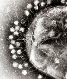
In this electron micrograph of bacteriophages attached to a bacterial cell, the viruses are the size and shape of coliphage T1

Bacterial cells are protected by a cell wall of polysaccharides, which are important virulence factors protecting bacterial cells against both immune host defenses and antibiotics. To enter a host cell, bacteriophages bind to specific receptors on the surface of bacteria, including lipopolysaccharides, teichoic acids, proteins, or even flagella. This specificity means a bacteriophage can infect only certain bacteria bearing receptors to which they can bind, which in turn, determines the phage's host range. Polysaccharide-degrading enzymes, like endolysins are virion-associated proteins to enzymatically degrade the capsular outer layer of their hosts, at the initial step of a tightly programmed phage infection process. Host growth conditions also influence the ability of the phage to attach and invade them. As phage virions do not move independently, they must rely on random encounters with the correct receptors when in solution, such as blood, lymphatic circulation, irrigation, soil water, etc.

Myovirus bacteriophages use a hypodermic syringe-like motion to inject their genetic material into the cell. After contacting the appropriate receptor, the tail fibers flex to bring the base plate closer to the surface of the cell. This is known as reversible binding. Once attached completely, irreversible binding is initiated and the tail contracts, possibly with the help of ATP, present in the tail, injecting genetic material through the bacterial membrane. The injection is accomplished through a sort of bending motion in the shaft by going to the side, contracting closer to the cell and pushing back up. Podoviruses lack an elongated tail sheath like that of a myovirus, so instead, they use their small, tooth-like tail fibers enzymatically to degrade a portion of the cell membrane before inserting their genetic material.

Synthesis of proteins and nucleic acid

Within minutes, bacterial ribosomes start translating viral mRNA into protein. For RNA-based phages, RNA replicase is synthesized early in the process. Proteins modify the bacterial RNA polymerase so it preferentially transcribes viral mRNA. The host's normal synthesis of proteins and nucleic acids is disrupted, and it is forced to manufacture viral products instead. These products go on to become part of new virions within the cell, helper proteins that contribute to the assemblage of new virions, or proteins involved in cell lysis. In 1972, Walter Fiers (University of Ghent, Belgium) was the first to establish the complete nucleotide sequence of a gene and in 1976, of the viral genome of bacteriophage MS2. Some dsDNA bacteriophages encode ribosomal proteins, which are thought to modulate protein translation during phage infection.

Virion assembly

In the case of the T4 phage, the construction of new virus particles involves the assistance of helper proteins that act catalytically during phage morphogenesis. The base plates are assembled first, with the tails being built upon them afterward. The head capsids, constructed separately, will spontaneously assemble with the tails. During assembly of the phage T4 virion, the morphogenetic proteins encoded by the phage genes interact with each other in a characteristic sequence. Maintaining an appropriate balance in the amounts of each of these proteins produced during viral infection appears to be critical for normal phage T4 morphogenesis. The DNA is packed efficiently within the heads. The whole process takes about 15 minutes.

Release of virions

Phages may be released via cell lysis, by extrusion, or, in a few cases, by budding. Lysis, by tailed phages, is achieved by an enzyme called endolysin, which attacks and breaks down the cell wall peptidoglycan. An altogether different phage type, the filamentous phage, make the host cell continually secrete new virus particles. Released virions are described as free, and, unless defective, are capable of infecting a new bacterium. Budding is associated with certain Mycoplasma phages. In contrast to virion release, phages displaying a lysogenic cycle do not kill the host but, rather, become long-term residents as prophage.

Communication

Research in 2017 revealed that the bacteriophage Φ3T makes a short viral protein that signals other bacteriophages to lie dormant instead of killing the host bacterium. Arbitrium is the name given to this protein by the researchers who discovered it.

Genome structure

Given the millions of different phages in the environment, phage genomes come in a variety of forms and sizes. RNA phage such as MS2 have the smallest genomes, of only a few kilobases. However, some DNA phage such as T4 may have large genomes with hundreds of genes; the size and shape of the capsid varies along with the size of the genome. The largest bacteriophage genomes reach a size of 735 kb.

Bacteriophage genomes can be highly mosaic, i.e. the genome of many phage species appear to be composed of numerous individual modules. These modules may be found in other phage species in different arrangements. Mycobacteriophages, bacteriophages with mycobacterial hosts, have provided excellent examples of this mosaicism. In these mycobacteriophages, genetic assortment may be the result of repeated instances of site-specific recombination and illegitimate recombination (the result of phage genome acquisition of bacterial host genetic sequences). Evolutionary mechanisms shaping the genomes of bacterial viruses vary between different families and depend upon the type of the nucleic acid, characteristics of the virion structure, as well as the mode of the viral life cycle.

Systems biology

The field of systems biology investigates the complex networks of interactions within an organism, usually using computational tools and modeling. For example, a phage genome that enters into a bacterial host cell may express hundreds of phage proteins which will affect the expression of numerous host gene or the host's metabolism. All of these complex interactions can be described and simulated in computer models.

For instance, infection of Pseudomonas aeruginosa by the temperate phage PaP3 changed the expression of 38% (2160/5633) of its host's genes. Many of these effects are probably indirect, hence the challenge becomes to identify the direct interactions among bacteria and phage.

Several attempts have been made to map protein–protein interactions among phage and their host. For instance, bacteriophage lambda was found to interact with its host, E. coli, by dozens of interactions. Again, the significance of many of these interactions remains unclear, but these studies suggest that there most likely are several key interactions and many indirect interactions whose role remains uncharacterized.

In the environment

Metagenomics has allowed the in-water detection of bacteriophages that was not possible previously.

Also, bacteriophages have been used in hydrological tracing and modelling in river systems, especially where surface water and groundwater interactions occur. The use of phages is preferred to the more conventional dye marker because they are significantly less absorbed when passing through ground waters and they are readily detected at very low concentrations. Non-polluted water may contain approximately 2×10⁸ bacteriophages per ml.

Bacteriophages are thought to contribute extensively to horizontal gene transfer in natural environments, principally via transduction, but also via transformation. Metagenomics-based studies also have revealed that viromes from a variety of environments harbor antibiotic-resistance genes, including those that could confer multidrug resistance.

Streptococcus

From Wikipedia, the free encyclopedia
https://en.wikipedia.org/wiki/Streptococcus

Streptococcus
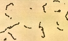
Streptococci.jpg
Scientific classification e
Domain: Bacteria
Phylum: Firmicutes
Class: Bacilli
Order: Lactobacillales
Family: Streptococcaceae
Genus: Streptococcus
Rosenbach, 1884

Streptococcus is a genus of gram-positive coccus (plural cocci) or spherical bacteria that belongs to the family Streptococcaceae, within the order Lactobacillales (lactic acid bacteria), in the phylum Firmicutes. Cell division in streptococci occurs along a single axis, so as they grow, they tend to form pairs or chains that may appear bent or twisted. This differs from staphylococci, which divide along multiple axes, thereby generating irregular, grape-like clusters of cells. Most streptococci are oxidase-negative and catalase-negative, and many are facultative anaerobes (capable of growth both aerobically and anaerobically).

The term was coined in 1877 by Viennese surgeon Albert Theodor Billroth (1829–1894), by combining the prefix "strepto-" (from Ancient Greek: στρεπτός, romanizedstreptós, lit.'easily twisted, pliant'), together with the suffix "-coccus" (from Modern Latin: coccus, from Ancient Greek: κόκκος, romanized: kókkos, lit.'grain, seed, berry'.) In 1984, many bacteria formerly grouped in the genus Streptococcus were separated out into the genera Enterococcus and Lactococcus. Currently, over 50 species are recognised in this genus. This genus has been found to be part of the salivary microbiome.

Pathogenesis and classification

In addition to streptococcal pharyngitis (strep throat), certain Streptococcus species are responsible for many cases of pink eye, meningitis, bacterial pneumonia, endocarditis, erysipelas, and necrotizing fasciitis (the 'flesh-eating' bacterial infections). However, many streptococcal species are not pathogenic, and form part of the commensal human microbiota of the mouth, skin, intestine, and upper respiratory tract. Streptococci are also a necessary ingredient in producing Emmentaler ("Swiss") cheese.

Species of Streptococcus are classified based on their hemolytic properties. Alpha-hemolytic species cause oxidization of iron in hemoglobin molecules within red blood cells, giving it a greenish color on blood agar. Beta-hemolytic species cause complete rupture of red blood cells. On blood agar, this appears as wide areas clear of blood cells surrounding bacterial colonies. Gamma-hemolytic species cause no hemolysis.

Beta-hemolytic streptococci are further classified by Lancefield grouping, a serotype classification (that is, describing specific carbohydrates present on the bacterial cell wall). The 21 described serotypes are named Lancefield groups A to W (excluding I and J). This system of classification was developed by Rebecca Lancefield, a scientist at Rockefeller University.

In the medical setting, the most important groups are the alpha-hemolytic streptococci S. pneumoniae and Streptococcus viridans group, and the beta-hemolytic streptococci of Lancefield groups A and B (also known as “group A strep” and “group B strep”).

Table: Medically relevant streptococci (not all are alpha-hemolytic)

Species Host Disease
S. pyogenes human pharyngitis, cellulitis, erysipelas
S. agalactiae human, cattle neonatal meningitis and sepsis
S. dysgalactiae human, animals endocarditis, bacteremia, pneumonia, meningitis, respiratory infections
S. gallolyticus human, animals biliary or urinary tract infections, endocarditis
S. anginosus human, animals subcutaneous/organ abscesses, meningitis, respiratory infections
S. sanguinis human endocarditis, dental caries
S. suis swine meningitis
S. mitis human endocarditis
S. mutans human dental caries
S. pneumoniae human pneumonia

Alpha-hemolytic

When alpha-hemolysis (α-hemolysis) is present, the agar under the colony will appear dark and greenish due to the conversion of hemoglobin to green biliverdin. Streptococcus pneumoniae and a group of oral streptococci (Streptococcus viridans or viridans streptococci) display alpha-hemolysis. Alpha-hemolysis is also termed incomplete hemolysis or partial hemolysis because the cell membranes of the red blood cells are left intact. This is also sometimes called green hemolysis because of the color change in the agar.

Pneumococci

  • S. pneumoniae (sometimes called pneumococcus), is a leading cause of bacterial pneumonia and occasional etiology of otitis media, sinusitis, meningitis, and peritonitis. Inflammation is thought to be the major cause of how pneumococci cause disease, hence the tendency of diagnoses associated with them to involve inflammation.

The viridans group: alpha-hemolytic

Beta-hemolytic

Beta hemolysis (β-hemolysis), sometimes called complete hemolysis, is a complete lysis of red cells in the media around and under the colonies: the area appears lightened (yellow) and transparent. Streptolysin, an exotoxin, is the enzyme produced by the bacteria which causes the complete lysis of red blood cells. There are two types of streptolysin: Streptolysin O (SLO) and streptolysin S (SLS). Streptolysin O is an oxygen-sensitive cytotoxin, secreted by most group A Streptococcus (GAS), and interacts with cholesterol in the membrane of eukaryotic cells (mainly red and white blood cells, macrophages, and platelets), and usually results in beta-hemolysis under the surface of blood agar. Streptolysin S is an oxygen-stable cytotoxin also produced by most GAS strains which results in clearing on the surface of blood agar. SLS affects immune cells, including polymorphonuclear leukocytes and lymphocytes, and is thought to prevent the host immune system from clearing infection. Streptococcus pyogenes, or GAS, displays beta hemolysis.

Some weakly beta-hemolytic species cause intense hemolysis when grown together with a strain of Staphylococcus. This is called the CAMP test. Streptococcus agalactiae displays this property. Clostridium perfringens can be identified presumptively with this test. Listeria monocytogenes is also positive on sheep's blood agar.

Alpha-hemolytic S. viridans (right) and beta-hemolytic S. pyogenes (left) streptococci growing on blood agar

Group A

Group A S. pyogenes is the causative agent in a wide range of group A streptococcal infections (GAS). These infections may be noninvasive or invasive. The noninvasive infections tend to be more common and less severe. The most common of these infections include streptococcal pharyngitis (strep throat) and impetigo. Scarlet fever is also a noninvasive infection, but has not been as common in recent years.

The invasive infections caused by group A beta-hemolytic streptococci tend to be more severe and less common. This occurs when the bacterium is able to infect areas where it is not usually found, such as the blood and the organs. The diseases that may be caused include streptococcal toxic shock syndrome, necrotizing fasciitis, pneumonia, and bacteremia. Globally, GAS has been estimated to cause more than 500,000 deaths every year, making it one of the world's leading pathogens.

Additional complications may be caused by GAS, namely acute rheumatic fever and acute glomerulonephritis. Rheumatic fever, a disease that affects the joints, kidneys, and heart valves, is a consequence of untreated strep A infection caused not by the bacterium itself. Rheumatic fever is caused by the antibodies created by the immune system to fight off the infection cross-reacting with other proteins in the body. This "cross-reaction" causes the body to essentially attack itself and leads to the damage above. A similar autoimmune mechanism initiated by Group A beta-hemolytic streptococcal (GABHS) infection is hypothesized to cause pediatric autoimmune neuropsychiatric disorders associated with streptococcal infections (PANDAS), wherein autoimmune antibodies affect the basal ganglia, causing rapid onset of psychiatric, motor, sleep, and other symptoms in pediatric patients.

GAS infection is generally diagnosed with a rapid strep test or by culture.

Group B

S. agalactiae, or group B streptococcus, GBS, causes pneumonia and meningitis in newborns and the elderly, with occasional systemic bacteremia. Importantly, Streptococcus agalactiae is the most common cause of meningitis in infants from one month to three months old. They can also colonize the intestines and the female reproductive tract, increasing the risk for premature rupture of membranes during pregnancy, and transmission of the organism to the infant. The American College of Obstetricians and Gynecologists, American Academy of Pediatrics, and the Centers for Disease Control recommend all pregnant women between 35 and 37 weeks gestation to be tested for GBS. Women who test positive should be given prophylactic antibiotics during labor, which will usually prevent transmission to the infant.

The United Kingdom has chosen to adopt a risk factor-based protocol, rather than the culture-based protocol followed in the US. Current guidelines state that if one or more of the following risk factors is present, then the woman should be treated with intrapartum antibiotics:

  • Preterm labour (<37 weeks)
  • Prolonged rupture of membranes (>18 hours)
  • Intrapartum fever (≥38 °C)
  • History of GBS disease in a previous infant
  • GBS bacteriuria during this pregnancy

This protocol results in the administration of intrapartum antibiotics to 15–20% of pregnant women and prevention of 65–70% of cases of early onset GBS sepsis.

Group C

This group includes S. equi, which causes strangles in horses, and S. zooepidemicusS. equi is a clonal descendant or biovar of the ancestral S. zooepidemicus—which causes infections in several species of mammals, including cattle and horses. S. dysgalactiae is also a member of group C, beta-haemolytic streptococci that can cause pharyngitis and other pyogenic infections similar to group A streptococci.

Group D (enterococci)

Many former group D streptococci have been reclassified and placed in the genus Enterococcus (including E. faecalis, E. faecium, E. durans, and E. avium). For example, Streptococcus faecalis is now Enterococcus faecalis. E. faecalis is sometimes alpha-hemolytic and E. faecium is sometimes beta hemolytic.

The remaining nonenterococcal group D strains include Streptococcus gallolyticus, Streptococcus bovis and Streptococcus equinus.

Nonhemolytic streptococci rarely cause illness. However, weakly hemolytic group D beta-hemolytic streptococci and Listeria monocytogenes (which is actually a gram-positive bacillus) should not be confused with nonhemolytic streptococci.

Group F streptococci

Group F streptococci were first described in 1934 by Long and Bliss amongst the "minute haemolytic streptococci". They are also known as Streptococcus anginosus (according to the Lancefield classification system) or as members of the S. milleri group (according to the European system).

Group G streptococci

These streptococci are usually, but not exclusively, beta-hemolytic. Streptococcus dysgalactiae is the predominant species encountered, particularly in human disease. S. canis is an example of a GGS which is typically found on animals, but can cause infection in humans. S. phocae is a GGS subspecies that has been found in marine mammals and marine fish species. In marine mammals it has been mainly associated with meningoencephalitis, sepsis, and endocarditis, but is also associated with many other pathologies. Its environmental reservoir and means of transmission in marine mammals is not well characterized.

Group H streptococci

Group H streptococci cause infections in medium-sized canines. Group H streptococci rarely cause human illness unless a human has direct contact with the mouth of a canine. One of the most common ways this can be spread is human-to-canine, mouth-to-mouth contact. However, the canine may lick the human's hand and infection can be spread, as well.

Molecular taxonomy and phylogenetics

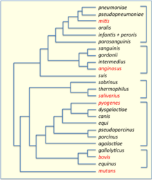
Phylogenetic tree of Streptococcus species, based on data from PATRIC. 16S groups are indicated by brackets and their key members are highlighted in red.

Streptococci have been divided into six groups on the basis of their 16S rDNA sequences: S. anginosus, S. gallolyticus, S. mitis, S. mutans, S. pyogenes and S. salivarius. The 16S groups have been confirmed by whole genome sequencing (see figure). The important pathogens S. pneumoniae and S. pyogenes belong to the S. mitis and S. pyogenes groups, respectively, while the causative agent of dental caries, Streptococcus mutans, is basal to the Streptococcus group.

Genomics

Common and species-specific genes among Streptococcus sanguinis, S. mutans, and S. pneumoniae. Modified after Xu et al. (2007)

The genomes of hundreds of species have been sequenced. Most Streptococcus genomes are 1.8 to 2.3 Mb in size and encode 1,700 to 2,300 proteins. Some important genomes are listed in the table. The four species shown in the table (S. pyogenes, S. agalactiae, S. pneumoniae, and S. mutans) have an average pairwise protein sequence identity of about 70%.

feature S. pyogenes S. agalactiae S. pneumoniae S. mutans
base pairs 1,852,442 2,211,488 2,160,837 2,030,921
ORFs 1792 2118 2236 1963
prophages yes no no no

Bacteriophage

Bacteriophages have been described for many species of Streptococcus. 18 prophages have been described in S. pneumoniae that range in size from 38 to 41 kb in size, encoding from 42 to 66 genes each. Some of the first Streptococcus phages discovered were Dp-1 and ω1 (alias ω-1). In 1981 the Cp (Complutense phage 1, officially Streptococcus virus Cp1, Picovirinae) family was discovered with Cp-1 as its first member. Dp-1 and Cp-1 infect both S. pneumoniae and S. mitis. However, the host ranges of most Streptococcus phages have not been investigated systematically.

Natural genetic transformation

Natural genetic transformation involves the transfer of DNA from one bacterium to another through the surrounding medium. Transformation is a complex process dependent on expression of numerous genes. To be capable of transformation a bacterium must enter a special physiologic state referred to as competence. S. pneumoniae, S. mitis and S. oralis can become competent, and as a result actively acquire homologous DNA for transformation by a predatory fratricidal mechanism.  This fratricidal mechanism mainly exploits non-competent siblings present in the same niche  Among highly competent isolates of S. pneumoniae, Li et al. showed that nasal colonization fitness and virulence (lung infectivity) depend on an intact competence system. Competence may allow the streptococcal pathogen to use external homologous DNA for recombinational repair of DNA damages caused by the hosts oxidative attack.

Citation signal

From Wikipedia, the free encyclopedia https://en.wikipedia.org/wiki/Cit...