Search This Blog

Thursday, July 25, 2019

RNA

From Wikipedia, the free encyclopedia

A hairpin loop from a pre-mRNA. Highlighted are the nucleobases (green) and the ribose-phosphate backbone (blue). This is a single strand of RNA that folds back upon itself.
 
Ribonucleic acid (RNA) is a polymeric molecule essential in various biological roles in coding, decoding, regulation and expression of genes. RNA and DNA are nucleic acids, and, along with lipids, proteins and carbohydrates, constitute the four major macromolecules essential for all known forms of life. Like DNA, RNA is assembled as a chain of nucleotides, but unlike DNA it is more often found in nature as a single-strand folded onto itself, rather than a paired double-strand. Cellular organisms use messenger RNA (mRNA) to convey genetic information (using the nitrogenous bases of guanine, uracil, adenine, and cytosine, denoted by the letters G, U, A, and C) that directs synthesis of specific proteins. Many viruses encode their genetic information using an RNA genome

Some RNA molecules play an active role within cells by catalyzing biological reactions, controlling gene expression, or sensing and communicating responses to cellular signals. One of these active processes is protein synthesis, a universal function in which RNA molecules direct the synthesis of proteins on ribosomes. This process uses transfer RNA (tRNA) molecules to deliver amino acids to the ribosome, where ribosomal RNA (rRNA) then links amino acids together to form coded proteins.

Comparison with DNA

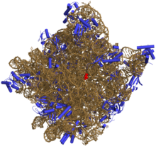
Three-dimensional representation of the 50S ribosomal subunit. Ribosomal RNA is in ochre, proteins in blue. The active site is a small segment of rRNA, indicated in red.
 
The chemical structure of RNA is very similar to that of DNA, but differs in three primary ways:
  • Unlike double-stranded DNA, RNA is a single-stranded molecule in many of its biological roles and consists of much shorter chains of nucleotides. However, a single RNA molecule can, by complementary base pairing, form intrastrand double helixes, as in tRNA.
  • While the sugar-phosphate "backbone" of DNA contains deoxyribose, RNA contains ribose instead. Ribose has a hydroxyl group attached to the pentose ring in the 2' position, whereas deoxyribose does not. The hydroxyl groups in the ribose backbone make RNA more chemically labile than DNA by lowering the activation energy of hydrolysis.
  • The complementary base to adenine in DNA is thymine, whereas in RNA, it is uracil, which is an unmethylated form of thymine.
Like DNA, most biologically active RNAs, including mRNA, tRNA, rRNA, snRNAs, and other non-coding RNAs, contain self-complementary sequences that allow parts of the RNA to fold and pair with itself to form double helices. Analysis of these RNAs has revealed that they are highly structured. Unlike DNA, their structures do not consist of long double helices, but rather collections of short helices packed together into structures akin to proteins. In this fashion, RNAs can achieve chemical catalysis (like enzymes). For instance, determination of the structure of the ribosome—an RNA-protein complex that catalyzes peptide bond formation—revealed that its active site is composed entirely of RNA.

Structure

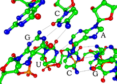
Watson-Crick base pairs in a siRNA (hydrogen atoms are not shown)
 
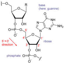
Each nucleotide in RNA contains a ribose sugar, with carbons numbered 1' through 5'. A base is attached to the 1' position, in general, adenine (A), cytosine (C), guanine (G), or uracil (U). Adenine and guanine are purines, cytosine and uracil are pyrimidines. A phosphate group is attached to the 3' position of one ribose and the 5' position of the next. The phosphate groups have a negative charge each, making RNA a charged molecule (polyanion). The bases form hydrogen bonds between cytosine and guanine, between adenine and uracil and between guanine and uracil. However, other interactions are possible, such as a group of adenine bases binding to each other in a bulge, or the GNRA tetraloop that has a guanine–adenine base-pair.

Chemical structure of RNA
 
An important structural component of RNA that distinguishes it from DNA is the presence of a hydroxyl group at the 2' position of the ribose sugar. The presence of this functional group causes the helix to mostly take the A-form geometry, although in single strand dinucleotide contexts, RNA can rarely also adopt the B-form most commonly observed in DNA. The A-form geometry results in a very deep and narrow major groove and a shallow and wide minor groove. A second consequence of the presence of the 2'-hydroxyl group is that in conformationally flexible regions of an RNA molecule (that is, not involved in formation of a double helix), it can chemically attack the adjacent phosphodiester bond to cleave the backbone.

RNA is transcribed with only four bases (adenine, cytosine, guanine and uracil), but these bases and attached sugars can be modified in numerous ways as the RNAs mature. Pseudouridine (Ψ), in which the linkage between uracil and ribose is changed from a C–N bond to a C–C bond, and ribothymidine (T) are found in various places (the most notable ones being in the TΨC loop of tRNA). Another notable modified base is hypoxanthine, a deaminated adenine base whose nucleoside is called inosine (I). Inosine plays a key role in the wobble hypothesis of the genetic code.

There are more than 100 other naturally occurring modified nucleosides. The greatest structural diversity of modifications can be found in tRNA, while pseudouridine and nucleosides with 2'-O-methylribose often present in rRNA are the most common. The specific roles of many of these modifications in RNA are not fully understood. However, it is notable that, in ribosomal RNA, many of the post-transcriptional modifications occur in highly functional regions, such as the peptidyl transferase center and the subunit interface, implying that they are important for normal function.

The functional form of single-stranded RNA molecules, just like proteins, frequently requires a specific tertiary structure. The scaffold for this structure is provided by secondary structural elements that are hydrogen bonds within the molecule. This leads to several recognizable "domains" of secondary structure like hairpin loops, bulges, and internal loops. Since RNA is charged, metal ions such as Mg2+ are needed to stabilise many secondary and tertiary structures.

The naturally occurring enantiomer of RNA is D-RNA composed of D-ribonucleotides. All chirality centers are located in the D-ribose. By the use of L-ribose or rather L-ribonucleotides, L-RNA can be synthesized. L-RNA is much more stable against degradation by RNase.

Like other structured biopolymers such as proteins, one can define topology of a folded RNA molecule. This is often done based on arrangement of intra-chain contacts within a folded RNA, termed as circuit topology.

Synthesis

Synthesis of RNA is usually catalyzed by an enzyme—RNA polymerase—using DNA as a template, a process known as transcription. Initiation of transcription begins with the binding of the enzyme to a promoter sequence in the DNA (usually found "upstream" of a gene). The DNA double helix is unwound by the helicase activity of the enzyme. The enzyme then progresses along the template strand in the 3’ to 5’ direction, synthesizing a complementary RNA molecule with elongation occurring in the 5’ to 3’ direction. The DNA sequence also dictates where termination of RNA synthesis will occur.

Primary transcript RNAs are often modified by enzymes after transcription. For example, a poly(A) tail and a 5' cap are added to eukaryotic pre-mRNA and introns are removed by the spliceosome

There are also a number of RNA-dependent RNA polymerases that use RNA as their template for synthesis of a new strand of RNA. For instance, a number of RNA viruses (such as poliovirus) use this type of enzyme to replicate their genetic material. Also, RNA-dependent RNA polymerase is part of the RNA interference pathway in many organisms.

Types of RNA

Overview

Structure of a hammerhead ribozyme, a ribozyme that cuts RNA
 
Messenger RNA (mRNA) is the RNA that carries information from DNA to the ribosome, the sites of protein synthesis (translation) in the cell. The coding sequence of the mRNA determines the amino acid sequence in the protein that is produced. However, many RNAs do not code for protein (about 97% of the transcriptional output is non-protein-coding in eukaryotes). 

These so-called non-coding RNAs ("ncRNA") can be encoded by their own genes (RNA genes), but can also derive from mRNA introns. The most prominent examples of non-coding RNAs are transfer RNA (tRNA) and ribosomal RNA (rRNA), both of which are involved in the process of translation. There are also non-coding RNAs involved in gene regulation, RNA processing and other roles. Certain RNAs are able to catalyse chemical reactions such as cutting and ligating other RNA molecules, and the catalysis of peptide bond formation in the ribosome; these are known as ribozymes.

In length

According to the length of RNA chain, RNA includes small RNA and long RNA. Usually, small RNAs are shorter than 200 nt in length, and long RNAs are greater than 200 nt long. Long RNAs, also called large RNAs, mainly include long non-coding RNA (lncRNA) and mRNA. Small RNAs mainly include 5.8S ribosomal RNA (rRNA), 5S rRNA, transfer RNA (tRNA), microRNA (miRNA), small interfering RNA (siRNA), small nucleolar RNA (snoRNAs), Piwi-interacting RNA (piRNA), tRNA-derived small RNA (tsRNA) and small rDNA-derived RNA (srRNA).

In translation

Messenger RNA (mRNA) carries information about a protein sequence to the ribosomes, the protein synthesis factories in the cell. It is coded so that every three nucleotides (a codon) corresponds to one amino acid. In eukaryotic cells, once precursor mRNA (pre-mRNA) has been transcribed from DNA, it is processed to mature mRNA. This removes its introns—non-coding sections of the pre-mRNA. The mRNA is then exported from the nucleus to the cytoplasm, where it is bound to ribosomes and translated into its corresponding protein form with the help of tRNA. In prokaryotic cells, which do not have nucleus and cytoplasm compartments, mRNA can bind to ribosomes while it is being transcribed from DNA. After a certain amount of time, the message degrades into its component nucleotides with the assistance of ribonucleases.

Transfer RNA (tRNA) is a small RNA chain of about 80 nucleotides that transfers a specific amino acid to a growing polypeptide chain at the ribosomal site of protein synthesis during translation. It has sites for amino acid attachment and an anticodon region for codon recognition that binds to a specific sequence on the messenger RNA chain through hydrogen bonding.

Ribosomal RNA (rRNA) is the catalytic component of the ribosomes. Eukaryotic ribosomes contain four different rRNA molecules: 18S, 5.8S, 28S and 5S rRNA. Three of the rRNA molecules are synthesized in the nucleolus, and one is synthesized elsewhere. In the cytoplasm, ribosomal RNA and protein combine to form a nucleoprotein called a ribosome. The ribosome binds mRNA and carries out protein synthesis. Several ribosomes may be attached to a single mRNA at any time. Nearly all the RNA found in a typical eukaryotic cell is rRNA. 

Transfer-messenger RNA (tmRNA) is found in many bacteria and plastids. It tags proteins encoded by mRNAs that lack stop codons for degradation and prevents the ribosome from stalling.

Regulatory RNA

The earliest known regulators of gene expression were proteins known as repressors and activators, regulators with specific short binding sites within enhancer regions near the genes to be regulated.  More recently, RNAs have been found to regulate genes as well.  There are several kinds of RNA-dependent processes in eukaryotes regulating the expression of genes at various points, such as RNAi repressing genes post-transcriptionally, long non-coding RNAs shutting down blocks of chromatin epigenetically, and enhancer RNAs inducing increased gene expression. In addition to these mechanisms in eukaryotes, both bacteria and archaea have been found to use regulatory RNAs extensively. Bacterial small RNA and the CRISPR system are examples of such prokaryotic regulatory RNA systems. Fire and Mello were awarded the 2006 Nobel Prize in Physiology or Medicine for discovering microRNAs (miRNAs), specific short RNA molecules that can base-pair with mRNAs.

RNA interference by miRNAs

Post-transcriptional expression levels of many genes can be controlled by RNA interference, in which miRNAs, specific short RNA molecules, pair with meRNA regions and target them for degradation. This antisense-based process involves steps that first process the RNA so that it can base-pair with a region of its target mRNAs. Once the base pairing occurs, other proteins direct the mRNA to be destroyed by nucleases. Fire and Mello were awarded the 2006 Nobel Prize in Physiology or Medicine for this discovery.

Long non-coding RNAs

Next to be linked to regulation were Xist and other long noncoding RNAs associated with X chromosome inactivation.  Their roles, at first mysterious, were shown by Jeannie T. Lee and others to be the silencing of blocks of chromatin via recruitment of Polycomb complex so that messenger RNA could not be transcribed from them.  Additional lncRNAs, currently defined as RNAs of more than 200 base pairs that do not appear to have coding potential, have been found associated with regulation of stem cell pluripotency and cell division.

Enhancer RNAs

The third major group of regulatory RNAs is called enhancer RNAs.  It is not clear at present whether they are a unique category of RNAs of various lengths or constitute a distinct subset of lncRNAs.  In any case, they are transcribed from enhancers, which are known regulatory sites in the DNA near genes they regulate.  They up-regulate the transcription of the gene(s) under control of the enhancer from which they are transcribed.

Regulatory RNA in prokaryotes

At first, regulatory RNA was thought to be a eukaryotic phenomenon, a part of the explanation for why so much more transcription in higher organisms was seen than had been predicted. But as soon as researchers began to look for possible RNA regulators in bacteria, they turned up there as well. Currently, the ubiquitous nature of systems of RNA regulation of genes has been discussed as support for the RNA World theory. Bacterial small RNAs generally act via antisense pairing with mRNA to down-regulate its translation, either by affecting stability or affecting cis-binding ability. Riboswitches have also been discovered. They are cis-acting regulatory RNA sequences acting allosterically. They change shape when they bind metabolites so that they gain or lose the ability to bind chromatin to regulate expression of genes.

Archaea also have systems of regulatory RNA. The CRISPR system, recently being used to edit DNA in situ, acts via regulatory RNAs in archaea and bacteria to provide protection against virus invaders.

In RNA processing

Uridine to pseudouridine is a common RNA modification.
 
Many RNAs are involved in modifying other RNAs. Introns are spliced out of pre-mRNA by spliceosomes, which contain several small nuclear RNAs (snRNA), or the introns can be ribozymes that are spliced by themselves. RNA can also be altered by having its nucleotides modified to nucleotides other than A, C, G and U. In eukaryotes, modifications of RNA nucleotides are in general directed by small nucleolar RNAs (snoRNA; 60–300 nt), found in the nucleolus and cajal bodies. snoRNAs associate with enzymes and guide them to a spot on an RNA by basepairing to that RNA. These enzymes then perform the nucleotide modification. rRNAs and tRNAs are extensively modified, but snRNAs and mRNAs can also be the target of base modification. RNA can also be methylated.

RNA genomes

Like DNA, RNA can carry genetic information. RNA viruses have genomes composed of RNA that encodes a number of proteins. The viral genome is replicated by some of those proteins, while other proteins protect the genome as the virus particle moves to a new host cell. Viroids are another group of pathogens, but they consist only of RNA, do not encode any protein and are replicated by a host plant cell's polymerase.

In reverse transcription

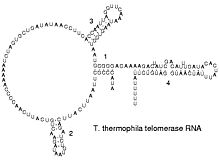
Reverse transcribing viruses replicate their genomes by reverse transcribing DNA copies from their RNA; these DNA copies are then transcribed to new RNA. Retrotransposons also spread by copying DNA and RNA from one another, and telomerase contains an RNA that is used as template for building the ends of eukaryotic chromosomes.

Double-stranded RNA

Double-stranded RNA
 
Double-stranded RNA (dsRNA) is RNA with two complementary strands, similar to the DNA found in all cells, but with the replacement of thymine by uracil. dsRNA forms the genetic material of some viruses (double-stranded RNA viruses). Double-stranded RNA, such as viral RNA or siRNA, can trigger RNA interference in eukaryotes, as well as interferon response in vertebrates.

Circular RNA

In the late 1970s, it was shown that there is a single stranded covalently closed, i.e. circular form of RNA expressed throughout the animal and plant kingdom. circRNAs are thought to arise via a "back-splice" reaction where the spliceosome joins a downstream donor to an upstream acceptor splice site. So far the function of circRNAs is largely unknown, although for few examples a microRNA sponging activity has been demonstrated.

Key discoveries in RNA biology

Robert W. Holley, left, poses with his research team.
 
Research on RNA has led to many important biological discoveries and numerous Nobel Prizes. Nucleic acids were discovered in 1868 by Friedrich Miescher, who called the material 'nuclein' since it was found in the nucleus. It was later discovered that prokaryotic cells, which do not have a nucleus, also contain nucleic acids. The role of RNA in protein synthesis was suspected already in 1939. Severo Ochoa won the 1959 Nobel Prize in Medicine (shared with Arthur Kornberg) after he discovered an enzyme that can synthesize RNA in the laboratory. However, the enzyme discovered by Ochoa (polynucleotide phosphorylase) was later shown to be responsible for RNA degradation, not RNA synthesis. In 1956 Alex Rich and David Davies hybridized two separate strands of RNA to form the first crystal of RNA whose structure could be determined by X-ray crystallography.

The sequence of the 77 nucleotides of a yeast tRNA was found by Robert W. Holley in 1965, winning Holley the 1968 Nobel Prize in Medicine (shared with Har Gobind Khorana and Marshall Nirenberg).
During the early 1970s, retroviruses and reverse transcriptase were discovered, showing for the first time that enzymes could copy RNA into DNA (the opposite of the usual route for transmission of genetic information). For this work, David Baltimore, Renato Dulbecco and Howard Temin were awarded a Nobel Prize in 1975. In 1976, Walter Fiers and his team determined the first complete nucleotide sequence of an RNA virus genome, that of bacteriophage MS2.

In 1977, introns and RNA splicing were discovered in both mammalian viruses and in cellular genes, resulting in a 1993 Nobel to Philip Sharp and Richard Roberts. Catalytic RNA molecules (ribozymes) were discovered in the early 1980s, leading to a 1989 Nobel award to Thomas Cech and Sidney Altman. In 1990, it was found in Petunia that introduced genes can silence similar genes of the plant's own, now known to be a result of RNA interference.

At about the same time, 22 nt long RNAs, now called microRNAs, were found to have a role in the development of C. elegans. Studies on RNA interference gleaned a Nobel Prize for Andrew Fire and Craig Mello in 2006, and another Nobel was awarded for studies on the transcription of RNA to Roger Kornberg in the same year. The discovery of gene regulatory RNAs has led to attempts to develop drugs made of RNA, such as siRNA, to silence genes. Adding to the Nobel prizes awarded for research on RNA in 2009 it was awarded for the elucidation of the atomic structure of the ribosome to Venki Ramakrishnan, Tom Steitz, and Ada Yonath.

Relevance for prebiotic chemistry and abiogenesis

In 1967, Carl Woese hypothesized that RNA might be catalytic and suggested that the earliest forms of life (self-replicating molecules) could have relied on RNA both to carry genetic information and to catalyze biochemical reactions—an RNA world.

In March 2015, complex DNA and RNA nucleotides, including uracil, cytosine and thymine, were reportedly formed in the laboratory under outer space conditions, using starter chemicals, such as pyrimidine, an organic compound commonly found in meteorites. Pyrimidine, like polycyclic aromatic hydrocarbons (PAHs), is one of the most carbon-rich compounds found in the Universe and may have been formed in red giants or in interstellar dust and gas clouds.

Neurofeedback

From Wikipedia, the free encyclopedia
 
Software real time data of neurofeedback training.
 
Neurofeedback (NFB), also called neurotherapy or neurobiofeedback, is a type of biofeedback that uses real-time displays of brain activity—most commonly electroencephalography (EEG)—in an attempt to teach self-regulation of brain function. Typically, sensors are placed on the scalp to measure electrical activity, with measurements displayed using video displays or sound.

Definition

Neurofeedback is a type of biofeedback that measures brain waves to produce a signal that can be used as feedback to teach self-regulation of brain function. Neurofeedback is commonly provided using video or sound, with positive feedback for desired brain activity and negative feedback for brain activity that is undesirable. Related technologies include hemoencephalography biofeedback (HEG) and functional magnetic resonance imaging (fMRI) biofeedback.

Uses

ADHD

Clinical guidelines on neurofeedback as a treatment for ADHD are mixed. However, the American Academy of Pediatrics does not list neurofeedback or biofeedback as a recommended treatment in their clinical practice guidelines for the diagnosis, evaluation, and treatment of ADHD in children and adolescents, instead mentioning EEG biofeedback as an area for future research. The NICE guideline for ADHD leaves the efficacy of biofeedback an open question (p. 412). In page 202 states "Biofeedback has been employed as a non-invasive treatment for children with ADHD since the 1970s but is probably not used as a significant intervention in UK clinical practice". However this is unsurprising since in the UK, NICE evaluates whether treatments should be recommended on the basis of the cost of a quality-adjusted life year. SIGN guideline no 112 in page 24 mentions "Neurofeedback is presently considered to be an experimental intervention in children and young people with ADHD/HKD. There are no standardised interventions". Institute for Clinical Systems Improvement guideline on Diagnosis and Management of Attention Deficit Hyperactivity Disorder in Primary Care for School-Age Children and Adolescents in page 41 mentions neurofeedback lacks enough research evidence for efficacy in ADHD.

Overall research into neurofeedback is considered to have been limited and of low quality, although others have disagreed.

It has been argued there is some indication on the effectiveness of biofeedback for ADHD but that it is not conclusive: several studies have yielded positive results, however the best designed ones have either shown absent or reduced effects. Other experts have proposed that standard neurofeedback protocols for ADHD, such as theta/beta, SMR and slow cortical potentials neurofeedback are well investigated and have demonstrated specificity. No serious adverse side effects from neurofeedback have been reported.

QEEG has been used to develop EEG models of ADHD. According to this model, persons with ADHD often have too many slow theta brain waves (associated with relaxation) and not enough fast beta wave activity (associated with mental focus). Neurofeedback therapies for ADHD generally attempt to increase the production of betawaves and decrease the number of slower brain waves. This can be accomplished by allowing the patient to view their levels of brain waves on a screen and attempt to alter them, or by integrating brain waves into a video game.

Other medical uses

Research shows neurofeedback may be a potentially useful intervention for a range of brain-related conditions. It has been used for pain, addiction, aggression, anxiety, autism, depression, Schizophrenia, epilepsy, headaches, insomnia, Tourette syndrome, and brain damage from stroke, trauma, and other conditions.

It is also used to treat other less well known disorders, such as Auditory Processing Disorder and working memory deficit.

Non-medical

The applications of neurofeedback to enhance performance extend to the arts in fields such as music, dance, and acting. A study with conservatoire musicians found that alpha-theta training benefitted the three music domains of musicality, communication, and technique. Historically, alpha-theta training, a form of neurofeedback, was created to assist creativity by inducing hypnagogia, a “borderline waking state associated with creative insights”, through facilitation of neural connectivity. Alpha-theta training has also been shown to improve novice singing in children. Alpha-theta neurofeedback, in conjunction with heart rate variability training, a form of biofeedback, has also produced benefits in dance by enhancing performance in competitive ballroom dancing and increasing cognitive creativity in contemporary dancers. Additionally, neurofeedback has also been shown to instil a superior flow state in actors, possibly due to greater immersion while performing.

However, randomized control trials have found that neurofeedback training (using either sensorimotor rhythm or theta/beta ratio training) did not enhance performance on attention-related tasks or creative tasks. It has been suggested that claims made by proponents of alpha wave neurofeedback training techniques have yet to be validated by randomized, double-blind, controlled studies, a view which even some supporters of alpha neurofeedback training have also expressed.

History and application

In 1924, the German psychiatrist Hans Berger connected a couple of electrodes (small round discs of metal) to a patient's scalp and detected a small current by using a ballistic galvanometer. During the years 1929-1938 he published 14 reports about his studies of EEGs, and much of our modern knowledge of the subject, especially in the middle frequencies, is due to his research. Berger analyzed EEGs qualitatively, but in 1932 G. Dietsch applied Fourier analysis to seven records of EEG and became the first researcher of what later is called QEEG (quantitative EEG).

Later, Joe Kamiya popularized neurofeedback in the 1960s when an article about the alpha brain wave experiments he had been conducting was published in Psychology Today in 1968. Kamiya’s experiment had two parts. In the first part, a subject was asked to keep his eyes closed and when a tone sounded to say whether he thought he was in alpha. He was then told whether he was correct or wrong. Initially the subject would get about fifty percent correct, but some subjects would eventually develop the ability to better distinguish between states. In the second part of the study, subjects were asked to go into alpha when a bell rang once and not go into the state when the bell rang twice. Once again some subjects were able to enter the state on command. Alpha states were connected with relaxation, and alpha training had the possibility to alleviate stress and stress-related conditions.

Despite these claims, the universal correlation of high alpha density to a subjective experience of calm cannot be assumed. Alpha states do not seem to have the universal stress-alleviating power indicated by early observations. At one point, Martin Orne and others challenged the claim that alpha biofeedback actually involved the training of an individual to voluntarily regulate brainwave activity. James Hardt and Joe Kamiya, then at UC San Francisco's Langley Porter Neuropsychiatric Institute published a paper that supported biofeedback.

In the late sixties and early seventies, Barbara Brown, one of the most effective popularizers of Biofeedback, wrote several books on biofeedback, making the public much more aware of the technology. The books included New Mind New Body, with a foreword from Hugh Downs, and Stress and the Art of Biofeedback. Brown took a creative approach to neurofeedback, linking brainwave self-regulation to a switching relay which turned on an electric train.

The work of Barry Sterman, Joel F. Lubar and others has been relevant on the study of beta training, involving the role of sensorimotor rhythmic EEG activity. This training has been used in the treatment of epilepsy, attention deficit disorder and hyperactive disorder. The sensorimotor rhythm (SMR) is rhythmic activity between 12 and 16 hertz that can be recorded from an area near the sensorimotor cortex. SMR is found in waking states and is very similar if not identical to the sleep spindles that are recorded in the second stage of sleep.

For example, Sterman has shown that both monkeys and cats who had undergone SMR training had elevated thresholds for the convulsant chemical monomethylhydrazine. These studies indicate that SMR may be associated with an inhibitory process in the motor system.

Within the last 5–10 years, neurofeedback has taken a new approach in taking a look at deep states. Alpha-theta training has been tried with patients with alcoholism, other addictions as well as anxiety. This low frequency training differs greatly from the high frequency beta and SMR training that has been practiced for over thirty years and is reminiscent of the original alpha training of Elmer Green and Joe Kamiya. Beta and SMR training can be considered a more directly physiological approach, strengthening sensorimotor inhibition in the cortex and inhibiting alpha patterns, which slow metabolism.  Alpha-theta training, however, derives from the psychotherapeutic model and involves accessing of painful or repressed memories through the alpha-theta state. The alpha-theta state is a term that comes from the representation on the EEG.

A recent development in the field is a conceptual approach called the Coordinated Allocation of Resource Model (CAR) of brain functioning which states that specific cognitive abilities are a function of specific electrophysiological variables which can overlap across different cognitive tasks. The activation database guided EEG biofeedback approach initially involves evaluating the subject on a number of academically relevant cognitive tasks and compares the subject's values on the QEEG measures to a normative database, in particular on the variables that are related to success at that task.

Organizations

The Association for Applied Psychophysiology and Biofeedback (AAPB) is a non-profit scientific and professional society for biofeedback and neurofeedback. The International Society for Neurofeedback and Research (ISNR) is a non-profit scientific and professional society for neurofeedback. The Biofeedback Federation of Europe (BFE) sponsors international education, training, and research activities in biofeedback and neurofeedback.

Certification

The Biofeedback Certification International Alliance (formerly the Biofeedback Certification Institute of America) is a non-profit organization that is a member of the Institute for Credentialing Excellence (ICE). BCIA certifies individuals who meet education and training standards in biofeedback and neurofeedback and progressively recertifies those who satisfy continuing education requirements. BCIA offers biofeedback certification, neurofeedback (also called EEG biofeedback) certification, and pelvic muscle dysfunction biofeedback certification. BCIA certification has been endorsed by the Mayo Clinic, the Association for Applied Psychophysiology and Biofeedback (AAPB), the International Society for Neurofeedback and Research (ISNR), and the Washington State Legislature.

The BCIA didactic education requirement includes a 36-hour course from a regionally accredited academic institution or a BCIA-approved training program that covers the complete Neurofeedback Blueprint of Knowledge and study of human anatomy and physiology. The Neurofeedback Blueprint of Knowledge areas include: I. Orientation to Neurofeedback, II. Basic Neurophysiology and Neuroanatomy, III. Instrumentation and Electronics, IV. Research, V. Psychopharmalogical Considerations, VI. Treatment Planning, and VII. Professional Conduct.

Applicants may demonstrate their knowledge of human anatomy and physiology by completing a course in biological psychology, human anatomy, human biology, human physiology, or neuroscience provided by a regionally accredited academic institution or a BCIA-approved training program or by successfully completing an Anatomy and Physiology exam covering the organization of the human body and its systems. 

Applicants must also document practical skills training that includes 25 contact hours supervised by a BCIA-approved mentor designed to teach them how to apply clinical biofeedback skills through self-regulation training, 100 patient/client sessions, and case conference presentations. Distance learning allows applicants to complete didactic course work over the internet. Distance mentoring trains candidates from their residence or office. They must recertify every 4 years, complete 55 hours of continuing education (30 hours for Senior Fellows) during each review period or complete the written exam, and attest that their license/credential (or their supervisor’s license/credential) has not been suspended, investigated, or revoked.

Neuroplasticity

In 2010, a study provided some evidence of neuroplastic changes occurring after brainwave training. Half an hour of voluntary control of brain rhythms led in this study to a lasting shift in cortical excitability and intracortical function. The authors observed that the cortical response to transcranial magnetic stimulation (TMS) was significantly enhanced after neurofeedback, persisted for at least 20 minutes, and was correlated with an EEG time-course indicative of activity-dependent plasticity.

Criticism

It has been suggested that the benefits of EEG neurofeedback come from placebo effects, and the effectiveness of the treatment remains controversial.

Although over 3,000 scientific articles have been published on EEG neurofeedback since 1968, EEG neurofeedback has failed to gain acceptance among the medical mainstream. The United States Food and Drug Administration (FDA) allows neurofeedback for relaxation training, but have never approved neurofeedback for any other purpose.

Creutzfeldt–Jakob disease

From Wikipedia, the free encyclopedia

Creutzfeldt–Jakob disease
Other namesClassic Creutzfeldt–Jakob disease
Practneurol-2016-001571f02.jpg
MRI of sporadic CJD
Pronunciation
SpecialtyNeurology
SymptomsEarly: memory problems, behavioral changes, poor coordination, visual disturbances
Later: dementia, involuntary movements, blindness, weakness, coma
Usual onsetAround 60
TypesSporadic, hereditary, acquired
CausesPrion
Diagnostic methodAfter ruling out other possible causes
Differential diagnosisEncephalitis, chronic meningitis, Huntington’s disease, Alzheimer's disease
TreatmentSupportive care
PrognosisUniversally fatal; 70% die within a year of diagnosis
Frequency1 per million per year

Creutzfeldt–Jakob disease (CJD), also known as classic Creutzfeldt–Jakob disease, is a fatal degenerative brain disorder. Early symptoms include memory problems, behavioral changes, poor coordination, and visual disturbances. Later dementia, involuntary movements, blindness, weakness, and coma occur. About 70% of people die within a year of diagnosis.

CJD is caused by a protein known as a prion. Infectious prions are misfolded proteins that can cause normally folded proteins to become misfolded. Most cases occur spontaneously, while about 7.5% of cases are inherited from a person's parents in an autosomal dominant manner. Exposure to brain or spinal tissue from an infected person may also result in spread. There is no evidence that it can spread between people via normal contact or blood transfusions. Diagnosis involves ruling out other potential causes. An electroencephalogram, spinal tap, or magnetic resonance imaging may support the diagnosis.

There is no specific treatment. Opioids may be used to help with pain, while clonazepam or sodium valproate may help with involuntary movements. CJD affects about one per million people per year. Onset is typically around 60 years of age. The condition was first described in 1920. It is classified as a type of transmissible spongiform encephalopathy. CJD is different from bovine spongiform encephalopathy (mad cow disease) and variant Creutzfeldt–Jakob disease (vCJD).

Signs and symptoms

The first symptom of CJD is usually rapidly progressive dementia, leading to memory loss, personality changes, and hallucinations. Myoclonus (jerky movements) typically occurs in 90% of cases, but may be absent at initial onset. Other frequently occurring features include anxiety, depression, paranoia, obsessive-compulsive symptoms, and psychosis. This is accompanied by physical problems such as speech impairment, balance and coordination dysfunction (ataxia), changes in gait, rigid posture. In most people with CJD, these symptoms are accompanied by involuntary movements and the appearance of an atypical, diagnostic electroencephalogram tracing. The duration of the disease varies greatly, but sporadic (non-inherited) CJD can be fatal within months or even weeks. Most victims die six months after initial symptoms appear, often of pneumonia due to impaired coughing reflexes. About 15% of people with CJD survive for two or more years.

The symptoms of CJD are caused by the progressive death of the brain's nerve cells, which is associated with the build-up of abnormal prion protein molecules forming amyloids. When brain tissue from a person with CJD is examined under a microscope, many tiny holes can be seen where whole areas of nerve cells have died. The word "spongiform" in "transmissible spongiform encephalopathies" refers to the sponge-like appearance of the brain tissue.

Cause

CJD, is a type of transmissible spongiform encephalopathy (TSE), which are caused by prions. Prions are proteins that occur normally in neurons of the central nervous system (CNS). These proteins, once misfolded, are thought to affect signaling processes, damaging neurons and resulting in degeneration that causes the spongiform appearance in the affected brain.

The CJD prion is dangerous because it promotes refolding of native prion protein into the diseased state. The number of misfolded protein molecules will increase exponentially and the process leads to a large quantity of insoluble protein in affected cells. This mass of misfolded proteins disrupts neuronal cell function and causes cell death. Mutations in the gene for the prion protein can cause a misfolding of the dominantly alpha helical regions into beta pleated sheets. This change in conformation disables the ability of the protein to undergo digestion. Once the prion is transmitted, the defective proteins invade the brain and induce other prion protein molecules to misfold in a self-sustaining feedback loop. These neurodegenerative diseases are commonly called prion diseases.

People can also develop CJD because they carry a mutation of the gene that codes for the prion protein (PRNP). This occurs in only 5–10% of all CJD cases. In sporadic cases the misfolding of the prion protein probably occurs as a natural, spontaneous process. An EU study determined that "87% of cases were sporadic, 8% genetic, 5% iatrogenic and less than 1% variant."

Transmission

MRI of iCJD because of growth hormone
 
The defective protein can be transmitted by contaminated harvested human brain products, corneal grafts, dural grafts, or electrode implants and human growth hormone.

It can be familial (fCJD); or it may appear without risk factors (sporadic form: sCJD). In the familial form, a mutation has occurred in the gene for PrP, PRNP, in that family. All types of CJD are transmissible irrespective of how they occur in the person.

It is thought that humans can contract the disease by consuming material from animals infected with the bovine form of the disease.

Cannibalism has also been implicated as a transmission mechanism for abnormal prions, causing the disease known as kuru, once found primarily among women and children of the Fore people in Papua New Guinea. While the men of the tribe ate the body of the deceased and rarely contracted the disease, the women and children, who ate the less desirable body parts, including the brain, were eight times more likely than men to contract kuru from infected tissue.

Prions, the infectious agent of CJD, may not be inactivated by means of routine surgical instrument sterilization procedures. The World Health Organization and the US Centers for Disease Control and Prevention recommend that instrumentation used in such cases be immediately destroyed after use; short of destruction, it is recommended that heat and chemical decontamination be used in combination to process instruments that come in contact with high-infectivity tissues. No cases of iatrogenic transmission of CJD have been reported subsequent to the adoption of current sterilization procedures, or since 1976. Copper-hydrogen peroxide has been suggested as an alternative to the current recommendation of sodium hydroxide or sodium hypochlorite. Thermal depolymerization also destroys prions in infected organic and inorganic matter, since the process chemically attacks protein at the molecular level, although more effective and practical methods involve destruction by combinations of detergents and enzymes similar to biological washing powders.

Blood products

As of 2018, evidence suggests that while there may be prions in the blood of individuals with vCJD, this is not the case in individuals with sporadic CJD.

Diagnosis

Testing for CJD has historically been problematic, due to nonspecific nature of early symptoms and difficulty in safely obtaining brain tissue for confirmation. The diagnosis may initially be suspected in a person with rapidly progressing dementia, particularly when they are also found with the characteristic medical signs and symptoms such as involuntary muscle jerking, difficulty with coordination/balance and walking, and visual disturbances. Further testing can support the diagnosis and may include:
  • Electroencephalography – may have characteristic generalized periodic sharp wave pattern. Periodic sharp wave complexes develop in half of the people with sporadic CJD, particularly in the later stages.
  • Cerebrospinal fluid (CSF) analysis for elevated levels of 14-3-3 protein could be supportive in the diagnosis of sCJD. However, a positive result should not be regarded as sufficient for the diagnosis. The Real-Time Quaking-Induced Conversion (RT-QuIC) assay has a diagnostic sensitivity of more than 80% and a specificity approaching 100%, tested in detecting PrPSc in CSF samples of people with CJD. It is therefore suggested as a high-value diagnostic method for the disease.
  • MRI of the brain – often shows high signal intensity in the caudate nucleus and putamen bilaterally on T2-weighted images.
In recent years, studies have shown that the tumour marker Neuron-specific enolase (NSE) is often elevated in CJD cases; however, its diagnostic utility is seen primarily when combined with a test for the 14-3-3 protein. As of 2010, screening tests to identify infected asymptomatic individuals, such as blood donors, are not yet available, though methods have been proposed and evaluated.

Imaging

Imaging of the brain may be performed during medical evaluation, both to rule out other causes and to obtain supportive evidence for diagnosis. Imaging findings are variable in their appearance, and also variable in sensitivity and specificity. While imaging plays a lesser role in diagnosis of CJD, characteristic findings on brain MRI in some cases may precede onset of clinical manifestations.

Brain MRI is most useful imaging modality for changes related to CJD. Of the MRI sequences, diffuse-weighted imaging sequences are most sensitive. Characteristic findings are as follows:
  • Focal or diffuse diffusion-restriction involving the cerebral cortex and/or basal ganglia. In about 24% of cases DWI shows only cortical hyperintensity; in 68%, cortical and subcortical abnormalities; and in 5%, only subcortical anomalies. The most iconic and striking cortical abnormality has been called "cortical ribboning" or "cortical ribbon sign" due to hyperintensities resembling ribbons appearing in the cortex on MRI. The involvement of the thalamus can be found in sCJD, is even stronger and constant in vCJD.
  • Varying degree of symmetric T2 hyperintense signal changes in the basal ganglia (i.e., caudate and putamen), and to a lesser extent globus pallidus and occipital cortex.
  • Cerebellar atrophy

Histopathology

Spongiform change in CJD
 
Testing of tissue remains the most definitive way of confirming the diagnosis of CJD, although it must be recognized that even biopsy is not always conclusive. 

In one-third of people with sporadic CJD, deposits of "prion protein (scrapie)," PrPSc, can be found in the skeletal muscle and/or the spleen. Diagnosis of vCJD can be supported by biopsy of the tonsils, which harbour significant amounts of PrPSc; however, biopsy of brain tissue is the definitive diagnostic test for all other forms of prion disease. Due to its invasiveness, biopsy will not be done if clinical suspicion is sufficiently high or low. A negative biopsy does not rule out CJD, since it may predominate in a specific part of the brain.

The classic histologic appearance is spongiform change in the gray matter: the presence of many round vacuoles from one to 50 micrometers in the neuropil, in all six cortical layers in the cerebral cortex or with diffuse involvement of the cerebellar molecular layer. These vacuoles appear glassy or eosinophilic and may coalesce. Neuronal loss and gliosis are also seen. Plaques of amyloid-like material can be seen in the neocortex in some cases of CJD. 

However, extra-neuronal vacuolization can also be seen in other disease states. Diffuse cortical vacuolization occurs in Alzheimer's disease, and superficial cortical vacuolization occurs in ischemia and frontotemporal dementia. These vacuoles appear clear and punched-out. Larger vacuoles encircling neurons, vessels, and glia are a possible processing artifact.

Classification

Types of CJD include:
  • Sporadic (sCJD), caused by the spontaneous misfolding of prion-protein in an individual. This accounts for 85% of cases of CJD.
  • Familial (fCJD), caused by an inherited mutation in the prion-protein gene. This accounts for the majority of the other 15% of cases of CJD.
  • Acquired CJD, caused by contamination with tissue from an infected person, usually as the result of a medical procedure (iatrogenic CJD). Medical procedures that are associated with the spread of this form of CJD include blood transfusion from the infected person, use of human-derived pituitary growth hormones, gonadotropin hormone therapy, and corneal and meningeal transplants. Variant Creutzfeldt–Jakob disease (vCJD) is a type of acquired CJD potentially acquired from bovine spongiform encephalopathy or caused by consuming food contaminated with prions.
Clinical and pathologic characteristics
Characteristic Classic CJD Variant CJD
Median age at death 68 years 28 years
Median duration of illness 4–5 months 13–14 months
Clinical signs and symptoms Dementia; early neurologic signs Prominent psychiatric/behavioral symptoms; painful dysesthesias; delayed neurologic signs
Periodic sharp waves on electroencephalogram Often present Often absent
Signal hyperintensity in the caudate nucleus and putamen on diffusion-weighted and FLAIR MRI Often present Often absent
Pulvinar sign-bilateral high signal intensities on axial FLAIR MRI. Also posterior thalamic involvement on sagittal T2 sequences Not reported Present in >75% of cases
Immunohistochemical analysis of brain tissue Variable accumulation. Marked accumulation of protease-resistant prion protein
Presence of agent in lymphoid tissue Not readily detected Readily detected
Increased glycoform ratio on immunoblot analysis of protease-resistant prion protein Not reported Marked accumulation of protease-resistant prion protein
Presence of amyloid plaques in brain tissue May be present May be present

Treatment

As of 2015 there was no cure for CJD. Some of the symptoms like twitching can be managed, but otherwise treatment is palliative care. Psychiatric symptoms like anxiety and depression can be treated with sedatives and antidepressants. Myoclonic jerks can be handled with clonazepam or sodium valproate. Opiates can help in pain. Seizures are very uncommon and can be treated with antiepileptic drugs.

Prognosis

The condition is universally fatal. As of 1981 people are not known to have lived longer than 2.5 years after the onset of CJD symptoms.

Epidemiology

Although CJD is the most common human prion disease, it is still believed to be rare, estimated to occur in about one out of every one million people every year. However, an autopsy study published in 1989 and others suggest that between 3–13% of people diagnosed with Alzheimer's were actually misdiagnosed and instead had CJD. Presumably, those afflicted have become infected through prion-contaminated beef from cattle with subclinical atypical BSE (bovine spongiform encephalopathy), which has a very long incubation period. CJD usually affects people aged 45–75, most commonly appearing in people between the ages of 60–65. The exception to this is the more recently recognised 'variant' CJD (vCJD), which occurs in younger people.

CDC monitors the occurrence of CJD in the United States through periodic reviews of national mortality data. According to the CDC:
  • CJD occurs worldwide at a rate of about 1 case per million population per year.
  • On the basis of mortality surveillance from 1979 to 1994, the annual incidence of CJD remained stable at approximately 1 case per million people in the United States.
  • In the United States, CJD deaths among people younger than 30 years of age are extremely rare (fewer than five deaths per billion per year).
  • The disease is found most frequently in people 55–65 years of age, but cases can occur in people older than 90 years and younger than 55 years of age.
  • In more than 85% of cases, the duration of CJD is less than 1 year (median: four months) after onset of symptoms.

History

The disease was first described by German neurologist Hans Gerhard Creutzfeldt in 1920 and shortly afterward by Alfons Maria Jakob, giving it the name Creutzfeldt–Jakob. Some of the clinical findings described in their first papers do not match current criteria for Creutzfeldt–Jakob disease, and it has been speculated that at least two of the people in initial studies were suffering from a different ailment. An early description of familial CJD stems from the German psychiatrist and neurologist Friedrich Meggendorfer (1880–1953). A study published in 1997 counted more than 100 cases worldwide of transmissible CJD and new cases continued to appear at the time.

The first report of suspected iatrogenic CJD was published in 1974. Animal experiments showed that corneas of infected animals could transmit CJD, and the causative agent spreads along visual pathways. A second case of CJD associated with a corneal transplant was reported without details. In 1977, CJD transmission caused by silver electrodes previously used in the brain of a person with CJD was first reported. Transmission occurred despite decontamination of the electrodes with ethanol and formaldehyde. Retrospective studies identified four other cases likely of similar cause. The rate of transmission from a single contaminated instrument is unknown, although it is not 100%. In some cases, the exposure occurred weeks after the instruments were used on a person with CJD. In the 1980s it was discovered that Lyodura, a dura mater transplant product was shown to transmit Creutzfeldt–Jakob disease from the donor to the recipient. This led to the product being banned in Canada but it was used in other countries as Japan until 1993.

A review article published in 1979 indicated that 25 dura mater cases had occurred by that date in Australia, Canada, Germany, Italy, Japan, New Zealand, Spain, the United Kingdom, and the United States.

By 1985, a series of case reports in the United States showed that when injected, cadaver-extracted pituitary human growth hormone could transmit CJD to humans.

In 1992, it was recognized that human gonadotropin administered by injection could also transmit CJD from person to person.

Stanley B. Prusiner of the University of California, San Francisco (UCSF) was awarded the Nobel Prize in physiology or medicine in 1997 "for his discovery of Prions—a new biological principle of infection". However, Yale University neuropathologist Laura Manuelidis has challenged the prion protein (PrP) explanation for the disease. In January 2007, she and her colleagues reported that they had found a virus-like particle in naturally and experimentally infected animals. "The high infectivity of comparable, isolated virus-like particles that show no intrinsic PrP by antibody labeling, combined with their loss of infectivity when nucleic acid–protein complexes are disrupted, make it likely that these 25-nm particles are the causal TSE virions".

Australia

There have been ten cases of healthcare-acquired CJD (iatrogenic or ICJD) in Australia. They consist of five deaths following treatment with pituitary extract hormone for either infertility or short stature, with no further cases since 1991. The five other deaths were caused by dura grafting during brain surgery, where the covering of the brain was repaired. There have been no other known healthcare-acquired ICJD deaths in Australia. 

However, the wife of Australian Reporter Mike Willesee died of the sporadic form of the disease in December 2006.

New Zealand

A case was reported in 1989 in a 25-year-old man from New Zealand, who also received dura mater transplant. Five New Zealanders have been confirmed to have died of the sporadic form of Creutzfeldt–Jakob disease (CJD) in 2012.

United States

In 1988, there was a confirmed death from CJD of a person from Manchester, New Hampshire. Massachusetts General Hospital believed the person acquired the disease from a surgical instrument at a podiatrist's office. In September 2013, another person in Manchester was posthumously determined to have died of the disease. The person had undergone brain surgery at Catholic Medical Center three months before his death, and a surgical probe used in the procedure was subsequently reused in other operations. Public health officials identified thirteen people at three hospitals who may have been exposed to the disease through the contaminated probe, but said the risk of anyone's contracting CJD is "extremely low." In January 2015, former speaker of the Utah House of Representatives Rebecca D. Lockhart died of the disease within a few weeks of diagnosis. John Carroll, former editor of The Baltimore Sun and Los Angeles Times, died of CJD in Kentucky in June 2015, after having been diagnosed in January. American actress Barbara Tarbuck (General Hospital, American Horror Story) died of the disease on December 26, 2016.

Research

Diagnosis

  • In 2010, a team from New York described detection of PrPSc in sheep's blood, even when initially present at only one part in one hundred billion (10−11) in sheep's brain tissue. The method combines amplification with a novel technology called surround optical fiber immunoassay (SOFIA) and some specific antibodies against PrPSc. The technique allowed improved detection and testing time for PrPSc.
  • In 2014, a human study showed a nasal brushing method that can accurately detect PrP in the olfactory epithelial of people with CJD.

Treatment

  • Pentosan polysulphate (PPS) was thought to slow the progression of the disease, and may have contributed to the longer than expected survival of the seven people studied. The CJD Therapy Advisory Group to the UK Health Departments advises that data are not sufficient to support claims that pentosan polysulphate is an effective treatment and suggests that further research in animal models is appropriate. A 2007 review of the treatment of 26 people with PPS finds no proof of efficacy because of the lack of accepted objective criteria.
  • Use of RNA interference to slow the progression of scrapie has been studied in mice. The RNA blocks production of the protein that the CJD process transforms into prions. This research is unlikely to lead to a human therapy for many years.
  • Both amphotericin B and doxorubicin have been investigated as treatments for CJD, but as yet there is no strong evidence that either drug is effective in stopping the disease. Further study has been taken with other medical drugs, but none are effective. However, anticonvulsants and anxiolytic agents, such as valproate or a benzodiazepine, may be administered to relieve associated symptoms.
  • Quinacrine, a medicine originally created for malaria, has been evaluated as a treatment for CJD. The efficacy of quinacrine was assessed in a rigorous clinical trial in the UK and the results were published in Lancet Neurology, and concluded that quinacrine had no measurable effect on the clinical course of CJD.
  • Astemizole, a medication approved for human use, has been found to have anti-prion activity and may lead to a treatment for Creutzfeldt–Jakob disease.

Behavioral modernity

From Wikipedia, the free encyclopedia https://en.wikipedia.org/wiki/Beh...