Search This Blog

Thursday, September 20, 2018

Oncogenomics

From Wikipedia, the free encyclopedia
 
Oncogenomics is a sub-field of genomics that characterizes cancer-associated genes. It focuses on genomic, epigenomic and transcript alterations in cancer.

Cancer is a genetic disease caused by accumulation of DNA mutations and epigenetic alterations leading to unrestrained cell proliferation and neoplasm formation. The goal of oncogenomics is to identify new oncogenes or tumor suppressor genes that may provide new insights into cancer diagnosis, predicting clinical outcome of cancers and new targets for cancer therapies. The success of targeted cancer therapies such as Gleevec, Herceptin and Avastin raised the hope for oncogenomics to elucidate new targets for cancer treatment.

Overall goals of oncogenomics

Besides understanding the underlying genetic mechanisms that initiate or drive cancer progression, oncogenomics targets personalized cancer treatment. Cancer develops due to DNA mutations and epigenetic alterations that accumulate randomly. Identifying and targeting the mutations in an individual patient may lead to increased treatment efficacy.

The completion of the Human Genome Project facilitated the field of oncogenomics and increased the abilities of researchers to find oncogenes. Sequencing technologies and global methylation profiling techniques have been applied to the study of oncogenomics.

History

The genomics era began in the 1990s, with the generation of DNA sequences of many organisms. In the 21st century, the completion of the Human Genome Project enabled the study of functional genomics and examining tumor genomes. Cancer is a main focus.

The epigenomics era largely began more recently, about 2000. One major source of epigenetic change is altered methylation of CpG islands at the promoter region of genes. A number of recently devised methods can assess the DNA methylation status in cancers versus normal tissues. Some methods assess methylation of CpGs located in different classes of loci, including CpG islands, shores, and shelves as well as promoters, gene bodies, and intergenic regions. Cancer is also a major focus of epigenetic studies.

Access to whole cancer genome sequencing is important to cancer (or cancer genome) research because:
  • Mutations are the immediate cause of cancer and define the tumor phenotype.
  • Access to cancerous and normal tissue samples from the same patient and the fact that most cancer mutations represent somatic events, allow the identification of cancer-specific mutations.
  • Cancer mutations are cumulative and sometimes are related to disease stage. Metastasis and drug resistance are distinguishable.
Access to methylation profiling is important to cancer research because:
  • Epi-drivers, along with Mut-drivers, can act as immediate causes of cancers
  • Cancer epimutations are cumulative and sometimes related to disease stage

Whole genome sequencing

The first cancer genome was sequenced in 2008. This study sequenced a typical acute myeloid leukaemia (AML) genome and its normal counterpart genome obtained from the same patient. The comparison revealed ten mutated genes. Two were already thought to contribute to tumor progression: an internal tandem duplication of the FLT3 receptor tyrosine kinase gene, which activates kinase signaling and is associated with a poor prognosis and a four base insertion in exon 12 of the NPM1 gene (NPMc). These mutations are found in 25-30% of AML tumors and are thought to contribute to disease progression rather than to cause it directly.

The remaining 8 were new mutations and all were single base changes: Four were in families that are strongly associated with cancer pathogenesis (PTPRT, CDH24, PCLKC and SLC15A1). The other four had no previous association with cancer pathogenesis. They did have potential functions in metabolic pathways that suggested mechanisms by which they could act to promote cancer (KNDC1, GPR124, EB12, GRINC1B).

These genes are involved in pathways known to contribute to cancer pathogenesis, but before this study most would not have been candidates for targeted gene therapy. This analysis validated the approach of whole cancer genome sequencing in identifying somatic mutations and the importance of parallel sequencing of normal and tumor cell genomes.

In 2011, the genome of an exceptional bladder cancer patient whose tumor had been eliminated by the drug everolimus was sequenced, revealing mutations in two genes, TSC1 and NF2. The mutations disregulated mTOR, the protein inhibited by everolimus, allowing it to reproduce without limit. As a result, in 2015, the Exceptional Responders Initiative was created at the National Cancer Institute. The initiative allows such exceptional patients (who have responded positively for at least six months to a cancer drug that usually fails) to have their genomes sequenced to identify the relevant mutations. Once identified, other patients could be screened for those mutations and then be given the drug. In 2016 To that end, a nationwide cancer drug trial began in 2015, involving up to twenty-four hundred centers. Patients with appropriate mutations are matched with one of more than forty drugs.

In 2014 the Center for Molecular Oncology rolled out the MSK-IMPACT test, a screening tool that looks for mutations in 341 cancer-associated genes. By 2015 more than five thousand patients had been screened. Patients with appropriate mutations are eligible to enroll in clinical trials that provide targeted therapy.

Technologies

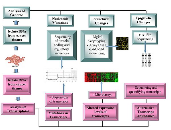
Current technologies being used in Oncogenomics.
Genomics technologies include:

Genome sequencing

  • DNA sequencing: Pyrosequencing-based sequencers offer a relatively low-cost method to generate sequence data.
  • Array Comparative Genome Hybridization: This technique measures the DNA copy number differences between normal and cancer genomes. It uses the fluorescence intensity from fluorescent-labeled samples, which are hybridized to known probes on a microarray.
  • Representational oligonucleotide microarray analysis: Detects copy number variation using amplified restriction-digested genomic fragments that are hybridized to human oligonucleotides, achieving a resolution between 30 and 35 kbit/s.
  • Digital Karyotyping: Detects copy number variation using genomics tags obtained via restriction enzyme digests. These tags are then linked to into ditags, concatenated, cloned, sequenced and mapped back to the reference genome to evaluate tag density.
  • Bacterial Artificial Chromosome (BAC)-end sequencing (end-sequence profiling): Identifies chromosomal breakpoints by generating a BAC library from a cancer genome and sequencing their ends. The BAC clones that contain chromosome aberrations have end sequences that do not map to a similar region of the reference genome, thus identifying a chromosomal breakpoint.

Transcriptomes

  • Microarrays: Assess transcript abundance. Useful in classification, prognosis, raise the possibility of differential treatment approaches and aid identification of mutations in the proteins' coding regions. The relative abundance of alternative transcripts has become an important feature of cancer research. Particular alternative transcript forms correlate with specific cancer types.
  • RNA-Seq

Bioinformatics and functional analysis of oncogenes

Bioinformatics technologies allow the statistical analysis of genomic data. The functional characteristics of oncogenes has yet to be established. Potential functions include their transformational capabilities relating to tumour formation and specific roles at each stage of cancer development.

After the detection of somatic cancer mutations across a cohort of cancer samples, bioinformatic computational analyses can be carried out to identify likely functional and likely driver mutations. There are three main approaches routinely used for this identification: mapping mutations, assessing the effect of mutation of the function of a protein or a regulatory element and finding signs of positive selection across a cohort of tumors. The approaches are not necessarily sequential however, there are important relationships of precedence between elements from the different approaches. Different tools are used at each step.

Operomics

Operomics aims to integrate genomics, transcriptomics and proteomics to understand the molecular mechanisms that underlie the cancer development.

Comparative oncogenomics

Comparative oncogenomics uses cross-species comparisons to identify oncogenes. This research involves studying cancer genomes, transcriptomes and proteomes in model organisms such as mice, identifying potential oncogenes and referring back to human cancer samples to see whether homologues of these oncogenes are important in causing human cancers. Genetic alterations in mouse models are similar to those found in human cancers. These models are generated by methods including retroviral insertion mutagenesis or graft transplantation of cancerous cells.

Source of cancer driver mutations, cancer mutagenesis

Mutations provide the raw material for natural selection in evolution and can be caused by errors of DNA replication, the action of exogenous mutagens or endogenous DNA damage. The machinery of replication and genome maintenance can be damaged by mutations, or altered by physiological conditions and differential levels of expression in cancer (see references in).

As pointed out by Gao et al., the stability and integrity of the human genome are maintained by the DNA-damage response (DDR) system. Un-repaired DNA damage is a major cause of mutations that drive carcinogenesis. If DNA repair is deficient, DNA damage tends to accumulate. Such excess DNA damage can increase mutational errors during DNA replication due to error-prone translesion synthesis. Excess DNA damage can also increase epigenetic alterations due to errors during DNA repair. Such mutations and epigenetic alterations can give rise to cancer. DDR genes are often repressed in human cancer by epigenetic mechanisms. Such repression may involve DNA methylation of promoter regions or repression of DDR genes by a microRNA. Epigenetic repression of DDR genes occurs more frequently than gene mutation in many types of cancer. Thus, epigenetic repression often plays a more important role than mutation in reducing expression of DDR genes. This reduced expression of DDR genes is likely an important driver of carcinogenesis.

Nucleotide sequence context influences mutation probability and analysis of mutational (mutable) DNA motifs can be essential for understanding the mechanisms of mutagenesis in cancer. Such motifs represent the fingerprints of interactions between DNA and mutagens, between DNA and repair/replication/modification enzymes. Examples of motifs are the AID motif WRCY/RGYW (W = A or T, R = purine and Y = pyrimidine) with C to T/G/A mutations, and error-prone DNA pol η attributed AID-related mutations (A to G/C/G) in WA/TW motifs.

Another (agnostic) way to analyze the observed mutational spectra and DNA sequence context of mutations in tumors involves pooling all mutations of different types and contexts from cancer samples into a discrete distribution. If multiple cancer samples are available, their context-dependent mutations can be represented in the form of a nonnegative matrix. This matrix can be further decomposed into components (mutational signatures) which ideally should describe individual mutagenic factors. Several computational methods have been proposed for solving this decomposition problem. The first implementation of Non-negative Matrix Factorization (NMF) method is available in Sanger Institute Mutational Signature Framework in the form of a MATLAB package. On the other hand, if mutations from a single tumor sample are only available, the DeconstructSigs R package and MutaGene server may provide the identification of contributions of different mutational signatures for a single tumor sample. In addition, MutaGene server provides mutagen or cancer-specific mutational background models and signatures that can be applied to calculate expected DNA and protein site mutability to decouple relative contributions of mutagenesis and selection in carcinogenesis.

Synthetic lethality

Synthetic lethality arises when a combination of deficiencies in the expression of two or more genes leads to cell death, whereas a deficiency in only one of these genes does not. The deficiencies can arise through mutations, epigenetic alterations or inhibitors of one of the genes.

The therapeutic potential of synthetic lethality as an efficacious anti-cancer strategy is continually improving. Recently, the applicability of synthetic lethality to targeted cancer therapy has heightened due to the recent work of scientists including Ronald A. DePinho and colleagues, in what is termed 'collateral lethality'. Muller et al. found that passenger genes, with chromosomal proximity to tumor suppressor genes, are collaterally deleted in some cancers. Thus, the identification of collaterally deleted redundant genes carrying out an essential cellular function may be the untapped reservoir for then pursuing a synthetic lethality approach. Collateral lethality therefore holds great potential in identification of novel and selective therapeutic targets in oncology. In 2012, Muller et al. identified that homozygous deletion of redundant-essential glycolytic ENO1 gene in human glioblastoma (GBM) is the consequence of proximity to 1p36 tumor suppressor locus deletions and may hold potential for a synthetic lethality approach to GBM inhibition. ENO1 is one of three homologous genes (ENO2, ENO3) that encodes the mammalian alpha-enolase enzyme. ENO2, which encodes enolase 2, is mostly expressed in neural tissues, leading to the postulation that in ENO1-deleted GBM, ENO2 may be the ideal target as the redundant homologue of ENO1. Muller found that both genetic and pharmacological ENO2 inhibition in GBM cells with homozygous ENO1 deletion elicits a synthetic lethality outcome by selective killing of GBM cells). In 2016, Muller and colleagues discovered antibiotic SF2312 as a highly potent nanomolar-range enolase inhibitor which preferentially inhibits glioma cell proliferation and glycolytic flux in ENO1-deleted cells. SF2312 was shown to be more efficacious than pan-enolase inhibitor PhAH and have more specificity for ENO2 inhibition over ENO1. Subsequent work by the same team showed that the same approach could be applied to pancreatic cancer, whereby homozygously deleted SMAD4 results in the collateral deletion of mitochondrial malic enzyme 2 (ME2), an oxidative decarboxylase essential for redox homeostasis. Dey et al. show that ME2 genomic deletion in pancreatic ductal adenocarcinoma cells results in high endogenous reactive oxygen species, consistent with KRAS-driven pancreatic cancer, and essentially primes ME2-null cells for synthetic lethality by depletion of redundant NAD(P)+-dependent isoform ME3. The effects of ME3 depletion were found to be mediated by inhibition of de novo nucleotide synthesis resulting from AMPK activation and mitochondrial ROS-mediated apoptosis. Meanwhile, Oike et al. demonstrated the generalizability of the concept by targeting redundant essential-genes in process other than metabolism, namely the SMARCA4 and SMARCA2 subunits in the chromatin-remodeling SWI/SNF complex.

Some oncogenes are essential for survival of all cells (not only cancer cells). Thus, drugs that knock out these oncogenes (and thereby kill cancer cells) may also damage normal cells, inducing significant illness. However, other genes may be essential to cancer cells but not to healthy cells.
Treatments based on the principle of synthetic lethality have prolonged the survival of cancer patients, and show promise for future advances in reversal of carcinogenesis. A major type of synthetic lethality operates on the DNA repair defect that often initiates a cancer, and is still present in the tumor cells. Some examples are given here.

BRCA1 or BRCA2 expression is deficient in a majority of high-grade breast and ovarian cancers, usually due to epigenetic methylation of its promoter or epigenetic repression by an over-expressed microRNA (see articles BRCA1 and BRCA2). BRCA1 and BRCA2 are important components of the major pathway for homologous recombinational repair of double-strand breaks. If one or the other is deficient, it increases the risk of cancer, especially breast or ovarian cancer. A back-up DNA repair pathway, for some of the damages usually repaired by BRCA1 and BRCA2, depends on PARP1. Thus, many ovarian cancers respond to an FDA-approved treatment with a PARP inhibitor, causing synthetic lethality to cancer cells deficient in BRCA1 or BRCA2. This treatment is also being evaluated for breast cancer and numerous other cancers in Phase III clinical trials in 2016.

There are two pathways for homologous recombinational repair of double-strand breaks. The major pathway depends on BRCA1, PALB2 and BRCA2 while an alternative pathway depends on RAD52. Pre-clinical studies, involving epigenetically reduced or mutated BRCA-deficient cells (in culture or injected into mice), show that inhibition of RAD52 is synthetically lethal with BRCA-deficiency.

Mutations in genes employed in DNA mismatch repair (MMR) cause a high mutation rate. In tumors, such frequent subsequent mutations often generate “non-self” immunogenic antigens. A human Phase II clinical trial, with 41 patients, evaluated one synthetic lethal approach for tumors with or without MMR defects. The product of gene PD-1 ordinarily represses cytotoxic immune responses. Inhibition of this gene allows a greater immune response. When cancer patients with a defect in MMR in their tumors were exposed to an inhibitor of PD-1, 67% - 78% of patients experienced immune-related progression-free survival. In contrast, for patients without defective MMR, addition of PD-1 inhibitor generated only 11% of patients with immune-related progression-free survival. Thus inhibition of PD-1 is primarily synthetically lethal with MMR defects.

ARID1A, a chromatin modifier, is required for non-homologous end joining, a major pathway that repairs double-strand breaks in DNA, and also has transcription regulatory roles. ARID1A mutations are one of the 12 most common carcinogenic mutations. Mutation or epigenetically decreased expression of ARID1A has been found in 17 types of cancer. Pre-clinical studies in cells and in mice show that synthetic lethality for ARID1A deficiency occurs by either inhibition of the methyltransferase activity of EZH2, or with addition of the kinase inhibitor dasatinib.

Another approach is to individually knock out each gene in a genome and observe the effect on normal and cancerous cells. If the knockout of an otherwise nonessential gene has little or no effect on healthy cells, but is lethal to cancerous cells containing a mutated oncogene, then the system-wide suppression of the suppressed gene can destroy cancerous cells while leaving healthy ones relatively undamaged. The technique was used to identify PARP-1 inhibitors to treat BRCA1/BRCA2-associated cancers. In this case, the combined presence of PARP-1 inhibition and of the cancer-associated mutations in BRCA genes is lethal only to the cancerous cells.

Databases for cancer research

The Cancer Genome Project is an initiative to map out all somatic mutations in cancer. The project systematically sequences the exons and flanking splice junctions of the genomes of primary tumors and cancerous cell lines. COSMIC software displays the data generated from these experiments. As of February 2008, the CGP had identified 4,746 genes and 2,985 mutations in 1,848 tumours. The Cancer Genome Anatomy Project includes information of research on cancer genomes, transcriptomes and proteomes.

Progenetix is an oncogenomic reference database, presenting cytogenetic and molecular-cytogenetic tumor data.

Oncomine has compiled data from cancer transcriptome profiles.

The integrative oncogenomics database IntOGen and the Gitools datasets integrate multidimensional human oncogenomic data classified by tumor type. The first version of IntOGen focused on the role of deregulated gene expression and CNV in cancer. A later version emphasized mutational cancer driver genes across 28 tumor types. All releases of IntOGen data are made available at the IntOGen database.

The International Cancer Genome Consortium is the biggest project to collect human cancer genome data. The data is accessible through the ICGC website. The BioExpress® Oncology Suite contains gene expression data from primary, metastatic and benign tumor samples and normal samples, including matched adjacent controls. The suite includes hematological malignancy samples for many well-known cancers.

Specific databases for model animals include the Retrovirus Tagged Cancer Gene Database (RTCGD) that compiled research on retroviral and transposon insertional mutagenesis in mouse tumors.

Gene families

Mutational analysis of entire gene families revealed that genes of the same family have similar functions, as predicted by similar coding sequences and protein domains. Two such classes are the kinase family, involved in adding phosphate groups to proteins and the phosphatase family, involved with removing phosphate groups from proteins. These families were first examined because of their apparent role in transducing cellular signals of cell growth or death. In particular, more than 50% of colorectal cancers carry a mutation in a kinase or phosphatase gene. Phosphatidylinositold 3-kinases (PIK3CA) gene encodes for lipid kinases that commonly contain mutations in colorectal, breast, gastric, lung and various other cancers. Drug therapies can inhibit PIK3CA. Another example is the BRAF gene, one of the first to be implicated in melanomas. BRAF encodes a serine/threonine kinase that is involved in the RAS-RAF-MAPK growth signaling pathway. Mutations in BRAF cause constitutive phosphorylation and activity in 59% of melanomas. Before BRAF, the genetic mechanism of melanoma development was unknown and therefore prognosis for patients was poor.

Mitochondrial DNA

Mitochondrial DNA (mtDNA) mutations are linked the formation of tumors. Four types of mtDNA mutations have been identified:

Point mutations

Point mutations have been observed in the coding and non-coding region of the mtDNA contained in cancer cells. In individuals with bladder, head/neck and lung cancers, the point mutations within the coding region show signs of resembling each other. This suggests that when a healthy cell transforms into a tumor cell (a neoplastic transformation) the mitochondria seem to become homogenous. Abundant point mutations located within the non-coding region, D-loop, of the cancerous mitochondria suggest that mutations within this region might be an important characteristic in some cancers.

Deletions

This type of mutation is sporadically detected due to its small size ( < 1kb). The appearance of certain specific mtDNA mutations (264-bp deletion and 66-bp deletion in the complex 1 subunit gene ND1) in multiple types of cancer provide some evidence that small mtDNA deletions might appear at the beginning of tumorigenesis. It also suggests that the amount of mitochondria containing these deletions increases as the tumor progresses. An exception is a relatively large deletion that appears in many cancers (known as the "common deletion"), but more mtDNA large scale deletions have been found in normal cells compared to tumor cells. This may be due to a seemingly adaptive process of tumor cells to eliminate any mitochondria that contain these large scale deletions (the "common deletion" is > 4kb).

Insertions

Two small mtDNA insertions of ~260 and ~520 bp can be present in breast cancer, gastric cancer, hepatocellular carcinoma (HCC) and colon cancer and in normal cells. No correlation between these insertions and cancer are established.

Copy number mutations

The characterization of mtDNA via real-time polymerase chain reaction assays shows the presence of quantitative alteration of mtDNA copy number in many cancers. Increase in copy number is expected to occur because of oxidative stress. On the other hand, decrease is thought to be caused by somatic point mutations in the replication origin site of the H-strand and/or the D310 homopolymeric c-stretch in the D-loop region, mutations in the p53 (tumor suppressor gene) mediated pathway and/or inefficient enzyme activity due to POLG mutations. Any increase/decrease in copy number then remains constant within tumor cells. The fact that the amount of mtDNA is constant in tumor cells suggests that the amount of mtDNA is controlled by a much more complicated system in tumor cells, rather than simply altered as a consequence of abnormal cell proliferation. The role of mtDNA content in human cancers apparently varies for particular tumor types or sites.

57.7% (500/867) contained somatic point putations and of the 1172 mutations surveyed 37.8% (443/1127) were located in the D-loop control region, 13.1% (154/1172) were located in the tRNA or rRNA genes and 49.1% (575/1127) were found in the mRNA genes needed for producing complexes required for mitochondrial respiration.

Diagnostic applications

Some anticancer drugs target mtDNA and have shown positive results in killing tumor cells. Research has used mitochondrial mutations as biomarkers for cancer cell therapy. It is easier to target mutation within mitochondrial DNA versus nuclear DNA because the mitochondrial genome is much smaller and easier to screen for specific mutations. MtDNA content alterations found in blood samples might be able to serve as a screening marker for predicting future cancer susceptibility as well as tracking malignant tumor progression. Along with these potential helpful characteristics of mtDNA, it is not under the control of the cell cycle and is important for maintaining ATP generation and mitochondrial homeostasis. These characteristics make targeting mtDNA a practical therapeutic strategy.

Cancer biomarkers

Several biomarkers can be useful in cancer staging, prognosis and treatment. They can range from single-nucleotide polymorphisms (SNPs), chromosomal aberrations, changes in DNA copy number, microsatellite instability, promoter region methylation, or even high or low protein levels.

Nanobacterium

From Wikipedia, the free encyclopedia
 
Structures found on meteorite fragment Allan Hills 84001

Nanobacterium (/ˌnænbækˈtɪəriəm/ NAN-oh-bak-TEER-ee-əm, pl. nanobacteria /ˌnænbækˈtɪəriə/ NAN-oh-bak-TEER-ee-ə) is the unit or member name of a proposed class of living organisms, specifically cell-walled microorganisms with a size much smaller than the generally accepted lower limit for life (about 200 nm for bacteria, like mycoplasma). Originally based on observed nano-scale structures in geological formations (including one meteorite), the status of nanobacteria has been controversial, with some researchers suggesting they are a new class of living organism capable of incorporating radiolabeled uridine, and others attributing to them a simpler, abiotic nature. One skeptic dubbed them "the cold fusion of microbiology", in reference to a notorious episode of supposed erroneous science. The term "calcifying nanoparticles" (CNPs) has also been used as a conservative name regarding their possible status as a life form.

Research tends to agree that these structures exist, and appear to replicate in some way. However, the idea that they are living entities has now largely been discarded, and the particles are instead thought to be nonliving crystallizations of minerals and organic molecules.

1981–2000

In 1981 Torella and Morita described very small cells called ultramicrobacteria. Defined as being smaller than 300 nm, by 1982 MacDonell and Hood found that some could pass through a 200 nm membrane. Early in 1989, geologist Robert L. Folk found what he later identified as nannobacteria (written with double "n"), that is, nanoparticles isolated from geological specimens in travertine from hot springs of Viterbo, Italy. Initially searching for a bacterial cause for travertine deposition, scanning electron microscope examination of the mineral where no bacteria were detectable revealed extremely small objects which appeared to be biological. His first oral presentation elicited what he called "mostly a stony silence", at the 1992 Geological Society of America's annual convention. He proposed that nanobacteria are the principal agents of precipitation of all minerals and crystals on Earth formed in liquid water, that they also cause all oxidation of metals, and that they are abundant in many biological specimens.

In 1996, NASA scientist David McKay published a study suggesting the existence of nanofossils — fossils of Martian nanobacteria — in ALH84001, a meteorite originating from Mars and found in Antarctica.

Nanobacterium sanguineum was proposed in 1998 as an explanation of certain kinds of pathologic calcification (apatite in kidney stones) by Finnish researcher Olavi Kajander and Turkish researcher Neva Ciftcioglu, working at the University of Kuopio in Finland. According to the researchers the particles self-replicated in microbiological culture, and the researchers further reported having identified DNA in these structures by staining.

A paper published in 2000 by a team led by an NIH scientist John Cisar further tested these ideas. It stated that what had previously been described as "self-replication" was a form of crystalline growth. The only DNA detected in his specimens was identified as coming from the bacteria Phyllobacterium myrsinacearum, which is a common contaminant in PCR reactions.

2001–present

In 2004 a Mayo Clinic team led by Franklin Cockerill, John Lieske, and Virginia M. Miller, reported to have isolated nanobacteria from diseased human arteries and kidney stones. Their results were published in 2004 and 2006 respectively.[3][13] Similar findings were obtained in 2005 by László Puskás at the DNA Lab, University of Szeged, Hungary. Dr. Puskás identified these particles in cultures obtained from human atherosclerotic aortic walls and blood samples of atherosclerotic patients but the group was unable to detect DNA in these samples.

In 2005, Ciftcioglu and her research team at NASA used a rotating cell culture flask, which simulates some aspects of low-gravity conditions, to culture nanobacteria suspected of rapidly forming kidney stones in astronauts. In this environment, they were found to multiply five times faster than in normal Earth gravity. The study concluded that nanobacteria might have a potential role in forming kidney stones and may need to be screened for in crews pre-flight.

The February 2008 Public Library of Science (PLoS) Pathogens article focused on the comprehensive characterization of nanobacteria. The authors say that their results rule out the existence of nanobacteria as living entities and that they are instead a unique self-propagating entity, namely self-propagating mineral-fetuin complexes.

An April 2008 Proceedings of the National Academy of Sciences (PNAS) article also reported that blood nanobacteria are not living organisms and stated that "CaCO3 precipitates prepared in vitro are remarkably similar to purported nanobacteria in terms of their uniformly sized, membrane-delineated vesicular shapes, with cellular division-like formations and aggregations in the form of colonies." The growth of such "biomorphic" inorganic precipitates was studied in detail in a 2009 Science paper, which showed that unusual crystal growth mechanisms can produce witherite precipitates from barium chloride and silica solutions that closely resemble primitive organisms. The authors commented on the close resemblance of these crystals to putative nanobacteria, stating that their results showed that evidence for life cannot rest on morphology alone.

Further work on the importance of nanobacteria in geology by R. L. Folk and co-workers includes study of calcium carbonate Bahama ooids, silicate clay minerals, metal sulfides, and iron oxides. In all these chemically diverse minerals, the putative nanobacteria are approximately the same size, mainly 0.05 to 0.2 μm. This suggests a commonality of origin. At least for the type locality at Viterbo, Italy, the biogenicity of these minute cells has been supported by transmission electron microscopy (TEM). Slices through a green bioslime showed entities from 0.4 down to as small as 0.09 μm with definite cell walls and interior dots resembling ribosomes; and even smaller objects with cell walls and lucent interiors with diameters of 0.05 μm. Culturable organisms on earth are the same 0.05 μm size as the supposed nanobacteria on Mars.

Prokaryote

From Wikipedia, the free encyclopedia
 
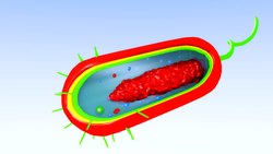
Diagram of a typical prokaryotic cell

A prokaryote is a unicellular organism that lacks a membrane-bound nucleus, mitochondria, or any other membrane-bound organelle. The word prokaryote comes from the Greek πρό (pro) "before" and κάρυον (karyon) "nut or kernel". Prokaryotes are divided into two domains, Archaea and Bacteria. In contrast, species with nuclei and organelles are placed in the third domain, Eukaryota. Prokaryotes reproduce without fusion of gametes. The first living organisms are thought to have been prokaryotes.

Phylogenetic tree showing the diversity of prokaryote.

In the prokaryotes, all the intracellular water-soluble components (proteins, DNA and metabolites) are located together in the cytoplasm enclosed by the cell membrane, rather than in separate cellular compartments. Bacteria, however, do possess protein-based bacterial microcompartments, which are thought to act as primitive organelles enclosed in protein shells. Some prokaryotes, such as cyanobacteria, may form large colonies. Others, such as myxobacteria, have multicellular stages in their life cycles.

Molecular studies have provided insight into the evolution and interrelationships of the three domains of biological species. Eukaryotes are organisms, including humans, whose cells have a well defined membrane-bound nucleus (containing chromosomal DNA) and organelles. The division between prokaryotes and eukaryotes reflects the existence of two very different levels of cellular organization. Distinctive types of prokaryotes include extremophiles and methanogens; these are common in some extreme environments.

Structure

Prokaryotes have a prokaryotic cytoskeleton, albeit more primitive than that of the eukaryotes. Besides homologues of actin and tubulin (MreB and FtsZ), the helically arranged building-block of the flagellum, flagellin, is one of the most significant cytoskeletal proteins of bacteria, as it provides structural backgrounds of chemotaxis, the basic cell physiological response of bacteria. At least some prokaryotes also contain intracellular structures that can be seen as primitive organelles. Membranous organelles (or intracellular membranes) are known in some groups of prokaryotes, such as vacuoles or membrane systems devoted to special metabolic properties, such as photosynthesis or chemolithotrophy. In addition, some species also contain carbohydrate-enclosed microcompartments, which have distinct physiological roles (e.g. carboxysomes or gas vacuoles).

Most prokaryotes are between 1 µm and 10 µm, but they can vary in size from 0.2 µm (Mycoplasma genitalium) to 750 µm (Thiomargarita namibiensis).

Prokaryotic cell structure
 
Flagellum (only in some types of prokaryotes)
Long, whip-like protrusion that aids cellular locomotion used by both gram positive and gram negative organisms.

Cell membrane
Surrounds the cell's cytoplasm and regulates the flow of substances in and out of the cell.

Cell wall (except genera Mycoplasma and Thermoplasma)
Outer covering of most cells that protects the bacterial cell and gives it shape.

Cytoplasm
A gel-like substance composed mainly of water that also contains enzymes, salts, cell components, and various organic molecules.

Ribosome
Cell structures responsible for protein production.

Nucleoid
Area of the cytoplasm that contains the prokaryote's single DNA molecule.

Glycocalyx (only in some types of prokaryotes)
A glycoprotein-polysaccharide covering that surrounds the cell membranes.

Inclusions
It contains the inclusion bodies like ribosomes and larger masses scattered in the cytoplasmic matrix.

Morphology

Prokaryotic cells have various shapes; the four basic shapes of bacteria are:
The archaeon Haloquadratum has flat square-shaped cells.

Reproduction

Bacteria and archaea reproduce through asexual reproduction, usually by binary fission. Genetic exchange and recombination still occur, but this is a form of horizontal gene transfer and is not a replicative process, simply involving the transference of DNA between two cells, as in bacterial conjugation.

DNA transfer

DNA transfer between prokaryotic cells occurs in bacteria and archaea, although it has been mainly studied in bacteria. In bacteria, gene transfer occurs by three processes. These are (1) bacterial virus (bacteriophage)-mediated transduction, (2) plasmid-mediated conjugation, and (3) natural transformation. Transduction of bacterial genes by bacteriophage appears to reflect an occasional error during intracellular assembly of virus particles, rather than an adaptation of the host bacteria. The transfer of bacterial DNA is under the control of the bacteriophage’s genes rather than bacterial genes. Conjugation in the well-studied E. coli system is controlled by plasmid genes, and is an adaptation for distributing copies of a plasmid from one bacterial host to another. Infrequently during this process, a plasmid may integrate into the host bacterial chromosome, and subsequently transfer part of the host bacterial DNA to another bacterium. Plasmid mediated transfer of host bacterial DNA (conjugation) also appears to be an accidental process rather than a bacterial adaptation.

3D animation of a prokaryotic cell that shows all the elements that compose it

Natural bacterial transformation involves the transfer of DNA from one bacterium to another through the intervening medium. Unlike transduction and conjugation, transformation is clearly a bacterial adaptation for DNA transfer, because it depends on numerous bacterial gene products that specifically interact to perform this complex process. For a bacterium to bind, take up and recombine donor DNA into its own chromosome, it must first enter a special physiological state called competence. About 40 genes are required in Bacillus subtilis for the development of competence. The length of DNA transferred during B. subtilis transformation can be as much as a third to the whole chromosome. Transformation is a common mode of DNA transfer, and 67 prokaryotic species are thus far known to be naturally competent for transformation.

Among archaea, Halobacterium volcanii forms cytoplasmic bridges between cells that appear to be used for transfer of DNA from one cell to another. Another archaeon, Sulfolobus solfataricus, transfers DNA between cells by direct contact. Frols et al. found that exposure of S. solfataricus to DNA damaging agents induces cellular aggregation, and suggested that cellular aggregation may enhance DNA transfer among cells to provide increased repair of damaged DNA via homologous recombination.

Sociality

While prokaryotes are considered strictly unicellular, most can form stable aggregate communities. When such communities are encased in a stabilizing polymer matrix ("slime"), they may be called "biofilms". Cells in biofilms often show distinct patterns of gene expression (phenotypic differentiation) in time and space. Also, as with multicellular eukaryotes, these changes in expression often appear to result from cell-to-cell signaling, a phenomenon known as quorum sensing.

Biofilms may be highly heterogeneous and structurally complex and may attach to solid surfaces, or exist at liquid-air interfaces, or potentially even liquid-liquid interfaces. Bacterial biofilms are often made up of microcolonies (approximately dome-shaped masses of bacteria and matrix) separated by "voids" through which the medium (e.g., water) may flow easily. The microcolonies may join together above the substratum to form a continuous layer, closing the network of channels separating microcolonies. This structural complexity—combined with observations that oxygen limitation (a ubiquitous challenge for anything growing in size beyond the scale of diffusion) is at least partially eased by movement of medium throughout the biofilm—has led some to speculate that this may constitute a circulatory system  and many researchers have started calling prokaryotic communities multicellular (for example). Differential cell expression, collective behavior, signaling, programmed cell death, and (in some cases) discrete biological dispersal events all seem to point in this direction. However, these colonies are seldom if ever founded by a single founder (in the way that animals and plants are founded by single cells), which presents a number of theoretical issues. Most explanations of co-operation and the evolution of multicellularity have focused on high relatedness between members of a group (or colony, or whole organism). If a copy of a gene is present in all members of a group, behaviors that promote cooperation between members may permit those members to have (on average) greater fitness than a similar group of selfish individuals.

Should these instances of prokaryotic sociality prove to be the rule rather than the exception, it would have serious implications for the way we view prokaryotes in general, and the way we deal with them in medicine. Bacterial biofilms may be 100 times more resistant to antibiotics than free-living unicells and may be nearly impossible to remove from surfaces once they have colonized them. Other aspects of bacterial cooperation—such as bacterial conjugation and quorum-sensing-mediated pathogenicity, present additional challenges to researchers and medical professionals seeking to treat the associated diseases.

Environment

Phylogenetic ring showing the diversity of prokaryotes, and symbiogenetic origins of eukaryotes

Prokaryotes have diversified greatly throughout their long existence. The metabolism of prokaryotes is far more varied than that of eukaryotes, leading to many highly distinct prokaryotic types. For example, in addition to using photosynthesis or organic compounds for energy, as eukaryotes do, prokaryotes may obtain energy from inorganic compounds such as hydrogen sulfide. This enables prokaryotes to thrive in harsh environments as cold as the snow surface of Antarctica, studied in cryobiology or as hot as undersea hydrothermal vents and land-based hot springs.

Prokaryotes live in nearly all environments on Earth. Some archaea and bacteria are extremophiles, thriving in harsh conditions, such as high temperatures (thermophiles) or high salinity (halophiles). Many archaea grow as plankton in the oceans. Symbiotic prokaryotes live in or on the bodies of other organisms, including humans.

Classification

Phylogenetic and symbiogenetic tree of living organisms, showing the origins of eukaryotes and prokaryotes

In 1977, Carl Woese proposed dividing prokaryotes into the Bacteria and Archaea (originally Eubacteria and Archaebacteria) because of the major differences in the structure and genetics between the two groups of organisms. Archaea were originally thought to be extremophiles, living only in inhospitable conditions such as extremes of temperature, pH, and radiation but have since been found in all types of habitats. The resulting arrangement of Eukaryota (also called "Eucarya"), Bacteria, and Archaea is called the three-domain system, replacing the traditional two-empire system.

Evolution

Diagram of the origin of life with the Eukaryotes appearing early, not derived from Prokaryotes, as proposed by Richard Egel in 2012. This view, one of many on the relative positions of Prokaryotes and Eukaryotes, implies that the universal common ancestor was relatively large and complex.

A widespread current model of the evolution of the first living organisms is that these were some form of prokaryotes, which may have evolved out of protocells, while the eukaryotes evolved later in the history of life. Some authors have questioned this conclusion, arguing that the current set of prokaryotic species may have evolved from more complex eukaryotic ancestors through a process of simplification. Others have argued that the three domains of life arose simultaneously, from a set of varied cells that formed a single gene pool. This controversy was summarized in 2005:
There is no consensus among biologists concerning the position of the eukaryotes in the overall scheme of cell evolution. Current opinions on the origin and position of eukaryotes span a broad spectrum including the views that eukaryotes arose first in evolution and that prokaryotes descend from them, that eukaryotes arose contemporaneously with eubacteria and archeabacteria and hence represent a primary line of descent of equal age and rank as the prokaryotes, that eukaryotes arose through a symbiotic event entailing an endosymbiotic origin of the nucleus, that eukaryotes arose without endosymbiosis, and that eukaryotes arose through a symbiotic event entailing a simultaneous endosymbiotic origin of the flagellum and the nucleus, in addition to many other models, which have been reviewed and summarized elsewhere.
The oldest known fossilized prokaryotes were laid down approximately 3.5 billion years ago, only about 1 billion years after the formation of the Earth's crust. Eukaryotes only appear in the fossil record later, and may have formed from endosymbiosis of multiple prokaryote ancestors. The oldest known fossil eukaryotes are about 1.7 billion years old. However, some genetic evidence suggests eukaryotes appeared as early as 3 billion years ago.

While Earth is the only place in the universe where life is known to exist, some have suggested that there is evidence on Mars of fossil or living prokaryotes. However, this possibility remains the subject of considerable debate and skepticism.

Relationship to eukaryotes

Comparison of eukaryotes vs. prokaryotes

The division between prokaryotes and eukaryotes is usually considered the most important distinction or difference among organisms. The distinction is that eukaryotic cells have a "true" nucleus containing their DNA, whereas prokaryotic cells do not have a nucleus. Both eukaryotes and prokaryotes contain large RNA/protein structures called ribosomes, which produce protein.

Another difference is that ribosomes in prokaryotes are smaller than in eukaryotes. However, two organelles found in many eukaryotic cells, mitochondria and chloroplasts, contain ribosomes similar in size and makeup to those found in prokaryotes. This is one of many pieces of evidence that mitochondria and chloroplasts are themselves descended from free-living bacteria. This theory holds that early eukaryotic cells took in primitive prokaryotic cells by phagocytosis and adapted themselves to incorporate their structures, leading to the mitochondria we see today.

The genome in a prokaryote is held within a DNA/protein complex in the cytosol called the nucleoid, which lacks a nuclear envelope. The complex contains a single, cyclic, double-stranded molecule of stable chromosomal DNA, in contrast to the multiple linear, compact, highly organized chromosomes found in eukaryotic cells. In addition, many important genes of prokaryotes are stored in separate circular DNA structures called plasmids. Like Eukaryotes, prokaryotes may partially duplicate genetic material, and can have a haploid chromosomal composition that is partially replicated, a condition known as merodiploidy.

Prokaryotes lack mitochondria and chloroplasts. Instead, processes such as oxidative phosphorylation and photosynthesis take place across the prokaryotic cell membrane. However, prokaryotes do possess some internal structures, such as prokaryotic cytoskeletons. It has been suggested that the bacterial order Planctomycetes have a membrane around their nucleoid and contain other membrane-bound cellular structures. However, further investigation revealed that Planctomycetes cells are not compartmentalized or nucleated and like the other bacterial membrane systems are all interconnected.

Prokaryotic cells are usually much smaller than eukaryotic cells. Therefore, prokaryotes have a larger surface-area-to-volume ratio, giving them a higher metabolic rate, a higher growth rate, and as a consequence, a shorter generation time than eukaryotes.

Targeted drug delivery

From Wikipedia, the free encyclopedia
 
Targeted drug delivery, sometimes called smart drug delivery, is a method of delivering medication to a patient in a manner that increases the concentration of the medication in some parts of the body relative to others. This means of delivery is largely founded on nanomedicine, which plans to employ nanoparticle-mediated drug delivery in order to combat the downfalls of conventional drug delivery. These nanoparticles would be loaded with drugs and targeted to specific parts of the body where there is solely diseased tissue, thereby avoiding interaction with healthy tissue. The goal of a targeted drug delivery system is to prolong, localize, target and have a protected drug interaction with the diseased tissue. The conventional drug delivery system is the absorption of the drug across a biological membrane, whereas the targeted release system releases the drug in a dosage form. The advantages to the targeted release system is the reduction in the frequency of the dosages taken by the patient, having a more uniform effect of the drug, reduction of drug side-effects, and reduced fluctuation in circulating drug levels. The disadvantage of the system is high cost, which makes productivity more difficult and the reduced ability to adjust the dosages.
 
Targeted drug delivery systems have been developed to optimize regenerative techniques. The system is based on a method that delivers a certain amount of a therapeutic agent for a prolonged period of time to a targeted diseased area within the body. This helps maintain the required plasma and tissue drug levels in the body, thereby preventing any damage to the healthy tissue via the drug. The drug delivery system is highly integrated and requires various disciplines, such as chemists, biologists, and engineers, to join forces to optimize this system.

Background

In traditional drug delivery systems such as oral ingestion or intravascular injection, the medication is distributed throughout the body through the systemic blood circulation. For most therapeutic agents, only a small portion of the medication reaches the organ to be affected, such as in chemotherapy where roughly 99% of the drugs administered do not reach the tumor site. Targeted drug delivery seeks to concentrate the medication in the tissues of interest while reducing the relative concentration of the medication in the remaining tissues. For example, by avoiding the host's defense mechanisms and inhibiting non-specific distribution in the liver and spleen, a system can reach the intended site of action in higher concentrations. Targeted delivery is believed to improve efficacy while reducing side-effects.

When implementing a targeted release system, the following design criteria for the system must be taken into account: the drug properties, side-effects of the drugs, the route taken for the delivery of the drug, the targeted site, and the disease.

Increasing developments to novel treatments requires a controlled microenvironment that is accomplished only through the implementation of therapeutic agents whose side-effects can be avoided with targeted drug delivery. Advances in the field of targeted drug delivery to cardiac tissue will be an integral component to regenerate cardiac tissue.

There are two kinds of targeted drug delivery: active targeted drug delivery, such as some antibody medications, and passive targeted drug delivery, such as the enhanced permeability and retention effect (EPR-effect).

Targeting Methods

This ability for nanoparticles to concentrate in areas of solely diseased tissue is accomplished through either one or both means of targeting: passive or active.

Passive Targeting

In passive targeting, the drug’s success is directly related to circulation time. This is achieved by cloaking the nanoparticle with some sort of coating. Several substances can achieve this, with one of them being polyethylene glycol (PEG). By adding PEG to the surface of the nanoparticle, it is rendered hydrophilic, thus allowing water molecules to bind to the oxygen molecules on PEG via hydrogen bonding. The result of this bond is a film of hydration around the nanoparticle which makes the substance antiphagocytic. The particles obtain this property due to the hydrophobic interactions that are natural to the reticuloendothelial system (RES), thus the drug-loaded nanoparticle is able to stay in circulation for a longer period of time. To work in conjunction with this mechanism of passive targeting, nanoparticles that are between 10 and 100 nanometers in size have been found to circulate systemically for longer periods of time.

Active Targeting

Active targeting of drug-loaded nanoparticles enhances the effects of passive targeting to make the nanoparticle more specific to a target site. There are several ways that active targeting can be accomplished. One way to actively target solely diseased tissue in the body is to know the nature of a receptor on the cell for which the drug will be targeted to. Researchers can then utilize cell-specific ligands that will allow for the nanoparticle to bind specifically to the cell that has the complementary receptor. This form of active targeting was found to be successful when utilizing transferrin as the cell-specific ligand. The transferrin was conjugated to the nanoparticle to target tumor cells that possess transferrin-receptor mediated endocytosis mechanisms on their membrane. This means of targeting was found to increase uptake, as opposed to non-conjugated nanoparticles.

Active targeting can also be achieved by utilizing magnetoliposomes, which usually serves as a contrast agent in magnetic resonance imaging. Thus, by grafting these liposomes with a desired drug to deliver to a region of the body, magnetic positioning could aid with this process.

Furthermore, a nanoparticle could possess the capability to be activated by a trigger that is specific to the target site, such as utilizing materials that are pH responsive. Most of the body has a consistent, neutral pH. However, some areas of the body are naturally more acidic than others, and, thus, nanoparticles can take advantage of this ability by releasing the drug when it encounters a specific pH. Another specific triggering mechanism is based on the redox potential. One of the side effects of tumors is hypoxia, which alters the redox potential in the vicinity of the tumor. By modifying the redox potential that triggers the payload release the vesicles can be selective to different types of tumors.

By utilizing both passive and active targeting, a drug-loaded nanoparticle has a heightened advantage over a conventional drug. It is able to circulate throughout the body for an extended period of time until it is successfully attracted to its target through the use of cell-specific ligands, magnetic positioning, or pH responsive materials. Because of these advantages, side effects from conventional drugs will be largely reduced as a result of the drug-loaded nanoparticles affecting only diseased tissue. However, an emerging field known as nanotoxicology has concerns that the nanoparticles themselves could pose a threat to both the environment and human health with side effects of their own. Active targeting can also be achieved through peptide based drug targeting system.

Delivery vehicles

There are different types of drug delivery vehicles, such as polymeric micelles, liposomes, lipoprotein-based drug carriers, nano-particle drug carriers, dendrimers, etc. An ideal drug delivery vehicle must be non-toxic, biocompatible, non-immunogenic, biodegradable, and must avoid recognition by the host's defense mechanisms.

Liposomes

Liposomes are composite structures made of phospholipids and may contain small amounts of other molecules. Though liposomes can vary in size from low micrometer range to tens of micrometers, unilamellar liposomes, as pictured here, are typically in the lower size range, with various targeting ligands attached to their surface, allowing for their surface-attachment and accumulation in pathological areas for treatment of disease.

The most common vehicle currently used for targeted drug delivery is the liposome. Liposomes are non-toxic, non-hemolytic, and non-immunogenic even upon repeated injections; they are biocompatible and biodegradable and can be designed to avoid clearance mechanisms (reticuloendothelial system (RES), renal clearance, chemical or enzymatic inactivation, etc.) Lipid-based, ligand-coated nanocarriers can store their payload in the hydrophobic shell or the hydrophilic interior depending on the nature of the drug/contrast agent being carried.

The only problem to using liposomes in vivo is their immediate uptake and clearance by the RES system and their relatively low stability in vitro. To combat this, polyethylene glycol (PEG) can be added to the surface of the liposomes. Increasing the mole percent of PEG on the surface of the liposomes by 4-10% significantly increased circulation time in vivo from 200 to 1000 minutes.

PEGylation of the liposomal nanocarrier elongates the half-life of the construct while maintaining the passive targeting mechanism that is commonly conferred to lipid-based nanocarriers. When used as a delivery system, the ability to induce instability in the construct is commonly exploited allowing the selective release of the encapsulated therapeutic agent in close proximity to the target tissue/cell in vivo. This nanocarrier system is commonly used in anti-cancer treatments as the acidity of the tumour mass caused by an over-reliance on glycolysis triggers drug release.

Micelles and dendrimers

Another type of drug delivery vehicle used is polymeric micelles. They are prepared from certain amphiphilic co-polymers consisting of both hydrophilic and hydrophobic monomer units. They can be used to carry drugs that have poor solubility. This method offers little in the terms of size control or function malleability. Techniques that utilize reactive polymers along with a hydrophobic additive to produce a larger micelle that create a range of sizes have been developed.

Dendrimers are also polymer-based delivery vehicles. They have a core that branches out in regular intervals to form a small, spherical, and very dense nanocarrier.

Biodegradable particles

Biodegradable particles have the ability to target diseased tissue as well as deliver their payload as a controlled-release therapy. Biodegradable particles bearing ligands to P-selectin, endothelial selectin (E-selectin) and ICAM-1 have been found to adhere to inflamed endothelium. Therefore, the use of biodegradable particles can also be used for cardiac tissue.

Artificial DNA nanostructures

The success of DNA nanotechnology in constructing artificially designed nanostructures out of nucleic acids such as DNA, combined with the demonstration of systems for DNA computing, has led to speculation that artificial nucleic acid nanodevices can be used to target drug delivery based upon directly sensing its environment. These methods make use of DNA solely as a structural material and a chemical, and do not make use of its biological role as the carrier of genetic information. Nucleic acid logic circuits that could potentially be used as the core of a system that releases a drug only in response to a stimulus such as a specific mRNA have been demonstrated. In addition, a DNA "box" with a controllable lid has been synthesized using the DNA origami method. This structure could encapsulate a drug in its closed state, and open to release it only in response to a desired stimulus.

Applications

Targeted drug delivery can be used to treat many diseases, such as the cardiovascular diseases and diabetes. However, the most important application of targeted drug delivery is to treat cancerous tumors. In doing so, the passive method of targeting tumors takes advantage of the enhanced permeability and retention (EPR) effect. This is a situation specific to tumors that results from rapidly forming blood vessels and poor lymphatic drainage. When the blood vessels form so rapidly, large fenestrae result that are 100 to 600 nanometers in size, which allows enhanced nanoparticle entry. Further, the poor lymphatic drainage means that the large influx of nanoparticles are rarely leaving, thus, the tumor retains more nanoparticles for successful treatment to take place.

The American Heart Association rates cardiovascular disease as the number one cause of death in the United States. Each year 1.5 million myocardial infarctions (MI), also known as heart attacks, occur in the United States, with 500,000 leading to deaths. The costs related to heart attacks exceed $60 billion per year. Therefore, there is a need to come up with an optimum recovery system. The key to solving this problem lies in the effective use of pharmaceutical drugs that can be targeted directly to the diseased tissue. This technique can help develop many more regenerative techniques to cure various diseases. The development of a number of regenerative strategies in recent years for curing heart disease represents a paradigm shift away from conventional approaches that aim to manage heart disease.

Stem cell therapy can be used to help regenerate myocardium tissue and return the contractile function of the heart by creating/supporting a microenvironment before the MI. Developments in targeted drug delivery to tumors have provided the groundwork for the burgeoning field of targeted drug delivery to cardiac tissue. Recent developments have shown that there are different endothelial surfaces in tumors, which has led to the concept of endothelial cell adhesion molecule-mediated targeted drug delivery to tumors.

Liposomes can be used as drug delivery for the treatment of tuberculosis. The traditional treatment for TB is skin to chemotherapy which is not overly effective, which may be due to the failure of chemotherapy to make a high enough concentration at the infection site. The liposome delivery system allows for better microphage penetration and better builds a concentration at the infection site. The delivery of the drugs works intravenously and by inhalation. Oral intake is not advised because the liposomes break down in the Gastrointestinal System.

3D printing is also used by doctors to investigate how to target cancerous tumors in a more efficient way. By printing a plastic 3D shape of the tumor and filling it with the drugs used in the treatment the flow of the liquid can be observed allowing the modification of the doses and targeting location of the drugs.

Delayed-choice quantum eraser

From Wikipedia, the free encyclopedia https://en.wikipedia.org/wiki/Delayed-choice_quantum_eraser A delayed-cho...