Search This Blog

Thursday, January 12, 2023

Turner syndrome

From Wikipedia, the free encyclopedia

Turner syndrome
Other namesUllrich–Turner syndrome, Bonnevie–Ullrich–Turner syndrome, gonadal dysgenesis; 45X, 45X0
Individuals of Latin American descent with Turner syndrome (centred).png
Five girls and women with Turner syndrome
SpecialtyPediatrics, medical genetics
SymptomsWebbed neck, short stature, swollen hands and feet
ComplicationsHeart defects, diabetes, low thyroid hormone
OnsetAt birth
DurationLong term
CausesMissing an X chromosome
Diagnostic methodPhysical signs, genetic testing
MedicationHuman growth hormone, estrogen replacement therapy
PrognosisReduced life expectancy
Frequency1 in 2,000 to 5,000

Turner syndrome (TS), also known as 45,X, or 45,X0, is a genetic condition in which a female is partially or completely missing an X chromosome. Signs and symptoms vary among those affected. Often, a short and webbed neck, low-set ears, low hairline at the back of the neck, short stature, and swollen hands and feet are seen at birth. Typically, those affected do not develop menstrual periods, or breasts without hormone treatment and are unable to have children without reproductive technology. Heart defects, diabetes, and low thyroid hormone occur in the disorder more frequently than average. Most people with Turner syndrome have normal intelligence; however, many have problems with spatial visualization that may be needed in order to learn mathematics. Vision and hearing problems also occur more often than average.

Turner syndrome is not usually inherited; rather, it occurs during formation of the reproductive cells in a parent or in early cell division during development. No environmental risks are known, and the mother's age does not play a role. While most people have 46 chromosomes, people with Turner syndrome usually have 45 in some or all cells. The chromosomal abnormality is often present in just some cells, in which case it is known as Turner syndrome with mosaicism. In these cases the symptoms are usually fewer, and possibly none occur at all. Diagnosis is based on physical signs and genetic testing.

No cure for Turner syndrome is known. Treatment may help with symptoms. Human growth hormone injections during childhood may increase adult height. Estrogen replacement therapy can promote development of the breasts and hips. Medical care is often required to manage other health problems with which Turner syndrome is associated.

Turner syndrome occurs in between one in 2,000 and one in 5,000 females at birth. All regions of the world and cultures are affected about equally. Generally people with Turner syndrome have a shorter life expectancy, mostly due to heart problems and diabetes. American endocrinologist Henry Turner first described the condition in 1938. In 1964, it was determined to be due to a chromosomal abnormality.

Presentation

Turner syndrome has a number of physical and psychological impacts, including short stature, heart defects, neck webbing, delayed or absent puberty, and infertility. The phenotype of Turner syndrome is affected by mosaicism, where cell lines with a single sex chromosome are combined with those with multiple. Around 40%–50% of cases of Turner syndrome are true "monosomy X" with a 45,X0 karyotype, while the remainder are mosaic for another cell line, most commonly 46,XX, or have other structural abnormalities of the X chromosome. The classic features of Turner syndrome, while distinctive, may be rarer than previously thought; incidental diagnosis, such as in biobank samples or prenatal testing for older mothers, finds many girls and women with few traditional signs of Turner syndrome.

Physiological

Height

Height comparison for women with full and mosaic Turner's compared to trisomy X and the general population

Turner syndrome is associated with short stature. The mean adult height of women with Turner syndrome without growth hormone therapy is around 20 cm (8 in) shorter than the mean of women in the general population. Mosaicism affects height in Turner syndrome; a large population sample drawn from the UK Biobank found women with 45,X0 karyotypes to have an average height of 145 cm (4 ft 9 in), while those with 45,X0/46,XX karyotypes averaged 159 cm (5 ft 2+12 in). The strength of the association between Turner syndrome and short stature is such that idiopathic short stature alone is a major diagnostic indication.

Growth delay in Turner syndrome does not begin at birth; most neonates with the condition have a birth weight in the lower end of the normal range. Height begins to lag in toddlerhood, with a delayed growth velocity becoming apparent as early as 18 months. When girls with Turner syndrome begin school, their height is usually still not remarkably unusual; marked short stature becomes obvious in mid-childhood. In undiagnosed preadolescents and adolescents, growth delay may be mistaken for a side effect of delayed puberty and improperly treated. Short stature in Turner syndrome and its counterpoint, tall stature in sex chromosome polysomy conditions such as Klinefelter syndrome, XYY syndrome, and trisomy X, is caused by the short-stature homeobox gene on the X and Y chromosomes. The absence of a copy of the SHOX gene in Turner's inhibits skeletal growth, resulting both in overall short stature and in a distinctive pattern of skeletal malformations including micrognathia (small chin), cubitus valgus (abnormal forearm angles), and short fingers.

When Turner syndrome is diagnosed in early life, growth hormone therapy can decrease the degree of short stature. The use of growth hormone therapy in Turner's originated from a series of studies in the 1980s finding it to substantially increase the height of treated girls, compared to prior adult height predictions and Turner's growth charts; treatment with human growth hormone appears to increase expected adult height by approximately 7 cm (3 in) from an otherwise expected norm of 142 cm (4 ft 8 in)–147 cm (4 ft 10 in). In some cases oxandrolone, a steroid with a relatively mild masculinizing effect, may be used alongside growth hormone. The addition of oxandrolone to a Turner's treatment regimen adds around 2 cm (1 in) to the final height. Oxandrolone is used particularly often in girls diagnosed later in their growth period, due to the reduced impact of growth hormone alone in this population. However, oxandrolone use runs the risk of delayed breast development, voice deepening, increased body hair, or clitoromegaly. The effects of growth hormone therapy are at their strongest during the first year of treatment and taper off over time.

Physical features

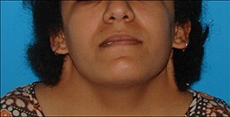
Webbed neck in a teenage girl with Turner syndrome

In addition to short stature, Turner syndrome is associated with a number of characteristic physical features. These include a webbed neck, a low hairline, a small chin and jaw, a high-arched palate, and a broad chest with wide-spaced nipples. Lymphedema (swelling) of the hands and feet is common at birth and sometimes persistent throughout the lifespan. Some Turner's stigmata, such as cubitus valgus and shortened fingers, are related to SHOX gene dosage effects.

A number of the external manifestations of Turner syndrome are focused on the limbs, hands, and feet. Lymphedema at birth is one of the classic features of the syndrome; though it often resolves during toddlerhood, recurrence in later life is frequent, often without apparent cause. Cases where the retained X chromosome was inherited from the mother more often experience lymphedema than those where it was from the father. As a consequence of lymphedema's effects on nail anatomy, females with Turner syndrome frequently have small hypoplastic nails. Shortened metacarpal bones, particularly the fourth metacarpal, are a frequent finding. The body shape of individuals with Turner syndrome is frequently quite broad and stocky, as the growth deficiency is more pronounced in the length of bones than in their width. Scoliosis is common in Turner syndrome, and is seen in 40% of girls without growth hormone treatment.

Facial features associated with Turner syndrome include prominent ears, a low hairline, a webbed neck, a small chin with dental malocclusion, and downslanting palpebral fissures (the opening between the eyelids). These are thought to be related to lymphedema during the fetal period, specifically to the presence and resorption of excess fluids in the head and neck region. Neck webbing is a particularly distinctive trait of Turner syndrome, leading to many neonatal diagnoses. The underlying etiology of neck webbing is related to prenatal blood flow issues, and even in populations without Turner's has broad health consequences; the rate of congenital heart disease in webbed neck is 150-fold higher than in the general population, while the feature is also associated with reduced height and minor developmental impairments. Some women with Turner syndrome have premature facial wrinkling.[14] Acne is less common in teenage girls and women with Turner syndrome, though the reasons why are unclear.

An infant with Turner syndrome

Other physical features connected to the condition include long eyelashes, sometimes including an additional set of eyelashes, and unusual dermatoglyphics (fingerprints). Some women with Turner's report being unable to create fingerprint passwords due to hypoplastic dermatoglyphics. Unusual dermatoglyphics are common to chromosome anomalies and in the case of Turner's may be a consequence of fetal lymphedema. Keloid scars, or raised hypertrophic scars growing beyond the boundaries of the original wound, are potentially associated with Turner syndrome; however, the association is underresearched. Though traditional medical counselling on the topic urges conservatism about elective procedures such as ear piercing due to the risk of severe scarring, the actual consequences are unclear. Keloids in Turner syndrome are particularly frequent following surgical procedures to reduce neck webbing. Turner syndrome has been associated with unusual patterns of hair growth, such as patches of short and long hair. Armpit and pubic hair is often sparse, while arm and leg hair is often thick. Though armpit hair is reduced in amount and thickness, the pattern in which it is implanted in the skin is as in men, rather than as in women.

Cardiac

Bicuspid aortic valve

Approximately half of individuals with Turner syndrome have congenital heart defects. CHDs associated with Turner syndrome include bicuspid aortic valves (30%), coarctation of the aorta (15%), and abnormalities of the arteries in the head and neck. A rare but potentially fatal complication of heart defects in Turner syndrome is aortic dissection, where the inner layer of the aorta tears open. Aortic dissection is six times as common in females with Turner syndrome as the general population and accounts for 8% of all deaths in the syndrome. The risk is substantially increased for individuals with bicuspid aortic valves, who make up 95% of patients with aortic dissection compared to 30% of all Turner's patients, and coarctation of the aorta, who make up 90% and 15% respectively.

Coronary artery disease onsets earlier in life in women with Turner syndrome compared to controls, and mortality from cardiac events is increased. This is thought to be in part a function of the relationship between Turner syndrome and obesity; women with Turner syndrome have a higher percentage of body fat for their weight than control women, and their short stature makes weight control more difficult. Though coronary artery disease is frequently thought a disease of older adults, young women with Turner syndrome are more likely to develop the disease than their 46,XX peers. Treatment recommendations for women with Turner syndrome and coronary artery disease are as in the general population, but as Turner's increases the risk of type 2 diabetes, women with insulin resistance must weigh up the benefits of prophylactic or early statin treatment with the risk of diabetes.

Internal medicine

Turner syndrome is associated with a broad variety of health considerations, such as liver and kidney issues, obesity, diabetes, and hypertension. Liver dysfunction is common in women with Turner syndrome, with 50%–80% having elevated liver enzymes. Non-alcoholic fatty liver disease is increased in prevalence in Turner syndrome, likely related in part to both conditions' associations with obesity. Hepatic vascular diseases are also seen in the syndrome as an aspect of Turner syndrome's broader vascular and cardiac impacts. Primary biliary cholangitis is more common in 45,X0 than 46,XX women. An unclear association exists between estrogen replacement therapy and liver dysfunction in Turner syndrome; some studies imply estrogen therapy worsens such conditions, while others imply improvement.

Duplicated ureter

Kidney issues, such as horseshoe kidney, are sometimes observed in Turner syndrome. Horseshoe kidney, where the kidneys are fused together in a U-shape, occurs in around 10% of Turner's cases compared to less than 0.5% of the general population. A missing kidney is observed in as many as 5% of individuals with Turner syndrome, compared to around 0.1% of the population. A duplicated ureter, where two ureters drain a single kidney, occurs in as much as 20%–30% of the Turner syndrome population. Kidney malformations in Turner syndrome may be more common in mosaicism than in the full 45,X0 karyotype. Serious complications of the kidney anomalies associated with Turner syndrome are rare, although there is some risk of issues such as obstructive uropathy, where the flow of urine from the kidneys is blocked.

Women with Turner syndrome are more likely than average to have high blood pressure; as many as 60% of women with the condition are hypertensive. Isolated diastolic hypertension often precedes systolic hypertension in the condition and may develop at a young age. Treatments for hypertension in Turner syndrome are as in the general population.

Approximately 25%–80% of women with Turner syndrome have some level of insulin resistance, and a minority develop type 2 diabetes. The risk of diabetes in Turner syndrome varies by karyotype and appears to be raised by specific deletions of the short arm of the X chromosome (Xp). One study found that while a relatively low 9% of women with Xq (long arm) deletions had type 2 diabetes, 18% of those with full 45,X0 karyotypes did, as well as 23% with Xp deletions. 43% of women with isochromosome Xq, who both lacked the short arm and had an additional copy of the long arm, developed type 2 diabetes. Though part of the diabetes risk in Turner syndrome is a function of weight control, some is independent; age- and weight-matched women with non-Turner's ovarian failure have a lower diabetes risk than in Turner syndrome. Growth hormone treatment plays an unclear role in diabetes risk, as does estrogen supplementation.

The association between Turner syndrome and other diseases, such as cancer, is unclear. Overall, women with Turner syndrome do not appear more likely to develop cancer than women with 46,XX karyotypes, but the specific pattern of what cancers are highest risk seems to differ. The risk of breast cancer appears lower in Turner's than in control women, perhaps due to decreased levels of estrogen. Neuroblastoma, a cancer of infancy and early childhood, has been reported in girls with Turner syndrome. Tumours of the nervous system, both the central nervous system and the peripheral nervous system, are overrepresented amongst cancers in Turner syndrome. Furthermore, about 5.5% of Turner syndrome individuals have an extra, abnormal small supernumerary marker chromosome (sSMC) which consists of part of a Y chromosome. This partial Y chromosome-bearing sSMC may include the SRY gene located on the p arm of the Y chromosome at band 11.2 (notated as Yp11.2). This gene encodes the testis-determining factor protein (also known as sex-determining region Y protein). Turner syndrome individuals with this SRY gene-containing sSMC have a very real increased risk of developing gonadal tissue neoplasms such as gonadoblastomas and in situ seminomas (also termed dysgerminomas to indicate that this tumor has the pathology of the testicular tumor, seminoma, but develops in ovaries). In one study, 34 Turner syndrome girls without overt evidence of these tumors were found at preventative surgery to have a gonadoblastoma (7 cases), dysgerminoma (1 case), or non-specific in situ gonadal neoplasm (1 case). Turner syndrome girls with this sSMC otherwise have typical features of the Turner syndrome except for a minority who also have hirsutism and/or clitoral enlargement. Surgical removal of the gonads has been recommended to remove the threat of developing these sSMC-associated neoplasms. Tuner syndrome individuals with an sSMC that lacks the SRY gene are not at an increased risk of developing these cancers.

Sensory

Hearing loss is common in Turner syndrome. Though at birth hearing is generally normal, chronic middle ear problems are frequent throughout childhood, which can cause permanent conductive hearing loss. In adulthood, sensorineural hearing loss occurs more often than in 46,XX women and at younger ages; though differing thresholds of hearing loss make it difficult to compare between studies, younger adult women with Turner syndrome are routinely found to have disproportionate rates of hearing issues, with sometimes up to half of women in their 20s and 30s having poor hearing. This hearing loss is progressive; at the age of 40, women with Turner syndrome have equivalent hearing loss to 46,XX women aged 60, on average. Cohort studies imply hearing loss may be more common in women who also have metabolic syndrome. The high prevalence of sensorineural hearing loss in Turner syndrome appears to be related to SHOX deficiency.

Ocular and visual disorders are also increased in prevalence in Turner syndrome. More than half of individuals with Turner syndrome have some form of eye disorder. This may be a consequence of shared genes on the X chromosome in both eye and ovary development. Nearly half of cases have hyperopia or myopia, usually mild. Strabismus, or misalignment of the eye, occurs in around one-fifth to one-third of girls with Turner syndrome. As with strabismus outside the Turner's context, it may be treated with glasses, patching, or surgical correction. Esotropia, where the eye turns inwards, is more common than exotropia, where it turns outwards. Ptosis, or a drooping eyelid, is a common facial manifestation of Turner syndrome; it usually has no appreciable impact on vision, but severe cases may limit visual range and require surgical correction. The rate of red-green colourblindness in Turner syndrome is 8%, the same as in men. This is due to red-green colourblindness being an X-linked recessive condition; in people with a single X chromosome, whether normal males or Turner females, only a single mutated X is necessary for symptoms. Red-green colourblindness may be underdiagnosed in the Turner context, as the rarity of the condition in females reduces the likelihood of screening, and practitioners may not connect that the karyotype of Turner syndrome increases the risk from the female baseline.

Autoimmune

Women with Turner syndrome are two to three times as likely to develop autoimmune disorders as the general population. Specific autoimmune disorders linked to Turner syndrome include Hashimoto's disease, vitiligo, psoriasis and psoriatic arthritis, alopecia, and celiac disease. Inflammatory bowel disease is also common, while the prevalence of type 1 diabetes is unclear, though appears increased.

Thyroid disease is common in Turner syndrome. Hypothyroidism is prevalent; 30%–50% of women with Turner syndrome have Hashimoto's disease, where the thyroid gland is slowly destroyed by an autoimmune reaction. By age 50, half of women with Turner syndrome have subclinical or clinical hypothyroidism. Hyperthyroidism and Graves' disease are also increased in prevalence, though more modestly. The Turner's presentation of hyperthyroidism is as in the general population, while the presentation of hypothyroidism is often atypical, with a mild early presentation yet a more severe progression. Women with isochromosome Xq are more likely to develop autoimmune thyroid disease than women with other forms of Turner syndrome.

The risk of irritable bowel syndrome is increased around fivefold in Turner syndrome, and that of ulcerative colitis around fourfold. Celiac disease is also increased in prevalence, with around 4%–8% of Turner's patients having comorbid celiac disease compared to 0.5%–1% of the general population. Diagnosis of such conditions is difficult due to their nonspecific early symptoms. In the Turner's context, diagnosis may in particular be missed due to growth delay; such conditions cause growth delay and failure to thrive when they onset in childhood, but as girls with Turner syndrome already have such delay, symptoms may be overlooked and ascribed to the original condition.

Alopecia areata, or recurrent patchy hair loss, is three times as common in Turner syndrome as the general population. Alopecia in the Turner syndrome context is frequently treatment-resistant, also seen in other chromosome aneuploidies such as Down syndrome. Psoriasis is common in Turner syndrome, although the precise prevalence is unclear. Turner's psoriasis may be related to growth hormone treatment, as psoriasis as a side effect of such therapies has been reported in patients without the karyotype. Psoriasis may progress to psoriatic arthritis, and this progression may be more common in Turner syndrome. Vitiligo has been reported in conjunction with Turner syndrome, but the risk is unclear and may be a side effect of increased clinical attention to autoimmune disease in this population.

Puberty

Histopathology of ovarian tissue in mosaic (A and B) and full (C) Turner syndrome

Puberty is delayed or absent in Turner syndrome. A 2019 literature review found that 13% of women with a 45,X0 karyotype could expect to experience spontaneous thelarche (breast development), while 9% would undergo spontaneous menarche (beginning of menstruation). These numbers were higher in women with mosaic Turner's; 63% with 45,X0/46,XX karyotypes experienced spontaneous thelarche and 39% spontaneous menarche, while 88% with 45,X0/47,XXX (the presence of a trisomy X cell line) experienced spontaneous thelarche and 66% spontaneous menarche. Unexpectedly, women with Y-chromosome cells also had increased rates of thelarche and menarche compared to the 45,X0 baseline, at 41% and 19%. However, few women with trisomy X or Y-chromosome cell lines were covered in the review, impeding extrapolation from these results. 6% of women with Turner syndrome have regular menstrual cycles; the rest experience primary or secondary amenorrhea or other menstrual dysfunction.

In girls with Turner syndrome who do not experience spontaneous puberty, exogenous estrogen is used to induce and maintain feminization. Estrogen replacement is recommended to begin at around age 11–12, although some parents prefer to delay the induction of puberty in girls with lower social and emotional preparedness. The dose of estrogen in induced puberty begins at 10% of adult estrogen levels and is steadily increased at six-month intervals, with a full adult dose attained two to three years after the beginning of treatment. Estrogen replacement may interfere with growth hormone therapy, due to the closing effects of estrogen on growth plates; individuals must weigh up their preferences for taller height versus greater feminization.

Fertility

Women with Turner syndrome are infertile. Only 2%–5% are capable of pregnancy without fertility treatment, most with mosaic karyotypes. Early in gestation, fetuses with Turner syndrome have a normal number of gametes in their developing ovaries, but this starts decreasingly rapidly as early as 18 weeks of pregnancy; by birth, girls with the condition have markedly reduced follicular counts. Women with Turner syndrome who wish to raise families but are incapable of conception with their own oocytes have the options of adoption or of pregnancy with donor eggs; the latter has a comparable success rate to donor pregnancy in women with 46,XX karyotypes.

Pregnancy in Turner syndrome is inherently high-risk; the maternal death rate is 2%.

Usually, estrogen replacement therapy is used to spur the growth of secondary sexual characteristics at the time when puberty should onset. While very few women with Turner syndrome menstruate spontaneously, estrogen therapy requires a regular shedding of the uterine lining ("withdrawal bleeding") to prevent its overgrowth. Withdrawal bleeding can be induced monthly, like menstruation, or less often, usually every three months, if the patient desires. Estrogen therapy does not make a woman with nonfunctional ovaries fertile, but it plays an important role in assisted reproduction; the health of the uterus must be maintained with estrogen if an eligible woman with Turner Syndrome wishes to use IVF (using donated oocytes).

Especially in mosaic cases of Turner syndrome that contains Y-chromosome (e.g. 45,X/46,XY) due to the risk of development of ovarian malignancy (most common is gonadoblastoma) gonadectomy is recommended. Turner syndrome is characterized by primary amenorrhoea, premature ovarian failure (hypergonadotropic hypogonadism), streak gonads and infertility (however, technology (especially oocyte donation) provides the opportunity of pregnancy in these patients). Failure to develop secondary sex characteristics (sexual infantilism) is typical.

Cognition

Neurodevelopmental

Individuals with Turner syndrome have normal intelligence. Verbal IQ is usually higher than performance IQ; one review of thirteen studies found an average verbal IQ of 101 compared to an average performance IQ of 89.

People with Turner syndrome have normal intelligence, and demonstrate relative strengths in verbal skills, but may exhibit weaker nonverbal skills – particularly in arithmetic, select visuospatial skills, and processing speed. Turner syndrome does not typically cause intellectual disability or impair cognition. However, learning difficulties are common among women with Turner syndrome, particularly a specific difficulty in perceiving spatial relationships, such as nonverbal learning disorder. This may also manifest itself as a difficulty with motor control or with mathematics. While it is not correctable, in most cases it does not cause difficulty in daily living. Most Turner syndrome patients are employed as adults and lead productive lives.

Also, a rare variety of Turner syndrome, known as "Ring-X Turner syndrome", has about a 60% association with intellectual disability. This variety accounts for around 2–4% of all Turner syndrome cases.

Psychological

Social difficulties appear to be an area of vulnerability for young women. Counseling affected individuals and their families about the need to carefully develop social skills and relationships may prove useful in advancing social adaptation. Women with Turner syndrome may experience adverse psychosocial outcomes which can be improved through early intervention and the provision of appropriate psychological and psychiatric care. Genetic, hormonal, and medical problems associated with Turner syndrome are likely to affect psychosexual development of female adolescent patients, and thus influence their psychological functioning, behavior patterns, social interactions, and learning ability. Although Turner syndrome constitutes a chronic medical condition, with possible physical, social, and psychological complications in a woman's life, hormonal and estrogen replacement therapy, and assisted reproduction, are treatments that can be helpful for Turner syndrome patients and improve their quality of life. Research shows a possible association between age at diagnosis and increased substance use and depressive symptoms.

Prenatal

Despite the excellent postnatal prognosis, 99% of Turner syndrome conceptions are thought to end in miscarriage or stillbirth, and as many as 15% of all spontaneous abortions have the 45,X karyotype. Among cases that are detected by routine amniocentesis or chorionic villus sampling, one study found that the prevalence of Turner syndrome among tested pregnancies was 5.58 and 13.3 times higher, respectively, than among live neonates in a similar population.

Cause

Turner syndrome is caused by the absence of one complete or partial copy of the X chromosome in some or all the cells. The abnormal cells may have only one X (monosomy) (45,X) or they may be affected by one of several types of partial monosomy like a deletion of the short p arm of one X chromosome (46,X,del(Xp)) or the presence of an isochromosome with two q arms (46,X,i(Xq)) Turner syndrome has distinct features due to the lack of pseudoautosomal regions, which are typically spared from X-inactivation. In mosaic individuals, cells with X monosomy (45,X) may occur along with cells that are normal (46,XX), cells that have partial monosomies, or cells that have a Y chromosome (46,XY). The presence of mosaicism is estimated to be relatively common in affected individuals (67–90%).

The (46,X,i(Xq) isochromosome in the Turner syndrome is classified as an small supernumerary marker chromosome (sSMC). Two of the types of sSMCs in this syndrome contain parts of the genetic material from either an X or, much less frequently, Y chromosome and may or may not contain an XIST gene. In normal females, the XIST gene occurs on the X chromosome inherited from her mother but not on the X chromosome inherited from her father. The gene is not present on Y chromosomes and in normal females resides on and functions to inactivate many of the genes located on its own maternal but not the father's X chromosome. Turner syndrome females with (46,X,i(Xq) sSMC consisting of a partial X chromosome that does not contain the XIST gene express at least some of this sSMC's genetic material and therefore contain excesses of this material. In consequence, they have a more serious form of the Turner syndrome that ranges form moderately severe to extremely severe. The extremely severe cases have anencephaly (absence of a major portion of the brain, skull, and scalp), agenesis of the corpus callosum (lack of the thick tract of nerve fibers that connect the left and right cerebral hemispheres), and complex heart deformities. Individuals with Turner syndrome that have partial X chromosome containing(46,X,i(Xq) sSMCs that have the XIST gene do not express this sSMC's genetic material and do not have the more severe manifestations of the syndrome.

Inheritance

In the majority of cases where monosomy occurs, the X chromosome comes from the mother. This may be due to a nondisjunction in the father. Meiotic errors that lead to the production of X with p arm deletions or abnormal Y chromosomes are also mostly found in the father. Isochromosome X or ring chromosome X on the other hand are formed equally often by both parents. Overall, the functional X chromosome usually comes from the mother.

In most cases, Turner syndrome is a sporadic event, and for the parents of an individual with Turner syndrome the risk of recurrence is not increased for subsequent pregnancies. Rare exceptions may include the presence of a balanced translocation of the X chromosome in a parent, or where the mother has 45,X mosaicism restricted to her germ cells.

Diagnosis

Prenatal

45,X karyotype, showing an unpaired X at the lower right

Turner syndrome may be diagnosed by amniocentesis or chorionic villus sampling during pregnancy.

Usually, fetuses with Turner syndrome can be identified by abnormal ultrasound findings (i.e., heart defect, kidney abnormality, cystic hygroma, ascites). In a study of 19 European registries, 67.2% of prenatally diagnosed cases of Turner syndrome were detected by abnormalities on ultrasound. 69.1% of cases had one anomaly present, and 30.9% had two or more anomalies.

An increased risk of Turner syndrome may also be indicated by abnormal triple or quadruple maternal serum screen. The fetuses diagnosed through positive maternal serum screening are more often found to have a mosaic karyotype than those diagnosed based on ultrasonographic abnormalities, and conversely, those with mosaic karyotypes are less likely to have associated ultrasound abnormalities.

Postnatal

Turner syndrome can be diagnosed postnatally at any age. Often, it is diagnosed at birth due to heart problems, an unusually wide neck or swelling of the hands and feet. However, it is also common for it to go undiagnosed for several years, often until the girl reaches the age of puberty and fails to develop typically (the changes associated with puberty do not occur). In childhood, a short stature can be indicative of Turner syndrome.

A test called a karyotype, also known as a chromosome analysis, analyzes the chromosomal composition of the individual. This is the test of choice to diagnose Turner syndrome.

Treatment

As a chromosomal condition, there is no cure for Turner syndrome. However, much can be done to minimize the symptoms. While most of the physical findings are harmless, significant medical problems can be associated with the syndrome. Most of these significant conditions are treatable with surgery and other therapies including hormonal therapy.

  • Growth hormone, either alone or with a low dose of androgen, will increase growth and probably final adult height. Growth hormone is approved by the U.S. Food and Drug Administration for treatment of Turner syndrome and is covered by many insurance plans. There is evidence that this is effective, even in toddlers. A 2019 systematic review comparing effects of adding oxandrolone to growth hormone treatment to growth hormone alone has found moderate-quality evidence that the addition of oxandrolone leads to an increase in final adult height of girls with Turner syndrome. When the same review assessed the effects of adding Oxandrolone to growth hormone treatment on speech, cognition and psychological status, the results were inconclusive due to very-low quality evidence.
  • Estrogen replacement therapy such as the birth control pill, has been used since the condition was described in 1938 to promote development of secondary sexual characteristics. Estrogens are crucial for maintaining good bone integrity, cardiovascular health and tissue health. Women with Turner syndrome who do not have spontaneous puberty and who are not treated with estrogen are at high risk for osteoporosis and heart conditions.
  • Modern reproductive technologies have also been used to help women with Turner syndrome become pregnant if they desire. For example, a donor egg can be used to create an embryo, which is carried by the Turner syndrome woman.
  • Uterine maturity is positively associated with years of estrogen use, history of spontaneous menarche, and negatively associated with the lack of current hormone replacement therapy.

Epidemiology

Turner syndrome occurs in between one in 2000 and one in 5000 females at birth.

Approximately 99 percent of fetuses with Turner syndrome spontaneously terminate during the first trimester. Turner syndrome accounts for about 10 percent of the total number of spontaneous abortions in the United States.

History

The syndrome is named after Henry Turner, an endocrinologist from Illinois, who described it in 1938. In Europe, it is often called Ullrich–Turner syndrome or Bonnevie–Ullrich–Turner syndrome to acknowledge that earlier cases had also been described by European doctors Kristine Bonnevie and Otto Ullrich. In Russian and USSR literature, it is called Shereshevsky–Turner syndrome to acknowledge that the condition was first described as hereditary in 1925 by the Soviet endocrinologist Nikolai Shereshevsky [ru], who believed that it was due to the underdevelopment of the gonads and the anterior pituitary gland and was combined with congenital malformations of internal development.

The first published report of a female with a 45,X karyotype was in 1959 by Charles Ford and colleagues in Harwell near Oxford, and Guy's Hospital in London. It was found in a 14-year-old girl with signs of Turner syndrome.

Hypothyroidism

From Wikipedia, the free encyclopedia
 
Hypothyroidism
Other namesUnderactive thyroid, low thyroid, hypothyreosis
Molecular structure of the thyroxine molecule
Molecular structure of thyroxine, the deficiency of which causes the symptoms of hypothyroidism
Pronunciation
SpecialtyEndocrinology
SymptomsPoor ability to tolerate cold, feeling tired, constipation, weight gain, depression, anxiety, irritability
ComplicationsDuring pregnancy can result in cretinism in the baby
Usual onset< 60 years old
CausesIodine deficiency, Hashimoto's thyroiditis
Diagnostic methodBlood tests (thyroid-stimulating hormone, thyroxine)
Differential diagnosisDepression, dementia, heart failure, chronic fatigue syndrome
PreventionSalt iodization
TreatmentLevothyroxine
Frequency0.3–0.4% (USA)

Hypothyroidism (also called underactive thyroid, low thyroid or hypothyreosis) is a disorder of the endocrine system in which the thyroid gland does not produce enough thyroid hormone. It can cause a number of symptoms, such as poor ability to tolerate cold, a feeling of tiredness, constipation, slow heart rate, depression, and weight gain. Occasionally there may be swelling of the front part of the neck due to goiter. Untreated cases of hypothyroidism during pregnancy can lead to delays in growth and intellectual development in the baby or congenital iodine deficiency syndrome.

Worldwide, too little iodine in the diet is the most common cause of hypothyroidism. Hashimoto's thyroiditis is the most common cause of hypothyroidism in countries with sufficient dietary iodine. Less common causes include previous treatment with radioactive iodine, injury to the hypothalamus or the anterior pituitary gland, certain medications, a lack of a functioning thyroid at birth, or previous thyroid surgery. The diagnosis of hypothyroidism, when suspected, can be confirmed with blood tests measuring thyroid-stimulating hormone (TSH) and thyroxine levels.

Salt iodization has prevented hypothyroidism in many populations. Thyroid hormone replacement with levothyroxine treats hypothyroidism. Medical professionals adjust the dose according to symptoms and normalization of the thyroxine and TSH levels. Thyroid medication is safe in pregnancy. Although an adequate amount of dietary iodine is important, too much may worsen specific forms of hypothyroidism.

Worldwide about one billion people are estimated to be iodine-deficient; however, it is unknown how often this results in hypothyroidism. In the United States, hypothyroidism occurs in 0.3–0.4% of people. Subclinical hypothyroidism, a milder form of hypothyroidism characterized by normal thyroxine levels and an elevated TSH level, is thought to occur in 4.3–8.5% of people in the United States. Hypothyroidism is more common in women than in men. People over the age of 60 are more commonly affected. Dogs are also known to develop hypothyroidism, as are cats and horses, albeit more rarely. The word hypothyroidism is from Greek hypo- 'reduced', thyreos 'shield', and eidos 'form'.

Signs and symptoms

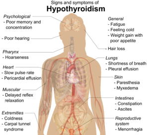
People with hypothyroidism often have no or only mild symptoms. Numerous symptoms and signs are associated with hypothyroidism and can be related to the underlying cause, or a direct effect of having not enough thyroid hormones. Hashimoto's thyroiditis may present with the mass effect of a goiter (enlarged thyroid gland). In middle-aged women, the symptoms may be mistaken for those of menopause.

Symptoms and signs of hypothyroidism
Symptoms Signs
Fatigue Dry, coarse skin
Feeling cold Cool extremities
Poor memory and concentration Myxedema (mucopolysaccharide deposits in the skin)
Constipation, dyspepsia Hair loss
Weight gain with poor appetite Slow pulse rate
Shortness of breath Swelling of the limbs
Hoarse voice Delayed relaxation of tendon reflexes
In females, heavy menstrual periods (and later light periods) Carpal tunnel syndrome
Abnormal sensation Pleural effusion, ascites, pericardial effusion
Poor hearing

Delayed relaxation after testing the ankle jerk reflex is a characteristic sign of hypothyroidism and is associated with the severity of the hormone deficit.

Myxedema coma

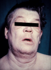
Man with myxedema or severe hypothyroidism showing an expressionless face, puffiness around the eyes and pallor
 
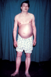
Additional symptoms include swelling of the arms and legs and ascites.

Myxedema coma is a rare but life-threatening state of extreme hypothyroidism. It may occur in those with established hypothyroidism when they develop an acute illness. Myxedema coma can be the first presentation of hypothyroidism. People with myxedema coma typically have a low body temperature without shivering, confusion, a slow heart rate and reduced breathing effort. There may be physical signs suggestive of hypothyroidism, such as skin changes or enlargement of the tongue.

Pregnancy

Even mild or subclinical hypothyroidism leads to possible infertility and an increased risk of miscarriage. Hypothyroidism in early pregnancy, even with limited or no symptoms, may increase the risk of pre-eclampsia, offspring with lower intelligence, and the risk of infant death around the time of birth. Women are affected by hypothyroidism in 0.3–0.5% of pregnancies. Subclinical hypothyroidism during pregnancy is associated with gestational diabetes, low birth-weight, placental abruption, and the birth of the baby before 37 weeks of pregnancy.

Children

Newborn children with hypothyroidism may have normal birth weight and height (although the head may be larger than expected and the posterior fontanelle may be open). Some may have drowsiness, decreased muscle tone, a hoarse-sounding cry, feeding difficulties, constipation, an enlarged tongue, umbilical hernia, dry skin, a decreased body temperature, and jaundice. A goiter is rare, although it may develop later in children who have a thyroid gland that does not produce functioning thyroid hormone. A goiter may also develop in children growing up in areas with iodine deficiency. Normal growth and development may be delayed, and not treating infants may lead to an intellectual impairment (IQ 6–15 points lower in severe cases). Other problems include the following: difficulty with large scale and fine motor skills and coordination, reduced muscle tone, squinting, decreased attention span, and delayed speaking. Tooth eruption may be delayed.

In older children and adolescents, the symptoms of hypothyroidism may include fatigue, cold intolerance, sleepiness, muscle weakness, constipation, a delay in growth, overweight for height, pallor, coarse and thick skin, increased body hair, irregular menstrual cycles in girls, and delayed puberty. Signs may include delayed relaxation of the ankle reflex and a slow heartbeat. A goiter may be present with a completely enlarged thyroid gland; sometimes only part of the thyroid is enlarged and it can be knobby.

Related disorders

Thyroid hormone abnormalities are common in major psychiatric disorders including bipolar disorder; clinical research has shown there is a high rate of thyroid dysfunction in mood disorders and schizophrenia-spectrum disorders, concluding that there is a case for screening for the latter among people with thyroid illness.

Causes

Hypothyroidism is caused by inadequate function of the gland itself (primary hypothyroidism), inadequate stimulation by thyroid-stimulating hormone from the pituitary gland (secondary hypothyroidism), or inadequate release of thyrotropin-releasing hormone from the brain's hypothalamus (tertiary hypothyroidism). Primary hypothyroidism is about a thousandfold more common than central hypothyroidism. Central hypothyroidism is the name used for secondary and tertiary, since hypothalamus and pituitary gland are at the center of thyroid hormone control.

Iodine deficiency is the most common cause of primary hypothyroidism and endemic goiter worldwide. In areas of the world with sufficient dietary iodine, hypothyroidism is most commonly caused by the autoimmune disease Hashimoto's thyroiditis (chronic autoimmune thyroiditis). Hashimoto's may be associated with a goiter. It is characterized by infiltration of the thyroid gland with T lymphocytes and autoantibodies against specific thyroid antigens such as thyroid peroxidase, thyroglobulin and the TSH receptor.

After women give birth, about 5% develop postpartum thyroiditis which can occur up to nine months afterwards. This is characterized by a short period of hyperthyroidism followed by a period of hypothyroidism; 20–40% remain permanently hypothyroid.

Autoimmune thyroiditis (Hashimoto's) is associated with other immune-mediated diseases such as diabetes mellitus type 1, pernicious anemia, myasthenia gravis, celiac disease, rheumatoid arthritis and systemic lupus erythematosus. It may occur as part of autoimmune polyendocrine syndrome (type 1 and type 2).

Iatrogenic hypothyroidism can be surgical (a result of thyroidectomy, usually for thyroid nodules or cancer) or following radioiodine ablation (usually for Graves' disease).

Group Causes
Primary hypothyroidism Iodine deficiency (developing countries), autoimmune thyroiditis, subacute granulomatous thyroiditis, subacute lymphocytic thyroiditis, postpartum thyroiditis, previous thyroidectomy, acute infectious thyroiditis, previous radioiodine treatment, previous external beam radiotherapy to the neck
Medication: lithium-based mood stabilizers, amiodarone, interferon alpha, tyrosine kinase inhibitors such as sunitinib
Central hypothyroidism Lesions compressing the pituitary (pituitary adenoma, craniopharyngioma, meningioma, glioma, Rathke's cleft cyst, metastasis, empty sella, aneurysm of the internal carotid artery), surgery or radiation to the pituitary, drugs, injury, vascular disorders (pituitary apoplexy, Sheehan syndrome, subarachnoid hemorrhage), autoimmune diseases (lymphocytic hypophysitis, polyglandular disorders), infiltrative diseases (iron overload due to hemochromatosis or thalassemia, neurosarcoidosis, Langerhans cell histiocytosis), particular inherited congenital disorders, and infections (tuberculosis, mycoses, syphilis)
Congenital hypothyroidism Thyroid dysgenesis (75%), thyroid dyshormonogenesis (20%), maternal antibody or radioiodine transfer
Syndromes: mutations (in GNAS complex locus, PAX8, TTF-1/NKX2-1, TTF-2/FOXE1), Pendred's syndrome (associated with sensorineural hearing loss)
Transiently: due to maternal iodine deficiency or excess, anti-TSH receptor antibodies, certain congenital disorders, neonatal illness
Central: pituitary dysfunction (idiopathic, septo-optic dysplasia, deficiency of PIT1, isolated TSH deficiency)

Pathophysiology

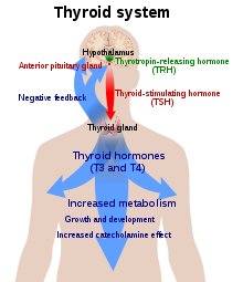
Diagram of a person with a large blue arrow representing the actions of thyroxine on the body and a green and red arrow representing actions of TSH and TRH respectively
Diagram of the hypothalamic–pituitary–thyroid axis. The hypothalamus secretes TRH (green), which stimulates the production of TSH (red) by the pituitary gland. This, in turn, stimulates the production of thyroxine by the thyroid (blue). Thyroxine levels decrease TRH and TSH production by a negative feedback process.

Thyroid hormone is required for the normal functioning of numerous tissues in the body. In healthy individuals, the thyroid gland predominantly secretes thyroxine (T4), which is converted into triiodothyronine (T3) in other organs by the selenium-dependent enzyme iodothyronine deiodinase. Triiodothyronine binds to the thyroid hormone receptor in the nucleus of cells, where it stimulates the turning on of particular genes and the production of specific proteins. Additionally, the hormone binds to integrin αvβ3 on the cell membrane, thereby stimulating the sodium–hydrogen antiporter and processes such as formation of blood vessels and cell growth. In blood, almost all thyroid hormone (99.97%) are bound to plasma proteins such as thyroxine-binding globulin; only the free unbound thyroid hormone is biologically active.

The thyroid gland is the only source of thyroid hormone in the body; the process requires iodine and the amino acid tyrosine. Iodine in the bloodstream is taken up by the gland and incorporated into thyroglobulin molecules. The process is controlled by the thyroid-stimulating hormone (TSH, thyrotropin), which is secreted by the pituitary. Not enough iodine, or not enough TSH, can result in decreased production of thyroid hormones.

The hypothalamic–pituitary–thyroid axis plays a key role in maintaining thyroid hormone levels within normal limits. Production of TSH by the anterior pituitary gland is stimulated in turn by thyrotropin-releasing hormone (TRH), released from the hypothalamus. Production of TSH and TRH is decreased by thyroxine by a negative feedback process. Not enough TRH, which is uncommon, can lead to not enough TSH and thereby to not enough thyroid hormone production.

Pregnancy leads to marked changes in thyroid hormone physiology. The gland is increased in size by 10%, thyroxine production is increased by 50%, and iodine requirements are increased. Many women have normal thyroid function but have immunological evidence of thyroid autoimmunity (as evidenced by autoantibodies) or are iodine deficient, and develop evidence of hypothyroidism before or after giving birth.

Diagnosis

Laboratory testing of thyroid stimulating hormone levels in the blood is considered the best initial test for hypothyroidism; a second TSH level is often obtained several weeks later for confirmation. Levels may be abnormal in the context of other illnesses, and TSH testing in hospitalized people is discouraged unless thyroid dysfunction is strongly suspected, as the cause of the acute illness. An elevated TSH level indicates that the thyroid gland is not producing enough thyroid hormone, and free T4 levels are then often obtained. Measuring T3 is discouraged by the AACE in the assessment for hypothyroidism. In England and Wales, the National Institute for Health and Care Excellence (NICE) recommends routine T4 testing in children, and T3 testing in both adults and children if central hypothyroidism is suspected and the TSH is low. There are a number of symptom rating scales for hypothyroidism; they provide a degree of objectivity but have limited use for diagnosis.

TSH T4 Interpretation
Normal Normal Normal thyroid function
Elevated Low Overt hypothyroidism
Normal/low Low Central hypothyroidism
Elevated Normal Subclinical hypothyroidism

Many cases of hypothyroidism are associated with mild elevations in creatine kinase and liver enzymes in the blood. They typically return to normal when hypothyroidism has been fully treated. Levels of cholesterol, low-density lipoprotein and lipoprotein (a) can be elevated; the impact of subclinical hypothyroidism on lipid parameters is less well-defined.

Very severe hypothyroidism and myxedema coma are characteristically associated with low sodium levels in the blood together with elevations in antidiuretic hormone, as well as acute worsening of kidney function due to a number of causes. In most causes, however, it is unclear if the relationship is causal.

A diagnosis of hypothyroidism without any lumps or masses felt within the thyroid gland does not require thyroid imaging; however, if the thyroid feels abnormal, diagnostic imaging is then recommended. The presence of antibodies against thyroid peroxidase (TPO) makes it more likely that thyroid nodules are caused by autoimmune thyroiditis, but if there is any doubt, a needle biopsy may be required.

Central

If the TSH level is normal or low and serum free T4 levels are low, this is suggestive of central hypothyroidism (not enough TSH or TRH secretion by the pituitary gland or hypothalamus). There may be other features of hypopituitarism, such as menstrual cycle abnormalities and adrenal insufficiency. There might also be symptoms of a pituitary mass such as headaches and vision changes. Central hypothyroidism should be investigated further to determine the underlying cause.

Overt

In overt primary hypothyroidism, TSH levels are high and T4 and T3 levels are low. Overt hypothyroidism may also be diagnosed in those who have a TSH on multiple occasions of greater than 5mIU/L, appropriate symptoms, and only a borderline low T4. It may also be diagnosed in those with a TSH of greater than 10mIU/L.

Subclinical

Subclinical hypothyroidism is a milder form of hypothyroidism characterized by an elevated serum TSH level, but with a normal serum free thyroxine level. This milder form of hypothyroidism is most commonly caused by Hashimoto's thyroiditis. In adults it is diagnosed when TSH levels are greater than 5 mIU/L and less than 10mIU/L. The presentation of subclinical hypothyroidism is variable and classic signs and symptoms of hypothyroidism may not be observed. Of people with subclinical hypothyroidism, a proportion will develop overt hypothyroidism each year. In those with detectable antibodies against thyroid peroxidase (TPO), this occurs in 4.3%, while in those with no detectable antibodies, this occurs in 2.6%. Those with subclinical hypothyroidism and detectable anti-TPO antibodies who do not require treatment should have repeat thyroid function tests more frequently (e.g. yearly) compared with those who do not have antibodies.

Pregnancy

During pregnancy, the thyroid gland must produce 50% more thyroid hormone to provide enough thyroid hormone for the developing fetus and the expectant mother. In pregnancy, free thyroxine levels may be lower than anticipated due to increased binding to thyroid binding globulin and decreased binding to albumin. They should either be corrected for the stage of pregnancy, or total thyroxine levels should be used instead for diagnosis. TSH values may also be lower than normal (particularly in the first trimester) and the normal range should be adjusted for the stage of pregnancy.

In pregnancy, subclinical hypothyroidism is defined as a TSH between 2.5 and 10 mIU/L with a normal thyroxine level, while those with TSH above 10 mIU/L are considered to be overtly hypothyroid even if the thyroxine level is normal. Antibodies against TPO may be important in making decisions about treatment, and should, therefore, be determined in women with abnormal thyroid function tests.

Determination of TPO antibodies may be considered as part of the assessment of recurrent miscarriage, as subtle thyroid dysfunction can be associated with pregnancy loss, but this recommendation is not universal, and presence of thyroid antibodies may not predict future outcome.

Prevention

A 3-month-old infant with untreated congenital hypothyroidism showing myxedematous facies, a big tongue, and skin mottling

Hypothyroidism may be prevented in a population by adding iodine to commonly used foods. This public health measure has eliminated endemic childhood hypothyroidism in countries where it was once common. In addition to promoting the consumption of iodine-rich foods such as dairy and fish, many countries with moderate iodine deficiency have implemented universal salt iodization. Encouraged by the World Health Organization, 70% of the world's population across 130 countries are receiving iodized salt. In some countries, iodized salt is added to bread. Despite this, iodine deficiency has reappeared in some Western countries as a result of attempts to reduce salt intake.

Pregnant and breastfeeding women, who require 66% more daily iodine than non-pregnant women, may still not be getting enough iodine. The World Health Organization recommends a daily intake of 250 µg for pregnant and breastfeeding women. As many women will not achieve this from dietary sources alone, the American Thyroid Association recommends a 150 µg daily supplement by mouth.

Screening

Screening for hypothyroidism is performed in the newborn period in many countries, generally using TSH. This has led to the early identification of many cases and thus the prevention of developmental delay. It is the most widely used newborn screening test worldwide. While TSH-based screening will identify the most common causes, the addition of T4 testing is required to pick up the rarer central causes of neonatal hypothyroidism. If T4 determination is included in the screening done at birth, this will identify cases of congenital hypothyroidism of central origin in 1:16,000 to 1:160,000 children. Considering that these children usually have other pituitary hormone deficiencies, early identification of these cases may prevent complications.

In adults, widespread screening of the general population is a matter of debate. Some organizations (such as the United States Preventive Services Task Force) state that evidence is insufficient to support routine screening, while others (such as the American Thyroid Association) recommend either intermittent testing above a certain age in all sexes or only in women. Targeted screening may be appropriate in a number of situations where hypothyroidism is common: other autoimmune diseases, a strong family history of thyroid disease, those who have received radioiodine or other radiation therapy to the neck, those who have previously undergone thyroid surgery, those with an abnormal thyroid examination, those with psychiatric disorders, people taking amiodarone or lithium, and those with a number of health conditions (such as certain heart and skin conditions). Yearly thyroid function tests are recommended in people with Down syndrome, as they are at higher risk of thyroid disease. Guidelines for England and Wales from the National Institute for Health and Care Excellence (NICE) recommend testing for thyroid disease in people with type 1 diabetes and new-onset atrial fibrillation, and suggests testing in those with depression or unexplained anxiety (all ages), in children with abnormal growth, or unexplained change in behaviour or school performance. On diagnosis of autoimmune thyroid disease, NICE also recommends screening for celiac disease.

Management

Hormone replacement

Most people with hypothyroidism symptoms and confirmed thyroxine deficiency are treated with a synthetic long-acting form of thyroxine, known as levothyroxine (L-thyroxine). In young and otherwise healthy people with overt hypothyroidism, a full replacement dose (adjusted by weight) can be started immediately; in the elderly and people with heart disease a lower starting dose is recommended to prevent over supplementation and risk of complications. Lower doses may be sufficient in those with subclinical hypothyroidism, while people with central hypothyroidism may require a higher than average dose.

Blood free thyroxine and TSH levels are monitored to help determine whether the dose is adequate. This is done 4–8 weeks after the start of treatment or a change in levothyroxine dose. Once the adequate replacement dose has been established, the tests can be repeated after 6 and then 12 months, unless there is a change in symptoms. Normalization of TSH does not mean that other abnormalities associated with hypothyroidism improve entirely, such as elevated cholesterol levels.

In people with central/secondary hypothyroidism, TSH is not a reliable marker of hormone replacement and decisions are based mainly on the free T4 level. Levothyroxine is best taken 30–60 minutes before breakfast, or four hours after food, as certain substances such as food and calcium can inhibit the absorption of levothyroxine. There is no direct way of increasing thyroid hormone secretion by the thyroid gland.

Liothyronine

Treatment with liothyronine alone has not received enough study to make a recommendation as to its use; due to its shorter half-life it would need to be taken more often than levothyroxine.

Adding liothyronine (synthetic T3) to levothyroxine has been suggested as a measure to provide better symptom control, but this has not been confirmed by studies. In 2007, the British Thyroid Association stated that combined T4 and T3 therapy carried a higher rate of side effects and no benefit over T4 alone. Similarly, American guidelines discourage combination therapy due to a lack of evidence, although they acknowledge that some people feel better when receiving combination treatment. Guidelines by NICE for England and Wales discourage liothyronine.

People with hypothyroidism who do not feel well despite optimal levothyroxine dosing may request adjunctive treatment with liothyronine. A 2012 guideline from the European Thyroid Association recommends that support should be offered with regards to the chronic nature of the disease and that other causes of the symptoms should be excluded. Addition of liothyronine should be regarded as experimental, initially only for a trial period of 3 months, and in a set ratio to the current dose of levothyroxine. The guideline explicitly aims to enhance the safety of this approach and to counter its indiscriminate use.

Desiccated animal thyroid

Desiccated thyroid extract is an animal-based thyroid gland extract, most commonly from pigs. It is a combination therapy, containing forms of T4 and T3. It also contains calcitonin (a hormone produced in the thyroid gland involved in the regulation of calcium levels), T1 and T2; these are not present in synthetic hormone medication. This extract was once a mainstream hypothyroidism treatment, but its use today is unsupported by evidence; British Thyroid Association and American professional guidelines discourage its use, as does NICE.

Subclinical hypothyroidism

There is no evidence of a benefit from treating subclinical hypothyroidism in those who are not pregnant, and there are potential risks of overtreatment. Untreated subclinical hypothyroidism may be associated with a modest increase in the risk of coronary artery disease when the TSH is over 10 mIU/L. A 2007 review found no benefit of thyroid hormone replacement except for "some parameters of lipid profiles and left ventricular function". There is no association between subclinical hypothyroidism and an increased risk of bone fractures, nor is there a link with cognitive decline.

Since 2008, consensus American opinion has been that in general people with TSH under 10 to 20 mIU/L do not require treatment.

American guidelines recommend that treatment should be considered in people with symptoms of hypothyroidism, detectable antibodies against thyroid peroxidase, a history of heart disease or are at an increased risk for heart disease, if the TSH is elevated but below 10 mIU/L. NICE recommends that those with a TSH above 10 mIU/L should be treated in the same way as overt hypothyroidism. Those with an elevated TSH but below 10 mIU/L who have symptoms suggestive of hypothyroidism should have a trial of treatment but with the aim to stopping this if the symptoms persist despite normalisation of the TSH.

A recent meta-analysis, however, found an increased risk for cardiovascular death in subclinical hypothyroidism.

Myxedema coma

Myxedema coma or severe decompensated hypothyroidism usually requires admission to the intensive care, close observation and treatment of abnormalities in breathing, temperature control, blood pressure, and sodium levels. Mechanical ventilation may be required, as well as fluid replacement, vasopressor agents, careful rewarming, and corticosteroids (for possible adrenal insufficiency which can occur together with hypothyroidism). Careful correction of low sodium levels may be achieved with hypertonic saline solutions or vasopressin receptor antagonists. For rapid treatment of the hypothyroidism, levothyroxine or liothyronine may be administered intravenously, particularly if the level of consciousness is too low to be able to safely swallow medication. While administration through a nasogastric tube is possible, this may be unsafe and is discouraged.

Pregnancy

In women with known hypothyroidism who become pregnant, it is recommended that serum TSH levels are closely monitored. Levothyroxine should be used to keep TSH levels within the normal range for that trimester. The first trimester normal range is below 2.5 mIU/L and the second and third trimesters normal range is below 3.0 mIU/L. Treatment should be guided by total (rather than free) thyroxine or by the free T4 index. Similarly to TSH, the thyroxine results should be interpreted according to the appropriate reference range for that stage of pregnancy. The levothyroxine dose often needs to be increased after pregnancy is confirmed, although this is based on limited evidence and some recommend that it is not always required; decisions may need to based on TSH levels.

Women with anti-TPO antibodies who are trying to become pregnant (naturally or by assisted means) may require thyroid hormone supplementation even if the TSH level is normal. This is particularly true if they have had previous miscarriages or have been hypothyroid in the past. Supplementary levothyroxine may reduce the risk of preterm birth and possibly miscarriage. The recommendation is stronger in pregnant women with subclinical hypothyroidism (defined as TSH 2.5–10 mIU/L) who are anti-TPO positive, in view of the risk of overt hypothyroidism. If a decision is made not to treat, close monitoring of the thyroid function (every 4 weeks in the first 20 weeks of pregnancy) is recommended. If anti-TPO is not positive, treatment for subclinical hypothyroidism is not currently recommended. It has been suggested that many of the aforementioned recommendations could lead to unnecessary treatment, in the sense that the TSH cutoff levels may be too restrictive in some ethnic groups; there may be little benefit from treatment of subclinical hypothyroidism in certain cases.

Alternative medicine

The effectiveness and safety of using Chinese herbal medicines to treat hypothyroidism is not known.

Epidemiology

Worldwide about one billion people are estimated to be iodine deficient; however, it is unknown how often this results in hypothyroidism. In large population-based studies in Western countries with sufficient dietary iodine, 0.3–0.4% of the population have overt hypothyroidism. A larger proportion, 4.3–8.5%, have subclinical hypothyroidism. Undiagnosed hypothyroidism is estimated to affect about 4–7% of community-derived populations in the US and Europe. Of people with subclinical hypothyroidism, 80% have a TSH level below the 10 mIU/L mark regarded as the threshold for treatment. Children with subclinical hypothyroidism often return to normal thyroid function, and a small proportion develops overt hypothyroidism (as predicted by evolving antibody and TSH levels, the presence of celiac disease, and the presence of a goiter).

Women are more likely to develop hypothyroidism than men. In population-based studies, women were seven times more likely than men to have TSH levels above 10 mU/L. 2–4% of people with subclinical hypothyroidism will progress to overt hypothyroidism each year. The risk is higher in those with antibodies against thyroid peroxidase. Subclinical hypothyroidism is estimated to affect approximately 2% of children; in adults, subclinical hypothyroidism is more common in the elderly, and in white people. There is a much higher rate of thyroid disorders, the most common of which is hypothyroidism, in individuals with Down syndrome and Turner syndrome.

Very severe hypothyroidism and myxedema coma are rare, with it estimated to occur in 0.22 per million people a year. The majority of cases occur in women over 60 years of age, although it may happen in all age groups.

Most hypothyroidism is primary in nature. Central/secondary hypothyroidism affects 1:20,000 to 1:80,000 of the population, or about one out of every thousand people with hypothyroidism.

History

In 1811, Bernard Courtois discovered iodine was present in seaweed, and iodine intake was linked with goiter size in 1820 by Jean-Francois Coindet. Gaspard Adolphe Chatin proposed in 1852 that endemic goiter was the result of not enough iodine intake, and Eugen Baumann demonstrated iodine in thyroid tissue in 1896.

The first cases of myxedema were recognized in the mid-19th century (the 1870s), but its connection to the thyroid was not discovered until the 1880s when myxedema was observed in people following the removal of the thyroid gland (thyroidectomy). The link was further confirmed in the late 19th century when people and animals who had had their thyroid removed showed improvement in symptoms with transplantation of animal thyroid tissue. The severity of myxedema, and its associated risk of mortality and complications, created interest in discovering effective treatments for hypothyroidism. Transplantation of thyroid tissue demonstrated some efficacy, but recurrences of hypothyroidism was relatively common, and sometimes required multiple repeat transplantations of thyroid tissue.

In 1891, the English physician George Redmayne Murray introduced subcutaneously injected sheep thyroid extract, followed shortly after by an oral formulation. Purified thyroxine was introduced in 1914 and in the 1930s synthetic thyroxine became available, although desiccated animal thyroid extract remained widely used. Liothyronine was identified in 1952.

Early attempts at titrating therapy for hypothyroidism proved difficult. After hypothyroidism was found to cause a lower basal metabolic rate, this was used as a marker to guide adjustments in therapy in the early 20th century (around 1915). However, a low basal metabolic rate was known to be non-specific, also present in malnutrition. The first laboratory test to be helpful in assessing thyroid status was the serum protein-bound iodine, which came into use around the 1950s.

In 1971, the thyroid stimulating hormone (TSH) radioimmunoassay was developed, which was the most specific marker for assessing thyroid status in patients. Many people who were being treated based on basal metabolic rate, minimizing hypothyroid symptoms, or based on serum protein-bound iodine, were found to have excessive thyroid hormone. The following year, in 1972, a T3 radioimmunoassay was developed, and in 1974, a T4 radioimmunoassay was developed.

Other animals

Photograph of a Labrador Retriever dog with sagging facial skin characteristic of hypothyroidism
Characteristic changes in the facial skin of a Labrador Retriever with hypothyroidism

In veterinary practice, dogs are the species most commonly affected by hypothyroidism. The majority of cases occur as a result of primary hypothyroidism, of which two types are recognized: lymphocytic thyroiditis, which is probably immune-driven and leads to destruction and fibrosis of the thyroid gland, and idiopathic atrophy, which leads to the gradual replacement of the gland by fatty tissue. There is often lethargy, cold intolerance, exercise intolerance, and weight gain. Furthermore, skin changes and fertility problems are seen in dogs with hypothyroidism, as well as a number of other symptoms. The signs of myxedema can be seen in dogs, with prominence of skin folds on the forehead, and cases of myxedema coma are encountered. The diagnosis can be confirmed by blood test, as the clinical impression alone may lead to overdiagnosis. Lymphocytic thyroiditis is associated with detectable antibodies against thyroglobulin, although they typically become undetectable in advanced disease. Treatment is with thyroid hormone replacement.

Other species that are less commonly affected include cats and horses, as well as other large domestic animals. In cats, hypothyroidism is usually the result of other medical treatment such as surgery or radiation. In young horses, congenital hypothyroidism has been reported predominantly in Western Canada and has been linked with the mother's diet.

Delayed-choice quantum eraser

From Wikipedia, the free encyclopedia https://en.wikipedia.org/wiki/Delayed-choice_quantum_eraser A delayed-cho...