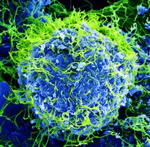
Search This Blog

Friday, August 2, 2019

Metagenomics

From Wikipedia, the free encyclopedia
 
Metagenomics allows the study of microbial communities like those present in this stream receiving acid drainage from surface coal mining.
 
Metagenomics is the study of genetic material recovered directly from environmental samples. The broad field may also be referred to as environmental genomics, ecogenomics or community genomics

While traditional microbiology and microbial genome sequencing and genomics rely upon cultivated clonal cultures, early environmental gene sequencing cloned specific genes (often the 16S rRNA gene) to produce a profile of diversity in a natural sample. Such work revealed that the vast majority of microbial biodiversity had been missed by cultivation-based methods.

Because of its ability to reveal the previously hidden diversity of microscopic life, metagenomics offers a powerful lens for viewing the microbial world that has the potential to revolutionize understanding of the entire living world. As the price of DNA sequencing continues to fall, metagenomics now allows microbial ecology to be investigated at a much greater scale and detail than before. Recent studies use either "shotgun" or PCR directed sequencing to get largely unbiased samples of all genes from all the members of the sampled communities.

Etymology

The term "metagenomics" was first used by Jo Handelsman, Jon Clardy, Robert M. Goodman, Sean F. Brady, and others, and first appeared in publication in 1998. The term metagenome referenced the idea that a collection of genes sequenced from the environment could be analyzed in a way analogous to the study of a single genome. In 2005, Kevin Chen and Lior Pachter (researchers at the University of California, Berkeley) defined metagenomics as "the application of modern genomics technique without the need for isolation and lab cultivation of individual species".

History

Conventional sequencing begins with a culture of identical cells as a source of DNA. However, early metagenomic studies revealed that there are probably large groups of microorganisms in many environments that cannot be cultured and thus cannot be sequenced. These early studies focused on 16S ribosomal RNA sequences which are relatively short, often conserved within a species, and generally different between species. Many 16S rRNA sequences have been found which do not belong to any known cultured species, indicating that there are numerous non-isolated organisms. These surveys of ribosomal RNA (rRNA) genes taken directly from the environment revealed that cultivation based methods find less than 1% of the bacterial and archaeal species in a sample. Much of the interest in metagenomics comes from these discoveries that showed that the vast majority of microorganisms had previously gone unnoticed. 

Early molecular work in the field was conducted by Norman R. Pace and colleagues, who used PCR to explore the diversity of ribosomal RNA sequences. The insights gained from these breakthrough studies led Pace to propose the idea of cloning DNA directly from environmental samples as early as 1985. This led to the first report of isolating and cloning bulk DNA from an environmental sample, published by Pace and colleagues in 1991 while Pace was in the Department of Biology at Indiana University. Considerable efforts ensured that these were not PCR false positives and supported the existence of a complex community of unexplored species. Although this methodology was limited to exploring highly conserved, non-protein coding genes, it did support early microbial morphology-based observations that diversity was far more complex than was known by culturing methods. Soon after that, Healy reported the metagenomic isolation of functional genes from "zoolibraries" constructed from a complex culture of environmental organisms grown in the laboratory on dried grasses in 1995. After leaving the Pace laboratory, Edward DeLong continued in the field and has published work that has largely laid the groundwork for environmental phylogenies based on signature 16S sequences, beginning with his group's construction of libraries from marine samples.

In 2002, Mya Breitbart, Forest Rohwer, and colleagues used environmental shotgun sequencing (see below) to show that 200 liters of seawater contains over 5000 different viruses. Subsequent studies showed that there are more than a thousand viral species in human stool and possibly a million different viruses per kilogram of marine sediment, including many bacteriophages. Essentially all of the viruses in these studies were new species. In 2004, Gene Tyson, Jill Banfield, and colleagues at the University of California, Berkeley and the Joint Genome Institute sequenced DNA extracted from an acid mine drainage system. This effort resulted in the complete, or nearly complete, genomes for a handful of bacteria and archaea that had previously resisted attempts to culture them.

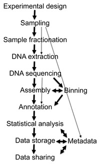
Flow diagram of a typical metagenome project
 
Beginning in 2003, Craig Venter, leader of the privately funded parallel of the Human Genome Project, has led the Global Ocean Sampling Expedition (GOS), circumnavigating the globe and collecting metagenomic samples throughout the journey. All of these samples are sequenced using shotgun sequencing, in hopes that new genomes (and therefore new organisms) would be identified. The pilot project, conducted in the Sargasso Sea, found DNA from nearly 2000 different species, including 148 types of bacteria never before seen. Venter has circumnavigated the globe and thoroughly explored the West Coast of the United States, and completed a two-year expedition to explore the Baltic, Mediterranean and Black Seas. Analysis of the metagenomic data collected during this journey revealed two groups of organisms, one composed of taxa adapted to environmental conditions of 'feast or famine', and a second composed of relatively fewer but more abundantly and widely distributed taxa primarily composed of plankton.

In 2005 Stephan C. Schuster at Penn State University and colleagues published the first sequences of an environmental sample generated with high-throughput sequencing, in this case massively parallel pyrosequencing developed by 454 Life Sciences. Another early paper in this area appeared in 2006 by Robert Edwards, Forest Rohwer, and colleagues at San Diego State University.

Sequencing

Recovery of DNA sequences longer than a few thousand base pairs from environmental samples was very difficult until recent advances in molecular biological techniques allowed the construction of libraries in bacterial artificial chromosomes (BACs), which provided better vectors for molecular cloning.

Environmental Shotgun Sequencing (ESS). (A) Sampling from habitat; (B) filtering particles, typically by size; (C) Lysis and DNA extraction; (D) cloning and library construction; (E) sequencing the clones; (F) sequence assembly into contigs and scaffolds.

Shotgun metagenomics

Advances in bioinformatics, refinements of DNA amplification, and the proliferation of computational power have greatly aided the analysis of DNA sequences recovered from environmental samples, allowing the adaptation of shotgun sequencing to metagenomic samples (known also as whole metagenome shotgun or WMGS sequencing). The approach, used to sequence many cultured microorganisms and the human genome, randomly shears DNA, sequences many short sequences, and reconstructs them into a consensus sequence. Shotgun sequencing reveals genes present in environmental samples. Historically, clone libraries were used to facilitate this sequencing. However, with advances in high throughput sequencing technologies, the cloning step is no longer necessary and greater yields of sequencing data can be obtained without this labour-intensive bottleneck step. Shotgun metagenomics provides information both about which organisms are present and what metabolic processes are possible in the community. Because the collection of DNA from an environment is largely uncontrolled, the most abundant organisms in an environmental sample are most highly represented in the resulting sequence data. To achieve the high coverage needed to fully resolve the genomes of under-represented community members, large samples, often prohibitively so, are needed. On the other hand, the random nature of shotgun sequencing ensures that many of these organisms, which would otherwise go unnoticed using traditional culturing techniques, will be represented by at least some small sequence segments. An emerging approach combines shotgun sequencing and chromosome conformation capture (Hi-C), which measures the proximity of any two DNA sequences within the same cell, to guide microbial genome assembly.

High-throughput sequencing

The first metagenomic studies conducted using high-throughput sequencing used massively parallel 454 pyrosequencing. Three other technologies commonly applied to environmental sampling are the Ion Torrent Personal Genome Machine, the Illumina MiSeq or HiSeq and the Applied Biosystems SOLiD system. These techniques for sequencing DNA generate shorter fragments than Sanger sequencing; Ion Torrent PGM System and 454 pyrosequencing typically produces ~400 bp reads, Illumina MiSeq produces 400-700bp reads (depending on whether paired end options are used), and SOLiD produce 25-75 bp reads. Historically, these read lengths were significantly shorter than the typical Sanger sequencing read length of ~750 bp, however the Illumina technology is quickly coming close to this benchmark. However, this limitation is compensated for by the much larger number of sequence reads. In 2009, pyrosequenced metagenomes generate 200–500 megabases, and Illumina platforms generate around 20–50 gigabases, but these outputs have increased by orders of magnitude in recent years. An additional advantage to high throughput sequencing is that this technique does not require cloning the DNA before sequencing, removing one of the main biases and bottlenecks in environmental sampling.

Bioinformatics

The data generated by metagenomics experiments are both enormous and inherently noisy, containing fragmented data representing as many as 10,000 species. The sequencing of the cow rumen metagenome generated 279 gigabases, or 279 billion base pairs of nucleotide sequence data, while the human gut microbiome gene catalog identified 3.3 million genes assembled from 567.7 gigabases of sequence data. Collecting, curating, and extracting useful biological information from datasets of this size represent significant computational challenges for researchers.

Sequence pre-filtering

The first step of metagenomic data analysis requires the execution of certain pre-filtering steps, including the removal of redundant, low-quality sequences and sequences of probable eukaryotic origin (especially in metagenomes of human origin). The methods available for the removal of contaminating eukaryotic genomic DNA sequences include Eu-Detect and DeConseq.

Assembly

DNA sequence data from genomic and metagenomic projects are essentially the same, but genomic sequence data offers higher coverage while metagenomic data is usually highly non-redundant. Furthermore, the increased use of second-generation sequencing technologies with short read lengths means that much of future metagenomic data will be error-prone. Taken in combination, these factors make the assembly of metagenomic sequence reads into genomes difficult and unreliable. Misassemblies are caused by the presence of repetitive DNA sequences that make assembly especially difficult because of the difference in the relative abundance of species present in the sample. Misassemblies can also involve the combination of sequences from more than one species into chimeric contigs.

There are several assembly programs, most of which can use information from paired-end tags in order to improve the accuracy of assemblies. Some programs, such as Phrap or Celera Assembler, were designed to be used to assemble single genomes but nevertheless produce good results when assembling metagenomic data sets. Other programs, such as Velvet assembler, have been optimized for the shorter reads produced by second-generation sequencing through the use of de Bruijn graphs. The use of reference genomes allows researchers to improve the assembly of the most abundant microbial species, but this approach is limited by the small subset of microbial phyla for which sequenced genomes are available. After an assembly is created, an additional challenge is "metagenomic deconvolution", or determining which sequences come from which species in the sample.

Gene prediction

Metagenomic analysis pipelines use two approaches in the annotation of coding regions in the assembled contigs. The first approach is to identify genes based upon homology with genes that are already publicly available in sequence databases, usually by BLAST searches. This type of approach is implemented in the program MEGAN4. The second, ab initio, uses intrinsic features of the sequence to predict coding regions based upon gene training sets from related organisms. This is the approach taken by programs such as GeneMark and GLIMMER. The main advantage of ab initio prediction is that it enables the detection of coding regions that lack homologs in the sequence databases; however, it is most accurate when there are large regions of contiguous genomic DNA available for comparison.

Species diversity

A 2016 representation of the tree of life

Gene annotations provide the "what", while measurements of species diversity provide the "who". In order to connect community composition and function in metagenomes, sequences must be binned. Binning is the process of associating a particular sequence with an organism. In similarity-based binning, methods such as BLAST are used to rapidly search for phylogenetic markers or otherwise similar sequences in existing public databases. This approach is implemented in MEGAN. Another tool, PhymmBL, uses interpolated Markov models to assign reads. MetaPhlAn and AMPHORA are methods based on unique clade-specific markers for estimating organismal relative abundances with improved computational performances. Other tools, like mOTUs and MetaPhyler, use universal marker genes to profile prokaryotic species. With the mOTUs profiler is possible to profile species without a reference genome, improving the estimation of microbial community diversity. Recent methods, such as SLIMM, use read coverage landscape of individual reference genomes to minimize false-positive hits and get reliable relative abundances. In composition based binning, methods use intrinsic features of the sequence, such as oligonucleotide frequencies or codon usage bias. Once sequences are binned, it is possible to carry out comparative analysis of diversity and richness.

Data integration

The massive amount of exponentially growing sequence data is a daunting challenge that is complicated by the complexity of the metadata associated with metagenomic projects. Metadata includes detailed information about the three-dimensional (including depth, or height) geography and environmental features of the sample, physical data about the sample site, and the methodology of the sampling. This information is necessary both to ensure replicability and to enable downstream analysis. Because of its importance, metadata and collaborative data review and curation require standardized data formats located in specialized databases, such as the Genomes OnLine Database (GOLD).

Several tools have been developed to integrate metadata and sequence data, allowing downstream comparative analyses of different datasets using a number of ecological indices. In 2007, Folker Meyer and Robert Edwards and a team at Argonne National Laboratory and the University of Chicago released the Metagenomics Rapid Annotation using Subsystem Technology server (MG-RAST) a community resource for metagenome data set analysis. As of June 2012 over 14.8 terabases (14x1012 bases) of DNA have been analyzed, with more than 10,000 public data sets freely available for comparison within MG-RAST. Over 8,000 users now have submitted a total of 50,000 metagenomes to MG-RAST. The Integrated Microbial Genomes/Metagenomes (IMG/M) system also provides a collection of tools for functional analysis of microbial communities based on their metagenome sequence, based upon reference isolate genomes included from the Integrated Microbial Genomes (IMG) system and the Genomic Encyclopedia of Bacteria and Archaea (GEBA) project.

One of the first standalone tools for analysing high-throughput metagenome shotgun data was MEGAN (MEta Genome ANalyzer). A first version of the program was used in 2005 to analyse the metagenomic context of DNA sequences obtained from a mammoth bone. Based on a BLAST comparison against a reference database, this tool performs both taxonomic and functional binning, by placing the reads onto the nodes of the NCBI taxonomy using a simple lowest common ancestor (LCA) algorithm or onto the nodes of the SEED or KEGG classifications, respectively.

With the advent of fast and inexpensive sequencing instruments, the growth of databases of DNA sequences is now exponential (e.g., the NCBI GenBank database). Faster and efficient tools are needed to keep pace with the high-throughput sequencing, because the BLAST-based approaches such as MG-RAST or MEGAN run slowly to annotate large samples (e.g., several hours to process a small/medium size dataset/sample). Thus, ultra-fast classifiers have recently emerged, thanks to more affordable powerful servers. These tools can perform the taxonomic annotation at extremely high speed, for example CLARK (according to CLARK's authors, it can classify accurately "32 million metagenomic short reads per minute"). At such a speed, a very large dataset/sample of a billion short reads can be processed in about 30 minutes. 

With the increasing availability of samples containing ancient DNA and due to the uncertainty associated with the nature of those samples (ancient DNA damage), FALCON, a fast tool capable of producing conservative similarity estimates has been made available. According to FALCON's authors, it can use relaxed thresholds and edit distances without affecting the memory and speed performance.

Comparative metagenomics

Comparative analyses between metagenomes can provide additional insight into the function of complex microbial communities and their role in host health. Pairwise or multiple comparisons between metagenomes can be made at the level of sequence composition (comparing GC-content or genome size), taxonomic diversity, or functional complement. Comparisons of population structure and phylogenetic diversity can be made on the basis of 16S and other phylogenetic marker genes, or—in the case of low-diversity communities—by genome reconstruction from the metagenomic dataset. Functional comparisons between metagenomes may be made by comparing sequences against reference databases such as COG or KEGG, and tabulating the abundance by category and evaluating any differences for statistical significance. This gene-centric approach emphasizes the functional complement of the community as a whole rather than taxonomic groups, and shows that the functional complements are analogous under similar environmental conditions. Consequently, metadata on the environmental context of the metagenomic sample is especially important in comparative analyses, as it provides researchers with the ability to study the effect of habitat upon community structure and function.

Additionally, several studies have also utilized oligonucleotide usage patterns to identify the differences across diverse microbial communities. Examples of such methodologies include the dinucleotide relative abundance approach by Willner et al. and the HabiSign approach of Ghosh et al. This latter study also indicated that differences in tetranucleotide usage patterns can be used to identify genes (or metagenomic reads) originating from specific habitats. Additionally some methods as TriageTools or Compareads detect similar reads between two read sets. The similarity measure they apply on reads is based on a number of identical words of length k shared by pairs of reads. 

A key goal in comparative metagenomics is to identify microbial group(s) which are responsible for conferring specific characteristics to a given environment. However, due to issues in the sequencing technologies artifacts need to be accounted for like in metagenomeSeq. Others have characterized inter-microbial interactions between the resident microbial groups. A GUI-based comparative metagenomic analysis application called Community-Analyzer has been developed by Kuntal et al.  which implements a correlation-based graph layout algorithm that not only facilitates a quick visualization of the differences in the analyzed microbial communities (in terms of their taxonomic composition), but also provides insights into the inherent inter-microbial interactions occurring therein. Notably, this layout algorithm also enables grouping of the metagenomes based on the probable inter-microbial interaction patterns rather than simply comparing abundance values of various taxonomic groups. In addition, the tool implements several interactive GUI-based functionalities that enable users to perform standard comparative analyses across microbiomes.

Data analysis

Community metabolism

In many bacterial communities, natural or engineered (such as bioreactors), there is significant division of labor in metabolism (Syntrophy), during which the waste products of some organisms are metabolites for others. In one such system, the methanogenic bioreactor, functional stability requires the presence of several syntrophic species (Syntrophobacterales and Synergistia) working together in order to turn raw resources into fully metabolized waste (methane). Using comparative gene studies and expression experiments with microarrays or proteomics researchers can piece together a metabolic network that goes beyond species boundaries. Such studies require detailed knowledge about which versions of which proteins are coded by which species and even by which strains of which species. Therefore, community genomic information is another fundamental tool (with metabolomics and proteomics) in the quest to determine how metabolites are transferred and transformed by a community.

Metatranscriptomics

Metagenomics allows researchers to access the functional and metabolic diversity of microbial communities, but it cannot show which of these processes are active. The extraction and analysis of metagenomic mRNA (the metatranscriptome) provides information on the regulation and expression profiles of complex communities. Because of the technical difficulties (the short half-life of mRNA, for example) in the collection of environmental RNA there have been relatively few in situ metatranscriptomic studies of microbial communities to date. While originally limited to microarray technology, metatranscriptomics studies have made use of transcriptomics technologies to measure whole-genome expression and quantification of a microbial community, first employed in analysis of ammonia oxidation in soils.

Viruses

Metagenomic sequencing is particularly useful in the study of viral communities. As viruses lack a shared universal phylogenetic marker (as 16S RNA for bacteria and archaea, and 18S RNA for eukarya), the only way to access the genetic diversity of the viral community from an environmental sample is through metagenomics. Viral metagenomes (also called viromes) should thus provide more and more information about viral diversity and evolution. For example, a metagenomic pipeline called Giant Virus Finder showed the first evidence of existence of giant viruses in a saline desert  and in Antarctic dry valleys .

Applications

Metagenomics has the potential to advance knowledge in a wide variety of fields. It can also be applied to solve practical challenges in medicine, engineering, agriculture, sustainability and ecology.

Agriculture

The soils in which plants grow are inhabited by microbial communities, with one gram of soil containing around 109-1010 microbial cells which comprise about one gigabase of sequence information. The microbial communities which inhabit soils are some of the most complex known to science, and remain poorly understood despite their economic importance. Microbial consortia perform a wide variety of ecosystem services necessary for plant growth, including fixing atmospheric nitrogen, nutrient cycling, disease suppression, and sequester iron and other metals. Functional metagenomics strategies are being used to explore the interactions between plants and microbes through cultivation-independent study of these microbial communities. By allowing insights into the role of previously uncultivated or rare community members in nutrient cycling and the promotion of plant growth, metagenomic approaches can contribute to improved disease detection in crops and livestock and the adaptation of enhanced farming practices which improve crop health by harnessing the relationship between microbes and plants.

Biofuel

Bioreactors allow the observation of microbial communities as they convert biomass into cellulosic ethanol.
 
Biofuels are fuels derived from biomass conversion, as in the conversion of cellulose contained in corn stalks, switchgrass, and other biomass into cellulosic ethanol. This process is dependent upon microbial consortia(association) that transform the cellulose into sugars, followed by the fermentation of the sugars into ethanol. Microbes also produce a variety of sources of bioenergy including methane and hydrogen.

The efficient industrial-scale deconstruction of biomass requires novel enzymes with higher productivity and lower cost. Metagenomic approaches to the analysis of complex microbial communities allow the targeted screening of enzymes with industrial applications in biofuel production, such as glycoside hydrolases. Furthermore, knowledge of how these microbial communities function is required to control them, and metagenomics is a key tool in their understanding. Metagenomic approaches allow comparative analyses between convergent microbial systems like biogas fermenters or insect herbivores such as the fungus garden of the leafcutter ants.

Biotechnology

Microbial communities produce a vast array of biologically active chemicals that are used in competition and communication. Many of the drugs in use today were originally uncovered in microbes; recent progress in mining the rich genetic resource of non-culturable microbes has led to the discovery of new genes, enzymes, and natural products. The application of metagenomics has allowed the development of commodity and fine chemicals, agrochemicals and pharmaceuticals where the benefit of enzyme-catalyzed chiral synthesis is increasingly recognized.

Two types of analysis are used in the bioprospecting of metagenomic data: function-driven screening for an expressed trait, and sequence-driven screening for DNA sequences of interest. Function-driven analysis seeks to identify clones expressing a desired trait or useful activity, followed by biochemical characterization and sequence analysis. This approach is limited by availability of a suitable screen and the requirement that the desired trait be expressed in the host cell. Moreover, the low rate of discovery (less than one per 1,000 clones screened) and its labor-intensive nature further limit this approach. In contrast, sequence-driven analysis uses conserved DNA sequences to design PCR primers to screen clones for the sequence of interest. In comparison to cloning-based approaches, using a sequence-only approach further reduces the amount of bench work required. The application of massively parallel sequencing also greatly increases the amount of sequence data generated, which require high-throughput bioinformatic analysis pipelines. The sequence-driven approach to screening is limited by the breadth and accuracy of gene functions present in public sequence databases. In practice, experiments make use of a combination of both functional and sequence-based approaches based upon the function of interest, the complexity of the sample to be screened, and other factors. An example of success using metagenomics as a biotechnology for drug discovery is illustrated with the malacidin antibiotics.

Ecology

Metagenomics can provide valuable insights into the functional ecology of environmental communities. Metagenomic analysis of the bacterial consortia found in the defecations of Australian sea lions suggests that nutrient-rich sea lion faeces may be an important nutrient source for coastal ecosystems. This is because the bacteria that are expelled simultaneously with the defecations are adept at breaking down the nutrients in the faeces into a bioavailable form that can be taken up into the food chain.

DNA sequencing can also be used more broadly to identify species present in a body of water, debris filtered from the air, or sample of dirt. This can establish the range of invasive species and endangered species, and track seasonal populations.

Environmental remediation

Metagenomics can improve strategies for monitoring the impact of pollutants on ecosystems and for cleaning up contaminated environments. Increased understanding of how microbial communities cope with pollutants improves assessments of the potential of contaminated sites to recover from pollution and increases the chances of bioaugmentation or biostimulation trials to succeed.

Gut Microbe Characterization

Microbial communities play a key role in preserving human health, but their composition and the mechanism by which they do so remains mysterious. Metagenomic sequencing is being used to characterize the microbial communities from 15-18 body sites from at least 250 individuals. This is part of the Human Microbiome initiative with primary goals to determine if there is a core human microbiome, to understand the changes in the human microbiome that can be correlated with human health, and to develop new technological and bioinformatics tools to support these goals.

Another medical study as part of the MetaHit (Metagenomics of the Human Intestinal Tract) project consisted of 124 individuals from Denmark and Spain consisting of healthy, overweight, and irritable bowel disease patients. The study attempted to categorize the depth and phylogenetic diversity of gastrointestinal bacteria. Using Illumina GA sequence data and SOAPdenovo, a de Bruijn graph-based tool specifically designed for assembly short reads, they were able to generate 6.58 million contigs greater than 500 bp for a total contig length of 10.3 Gb and a N50 length of 2.2 kb.

The study demonstrated that two bacterial divisions, Bacteroidetes and Firmicutes, constitute over 90% of the known phylogenetic categories that dominate distal gut bacteria. Using the relative gene frequencies found within the gut these researchers identified 1,244 metagenomic clusters that are critically important for the health of the intestinal tract. There are two types of functions in these range clusters: housekeeping and those specific to the intestine. The housekeeping gene clusters are required in all bacteria and are often major players in the main metabolic pathways including central carbon metabolism and amino acid synthesis. The gut-specific functions include adhesion to host proteins and the harvesting of sugars from globoseries glycolipids. Patients with irritable bowel syndrome were shown to exhibit 25% fewer genes and lower bacterial diversity than individuals not suffering from irritable bowel syndrome indicating that changes in patients’ gut biome diversity may be associated with this condition. 

While these studies highlight some potentially valuable medical applications, only 31-48.8% of the reads could be aligned to 194 public human gut bacterial genomes and 7.6-21.2% to bacterial genomes available in GenBank which indicates that there is still far more research necessary to capture novel bacterial genomes.

Infectious disease diagnosis

Differentiating between infectious and non-infectious illness, and identifying the underlying etiology of infection, can be quite challenging. For example, more than half of cases of encephalitis remain undiagnosed, despite extensive testing using state-of-the-art clinical laboratory methods. Metagenomic sequencing shows promise as a sensitive and rapid method to diagnose infection by comparing genetic material found in a patient's sample to a database of thousands of bacteria, viruses, and other pathogens

Third-generation sequencing

From Wikipedia, the free encyclopedia
Third-generation sequencing (also known as long-read sequencing) is a class of DNA sequencing methods currently under active development. Third generation sequencing works by reading the nucleotide sequences at the single molecule level, in contrast to existing methods that require breaking long strands of DNA into small segments then inferring nucleotide sequences by amplification and synthesis. Critical challenges exist in the engineering of the necessary molecular instruments for whole genome sequencing to make the technology commercially available.

Second-generation sequencing, often referred to as Next-generation sequencing (NGS), has dominated the DNA sequencing space since its development. It has dramatically reduced the cost of DNA sequencing by enabling a massively-paralleled approach capable of producing large numbers of reads at exceptionally high coverages throughout the genome.

Since eukaryotic genomes contain many repetitive regions, a major limitation to this class of sequencing methods is the length of reads it produces. Briefly, second generation sequencing works by first amplifying the DNA molecule and then conducting sequencing by synthesis. The collective fluorescent signal resulting from synthesizing a large number of amplified identical DNA strands allows the inference of nucleotide identity. However, due to random errors, DNA synthesis between the amplified DNA strands would become progressively out-of-sync. Quickly, the signal quality deteriorates as the read-length grows. In order to preserve read quality, long DNA molecules must be broken up into small segments, resulting in a critical limitation of second generation sequencing technologies. Computational efforts aimed to overcome this challenge often rely on approximative heuristics that may not result in accurate assemblies.

By enabling direct sequencing of single DNA molecules, third generation sequencing technologies have the capability to produce substantially longer reads than second generation sequencing. Such an advantage has critical implications for both genome science and the study of biology in general. However, third generation sequencing data have much higher error rates than previous technologies, which can complicate downstream genome assembly and analysis of the resulting data. These technologies are undergoing active development and it is expected that there will be improvements to the high error rates. For applications that are more tolerant to error rates, such as structural variant calling, third generation sequencing has been found to outperform existing methods.

Current technologies

Sequencing technologies with a different approach than second-generation platforms were first described as "third-generation" in 2008-2009.

There are several companies currently at the heart of third generation sequencing technology development, namely, Pacific Biosciences, Oxford Nanopore Technology, Quantapore (CA-USA), and Stratos (WA-USA). These companies are taking fundamentally different approaches to sequencing single DNA molecules. 

PacBio developed the sequencing platform of single molecule real time sequencing (SMRT), based on the properties of zero-mode waveguides. Signals are in the form of fluorescent light emission from each nucleotide incorporated by a DNA polymerase bound to the bottom of the zL well. 

Oxford Nanopore’s technology involves passing a DNA molecule through a nanoscale pore structure and then measuring changes in electrical field surrounding the pore; while Quantapore has a different proprietary nanopore approach. Stratos Genomics spaces out the DNA bases with polymeric inserts, "Xpandomers", to circumvent the signal to noise challenge of nanopore ssDNA reading. 

Also notable is Helicos's single molecule fluorescence approach, but the company entered bankruptcy in the fall of 2015.

Advantages

Longer reads

In comparison to the current generation of sequencing technologies, third generation sequencing has the obvious advantage of producing much longer reads. It is expected that these longer read lengths will alleviate numerous computational challenges surrounding genome assembly, transcript reconstruction, and metagenomics among other important areas of modern biology and medicine.

It is well known that eukaryotic genomes including primates and humans are complex and have large numbers of long repeated regions. Short reads from second generation sequencing must resort to approximative strategies in order to infer sequences over long ranges for assembly and genetic variant calling. Pair end reads have been leveraged by second generation sequencing to combat these limitations. However, exact fragment lengths of pair ends are often unknown and must also be approximated as well. By making long reads lengths possible, third generation sequencing technologies have clear advantages.

Epigenetics

Epigenetic markers are stable and potentially heritable modifications to the DNA molecule that are not in its sequence. An example is DNA methylation at CpG sites, which has been found to influence gene expression. Histone modifications are another example. The current generation of sequencing technologies rely on laboratory techniques such as ChIP-sequencing for the detection of epigenetic markers. These techniques involve tagging the DNA strand, breaking and filtering fragments that contain markers, followed by sequencing. Third generation sequencing may enable direct detection of these markers due to their distinctive signal from the other four nucleotide bases.

Portability and speed

Other important advantages of third generation sequencing technologies include portability and sequencing speed. Since minimal sample preprocessing is required in comparison to second generation sequencing, smaller equipments could be designed. Oxford Nanopore Technology has recently commercialized the MinION sequencer. This sequencing machine is roughly the size of a regular USB flash drive and can be used readily by connecting to a laptop. In addition, since the sequencing process is not parallelized across regions of the genome, data could be collected and analyzed in real time. These advantages of third generation sequencing may be well-suited in hospital settings where quick and on-site data collection and analysis is demanded.

Challenges

Third generation sequencing, as it currently stands, faces important challenges mainly surrounding accurate identification of nucleotide bases; error rates are still much higher compared to second generation sequencing. This is generally due to instability of the molecular machinery involved. For example, in PacBio’s single molecular and real time sequencing technology, the DNA polymerase molecule becomes increasingly damaged as the sequencing process occurs. Additionally, since the process happens quickly, the signals given off by individual bases may be blurred by signals from neighbouring bases. This poses a new computational challenge for deciphering the signals and consequently inferring the sequence. Methods such as Hidden Markov Models, for example, have been leveraged for this purpose with some success.

On average, different individuals of the human population share about 99.9% of their genes. In other words, approximately only one out of every thousand bases would differ between any two person. The high error rates involved with third generation sequencing are inevitably problematic for the purpose of characterizing individual differences that exist between members of the same species.

Genome assembly

Genome assembly is the reconstruction of whole genome DNA sequences. This is generally done with two fundamentally different approaches.

Reference alignment

When a reference genome is available, as one is in the case of human, newly sequenced reads could simply be aligned to the reference genome in order to characterize its properties. Such reference based assembly is quick and easy but has the disadvantage of “hiding" novel sequences and large copy number variants. In addition, reference genomes do not yet exist for most organisms.

De novo assembly

De novo assembly is the alternative genome assembly approach to reference alignment. It refers to the reconstruction of whole genome sequences entirely from raw sequence reads. This method would be chosen when there is no reference genome, when the species of the given organism is unknown as in metagenomics, or when there exist genetic variants of interest that may not be detected by reference genome alignment. 

Given the short reads produced by the current generation of sequencing technologies, de novo assembly is a major computational problem. It is normally approached by an iterative process of finding and connecting sequence reads with sensible overlaps. Various computational and statistical techniques, such as de bruijn graphs and overlap layout consensus graphs, have been leveraged to solve this problem. Nonetheless, due to the highly repetitive nature of eukaryotic genomes, accurate and complete reconstruction of genome sequences in de novo assembly remains challenging. Pair end reads have been posed as a possible solution, though exact fragment lengths are often unknown and must be approximated.

Hybrid assembly - the use of reads from 3rd gen sequencing platforms with shorts reads from 2nd gen platforms - may be used to resolve ambiguities that exist in genomes previously assembled using second generation sequencing. Short second generation reads have also been used to correct errors that exist in the long third generation reads.

Hybrid assembly

Long read lengths offered by third generation sequencing may alleviate many of the challenges currently faced by de novo genome assemblies. For example, if an entire repetitive region can be sequenced unambiguously in a single read, no computation inference would be required. Computational methods have been proposed to alleviate the issue of high error rates. For example, in one study, it was demonstrated that de novo assembly of a microbial genome using PacBio sequencing alone performed superior to that of second generation sequencing.

Third generation sequencing may also be used in conjunction with second generation sequencing. This approach is often referred to as hybrid sequencing. For example, long reads from third generation sequencing may be used to resolve ambiguities that exist in genomes previously assembled using second generation sequencing. On the other hand, short second generation reads have been used to correct errors in that exist in the long third generation reads. In general, this hybrid approach has been shown to improve de novo genome assemblies significantly.

Epigenetic markers

DNA methylation (DNAm) – the covalent modification of DNA at CpG sites resulting in attached methyl groups – is the best understood component of epigenetic machinery. DNA modifications and resulting gene expression can vary across cell types, temporal development, with genetic ancestry, can change due to environmental stimuli and are heritable. After the discovery of DNAm, researchers have also found its correlation to diseases like cancer and autism. In this disease etiology context DNAm is an important avenue of further research.

Advantages

The current most common methods for examining methylation state require an assay that fragments DNA before standard second generation sequencing on the Illumina platform. As a result of short read length, information regarding the longer patterns of methylation are lost. Third generation sequencing technologies offer the capability for single molecule real-time sequencing of longer reads, and detection of DNA modification without the aforementioned assay.

PacBio SMRT technology and Oxford Nanopore can use unaltered DNA to detect methylation.
 
Oxford Nanopore Technologies’ MinION has been used to detect DNAm. As each DNA strand passes through a pore, it produces electrical signals which have been found to be sensitive to epigenetic changes in the nucleotides, and a hidden Markov model (HMM) was used to analyze MinION data to detect 5-methylcytosine (5mC) DNA modification. The model was trained using synthetically methylated E. coli DNA and the resulting signals measured by the nanopore technology. Then the trained model was used to detect 5mC in MinION genomic reads from a human cell line which already had a reference methylome. The classifier has 82% accuracy in randomly sampled singleton sites, which increases to 95% when more stringent thresholds are applied.

Other methods address different types of DNA modifications using the MinION platform. Stoiber et al. examined 4-methylcytosine (4mC) and 6-methyladenine (6mA), along with 5mC, and also created a software to directly visualize the raw MinION data in human-friendly way. Here they found that in E. coli, which has a known methylome, event windows of 5 base pairs long can be used to divide and statistically analyze the raw MinION electrical signals. A straightforward Mann-Whitney U test can detect modified portions of the E. coli sequence, as well as further split the modifications into 4mC, 6mA or 5mC regions.

It seems likely that in the future, MinION raw data will be used to detect many different epigenetic marks in DNA. 

PacBio sequencing has also been used to detect DNA methylation. In this platform the pulse width - the width of a fluorescent light pulse - corresponds to a specific base. In 2010 it was shown that the interpulse distance in control and methylated samples are different, and there is a "signature" pulse width for each methylation type. In 2012 using the PacBio platform the binding sites of DNA methyltransferases were characterized. The detection of N6-methylation in C Elegans was shown in 2015. DNA methylation on N6-adenine using the PacBio platform in mouse embryonic stem cells was shown in 2016.

Other forms of DNA modifications – from heavy metals, oxidation, or UV damage – are also possible avenues of research using Oxford Nanopore and PacBio third generation sequencing.

Drawbacks

Processing of the raw data – such as normalization to the median signal – was needed on MinION raw data, reducing real-time capability of the technology. Consistency of the electrical signals is still an issue, making it difficult to accurately call a nucleotide. MinION has low throughput; since multiple overlapping reads are hard to obtain, this further leads to accuracy problems of downstream DNA modification detection. Both the hidden Markov model and statistical methods used with MinION raw data require repeated observations of DNA modifications for detection, meaning that individual modified nucleotides need to be consistently present in multiple copies of the genome, e.g. in multiple cells or plasmids in the sample. 

For the PacBio platform, too, depending on what methylation you expect to find, coverage needs can vary. As of March 2017, other epigenetic factors like histone modifications have not been discoverable using third-generation technologies. Longer patterns of methylation are often lost because smaller contigs still need to be assembled.

Transcriptomics

Transcriptomics is the study of the transcriptome, usually by characterizing the relative abundances of messenger RNA molecules the tissue under study. According to the central dogma of molecular biology, genetic information flows from double stranded DNA molecules to single stranded mRNA molecules where they can be readily translated into function protein molecules. By studying the transcriptome, one can gain valuable insight into the regulation of gene expressions.

While expression levels as the gene level can be more or less accurately depicted by second generation sequencing, transcript level information is still an important challenge. As a consequence, the role of alternative splicing in molecular biology remains largely elusive. Third generation sequencing technologies hold promising prospects in resolving this issue by enabling sequencing of mRNA molecules at their full lengths.

Alternative splicing

Alternative splicing (AS) is the process by which a single gene may give rise to multiple distinct mRNA transcripts and consequently different protein translations. Some evidence suggests that AS is a ubiquitous phenomenon and may play a key role in determining the phenotypes of organisms, especially in complex eukaryotes; all eukaryotes contain genes consisting of introns that may undergo AS. In particular, it has been estimated that AS occurs in 95% of all human multi-exon genes. AS has undeniable potential to influence myriad biological processes. Advancing knowledge in this area has critical implications for the study of biology in general.

Transcript reconstruction

The current generation of sequencing technologies produce only short reads, putting tremendous limitation on the ability to detect distinct transcripts; short reads must be reverse engineered into original transcripts that could have given rise to the resulting read observations. This task is further complicated by the highly variable expression levels across transcripts, and consequently variable read coverages across the sequence of the gene. In addition, exons may be shared among individual transcripts, rendering unambiguous inferences essentially impossible. Existing computational methods make inferences based on the accumulation of short reads at various sequence locations often by making simplifying assumptions. Cufflinks takes a parsimonious approach, seeking to explain all the reads with the fewest possible number of transcripts. On the other hand, StringTie attempts to simultaneously estimate transcript abundances while assembling the reads. These methods, while reasonable, may not always identify real transcripts. 

A study published in 2008 surveyed 25 different existing transcript reconstruction protocols. Its evidence suggested that existing methods are generally weak in assembling transcripts, though the ability to detect individual exons are relatively intact. According to the estimates, average sensitivity to detect exons across the 25 protocols is 80% for Caenorhabditis elegans genes. In comparison, transcript identification sensitivity decreases to 65%. For human, the study reported an exon detection sensitivity averaging to 69% and transcript detection sensitivity had an average of mere 33%. In other words, for human, existing methods are able to identify less than half of all existing transcript.

Third generation sequencing technologies have demonstrated promising prospects in solving the problem of transcript detection as well as mRNA abundance estimation at the level of transcripts. While error rates remain high, third generation sequencing technologies have the capability to produce much longer read lengths. Pacific Bioscience has introduced the iso-seq platform, proposing to sequence mRNA molecules at their full lengths. It is anticipated that Oxford Nanopore will put forth similar technologies. The trouble with higher error rates may be alleviated by supplementary high quality short reads. This approach has been previously tested and reported to reduce the error rate by more than 3 folds.

Metagenomics

Metagenomics is the analysis of genetic material recovered directly from environmental samples.

Advantages

The main advantage for third-generation sequencing technologies in metagenomics is their speed of sequencing in comparison to second generation techniques. Speed of sequencing is important for example in the clinical setting (i.e. pathogen identification), to allow for efficient diagnosis and timely clinical actions. 

Oxford Nanopore's MinION was used in 2015 for real-time metagenomic detection of pathogens in complex, high-background clinical samples. The first Ebola virus (EBV) read was sequenced 44 seconds after data acquisition. There was uniform mapping of reads to genome; at least one read mapped to >88% of the genome. The relatively long reads allowed for sequencing of a near-complete viral genome to high accuracy (97–99% identity) directly from a primary clinical sample.

A common phylogenetic marker for microbial community diversity studies is the 16S ribosomal RNA gene. Both MinION and PacBio's SMRT platform have been used to sequence this gene. In this context the PacBio error rate was comparable to that of shorter reads from 454 and Illumina's MiSeq sequencing platforms.

Drawbacks

MinION's high error rate (~10-40%) prevented identification of antimicrobial resistance markers, for which single nucleotide resolution is necessary. For the same reason eukaryotic pathogens were not identified. Ease of carryover contamination when re-using same the flow cell (standard wash protocols don’t work) is also a concern. Unique barcodes may allow for more multiplexing. Furthermore, performing accurate species identification for bacteria, fungi and parasites is very difficult, as they share a larger portion of the genome, and some only differ by <5 nbsp="" p="">

The per base sequencing cost is still significantly more than that of MiSeq. However, the prospect of supplementing reference databases with full-length sequences from organisms below the limit of detection from the Sanger approach; this could possibly greatly help the identification of organisms in metagenomics. 

Before third generation sequencing can be used reliably in the clinical context, there is a need for standardization of lab protocols. These protocols are not yet as optimized as PCR methods.

Zaire ebolavirus

From Wikipedia, the free encyclopedia

Zaire ebolavirus
False color scanning electron microscope image of a single filamentous Ebola virus particle
Virus classification
(unranked): Virus
Realm: Riboviria
Phylum: Negarnaviricota
Class: Monjiviricetes
Order: Mononegavirales
Family: Filoviridae
Genus: Ebolavirus
Species:
Zaire ebolavirus

Zaire ebolavirus, more commonly known as simply Ebola virus (EBOV), is one of six known species within the genus Ebolavirus. Four of the six known ebolaviruses, including EBOV, cause a severe and often fatal hemorrhagic fever in humans and other mammals, known as Ebola virus disease (EVD). Ebola virus has caused the majority of human deaths from EVD and is the cause of the 2013–2016 Ebola virus epidemic in West Africa, which resulted in at least 28,646 suspected cases and 11,323 confirmed deaths.

Ebola virus and its genus were both originally named for Zaire (now the Democratic Republic of Congo), the country where it was first described, and was at first suspected to be a new "strain" of the closely related Marburg virus. The virus was renamed "Ebola virus" in 2010 to avoid confusion. Ebola virus is the single member of the species Zaire ebolavirus, which is the type species for the genus Ebolavirus, family Filoviridae, order Mononegavirales. The members of the species are called Zaire ebolaviruses. The natural reservoir of Ebola virus is believed to be bats, particularly fruit bats, and it is primarily transmitted between humans and from animals to humans through body fluids.

The EBOV genome is a single-stranded RNA approximately 19,000 nucleotides long. It encodes seven structural proteins: nucleoprotein (NP), polymerase cofactor (VP35), (VP40), GP, transcription activator (VP30), VP24, and RNA-dependent RNA polymerase (L).

Because of its high mortality rate (up to 83-90%), EBOV is also listed as a select agent, World Health Organization Risk Group 4 Pathogen (requiring Biosafety Level 4-equivalent containment), a US National Institutes of Health/National Institute of Allergy and Infectious Diseases Category A Priority Pathogen, US CDC Centers for Disease Control and Prevention Category A Bioterrorism Agent, and a Biological Agent for Export Control by the Australia Group.

Structure

Phylogenetic tree comparing ebolaviruses and marburgviruses. Numbers indicate percent confidence of branches.
 
EBOV carries a negative-sense RNA genome in virions that are cylindrical/tubular, and contain viral envelope, matrix, and nucleocapsid components. The overall cylinders are generally approximately 80 nm in diameter, and have a virally encoded glycoprotein (GP) projecting as 7-10 nm long spikes from its lipid bilayer surface. The cylinders are of variable length, typically 800 nm, but sometimes up to 1000 nm long. The outer viral envelope of the virion is derived by budding from domains of host cell membrane into which the GP spikes have been inserted during their biosynthesis. Individual GP molecules appear with spacings of about 10 nm. Viral proteins VP40 and VP24 are located between the envelope and the nucleocapsid (see following), in the matrix space. At the center of the virion structure is the nucleocapsid, which is composed of a series of viral proteins attached to an 18–19 kb linear, negative-sense RNA without 3′-polyadenylation or 5′-capping (see following); the RNA is helically wound and complexed with the NP, VP35, VP30, and L proteins; this helix has a diameter of 80 nm.

The overall shape of the virions after purification and visualization (e.g., by ultracentrifugation and electron microscopy, respectively) varies considerably; simple cylinders are far less prevalent than structures showing reversed direction, branches, and loops (e.g., U-, shepherd's crook-, 9-, or eye bolt-shapes, or other or circular/coiled appearances), the origin of which may be in the laboratory techniques applied. The characteristic "threadlike" structure is, however, a more general morphologic characteristic of filoviruses (alongside their GP-decorated viral envelope, RNA nucleocapsid, etc.).

Genome

Each virion contains one molecule of linear, single-stranded, negative-sense RNA, 18,959 to 18,961 nucleotides in length. The 3′ terminus is not polyadenylated and the 5′ end is not capped. This viral genome codes for seven structural proteins and one non-structural protein. The gene order is 3′ – leader – NP – VP35 – VP40 – GP/sGP – VP30 – VP24 – L – trailer – 5′; with the leader and trailer being non-transcribed regions, which carry important signals to control transcription, replication, and packaging of the viral genomes into new virions. Sections of the NP, VP35 and the L genes from filoviruses have been identified as endogenous in the genomes of several groups of small mammals.

Genome structure of Ebola virus, mostly after GenBank KJ660346.2

It was found that 472 nucleotides from the 3' end and 731 nucleotides from the 5' end are sufficient for replication of a viral "minigenome", though not sufficient for infection. Virus sequencing from 78 patients with confirmed Ebola virus disease, representing more than 70% of cases diagnosed in Sierra Leone from late May to mid-June 2014, provided evidence that the 2014 outbreak was no longer being fed by new contacts with its natural reservoir. Using third-generation sequencing technology, investigators were able to sequence samples as quickly as 48 hours. Like other RNA viruses, Ebola virus mutates rapidly, both within a person during the progression of disease and in the reservoir among the local human population. The observed mutation rate of 2.0 x 10⁻³ substitutions per site per year is as fast as that of seasonal influenza. This is likely to represent incomplete purifying selection as the virus is repeatedly passed from human to human, and may pose challenges for the development of a vaccine to the virus.

Entry

NPC1
 
There are two candidates for host cell entry proteins. The first is a cholesterol transporter protein, the host-encoded Niemann–Pick C1 (NPC1), which appears to be essential for entry of Ebola virions into the host cell and for its ultimate replication. In one study, mice with one copy of the NPC1 gene removed showed an 80 percent survival rate fifteen days after exposure to mouse-adapted Ebola virus, while only 10 percent of unmodified mice survived this long. In another study, small molecules were shown to inhibit Ebola virus infection by preventing viral envelope glycoprotein (GP) from binding to NPC1. Hence, NPC1 was shown to be critical to entry of this filovirus, because it mediates infection by binding directly to viral GP.

When cells from Niemann–Pick Type C individuals lacking this transporter were exposed to Ebola virus in the laboratory, the cells survived and appeared impervious to the virus, further indicating that Ebola relies on NPC1 to enter cells; mutations in the NPC1 gene in humans were conjectured as a possible mode to make some individuals resistant to this deadly viral disease. The same studies described similar results regarding NPC1's role in virus entry for Marburg virus, a related filovirus. A further study has also presented evidence that NPC1 is the critical receptor mediating Ebola infection via its direct binding to the viral GP, and that it is the second "lysosomal" domain of NPC1 that mediates this binding.

The second candidate is TIM-1 (a.k.a. HAVCR1). TIM-1 was shown to bind to the receptor binding domain of the EBOV glycoprotein, to increase the receptivity of Vero cells. Silencing its effect with siRNA prevented infection of Vero cells. TIM1 is expressed in tissues known to be seriously impacted by EBOV lysis (trachea, cornea, and conjunctiva). A monoclonal antibody against the IgV domain of TIM-1, ARD5, blocked EBOV binding and infection. Together, these studies suggest NPC1 and TIM-1 may be potential therapeutic targets for an Ebola anti-viral drug and as a basis for a rapid field diagnostic assay.

Replication

Colorized scanning electron micrograph of Ebola virus particles (green) found both as extracellular particles and budding particles from a chronically infected African Green Monkey Kidney cell (blue); 20,000x magnification
 
Being acellular, viruses such as Ebola do not replicate through any type of cell division; rather, they use a combination of host- and virally encoded enzymes, alongside host cell structures, to produce multiple copies of themselves. These then self-assemble into viral macromolecular structures in the host cell. The virus completes a set of steps when infecting each individual cell. The virus begins its attack by attaching to host receptors through the glycoprotein (GP) surface peplomer and is endocytosed into macropinosomes in the host cell. To penetrate the cell, the viral membrane fuses with vesicle membrane, and the nucleocapsid is released into the cytoplasm. Encapsidated, negative-sense genomic ssRNA is used as a template for the synthesis (3'-5') of polyadenylated, monocistronic mRNAs and, using the host cell's ribosomes, tRNA molecules, etc., the mRNA is translated into individual viral proteins.

These viral proteins are processed: a glycoprotein precursor (GP0) is cleaved to GP1 and GP2, which are then heavily glycosylated using cellular enzymes and substrates. These two molecules assemble, first into heterodimers, and then into trimers to give the surface peplomers. Secreted glycoprotein (sGP) precursor is cleaved to sGP and delta peptide, both of which are released from the cell. As viral protein levels rise, a switch occurs from translation to replication. Using the negative-sense genomic RNA as a template, a complementary +ssRNA is synthesized; this is then used as a template for the synthesis of new genomic (-)ssRNA, which is rapidly encapsidated. The newly formed nucleocapsids and envelope proteins associate at the host cell's plasma membrane; budding occurs, destroying the cell.

Ecology

Ebola virus is a zoonotic pathogen. Intermediary hosts have been reported to be "various species of fruit bats ... throughout central and sub-Saharan Africa". Evidence of infection in bats has been detected through molecular and serologic means. However, ebolaviruses have not been isolated in bats. End hosts are humans and great apes, infected through bat contact or through other end hosts. Pigs in the Philippines have been reported to be infected with Reston virus, so other interim or amplifying hosts may exist. Ebola virus outbreaks tend to occur when temperatures are lower and humidity is higher than usual for Africa. Even after a person recovers from the acute phase of the disease, Ebola virus survives for months in certain organs such as the eyes and testes.

Ebola virus disease

Ebola virus is one of the four ebolaviruses known to cause disease in humans. It has the highest case-fatality rate of these ebolaviruses, averaging 83 percent since the first outbreaks in 1976, although fatality rates up to 90 percent have been recorded in one outbreak (2002–03). There have also been more outbreaks of Ebola virus than of any other ebolavirus. The first outbreak occurred on 26 August 1976 in Yambuku. The first recorded case was Mabalo Lokela, a 44‑year-old schoolteacher. The symptoms resembled malaria, and subsequent patients received quinine. Transmission has been attributed to reuse of unsterilized needles and close personal contact, body fluids and places where the person has touched. During the 1976 Ebola outbreak in Zaire, Ngoy Mushola travelled from Bumba to Yambuku, where he recorded the first clinical description of the disease in his daily log:
The illness is characterized with a high temperature of about 39°C, hematemesis, diarrhea with blood, retrosternal abdominal pain, prostration with "heavy" articulations, and rapid evolution death after a mean of three days.
Since the first recorded clinical description of the disease during 1976 in Zaire, the recent Ebola outbreak that started in March 2014, in addition, reached epidemic proportions and has killed more than 8000 people as of January 2015. This outbreak was centered in West Africa, an area that had not previously been affected by the disease. The toll was particularly grave in three countries: Guinea, Liberia, and Sierra Leone. A few cases were also reported in countries outside of West Africa, all related to international travelers who were exposed in the most affected regions and later showed symptoms of Ebola fever after reaching their destinations.

The severity of the disease in humans varies widely, from rapid fatality to mild illness or even asymptomatic response. Studies of outbreaks in the late twentieth century failed to find a correlation between the disease severity and the genetic nature of the virus. Hence the variability in the severity of illness was suspected to correlate with genetic differences in the victims. This has been difficult to study in animal models that respond to the virus with hemorrhagic fever in a similar manner as humans, because typical mouse models do not so respond, and the required large numbers of appropriate test subjects are not easily available. In late October 2014, a publication reported a study of the response to a mouse-adapted strain of Zaire ebolavirus presented by a genetically diverse population of mice that was bred to have a range of responses to the virus that includes fatality from hemorrhagic fever.

Vaccine

In December 2016, a study found the VSV-EBOV vaccine to be 70–100% effective against the Ebola virus, making it the first vaccine against the disease.

Many Ebola vaccine candidates had been developed in the decade prior to 2014, but as of October 2014, none had yet been approved by the United States Food and Drug Administration (FDA) for use in humans.

History and nomenclature

Marburg virus
 
Ebola virus was first identified as a possible new "strain" of Marburg virus in 1976. At the same time, a third team introduced the name "Ebola virus", derived from the Ebola River, where the 1976 outbreak occurred. The International Committee on Taxonomy of Viruses (ICTV) identifies Ebola virus as species Zaire ebolavirus, which is part of the genus Ebolavirus, family Filoviridae, order Mononegavirales. The name "Ebola virus" is derived from the Ebola River—a river that was at first thought to be in close proximity to the area in Democratic Republic of Congo, previously called Zaire, where the 1976 Zaire Ebola virus outbreak occurred—and the taxonomic suffix virus.

In 1998, the virus name was changed to "Zaire Ebola virus" and in 2002 to species Zaire ebolavirus. However, most scientific articles continued to refer to "Ebola virus" or used the terms "Ebola virus" and "Zaire ebolavirus" in parallel. Consequently, in 2010, a group of researchers recommended that the name "Ebola virus" be adopted for a subclassification within the species Zaire ebolavirus, with the corresponding abbreviation EBOV. Previous abbreviations for the virus were EBOV-Z (for "Ebola virus Zaire") and ZEBOV (for "Zaire Ebola virus" or "Zaire ebolavirus"). In 2011, the ICTV explicitly rejected a proposal (2010.010bV) to recognize this name, as ICTV does not designate names for subtypes, variants, strains, or other subspecies level groupings. At present, ICTV does not officially recognize "Ebola virus" as a taxonomic rank, but rather continues to use and recommend only the species designation Zaire ebolavirus. The prototype Ebola virus, variant Mayinga (EBOV/May), was named for Mayinga N'Seka, a nurse who died during the 1976 Zaire outbreak.

The name Zaire ebolavirus is derived from Zaire and the taxonomic suffix ebolavirus (which denotes an ebolavirus species and refers to the Ebola River). According to the rules for taxon naming established by the International Committee on Taxonomy of Viruses (ICTV), the name Zaire ebolavirus is always to be capitalized, italicized, and to be preceded by the word "species". The names of its members (Zaire ebolaviruses) are to be capitalized, are not italicized, and used without articles.

Virus inclusion criteria

A virus of the genus Ebolavirus is a member of the species Zaire ebolavirus if:

Evolution

Zaire ebolavirus diverged from its ancestors between 1960-1976. The genetic diversity of Ebolavirus remained constant before 1900. Then, around the 1960s, most likely due to climate change or human activities, the genetic diversity of the virus dropped rapidly and most lineages became extinct. As the number of susceptible hosts declines, so does the effective population size and its genetic diversity. This genetic bottleneck effect has implications for the species' ability to cause Ebola virus disease in human hosts.

Zaire ebolavirus – Makona variant caused the 2014 West Africa outbreak. The outbreak was characterized by the longest instance of human-to-human transmission of the viral species. Pressures to adapt to the human host were seen at this time, however, no phenotypic changes in the virus (such as increased transmission, increased immune evasion by the virus) were seen.

In literature

  • William Close's 1995 Ebola: A Documentary Novel of Its First Explosion and 2002 Ebola: Through the Eyes of the People focused on individuals' reactions to the 1976 Ebola outbreak in Zaire.
  • The Hot Zone: A Terrifying True Story: A 1994 best-selling book by Richard Preston about Ebola virus and related viruses, including an account of the outbreak of an Ebolavirus in primates housed in a quarantine facility in Reston, Virginia, USA
  • Tom Clancy's 1996 novel, Executive Orders, involves a Middle Eastern terrorist attack on the United States using an airborne form of a deadly Ebola virus named "Ebola Mayinga".

Infanticide (zoology)

From Wikipedia, the free encyclopedia https://en.wikipedia.org/wiki/Infanticide_(zoology) Lion cubs may be...