Search This Blog

Monday, May 7, 2018

Extinction

From Wikipedia, the free encyclopedia
Conservation status
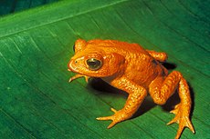
Bufo periglenes, the Golden Toad, was last recorded on May 15, 1989
Extinct
Threatened
Lower Risk

Other categories

Related topics

IUCN Red List category abbreviations (version 3.1, 2001)

In biology and ecology, extinction is the termination of an organism or of a group of organisms (taxon), normally a species. The moment of extinction is generally considered to be the death of the last individual of the species, although the capacity to breed and recover may have been lost before this point. Because a species' potential range may be very large, determining this moment is difficult, and is usually done retrospectively. This difficulty leads to phenomena such as Lazarus taxa, where a species presumed extinct abruptly "reappears" (typically in the fossil record) after a period of apparent absence.

More than 99 percent of all species, amounting to over five billion species,[1] that ever lived on Earth are estimated to be extinct.[2][3][4] Estimates on the number of Earth's current species range from 10 million to 14 million,[5] of which about 1.2 million have been documented and over 86 percent have not yet been described.[6] More recently, in May 2016, scientists reported that 1 trillion species are estimated to be on Earth currently with only one-thousandth of one percent described.[7]

Through evolution, species arise through the process of speciation—where new varieties of organisms arise and thrive when they are able to find and exploit an ecological niche—and species become extinct when they are no longer able to survive in changing conditions or against superior competition. The relationship between animals and their ecological niches has been firmly established.[8] A typical species becomes extinct within 10 million years of its first appearance,[4] although some species, called living fossils, survive with virtually no morphological change for hundreds of millions of years.

Mass extinctions are relatively rare events; however, isolated extinctions are quite common. Only recently have extinctions been recorded and scientists have become alarmed at the current high rate of extinctions.[9][10][11][12] Most species that become extinct are never scientifically documented. Some scientists estimate that up to half of presently existing plant and animal species may become extinct by 2100.[13]

A dagger symbol (†) placed next to the name of a species or other taxon is often done to indicate its status as extinct.

Definition


External mold of the extinct Lepidodendron from the Upper Carboniferous of Ohio[14]

A species is extinct when the last existing member dies. Extinction therefore becomes a certainty when there are no surviving individuals that can reproduce and create a new generation. A species may become functionally extinct when only a handful of individuals survive, which cannot reproduce due to poor health, age, sparse distribution over a large range, a lack of individuals of both sexes (in sexually reproducing species), or other reasons.

Pinpointing the extinction (or pseudoextinction) of a species requires a clear definition of that species. If it is to be declared extinct, the species in question must be uniquely distinguishable from any ancestor or daughter species, and from any other closely related species. Extinction of a species (or replacement by a daughter species) plays a key role in the punctuated equilibrium hypothesis of Stephen Jay Gould and Niles Eldredge.[15]


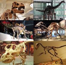
Skeleton of various extinct dinosaurs; some other dinosaur lineages still flourish in the form of birds

In ecology, extinction is often used informally to refer to local extinction, in which a species ceases to exist in the chosen area of study, but may still exist elsewhere. This phenomenon is also known as extirpation. Local extinctions may be followed by a replacement of the species taken from other locations; wolf reintroduction is an example of this. Species which are not extinct are termed extant. Those that are extant but threatened by extinction are referred to as threatened or endangered species.


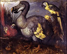
The dodo of Mauritius, shown here in a 1626 illustration by Roelant Savery, is an often-cited example of modern extinction[16]

Currently an important aspect of extinction is human attempts to preserve critically endangered species. These are reflected by the creation of the conservation status "extinct in the wild" (EW). Species listed under this status by the International Union for Conservation of Nature (IUCN) are not known to have any living specimens in the wild, and are maintained only in zoos or other artificial environments. Some of these species are functionally extinct, as they are no longer part of their natural habitat and it is unlikely the species will ever be restored to the wild.[17] When possible, modern zoological institutions try to maintain a viable population for species preservation and possible future reintroduction to the wild, through use of carefully planned breeding programs.

The extinction of one species' wild population can have knock-on effects, causing further extinctions. These are also called "chains of extinction".[18] This is especially common with extinction of keystone species.

Pseudoextinction

Extinction of a parent species where daughter species or subspecies are still extant is called pseudoextinction or phyletic extinction. Effectively, the old taxon vanishes, transformed (anagenesis) into a successor,[19] or split into more than one (cladogenesis).[20]

Pseudoextinction is difficult to demonstrate unless one has a strong chain of evidence linking a living species to members of a pre-existing species. For example, it is sometimes claimed that the extinct Hyracotherium, which was an early horse that shares a common ancestor with the modern horse, is pseudoextinct, rather than extinct, because there are several extant species of Equus, including zebra and donkey. However, as fossil species typically leave no genetic material behind, one cannot say whether Hyracotherium evolved into more modern horse species or merely evolved from a common ancestor with modern horses. Pseudoextinction is much easier to demonstrate for larger taxonomic groups.

Lazarus taxa

The coelacanth, a fish related to lungfish and tetrapods, was considered to have been extinct since the end of the Cretaceous Period until 1938 when a specimen was found, off the Chalumna River (now Tyolomnqa) on the east coast of South Africa.[21] Museum curator Marjorie Courtenay-Latimer discovered the fish among the catch of a local angler, Captain Hendrick Goosen, on December 23, 1938.[21] A local chemistry professor, JLB Smith, confirmed the fish's importance with a famous cable: "MOST IMPORTANT PRESERVE SKELETON AND GILLS = FISH DESCRIBED".[21]

Far more recent possible or presumed extinctions of species which may turn out still to exist include the thylacine, or Tasmanian tiger (Thylacinus cynocephalus), the last known example of which died in Hobart Zoo in Tasmania in 1936; the Japanese wolf (Canis lupus hodophilax), last sighted over 100 years ago; the ivory-billed woodpecker (Campephilus principalis), last sighted for certain in 1944; and the slender-billed curlew (Numenius tenuirostris), not seen since 2007.[22]

Causes


The passenger pigeon, one of hundreds of species of extinct birds, was hunted to extinction over the course of a few decades

As long as species have been evolving, species have been going extinct. It is estimated that over 99.9% of all species that ever lived are extinct. The average lifespan of a species is 1–10 million years,[23] although this varies widely between taxa. There are a variety of causes that can contribute directly or indirectly to the extinction of a species or group of species. "Just as each species is unique", write Beverly and Stephen C. Stearns, "so is each extinction ... the causes for each are varied—some subtle and complex, others obvious and simple".[24] Most simply, any species that cannot survive and reproduce in its environment and cannot move to a new environment where it can do so, dies out and becomes extinct. Extinction of a species may come suddenly when an otherwise healthy species is wiped out completely, as when toxic pollution renders its entire habitat unliveable; or may occur gradually over thousands or millions of years, such as when a species gradually loses out in competition for food to better adapted competitors. Extinction may occur a long time after the events that set it in motion, a phenomenon known as extinction debt.

Assessing the relative importance of genetic factors compared to environmental ones as the causes of extinction has been compared to the debate on nature and nurture.[25] The question of whether more extinctions in the fossil record have been caused by evolution or by catastrophe is a subject of discussion; Mark Newman, the author of Modeling Extinction, argues for a mathematical model that falls between the two positions.[4] By contrast, conservation biology uses the extinction vortex model to classify extinctions by cause. When concerns about human extinction have been raised, for example in Sir Martin Rees' 2003 book Our Final Hour, those concerns lie with the effects of climate change or technological disaster.

Currently, environmental groups and some governments are concerned with the extinction of species caused by humanity, and they try to prevent further extinctions through a variety of conservation programs.[9] Humans can cause extinction of a species through overharvesting, pollution, habitat destruction, introduction of invasive species (such as new predators and food competitors), overhunting, and other influences. Explosive, unsustainable human population growth is an essential cause of the extinction crisis.[26] According to the International Union for Conservation of Nature (IUCN), 784 extinctions have been recorded since the year 1500, the arbitrary date selected to define "recent" extinctions, up to the year 2004; with many more likely to have gone unnoticed. Several species have also been listed as extinct since 2004.[27]

Genetics and demographic phenomena

If adaptation increasing population fitness is slower than environmental degradation plus the accumulation of slightly deleterious mutations, then a population will go extinct.[28] Smaller populations have fewer beneficial mutations entering the population each generation, slowing adaptation. It is also easier for slightly deleterious mutations to fix in small populations; the resulting positive feedback loop between small population size and low fitness can cause mutational meltdown.

Limited geographic range is the most important determinant of genus extinction at background rates but becomes increasingly irrelevant as mass extinction arises.[29] Limited geographic range is a cause both of small population size and of greater vulnerability to local environmental catastrophes.

Extinction rates can be affected not just by population size, but by any factor that affects evolvability, including balancing selection, cryptic genetic variation, phenotypic plasticity, and robustness. A diverse or deep gene pool gives a population a higher chance in the short term of surviving an adverse change in conditions. Effects that cause or reward a loss in genetic diversity can increase the chances of extinction of a species. Population bottlenecks can dramatically reduce genetic diversity by severely limiting the number of reproducing individuals and make inbreeding more frequent.

Genetic pollution

Purebred wild species evolved to a specific ecology can be threatened with extinction[30] through the process of genetic pollution—i.e., uncontrolled hybridization, introgression genetic swamping which leads to homogenization or out-competition from the introduced (or hybrid) species.[31] Endemic populations can face such extinctions when new populations are imported or selectively bred by people, or when habitat modification brings previously isolated species into contact. Extinction is likeliest for rare species coming into contact with more abundant ones;[32] interbreeding can swamp the rarer gene pool and create hybrids, depleting the purebred gene pool (for example, the endangered wild water buffalo is most threatened with extinction by genetic pollution from the abundant domestic water buffalo). Such extinctions are not always apparent from morphological (non-genetic) observations. Some degree of gene flow is a normal evolutionarily process, nevertheless, hybridization (with or without introgression) threatens rare species' existence.[33][34]

The gene pool of a species or a population is the variety of genetic information in its living members. A large gene pool (extensive genetic diversity) is associated with robust populations that can survive bouts of intense selection. Meanwhile, low genetic diversity (see inbreeding and population bottlenecks) reduces the range of adaptions possible.[35] Replacing native with alien genes narrows genetic diversity within the original population,[32][36] thereby increasing the chance of extinction.


Scorched land resulting from slash-and-burn agriculture

Habitat degradation

Habitat degradation is currently the main anthropogenic cause of species extinctions. The main cause of habitat degradation worldwide is agriculture, with urban sprawl, logging, mining and some fishing practices close behind. The degradation of a species' habitat may alter the fitness landscape to such an extent that the species is no longer able to survive and becomes extinct. This may occur by direct effects, such as the environment becoming toxic, or indirectly, by limiting a species' ability to compete effectively for diminished resources or against new competitor species.

Habitat degradation through toxicity can kill off a species very rapidly, by killing all living members through contamination or sterilizing them. It can also occur over longer periods at lower toxicity levels by affecting life span, reproductive capacity, or competitiveness.

Habitat degradation can also take the form of a physical destruction of niche habitats. The widespread destruction of tropical rainforests and replacement with open pastureland is widely cited as an example of this;[13] elimination of the dense forest eliminated the infrastructure needed by many species to survive. For example, a fern that depends on dense shade for protection from direct sunlight can no longer survive without forest to shelter it. Another example is the destruction of ocean floors by bottom trawling.[37]

Diminished resources or introduction of new competitor species also often accompany habitat degradation. Global warming has allowed some species to expand their range, bringing unwelcome[according to whom?] competition to other species that previously occupied that area. Sometimes these new competitors are predators and directly affect prey species, while at other times they may merely outcompete vulnerable species for limited resources. Vital resources including water and food can also be limited during habitat degradation, leading to extinction.


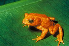
The golden toad was last seen on May 15, 1989. Decline in amphibian populations is ongoing worldwide

Predation, competition, and disease

In the natural course of events, species become extinct for a number of reasons, including but not limited to: extinction of a necessary host, prey or pollinator, inter-species competition, inability to deal with evolving diseases and changing environmental conditions (particularly sudden changes) which can act to introduce novel predators, or to remove prey. Recently in geological time, humans have become an additional cause of extinction (many people would say premature extinction) of some species, either as a new mega-predator or by transporting animals and plants from one part of the world to another. Such introductions have been occurring for thousands of years, sometimes intentionally (e.g. livestock released by sailors on islands as a future source of food) and sometimes accidentally (e.g. rats escaping from boats). In most cases, the introductions are unsuccessful, but when an invasive alien species does become established, the consequences can be catastrophic. Invasive alien species can affect native species directly by eating them, competing with them, and introducing pathogens or parasites that sicken or kill them; or indirectly by destroying or degrading their habitat. Human populations may themselves act as invasive predators. According to the "overkill hypothesis", the swift extinction of the megafauna in areas such as Australia (40,000 years before present), North and South America (12,000 years before present), Madagascar, Hawaii (300–1000 CE), and New Zealand (1300–1500 CE), resulted from the sudden introduction of human beings to environments full of animals that had never seen them before, and were therefore completely unadapted to their predation techniques.[38]

Coextinction


The large Haast's eagle and moa from New Zealand

Coextinction refers to the loss of a species due to the extinction of another; for example, the extinction of parasitic insects following the loss of their hosts. Coextinction can also occur when a species loses its pollinator, or to predators in a food chain who lose their prey. "Species coextinction is a manifestation of the interconnectedness of organisms in complex ecosystems ... While coextinction may not be the most important cause of species extinctions, it is certainly an insidious one".[39] Coextinction is especially common when a keystone species goes extinct. Models suggest that coextinction is the most common form of biodiversity loss. There may be a cascade of coextinction across the trophic levels. Such effects are most severe in mutualistic and parasitic relationships. An example of coextinction is the Haast's eagle and the moa: the Haast's eagle was a predator that became extinct because its food source became extinct. The moa were several species of flightless birds that were a food source for the Haast's eagle.[40]

Climate change

Extinction as a result of climate change has been confirmed by fossil studies.[41] Particularly, the extinction of amphibians during the Carboniferous Rainforest Collapse, 305 million years ago.[41] A 2003 review across 14 biodiversity research centers predicted that, because of climate change, 15–37% of land species would be "committed to extinction" by 2050.[42][43] The ecologically rich areas that would potentially suffer the heaviest losses include the Cape Floristic Region, and the Caribbean Basin. These areas might see a doubling of present carbon dioxide levels and rising temperatures that could eliminate 56,000 plant and 3,700 animal species.[44] Climate change has also been found to be a factor in habitat loss and desertification.[45]

Mass extinctions

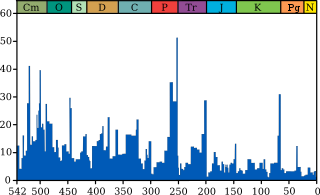
Extinction intensity.svg Cambrian Ordovician Silurian Devonian Carboniferous Permian Triassic Jurassic Cretaceous Paleogene Neogene
Marine extinction intensity during the Phanerozoic
%
Millions of years ago
Extinction intensity.svg
The blue graph shows the apparent percentage (not the absolute number) of marine animal genera becoming extinct during any given time interval. It does not represent all marine species, just those that are readily fossilized. The labels of the traditional "Big Five" extinction events and the more recently recognised End-Capitanian extinction event are clickable hyperlinks; see Extinction event for more details. (source and image info)

There have been at least five mass extinctions in the history of life on earth, and four in the last 350 million years in which many species have disappeared in a relatively short period of geological time. A massive eruptive event is considered to be one likely cause of the "Permian–Triassic extinction event" about 250 million years ago,[46] which is estimated to have killed 90% of species then existing.[47] There is also evidence to suggest that this event was preceded by another mass extinction, known as Olson's Extinction.[46] The Cretaceous–Paleogene extinction event (K-Pg) occurred 66 million years ago, at the end of the Cretaceous period, and is best known for having wiped out non-avian dinosaurs, among many other species.

Modern extinctions

According to a 1998 survey of 400 biologists conducted by New York's American Museum of Natural History, nearly 70% believed that the Earth is currently in the early stages of a human-caused mass extinction,[48] known as the Holocene extinction. In that survey, the same proportion of respondents agreed with the prediction that up to 20% of all living populations could become extinct within 30 years (by 2028). A 2014 special edition of Science declared there is widespread consensus on the issue of human-driven mass species extinctions.[49]

Biologist E. O. Wilson estimated [13] in 2002 that if current rates of human destruction of the biosphere continue, one-half of all plant and animal species of life on earth will be extinct in 100 years.[50] More significantly, the current rate of global species extinctions is estimated as 100 to 1000 times "background" rates (the average extinction rates in the evolutionary time scale of planet Earth),[51][52] while future rates are likely 10,000 times higher.[52] However, some groups are going extinct much faster. Biologists Paul R. Ehrlich and Stuart Pimm, among others, contend that human population growth and overconsumption are the main drivers of the modern extinction crisis.[53][54][55][56]

History of scientific understanding


Dilophosaurus, one of the many extinct dinosaur genera. The cause of the Cretaceous–Paleogene extinction event is a subject of much debate amongst researchers

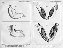
Georges Cuvier compared fossil mammoth jaws to those of living elephants, concluding that they were distinct from any known living species.[57]

For much of history, the modern understanding of extinction as the end of a species was incompatible with the prevailing worldview. Through the 18th century, much of Western society adhered to the belief that the world was created by God and as such was complete and perfect.[58] This concept reached its heyday in the 1700s with the peak popularity of a theological concept called the Great Chain of Being, in which all life on earth, from the tiniest microorganism to God, is linked in a continuous chain.[59] The extinction of a species was impossible under this model, as it would create gaps or missing links in the chain and destroy the natural order.[58][59] Thomas Jefferson was a firm supporter of the Great Chain of Being and an opponent of extinction,[58][60] famously denying the extinction of the wooly mammoth on the grounds that nature never allows a race of animals to become extinct.[61]

A series of fossils were discovered in the late 17th century that appeared unlike any living species. As a result, the scientific community embarked on a voyage of creative rationalization, seeking to understand what had happened to these species within a framework that did not account for total extinction. In October 1686, Robert Hooke presented an impression of a nautilus to the Royal Society that was more than two feet in diameter,[62] and morphologically distinct from any known living species. Hooke theorized that this was simply because the species lived in the deep ocean and no one had discovered them yet.[59] While he contended that it was possible a species could be "lost", he thought this highly unlikely.[59] Similarly, in 1695, Thomas Molyneux published an account of enormous antlers found in Ireland that did not belong to any extant taxa in that area.[63][60] Molyneux reasoned that they came from the North American moose and that the animal had once been common on the British Isles.[63] Rather than suggest that this indicated the possibility of species going extinct, he argued that although organisms could become locally extinct, they could never be entirely lost and would continue to exist in some unknown region of the globe.[63] Using the antlers as evidence for this position, Molyneux described how moose had continued to exist in North America even as they were lost to the British Isles.[60] The antlers were later confirmed to be from the extinct Irish elk Megaloceros.[60] Hooke and Molyneux's line of thinking was difficult to disprove. When parts of the world had not been thoroughly examined and charted, scientists could not rule out that animals found only in the fossil record were not simply "hiding" in unexplored regions of the Earth.[64]

Georges Cuvier is credited with establishing the modern conception of extinction in a 1796 lecture to the French Institute,[61][57] though he would spend most of his career trying to convince the wider scientific community of his theory.[65] Cuvier was a well-regarded geologist, lauded for his ability to reconstruct the anatomy of an unknown species from a few fragments of bone.[57] His primary evidence for extinction came from mammoth skulls found in the Paris basin.[57] Cuvier recognized them as distinct from any known living species of elephant, and argued that it was highly unlikely such an enormous animal would go undiscovered.[57] In 1812, Cuvier, along with Alexandre Bronigniart & Geoffroy Saint-Hilaire, mapped the strata of the Paris basin.[59] They saw alternating saltwater and freshwater deposits, as well as patterns of the appearance and disappearance of fossils throughout the record.[60][65] From these patterns, Cuvier inferred historic cycles of catastrophic flooding, extinction, and repopulation of the earth with new species.[60][65]

Cuvier’s fossil evidence showed that very different life forms existed in the past than those that exist today, a fact that was accepted by most scientists.[58] The primary debate focused whether this turnover caused by extinction was gradual or abrupt in nature.[65] Cuvier understood extinction to be the result of cataclysmic events that wipe out huge numbers of species, as opposed to the gradual decline of a species over time.[66] His catastrophic view of the nature of extinction garnered him many opponents in the newly emerging school of uniformitarianism.[66]

Jean-Baptist Lamarck, a gradualist and colleague of Cuvier, saw the fossils of different life forms as evidence of the mutable character of species.[65] While Lamarck did not deny the possibility of extinction, he believed that it was exceptional and rare and that most of the change in species over time was due to gradual change.[65] Unlike Cuvier, Lamarck was skeptical that catastrophic events of a scale large enough to cause total extinction were possible. In his geological history of the earth titled Hydrogeologie, Lamarck instead argued that the surface of the earth was shaped by gradual erosion and deposition by water, and that species changed over time in response to the changing environment.[65][67]

Charles Lyell, a noted geologist and founder of uniformitarianism, believed that past processes should be understood using present day processes. Like Lamarck, Lyell acknowledged that extinction could occur, noting the total extinction of the dodo and the extirpation of indigenous horses to the British Isles.[59] He similarly argued against mass extinctions, believing that any extinction must be a gradual process.[57][61] Lyell also showed that Cuvier’s original interpretation of the Parisian strata was incorrect. Instead of the catastrophic floods inferred by Cuvier, Lyell demonstrated that patterns of saltwater and freshwater deposits, like those seen in the Paris basin, could be formed by a slow rise and fall of sea levels.[60]

The concept of extinction was integral to Charles Darwin’s On the Origin of Species, with less fit lineages disappearing over time. For Darwin, extinction was a constant side effect of competition.[68] Because of the wide reach of On the Origin of Species, it was widely accepted that extinction occurred gradually and evenly (a concept we now refer to as background extinction).[61] It was not until 1982, when David Raup and Jack Sepkoski published their seminal paper on mass extinctions, that Cuvier was vindicated and catastrophic extinction was accepted as an important mechanism. The current understanding of extinction is a synthesis of the cataclysmic extinction events proposed by Cuvier, and the background extinction events proposed by Lyell and Darwin.

Human attitudes and interests

Extinction is an important research topic in the field of zoology, and biology in general, and has also become an area of concern outside the scientific community. A number of organizations, such as the Worldwide Fund for Nature, have been created with the goal of preserving species from extinction. Governments have attempted, through enacting laws, to avoid habitat destruction, agricultural over-harvesting, and pollution. While many human-caused extinctions have been accidental, humans have also engaged in the deliberate destruction of some species, such as dangerous viruses, and the total destruction of other problematic species has been suggested. Other species were deliberately driven to extinction, or nearly so, due to poaching or because they were "undesirable", or to push for other human agendas. One example was the near extinction of the American bison, which was nearly wiped out by mass hunts sanctioned by the United States government, to force the removal of Native Americans, many of whom relied on the bison for food.[69]

Biologist Bruce Walsh of the University of Arizona states three reasons for scientific interest in the preservation of species: genetic resources, ecosystem stability, and ethics;[70] and today the scientific community "stress[es] the importance" of maintaining biodiversity.[70][71]

In modern times, commercial and industrial interests often have to contend with the effects of production on plant and animal life. However, some technologies with minimal, or no, proven harmful effects on Homo sapiens can be devastating to wildlife (for example, DDT).[72]  Biogeographer Jared Diamond notes that while big business may label environmental concerns as "exaggerated", and often cause "devastating damage", some corporations find it in their interest to adopt good conservation practices, and even engage in preservation efforts that surpass those taken by national parks.[73]

Governments sometimes see the loss of native species as a loss to ecotourism,[74] and can enact laws with severe punishment against the trade in native species in an effort to prevent extinction in the wild. Nature preserves are created by governments as a means to provide continuing habitats to species crowded by human expansion. The 1992 Convention on Biological Diversity has resulted in international Biodiversity Action Plan programmes, which attempt to provide comprehensive guidelines for government biodiversity conservation. Advocacy groups, such as The Wildlands Project[75] and the Alliance for Zero Extinctions,[76] work to educate the public and pressure governments into action.

People who live close to nature can be dependent on the survival of all the species in their environment, leaving them highly exposed to extinction risks. However, people prioritize day-to-day survival over species conservation; with human overpopulation in tropical developing countries, there has been enormous pressure on forests due to subsistence agriculture, including slash-and-burn agricultural techniques that can reduce endangered species's habitats.[77]

Antinatalist philosopher David Benatar concludes that any popular concern about non-human species extinction usually arises out of concern about how the loss of a species will impact human wants and needs, that "we shall live in a world impoverished by the loss of one aspect of faunal diversity, that we shall no longer be able to behold or use that species of animal." He notes that typical concerns about possible human extinction, such as the loss of individual members, are not considered in regards to non-human species extinction.[78]

Planned extinction

Completed

  • The smallpox virus is now extinct in the wild,[79] although samples are retained in laboratory settings.
  • The rinderpest virus, which infected domestic cattle, is now extinct in the wild.[80]

Proposed

The poliovirus is now confined to small parts of the world due to extermination efforts.[81]

Dracunculus medinensis, a parasitic worm which causes the disease dracunculiasis, is now close to eradication thanks to efforts led by the Carter Center.[82]

Treponema pallidum pertenue, a bacterium which causes the disease yaws, is in the process of being eradicated.

Biologist Olivia Judson has advocated the deliberate extinction of certain disease-carrying mosquito species. In a September 25, 2003 New York Times article, she advocated "specicide" of thirty mosquito species by introducing a genetic element which can insert itself into another crucial gene, to create recessive "knockout genes".[83] She says that the Anopheles mosquitoes (which spread malaria) and Aedes mosquitoes (which spread dengue fever, yellow fever, elephantiasis, and other diseases) represent only 30 species; eradicating these would save at least one million human lives per annum, at a cost of reducing the genetic diversity of the family Culicidae by only 1%. She further argues that since species become extinct "all the time" the disappearance of a few more will not destroy the ecosystem: "We're not left with a wasteland every time a species vanishes. Removing one species sometimes causes shifts in the populations of other species—but different need not mean worse." In addition, anti-malarial and mosquito control programs offer little realistic hope to the 300 million people in developing nations who will be infected with acute illnesses this year. Although trials are ongoing, she writes that if they fail: "We should consider the ultimate swatting."[83]

Biologist E. O. Wilson has advocated the eradication of several species of mosquito, including malaria vector Anopheles gambiae. Wilson stated, "I'm talking about a very small number of species that have co-evolved with us and are preying on humans, so it would certainly be acceptable to remove them. I believe it's just common sense."[84]

Cloning

Some, such as Harvard geneticist George M. Church, believe that ongoing technological advances will let us "bring back to life" an extinct species by cloning, using DNA from the remains of that species. Proposed targets for cloning include the mammoth, the thylacine, and the Pyrenean ibex. For this to succeed, enough individuals would have to be cloned, from the DNA of different individuals (in the case of sexually reproducing organisms) to create a viable population. Though bioethical and philosophical objections have been raised,[85] the cloning of extinct creatures seems theoretically possible.[86]

In 2003, scientists tried to clone the extinct Pyrenean ibex (C. p. pyrenaica). This attempt failed: of the 285 embryos reconstructed, 54 were transferred to 12 mountain goats and mountain goat-domestic goat hybrids, but only two survived the initial two months of gestation before they too died.[87] In 2009, a second attempt was made to clone the Pyrenean ibex: one clone was born alive, but died seven minutes later, due to physical defects in the lungs.[88]

Homeostasis

From Wikipedia, the free encyclopedia
Homeostasis can be defined as the stable state of an organism and of its internal environment;[1] as the maintenance or regulation of the stable condition, or its equilibrium;[2] or simply as the balance of bodily functions.[3] The stable condition is the condition of optimal functioning for the organism, and is dependent on many variables, such as body temperature and fluid balance, being kept within certain pre-set limits.[4] Other variables include the pH of extracellular fluid, the concentrations of sodium, potassium and calcium ions, as well as that of the blood sugar level, and these need to be regulated despite changes in the environment, diet, or level of activity. Each of these variables is controlled by one or more regulators or homeostatic mechanisms, which together maintain life.

Homeostasis is brought about by a natural resistance to change in the optimal conditions,[2] and equilibrium is maintained by many regulatory mechanisms. All homeostatic control mechanisms have at least three interdependent components for the variable being regulated: a receptor, a control centre, and an effector. The receptor is the sensing component that monitors and responds to changes in the environment, either external or internal. Receptors include thermoreceptors, and mechanoreceptors. Control centres include the respiratory centre, and the renin–angiotensin system. An effector is the target acted on, to bring about the change back to the normal state. At the cellular level, receptors include nuclear receptors that bring about changes in gene expression through up-regulation or down-regulation, and act in negative feedback mechanisms. An example of this is in the control of bile acids in the liver.[5]

Some centres, such as the renin–angiotensin system, control more than one variable. When the receptor senses a stimulus, it reacts by sending action potentials to a control centre. The control centre sets the maintenance range—the acceptable upper and lower limits—for the particular variable, such as temperature. The control center responds to the signal by determining an appropriate response and sending signals to an effector, which can be one or more muscles, an organ, or a gland. When the signal is received and acted on, negative feedback is fed back to the receptor that stops the need for further signalling.[6]

The concept of the regulation of the internal environment was described by French physiologist Claude Bernard in 1865, and the word homeostasis was coined by Walter Bradford Cannon in 1926.[7][8] Homeostasis is an almost exclusively biological term, referring to the concepts described by Bernard and Cannon, concerning the constancy of the internal environment in which the cells of the body live and survive.[4][7][8] The term cybernetics is applied to technological control systems such as thermostats, which function as homeostatic mechanisms, but is often defined much more broadly than the biological term of homeostasis.[9][6][10][11]

Etymology

The word homeostasis (/ˌhoʊmioʊˈsteɪsɪs/[12][13]) uses combining forms of homeo- and -stasis, New Latin from Greek: ᜅΌοιος homoios, "similar" and στάσις stasis, "standing still", yielding the idea of "staying the same".

Overview

The metabolic processes of all organisms can only take place in very specific physical and chemical environments. The conditions vary with each organism, and with whether the chemical processes take place inside the cell or in the interstitial fluid bathing the cells. The best known homeostatic mechanisms in humans and other mammals are regulators that keep the composition of the extracellular fluid (or the "internal environment") constant, especially with regard to the temperature, pH, osmolality, and the concentrations of sodium, potassium, glucose, carbon dioxide, and oxygen. However, a great many other homeostatic mechanisms, encompassing many aspects of human physiology, control other entities in the body. Where the levels of variables are higher or lower than those needed, they are often prefixed with hyper- and hypo-, respectively such as hyperthermia and hypothermia and hypertension and hypotension.


Circadian variation in body temperature, ranging from about 37.5 °C from 10 a.m. to 6 p.m., and falling to about 36.4 °C from 2 a.m. to 6 a.m.

If an entity is homeostatically controlled it does not imply that its value is necessarily absolutely steady in health. Core body temperature is, for instance, regulated by a homeostatic mechanism with temperature sensors in, amongst others, the hypothalamus of the brain.[14] However, the set point of the regulator is regularly reset.[citation needed] For instance, core body temperature in humans varies during the course of the day (i.e. has a circadian rhythm), with the lowest temperatures occurring at night, and the highest in the afternoons. Other normal temperature variations include those related to the menstrual cycle.[15][16] The temperature regulator's set point is reset during infections to produce a fever.[14][17][18] Organisms are capable of adjusting somewhat to varied conditions such as temperature changes or oxygen levels at altitude, by a process of acclimatisation.

Homeostasis does not govern every activity in the body.[19][20] For instance the signal (be it via neurons or hormones) from the sensor to the effector is, of necessity, highly variable in order to convey information about the direction and magnitude of the error detected by the sensor.[21][22][23] Similarly the effector's response needs to be highly adjustable to reverse the error – in fact it should be very nearly in proportion (but in the opposite direction) to the error that is threatening the internal environment.[10][11] For instance, the arterial blood pressure in mammals is homeostatically controlled, and measured by stretch receptors in the walls of the aortic arch and carotid sinuses at beginnings of the internal carotid arteries.[14] The sensors send messages via sensory nerves to the medulla oblongata of the brain indicating whether the blood pressure has fallen or risen, and by how much. The medulla oblongata then distributes messages along motor or efferent nerves belonging to the autonomic nervous system to a wide variety of effector organs, whose activity is consequently changed to reverse the error in the blood pressure. One of the effector organs is the heart whose rate is stimulated to rise (tachycardia) when the arterial blood pressure falls, or to slow down (bradycardia) when the pressure rises above set point.[14] Thus the heart rate (for which there is no sensor in the body) is not homeostatically controlled, but is one of effector responses to errors in the arterial blood pressure. Another example is the rate of sweating. This is one of the effectors in the homeostatic control of body temperature, and therefore highly variable in rough proportion to the heat load that threatens to destabilize the body's core temperature, for which there is a sensor in the hypothalamus of the brain.

Controls of variables

Core temperature


Birds huddling for warmth

Mammals regulate their core temperature using input from thermoreceptors in the hypothalamus, brain,[14][24] spinal cord, internal organs, and great veins.[25][26] Apart from the internal regulation of temperature, a process called allostasis can come into play that adjusts behaviour to adapt to the challenge of very hot or cold extremes (and to other challenges).[27] These adjustments may include seeking shade and reducing activity, or seeking warmer conditions and increasing activity, or huddling.[28] Behavioural thermoregulation takes precedence over physiological thermoregulation since necessary changes can be affected more quickly and physiological thermoregulation is limited in its capacity to respond to extreme temperatures.[29]

When core temperature falls, the blood supply to the skin is reduced by intense vasoconstriction.[14] The blood flow to the limbs (which have a large surface area) is similarly reduced, and returned to the trunk via the deep veins which lie alongside the arteries (forming venae comitantes).[24][28][30] This acts as a counter-current exchange system which short-circuits the warmth from the arterial blood directly into the venous blood returning into the trunk, causing minimal heat loss from the extremities in cold weather.[24][28][31] The subcutaneous limb veins are tightly constricted,[14] not only reducing heat loss from this source, but also forcing the venous blood into the counter-current system in the depths of the limbs.

The metabolic rate is increased, initially by non-shivering thermogenesis,[32] followed by shivering thermogenesis if the earlier reactions are insufficient to correct the hypothermia.

When core temperature rises are detected by thermoreceptors, the sweat glands in the skin are stimulated via cholinergic sympathetic nerves to secrete sweat onto the skin, which, when it evaporates, cools the skin and the blood flowing through it. Panting is an alternative effector in many vertebrates, which cools the body also by the evaporation of water, but this time from the mucous membranes of the throat and mouth.

Blood glucose


Negative feedback at work in the regulation of blood sugar. Flat line is the set-point of glucose level and sine wave the fluctuations of glucose.

Blood sugar levels are regulated within fairly narrow limits.[33] In mammals the primary sensors for this are the beta cells of the pancreatic islets.[34][35] The beta cells respond to a rise in the blood sugar level by secreting insulin into the blood, and simultaneously inhibiting their neighboring alpha cells from secreting glucagon into the blood.[34] This combination (high blood insulin levels and low glucagon levels) act on effector tissues, chief of which are the liver, fat cells and muscle cells. The liver is inhibited from producing glucose, taking it up instead, and converting it to glycogen and triglycerides. The glycogen is stored in the liver, but the triglycerides are secreted into the blood as very low-density lipoprotein (VLDL) particles which are taken up by adipose tissue, there to be stored as fats. The fat cells take up glucose through special glucose transporters (GLUT4), whose numbers in the cell wall are increased as a direct effect of insulin acting on these cells. The glucose that enters the fat cells in this manner is converted into triglycerides (via the same metabolic pathways as are used by the liver) and then stored in those fat cells together with the VLDL-derived triglycerides that were made in the liver. Muscle cells also take glucose up through insulin-sensitive GLUT4 glucose channels, and convert it into muscle glycogen.

A fall in blood glucose, causes insulin secretion to be stopped, and glucagon to be secreted from the alpha cells into the blood. This inhibits the uptake of glucose from the blood by the liver, fats cells and muscle. Instead the liver is strongly stimulated to manufacture glucose from glycogen (through glycogenolysis) and from non-carbohydrate sources (such as lactate and de-aminated amino acids) using a process known as gluconeogenesis. The glucose thus produced is discharged into the blood correcting the detected error (hypoglycemia). The glycogen stored in muscles remains in the muscles, and is only broken down, during exercise, to glucose-6-phosphate and thence to pyruvate to be fed into the citric acid cycle or turned into lactate. It is only the lactate and the waste products of the citric acid cycle that are returned to the blood. The liver can take up only the lactate, and by the process of energy consuming gluconeogenesis convert it back to glucose.

Levels of blood gases


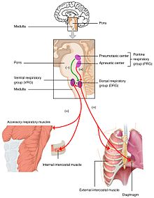
The respiratory centre

Changes in the levels of oxygen, carbon dioxide and plasma pH are sent to the respiratory center, in the brainstem where they are regulated. The partial pressure of oxygen and carbon dioxide in the arterial blood is monitored by the peripheral chemoreceptors (PNS) in the carotid artery and aortic arch. A change in the partial pressure of carbon dioxide is detected as altered pH in the cerebrospinal fluid by central chemoreceptors (CNS) in the medulla oblongata of the brainstem. Information from these sets of sensors is sent to the respiratory center which activates the effector organs – the diaphragm and other muscles of respiration. An increased level of carbon dioxide in the blood, or a decreased level of oxygen, will result in a deeper breathing pattern and increased respiratory rate to bring the blood gases back to equilibrium.

Too little carbon dioxide, and, to a lesser extent, too much oxygen in the blood can temporarily halt breathing, a condition known as apnea, which freedivers use to prolong the time they can stay underwater.

The partial pressure of carbon dioxide is more of a deciding factor in the monitoring of pH.[36] However, at high altitude (above 2500 m) the monitoring of the partial pressure of oxygen takes priority, and hyperventilation keeps the oxygen level constant. With the lower level of carbon dioxide, to keep the pH at 7.4 the kidneys secrete hydrogen ions into the blood, and excrete bicarbonate into the urine.[37][38] This is important in the acclimatization to high altitude.[39]

Blood oxygen content

The kidneys measure the oxygen content rather than the partial pressure of oxygen in the arterial blood. When the oxygen content of the blood is chronically low, oxygen-sensitive cells secrete erythropoietin (EPO) into the blood.[40] The effector tissue is the red bone marrow which produces red blood cells (RBCs)(erythrocytes). The increase in RBCs leads to an increased hematocrit in the blood, and subsequent increase in hemoglobin that increases the oxygen carrying capacity. This is the mechanism whereby high altitude dwellers have higher hematocrits than sea-level residents, and also why persons with pulmonary insufficiency or right-to-left shunts in the heart (through which venous blood by-passes the lungs and goes directly into the systemic circulation) have similarly high hematocrits.[41][42]

Regardless of the partial pressure of oxygen in the blood, the amount of oxygen that can be carried, depends on the hemoglobin content. The partial pressure of oxygen may be sufficient for example in anemia, but the hemoglobin content will be insufficient and subsequently as will be the oxygen content. Given enough supply of iron, vitamin B12 and folic acid, EPO can stimulate RBC production, and hemoglobin and oxygen content restored to normal.[41][43]

Arterial blood pressure

The brain can regulate blood flow over a range of blood pressure values by vasoconstriction and vasodilation of the arteries.[44]
High pressure receptors called baroreceptors in the walls of the aortic arch and carotid sinus (at the beginning of the internal carotid artery) monitor the arterial blood pressure.[45] Rising pressure is detected when the walls of the arteries stretch due to an increase in blood volume. This causes heart muscle cells to secrete the hormone atrial natriuretic peptide (ANP) into the blood. This acts on the kidneys to inhibit the secretion of renin and aldosterone causing the release of sodium, and accompanying water into the urine, thereby reducing the blood volume.[46] This information is then conveyed, via afferent nerve fibers, to the solitary nucleus in the medulla oblongata.[47] From here motor nerves belonging to the autonomic nervous system are stimulated to influence the activity of chiefly the heart and the smallest diameter arteries, called arterioles. The arterioles are the main resistance vessels in the arterial tree, and small changes in diameter cause large changes in the resistance to flow through them. When the arterial blood pressure rises the arterioles are stimulated to dilate making it easier for blood to leave the arteries, thus deflating them, and bringing the blood pressure down, back to normal. At the same time the heart is stimulated via cholinergic parasympathetic nerves to beat more slowly (called bradycardia), ensuring that the inflow of blood into the arteries is reduced, thus adding to the reduction in pressure, and correction of the original error.

Low pressure in the arteries, causes the opposite reflex of constriction of the arterioles, and a speeding up of the heart rate (called tachycardia). If the drop in blood pressure is very rapid or excessive, the medulla oblongata stimulates the adrenal medulla, via "preganglionic" sympathetic nerves, to secrete epinephrine (adrenaline) into the blood. This hormone enhances the tachycardia and causes severe vasoconstriction of the arterioles to all but the essential organ in the body (especially the heart, lungs and brain). These reactions usually correct the low arterial blood pressure (hypotension) very effectively.

Calcium levels

Calcium homeostasis

The plasma ionized calcium (Ca2+) concentration is very tightly controlled by a pair of homeostatic mechanisms.[48] The sensor for the first one is situated in the parathyroid glands, where the chief cells sense the Ca2+ level by means of specialized calcium receptors in their membranes. The sensors for the second are the parafollicular cells in the thyroid gland. The parathyroid chief cells secrete parathyroid hormone (PTH) in response to a fall in the plasma ionized calcium level; the parafollicular cells of the thyroid gland secrete calcitonin in response to a rise in the plasma ionized calcium level.

The effector organs of the first homeostatic mechanism are the bones, the kidney, and, via a hormone released into the blood by the kidney in response to high PTH levels in the blood, the duodenum and jejunum. Parathyroid hormone (in high concentrations in the blood) causes bone resorption, releasing calcium into the plasma. This is a very rapid action which can correct a threatening hypocalcemia within minutes. High PTH concentrations cause the excretion of phosphate ions via the urine. Since phosphates combine with calcium ions to form insoluble salts, a decrease in the level of phosphates in the blood, releases free calcium ions into the plasma ionized calcium pool. PTH has a second action on the kidneys. It stimulates the manufacture and release, by the kidneys, of calcitriol into the blood. This steroid hormone acts on the epithelial cells of the upper small intestine, increasing their capacity to absorb calcium from the gut contents into the blood.[49]

The second homeostatic mechanism, with its sensors in the thyroid gland, releases calcitonin into the blood when the blood ionized calcium rises. This hormone acts primarily on bone, causing the rapid removal of calcium from the blood and depositing it, in insoluble form, in the bones.

The two homeostatic mechanisms working through PTH on the one hand, and calcitonin on the other can very rapidly correct any impending error in the plasma ionized calcium level by either removing calcium from the blood and depositing it in the skeleton, or by removing calcium from it. The skeleton acts as an extremely large calcium store (about 1 kg) compared with the plasma calcium store (about 180 mg). Longer term regulation occurs through calcium absorption or loss from the gut.

Sodium concentration

The homeostatic mechanism which controls the plasma sodium concentration is rather more complex than most of the other homeostatic mechanisms described on this page.
The sensor is situated in the juxtaglomerular apparatus of kidneys, which senses the plasma sodium concentration in a surprisingly indirect manner. Instead of measuring it directly in the blood flowing past the juxtaglomerular cells, these cells respond to the sodium concentration in the renal tubular fluid after it has already undergone a certain amount of modification in the proximal convoluted tubule and loop of Henle.[50] These cells also respond to rate of blood flow through the juxtaglomerular apparatus, which, under normal circumstances, is directly proportional to the arterial blood pressure, making this tissue an ancillary arterial blood pressure sensor.

In response to a lowering of the plasma sodium concentration, or to a fall in the arterial blood pressure, the juxtaglomerular cells release renin into the blood.[50][51][52] Renin is an enzyme which cleaves a decapeptide (a short protein chain, 10 amino acids long) from a plasma α-2-globulin called angiotensinogen. This decapeptide is known as angiotensin I.[50] It has no known biological activity. However, when the blood circulates through the lungs a pulmonary capillary endothelial enzyme called angiotensin-converting enzyme (ACE) cleaves a further two amino acids from angiotensin I to form an octapeptide known as angiotensin II. Angiotensin II is a hormone which acts on the adrenal cortex, causing the release into the blood of the steroid hormone, aldosterone. Angiotensin II also acts on the smooth muscle in the walls of the arterioles causing these small diameter vessels to constrict, thereby restricting the outflow of blood from the arterial tree, causing the arterial blood pressure to rise. This therefore reinforces the measures described above (under the heading of "Arterial blood pressure"), which defend the arterial blood pressure against changes, especially hypotension.

The angiotensin II-stimulated aldosterone released from the zona glomerulosa of the adrenal glands has an effect on particularly the epithelial cells of the distal convoluted tubules and collecting ducts of the kidneys. Here it causes the reabsorption of sodium ions from the renal tubular fluid, in exchange for potassium ions which are secreted from the blood plasma into the tubular fluid to exit the body via the urine.[50][53] The reabsorption of sodium ions from the renal tubular fluid halts further sodium ion losses from the body, and therefore preventing the worsening of hyponatremia. The hyponatremia can only be corrected by the consumption of salt in the diet. However, it is not certain whether a "salt hunger" can be initiated by hyponatremia, or by what mechanism this might come about.

When the plasma sodium ion concentration is higher than normal (hypernatremia), the release of renin from the juxtaglomerular apparatus is halted, ceasing the production of angiotensin II, and its consequent aldosterone-release into the blood. The kidneys respond by excreting sodium ions into the urine, thereby normalizing the plasma sodium ion concentration. The low angiotensin II levels in the blood lower the arterial blood pressure as an inevitable concomitant response.

The reabsorption of sodium ions from the tubular fluid as a result of high aldosterone levels in the blood does not, of itself, cause renal tubular water to be returned to the blood from the distal convoluted tubules or collecting ducts. This is because sodium is reabsorbed in exchange for potassium and therefore causes only a modest change in the osmotic gradient between the blood and the tubular fluid. Furthermore, the epithelium of the distal convoluted tubules and collecting ducts is impermeable to water in the absence of antidiuretic hormone (ADH) in the blood. ADH is part of the control of fluid balance. Its levels in the blood vary with the osmolality of the plasma, which is measured in the hypothalamus of the brain. Aldosterone's action on the kidney tubules prevents sodium loss to the extracellular fluid (ECF). So there is no change in the osmolality of the ECF, and therefore no change in the ADH concentration of the plasma. However, low aldosterone levels cause a loss of sodium ions from the ECF, which could potentially cause a change in extracellular osmolality and therefore of ADH levels in the blood.

Potassium concentration

High potassium concentrations in the plasma cause depolarization of the zona glomerulosa cells' membranes in the outer layer of the adrenal cortex.[54] This causes the release of aldosterone into the blood.
Aldosterone acts primarily on the distal convoluted tubules and collecting ducts of the kidneys, stimulating the excretion of potassium ions into the urine.[50] It does so, however, by activating the basolateral Na+/K+ pumps of the tubular epithelial cells. These sodium/potassium exchangers pump three sodium ions out of the cell, into the interstitial fluid and two potassium ions into the cell from the interstitial fluid. This creates an ionic concentration gradient which results in the reabsorption of sodium (Na+) ions from the tubular fluid into the blood, and secreting potassium (K+) ions from the blood into the urine (lumen of collecting duct).[55][56]

Fluid balance

The total amount of water in the body needs to be kept in balance. Fluid balance involves keeping the fluid volume stabilised, and also keeping the levels of electrolytes in the extracellular fluid stable. Fluid balance is maintained by the process of osmoregulation and by behaviour. Osmotic pressure is detected by osmoreceptors in the median preoptic nucleus in the hypothalamus. Measurement of the plasma osmolality to give an indication of the water content of the body, relies on the fact that water losses from the body, (through unavoidable water loss through the skin which is not entirely waterproof and therefore always slightly moist, water vapor in the exhaled air, sweating, vomiting, normal feces and especially diarrhea) are all hypotonic, meaning that they are less salty than the body fluids (compare, for instance, the taste of saliva with that of tears. The latter have almost the same salt content as the extracellular fluid, whereas the former is hypotonic with respect to plasma. Saliva does not taste salty, whereas tears are decidedly salty). Nearly all normal and abnormal losses of body water therefore cause the extracellular fluid to become hypertonic. Conversely excessive fluid intake dilutes the extracellular fluid causing the hypothalamus to register hypotonic hyponatremia conditions.

When the hypothalamus detects a hypertonic extracellular environment, it causes the secretion of an antidiuretic hormone (ADH) called vasopressin which acts on the effector organ, which in this case is the kidney. The effect of vasopressin on the kidney tubules is to reabsorb water from the distal convoluted tubules and collecting ducts, thus preventing aggravation of the water loss via the urine. The hypothalamus simultaneously stimulates the nearby thirst center causing an almost irresistible (if the hypertonicity is severe enough) urge to drink water. The cessation of urine flow prevents the hypovolemia and hypertonicity from getting worse; the drinking of water corrects the defect.

Hypo-osmolality results in very low plasma ADH levels. This results in the inhibition of water reabsorption from the kidney tubules, causing high volumes of very dilute urine to be excreted, thus getting rid of the excess water in the body.

Urinary water loss, when the body water homeostat is intact, is a compensatory water loss, correcting any water excess in the body. However, since the kidneys cannot generate water, the thirst reflex is the all important second effector mechanism of the body water homeostat, correcting any water deficit in the body.

Blood pH

2714 Respiratory Regulation of Blood.jpg

The plasma pH can be altered by respiratory changes in the partial pressure of carbon dioxide; or altered by metabolic changes in the carbonic acid to bicarbonate ion ratio. The bicarbonate buffer system regulates the ratio of carbonic acid to bicarbonate to be equal to 1:20, at which ratio the blood pH is 7.4 (as explained in the Henderson–Hasselbalch equation). A change in the plasma pH gives an acid–base imbalance. In acid–base homeostasis there are two mechanisms that can help regulate the pH. Respiratory compensation a mechanism of the respiratory center, adjusts the partial pressure of carbon dioxide by changing the rate and depth of breathing, to bring the pH back to normal. The partial pressure of carbon dioxide also determines the concentration of carbonic acid, and the bicarbonate buffer system can also come into play. Renal compensation can help the bicarbonate buffer system. The sensor for the plasma bicarbonate concentration is not known for certain. It is very probable that the renal tubular cells of the distal convoluted tubules are themselves sensitive to the pH of the plasma.[citation needed] The metabolism of these cells produces carbon dioxide, which is rapidly converted to hydrogen and bicarbonate through the action of carbonic anhydrase.[57] When the ECF pH falls (becoming more acidic) the renal tubular cells excrete hydrogen ions into the tubular fluid to leave the body via urine. Bicarbonate ions are simultaneously secreted into the blood that decreases the carbonic acid, and consequently raises the plasma pH.[57] The converse happens when the plasma pH rises above normal: bicarbonate ions are excreted into the urine, and hydrogen ions released into the plasma.

When hydrogen ions are excreted into the urine, and bicarbonate into the blood, the latter combine with the excess hydrogen ions in the plasma that stimulated the kidneys to perform this operation. The resulting reaction in the plasma is the formation of carbonic acid which is in equilibrium with the plasma partial pressure of carbon dioxide. This is tightly regulated to ensure that there is no excessive build-up of carbonic acid or bicarbonate. The overall effect is therefore that hydrogen ions are lost in the urine when the pH of the plasma falls. The concomitant rise in the plasma bicarbonate mops up the increased hydrogen ions (caused by the fall in plasma pH) and the resulting excess carbonic acid is disposed of in the lungs as carbon dioxide. This restores the normal ratio between bicarbonate and the partial pressure of carbon dioxide and therefore the plasma pH. The converse happens when a high plasma pH stimulates the kidneys to secrete hydrogen ions into the blood and to excrete bicarbonate into the urine. The hydrogen ions combine with the excess bicarbonate ions in the plasma, once again forming an excess of carbonic acid which can be exhaled, as carbon dioxide, in the lungs, keeping the plasma bicarbonate ion concentration, the partial pressure of carbon dioxide and, therefore, the plasma pH, constant.

Cerebrospinal fluid

Cerebrospinal fluid (CSF) allows for regulation of the distribution of substances between cells of the brain,[58] and neuroendocrine factors, to which slight changes can cause problems or damage to the nervous system. For example, high glycine concentration disrupts temperature and blood pressure control, and high CSF pH causes dizziness and syncope.[59]

Neurotransmission

Inhibitory neurons in the central nervous system play a homeostatic role in the balance of neuronal activity between excitation and inhibition. Inhibitory neurons using GABA, make compensating changes in the neuronal networks preventing runaway levels of excitation.[60] An imbalance between excitation and inhibition is seen to be implicated in a number of neuropsychiatric disorders.[61]

Neuroendocrine system

The neuroendocrine system is the mechanism by which the hypothalamus maintains homeostasis, regulating metabolism, reproduction, eating and drinking behaviour, energy utilization, osmolarity and blood pressure.
The regulation of metabolism, is carried out by hypothalamic interconnections to other glands.[62] Three endocrine glands of the hypothalamic–pituitary–gonadal axis (HPG axis) often work together and have important regulatory functions. Two other regulatory endocrine axes are the hypothalamic–pituitary–adrenal axis (HPA axis) and the hypothalamic–pituitary–thyroid axis (HPT axis).

The liver also has many regulatory functions of the metabolism. An important function is the production and control of bile acids. Too much bile acid can be toxic to cells and its synthesis can be inhibited by activation of FXR a nuclear receptor.[5]

Gene regulation

At the cellular level, homeostasis is carried out by several mechanisms including transcriptional regulation that can alter the activity of genes in response to changes.

Energy balance


The amount of energy taken in through nutrition needs to match the amount of energy used. To achieve energy homeostasis appetite is regulated by two hormones, grehlin and leptin. Grehlin stimulates hunger and the intake of food and leptin acts to signal satiety (fullness).

Clinical significance

Many diseases are the result of a homeostatic failure. Almost any homeostatic component can malfunction, either as a result of an inherited defect, an inborn error of metabolism, or an acquired disease. Some homeostatic mechanisms have inbuilt redundancies, which ensures that life is not immediately threatened if a component malfunctions; but sometimes a homeostatic malfunction can result in serious disease, which can be fatal if not treated. A well known example of a homeostatic failure is shown in type 1 diabetes mellitus. Here blood sugar regulation is unable to function because the beta cells of the pancreatic islets are destroyed and cannot produce the necessary insulin. The blood sugar rises in a condition known as hyperglycemia.

The plasma ionized calcium homeostat can be disrupted by the constant, unchanging, over-production of parathyroid hormone by a parathyroid adenoma resulting in the typically features of hyperparathyroidism, namely high plasma ionized Ca2+ levels and the resorption of bone, which can lead to spontaneous fractures. The abnormally high plasma ionized calcium concentrations cause conformational changes in many cell-surface proteins (especially ion channels and hormone or neurotransmitter receptors)[63] giving rise to lethargy, muscle weakness, anorexia, constipation and labile emotions.[64]

The body water homeostat can be compromised by the inability to secrete ADH in response to even the normal daily water losses via the exhaled air, the feces, and insensible sweating. On receiving a zero blood ADH signal, the kidneys produce huge unchanging volumes of very dilute urine, causing dehydration and death if not treated.

As organisms age, the efficiency of their control systems becomes reduced. The inefficiencies gradually result in an unstable internal environment that increases the risk of illness, and leads to the physical changes associated with aging.[6]

Various chronic diseases are kept under control by homeostatic compensation, which masks a problem by compensating for it (making up for it) in another way. However, the compensating mechanisms eventually wear out or are disrupted by a new complicating factor (such as the advent of a concurrent acute viral infection), which sends the body reeling through a new cascade of events. Such decompensation unmasks the underlying disease, worsening its symptoms. Common examples include decompensated heart failure, kidney failure, and liver failure.

Biosphere

In the Gaia hypothesis, James Lovelock[65] stated that the entire mass of living matter on Earth (or any planet with life) functions as a vast homeostatic superorganism that actively modifies its planetary environment to produce the environmental conditions necessary for its own survival. In this view, the entire planet maintains several homeostats (the primary one being temperature homeostasis). Whether this sort of system is present on Earth is open to debate. However, some relatively simple homeostatic mechanisms are generally accepted. For example, it is sometimes claimed that when atmospheric carbon dioxide levels rise, certain plants may be able to grow better and thus act to remove more carbon dioxide from the atmosphere. However, warming has exacerbated droughts, making water the actual limiting factor on land. When sunlight is plentiful and atmospheric temperature climbs, it has been claimed that the phytoplankton of the ocean surface waters, acting as global sunshine, and therefore heat sensors, may thrive and produce more dimethyl sulfide (DMS). The DMS molecules act as cloud condensation nuclei, which produce more clouds, and thus increase the atmospheric albedo, and this feeds back to lower the temperature of the atmosphere. However, rising sea temperature has stratified the oceans, separating warm, sunlit waters from cool, nutrient-rich waters. Thus, nutrients have become the limiting factor, and plankton levels have actually fallen over the past 50 years, not risen. As scientists discover more about Earth, vast numbers of positive and negative feedback loops are being discovered, that, together, maintain a metastable condition, sometimes within very broad range of environmental conditions.

Predictive

Predictive homeostasis is an anticipatory response to an expected challenge in the future, such as the stimulation of insulin secretion by gut hormones which enter the blood in response to a meal.[34] This insulin secretion occurs before the blood sugar level rises, lowering the blood sugar level in anticipation of a large influx into the blood of glucose resulting from the digestion of carbohydrates in the gut.[66] Such anticipatory reactions are open loop systems which are based, essentially, on "guess work", and are not self-correcting.[67] Anticipatory responses always require a closed loop negative feedback system to correct the 'over-shoots' and 'under-shoots' to which the anticipatory systems are prone.

Other fields

The term has come to be used in other fields, for example:

Risk

An actuary may refer to risk homeostasis, where (for example) people who have anti-lock brakes have no better safety record than those without anti-lock brakes, because the former unconsciously compensate for the safer vehicle via less-safe driving habits. Previous to the innovation of anti-lock brakes, certain maneuvers involved minor skids, evoking fear and avoidance: Now the anti-lock system moves the boundary for such feedback, and behavior patterns expand into the no-longer punitive area. It has also been suggested that ecological crises are an instance of risk homeostasis in which a particular behavior continues until proven dangerous or dramatic consequences actually occur.[68]

Stress

Sociologists and psychologists may refer to stress homeostasis, the tendency of a population or an individual to stay at a certain level of stress, often generating artificial stresses if the "natural" level of stress is not enough.[69]

Jean-François Lyotard, a postmodern theorist, has applied this term to societal 'power centers' that he describes in The Postmodern Condition, as being 'governed by a principle of homeostasis,' for example, the scientific hierarchy, which will sometimes ignore a radical new discovery for years because it destabilizes previously accepted norms.

Technology

Familiar technological homeostatic mechanisms include:
  • A thermostat operates by switching heaters or air-conditioners on and off in response to the output of a temperature sensor.
  • Cruise control adjusts a car's throttle in response to changes in speed.[70][71]
  • An autopilot operates the steering controls of an aircraft or ship in response to deviation from a pre-set compass bearing or route.[72]
  • Process control systems in a chemical plant or oil refinery maintain fluid levels, pressures, temperature, chemical composition, etc. by controlling heaters, pumps and valves.[citation needed]
  • The centrifugal governor of a steam engine, as designed by James Watt in 1788, reduces the throttle valve in response to increases in the engine speed, or opens the valve if the speed falls below the pre-set rate.[73][74]

Political psychology

From Wikipedia, the free encyclopedia ...