Search This Blog

Tuesday, November 13, 2018

Cerebral cortex

From Wikipedia, the free encyclopedia

Cerebral cortex
Brainmaps-macaque-hippocampus.jpg
Tissue slice from the brain of an adult macaque monkey (Macaca mulatta). The cerebral cortex is the outer layer depicted in dark violet. Source: BrainMaps.org
NeuronGolgi.png
Golgi-stained neurons in the cortex
Details
Part ofCerebrum
Identifiers
LatinCortex cerebri
MeSHD002540
NeuroNames39
NeuroLex IDbirnlex_1494
TAA14.1.09.003
A14.1.09.301
FMA61830

The cerebral cortex is the largest region of the cerebrum in the mammalian brain and plays a key role in memory, attention, perception, cognition, awareness, thought, language, and consciousness. The cerebral cortex is the most anterior (rostral) brain region and consists of an outer zone of neural tissue called gray matter, which contains neuronal cell bodies. It is also divided into left and right cerebral hemispheres by the longitudinal fissure, but the two hemispheres are joined at the midline by the corpus callosum.

At the cellular and circuit level, the cerebral cortex is characterized by two primary organizational features:
  1. across its surface it is divided into functional areas that serve various sensory, motor, and cognitive functions, and
  2. it is subdivided into several layers that organize the input and output connectivity of resident neurons.
These two fundamental properties provide modular functionality.

In large mammals, the cerebral cortex is usually folded, providing a greater surface area in the confined volume of the cranium. Increased surface area is thought to be important because it allows for the addition and evolution of a greater diversity of functional modules, or areas. A fold or ridge in the cortex is termed a gyrus (plural gyri) and a groove is termed a sulcus (plural sulci). These surface convolutions appear during fetal development and continue to mature after birth through the process of gyrification. In the human brain the majority of the cerebral cortex is not visible from the outside, but buried in the sulci.

The cerebral cortex contains a large number of neuronal and glial cell bodies, as well as their intricate dendritic formations and axonal projections, which connect at synapses to form basic functional circuits. The cerebral cortex is entirely made of gray matter, contrasting with the underlying white matter, which consists mainly of axons traveling to and from the cortex, their myelinated sheaths, and the cell bodies of oligodendrocytes.

Structure

Layers

Cerebral cortex. (Poirier.) To the left, the groups of cells; to the right, the systems of fibers. Quite to the left of the figure a sensory nerve fiber is shown. Cell body layers are labeled on the left, and fiber layers are labeled on the right.
Three drawings of cortical lamination by Santiago Ramon y Cajal, each showing a vertical cross-section, with the surface of the cortex at the top. Left: Nissl-stained visual cortex of a human adult. Middle: Nissl-stained motor cortex of a human adult. Right: Golgi-stained cortex of a ​1 12 month old infant. The Nissl stain shows the cell bodies of neurons; the Golgi stain shows the dendrites and axons of a random subset of neurons.
Micrograph showing the visual cortex (predominantly pink). Subcortical white matter (predominantly blue) is seen at the bottom of the image. HE-LFB stain.
The different cortical layers each contain a characteristic distribution of neuronal cell types and connections with other cortical and subcortical regions. There are direct connections between different cortical areas and indirect connections via the thalamus, for example. One of the clearest examples of cortical layering is the line of Gennari in the primary visual cortex. This is a band of whiter tissue that can be observed with the naked eye in the fundus of the calcarine sulcus of the occipital lobe. The line of Gennari is composed of axons bringing visual information from the thalamus into layer four of the visual cortex.

Staining cross-sections of the cortex to reveal the position of neuronal cell bodies and the intracortical axon tracts allowed neuroanatomists in the early 20th century to produce a detailed description of the laminar structure of the cortex in different species. After the work of Korbinian Brodmann (1909) the neurons of the cerebral cortex are grouped into six main layers, from outside (pial surface) to inside (white matter):
  1. Layer I, the molecular layer, contains few scattered neurons and consists mainly of extensions of apical dendritic tufts of pyramidal neurons and horizontally oriented axons, as well as glial cells. During development Cajal-Retzius and subpial granular layer cells are present in this layer. Also, some spiny stellate cells can be found here. Inputs to the apical tufts are thought to be crucial for the feedback interactions in the cerebral cortex involved in associative learning and attention. While it was once thought that the input to layer I came from the cortex itself, it is now realized that layer I across the cerebral cortex mantle receives substantial input from matrix or M-type thalamus cells (in contrast to core or C-type that go to layer IV).
  2. Layer II, the external granular layer, contains small pyramidal neurons and numerous stellate neurons.
  3. Layer III, the external pyramidal layer, contains predominantly small and medium-size pyramidal neurons, as well as non-pyramidal neurons with vertically oriented intracortical axons; layers I through III are the main target of interhemispheric corticocortical afferents, and layer III is the principal source of corticocortical efferents.
  4. Layer IV, the internal granular layer, contains different types of stellate and pyramidal neurons, and is the main target of thalamocortical afferents from thalamus type C neurons as well as intra-hemispheric corticocortical afferents. The layers above layer IV are also referred to as supragranular layers (layers I-III), whereas the layers below are referred to as infragranular layers (layers V and VI).
  5. Layer V, the internal pyramidal layer, contains large pyramidal neurons which give rise to axons leaving the cortex and running down to subcortical structures (such as the basal ganglia). In the primary motor cortex of the frontal lobe, layer V contains Betz cells, whose axons travel through the internal capsule, the brain stem and the spinal cord forming the corticospinal tract, which is the main pathway for voluntary motor control.
  6. Layer VI, the polymorphic or multiform layer, contains few large pyramidal neurons and many small spindle-like pyramidal and multiform neurons; layer VI sends efferent fibers to the thalamus, establishing a very precise reciprocal interconnection between the cortex and the thalamus. That is, layer VI neurons from one cortical column connect with thalamus neurons that provide input to the same cortical column. These connections are both excitatory and inhibitory. Neurons send excitatory fibers to neurons in the thalamus and also send collaterals to the thalamic reticular nucleus that inhibit these same thalamus neurons or ones adjacent to them. One theory is that because the inhibitory output is reduced by cholinergic input to the cerebral cortex, this provides the brainstem with adjustable "gain control for the relay of lemniscal inputs".
The cortical layers are not simply stacked one over the other; there exist characteristic connections between different layers and neuronal types, which span all the thickness of the cortex. These cortical microcircuits are grouped into cortical columns and minicolumns. It has been proposed that the minicolumns are the basic functional units of the cortex. In 1957, Vernon Mountcastle showed that the functional properties of the cortex change abruptly between laterally adjacent points; however, they are continuous in the direction perpendicular to the surface. Later works have provided evidence of the presence of functionally distinct cortical columns in the visual cortex (Hubel and Wiesel, 1959), auditory cortex, and associative cortex.

Cortical areas that lack a layer IV are called agranular. Cortical areas that have only a rudimentary layer IV are called dysgranular. Information processing within each layer is determined by different temporal dynamics with that in the layers II/III having a slow 2 Hz oscillation while that in layer V having a fast 10–15 Hz one.

Areas

Based on the differences in lamination the cerebral cortex can be classified into two parts, the large area of neocortex and the much smaller area of allocortex:
  • The neocortex (also known as the isocortex or neopallium) is the part of the mature cerebral cortex with six distinct layers. Examples of neocortical areas include the granular primary motor cortex, also known as Brodmann area 4, and the striate primary visual cortex, or Brodmann area 17. The neocortex has two types of cortices, the true isocortex and the proisocortex. The proisocortex contains Brodmann areas 24, 25, and 32
  • The allocortex is the part of the cerebral cortex with less than six layers and has three regions, the paleocortex with three cortical laminae and the archicortex which has four or five, and a transitional area adjacent to the allocortex, the periallocortex. Examples of allocortex are the olfactory cortex and the hippocampus.
There is a transitional area between the neocortex and the allocortex called the paralimbic cortex, where layers 2, 3 and 4 are merged. This area incorporates the proisocortex of the neocortex and the periallocortex of the allocortex. In addition, the cerebral cortex may be classified on the basis of gross topographical conventions into four lobes (named after the four skull bones protecting them): the temporal lobe, the occipital lobe, the parietal lobe, and the frontal lobe.

Brodmann areas

Different parts of the cerebral cortex are involved in different cognitive and behavioral functions. The differences show up in a number of ways: the effects of localized brain damage, regional activity patterns exposed when the brain is examined using functional imaging techniques, connectivity with subcortical areas, and regional differences in the cellular architecture of the cortex. Neuroscientists describe most of the cortex—the part they call the neocortex—as having six layers, but not all layers are apparent in all areas, and even when a layer is present, its thickness and cellular organization may vary. Scientists have constructed maps of cortical areas on the basis of variations in the appearance of the layers as seen with a microscope. One of the most widely used schemes came from Korbinian Brodmann, who split the cortex into 51 different areas and assigned each a number (many of these Brodmann areas have since been subdivided). For example, Brodmann area 1 is the primary somatosensory cortex, Brodmann area 17 is the primary visual cortex, and Brodmann area 25 is the anterior cingulate cortex.

Thickness

For mammals, species with larger brains (in absolute terms, not just in relation to body size) tend to have thicker cortices. The range, however, is not very great; only a factor of 7 differentiates between the thickest and thinnest cortices. The smallest mammals, such as shrews, have a neocortical thickness of about 0.5 mm; the ones with the largest brains, such as humans and fin whales, have thicknesses of 2.3–2.8 mm. There is an approximately logarithmic relationship between brain weight and cortical thickness.

Cortical blood supply

Magnetic resonance imaging of the brain (MRI) makes it possible to get a measure for the thickness of the human cerebral cortex and relate it to other measures. The thickness of different cortical areas varies but in general, sensory cortex is thinner than motor cortex. One study has found some positive association between the cortical thickness and intelligence. Another study has found that the somatosensory cortex is thicker in migraine sufferers, though it is not known if this is the result of migraine attacks or the cause of them. A later study using a larger patient population reports no change in the cortical thickness in migraine sufferers. A genetic disorder of the cerebral cortex, whereby decreased folding in certain areas results in a microgyrus, where there are four layers instead of six, is in some instances seen to be related to dyslexia.

Folds

The cerebral cortex is folded in a way that allows a large surface area to fit within the confines of the skull. When unfolded, each cerebral hemisphere cortex has a total surface area of about 1.3 square feet (0.12 m2). The folding is inward away from the surface of the brain, and is also present on the medial surface of the brain within the longitudinal fissure. The folding creates a looping, meandering, 'worm-pile' appearance of the surface of the brain. The peaks of folds are called gyri (singular gyrus), and the valleys are called sulci (singular sulcus).

Development

Human cortical development between 26 and 39 week gestational age

The ontogenic development of the cerebral cortex is a complex and finely tuned process influenced by the interplay between genes and environment. The cerebral cortex develops from the most anterior part of the neural plate, a specialized part of the embryonic ectoderm. The neural plate folds and closes to form the neural tube. From the cavity inside the neural tube develops the ventricular system, and, from the epithelial cells of its walls, the neurons and glia of the nervous system. The most anterior (front, or cranial) part of the neural plate, the prosencephalon, which is evident before neurulation begins, gives rise to the cerebral hemispheres and its later cortex.

Cortical neurons are generated within the ventricular zone, next to the ventricles. At first, this zone contains progenitor cells, which divide to produce glial cells and neurons. The glial fibers produced in the first divisions of the progenitor cells are radially oriented, spanning the thickness of the cortex from the ventricular zone to the outer, pial surface, and provide scaffolding for the migration of neurons outwards from the ventricular zone. The first divisions of the progenitor cells are symmetric, which duplicates the total number of progenitor cells at each mitotic cycle. Then, some progenitor cells begin to divide asymmetrically, producing one postmitotic cell that migrates along the radial glial fibers, leaving the ventricular zone, and one progenitor cell, which continues to divide until the end of development, when it differentiates into a glial cell or an ependymal cell. As the G1 phase of mitosis is elongated, in what is seen as selective cell-cycle lengthening, the newly-born neurons migrate to more superficial layers of the cortex. The migrating daughter cells become the pyramidal cells of the cerebral cortex. The development process is time ordered and regulated by hundreds of genes and epigenetic regulatory mechanisms.

The layered structure of the mature cerebral cortex is formed during development. The first pyramidal neurons generated migrate out of the ventricular zone and subventricular zone, together with reelin producing Cajal–Retzius neurons, from the preplate. Next, a cohort of neurons migrating into the middle of the preplate divides this transient layer into the superficial marginal zone, which will become layer one of the mature neocortex, and the subplate, forming a middle layer called the cortical plate. These cells will form the deep layers of the mature cortex, layers five and six. Later born neurons migrate radially into the cortical plate past the deep layer neurons, and become the upper layers (two to four). Thus, the layers of the cortex are created in an inside-out order. The only exception to this inside-out sequence of neurogenesis occurs in the layer I of primates, in which, in contrast to rodents, neurogenesis continues throughout the entire period of corticogenesis.

Depicted in blue, Emx2 is highly expressed at the caudomedial pole and dissipates outward. Pax6 expression is represented in purple and is highly expressed at the rostral lateral pole. (Adapted from Sanes, D., Reh, T., & Harris, W. (2012). Development of the Nervous System (3rd ed.). Burlington: Elsevier Science)

The map of functional cortical areas, which include primary motor and visual cortex, originates from a 'protomap', which is regulated by molecular signals such as fibroblast growth factor FGF8 early in embryonic development. These signals regulate the size, shape, and position of cortical areas on the surface of the cortical primordium, in part by regulating gradients of transcription factor expression, through a process called cortical patterning. Examples of such transcription factors include the genes EMX2 and PAX6. Together, both transcription factors form an opposing gradient of expression. Pax6 is highly expressed at the rostral lateral pole, while Emx2 is highly expressed in the caudomedial pole. The establishment of this gradient is important for proper development. For example, mutations in Pax6 can cause expression levels of Emx2 to expand out of its normal expression domain, which would ultimately lead to an expansion of the areas normally derived from the caudal medial cortex, such as the visual cortex. On the contrary, if mutations in Emx2 occur, it can cause the Pax6-expressing domain to expand and result in the frontal and motor cortical regions enlarging. Therefore, researchers believe that similar gradients and signaling centers next to the cortex could contribute to the regional expression of these transcription factors.

Two very well studied patterning signals for the cortex include FGF and retinoic acid. If FGFs are misexpressed in different areas of the developing cortex, cortical patterning is disrupted. Specifically, when Fgf8 is increased in the anterior pole, Emx2 is downregulated and a caudal shift in the cortical region occurs. This ultimately causes an expansion of the rostral regions. Therefore, Fgf8 and other FGFs play a role in the regulation of expression of Emx2 and Pax6 and represent how the cerebral cortex can become specialized for different functions.

Rapid expansion of the cortical surface area is regulated by the amount of self-renewal of radial glial cells and is partly regulated by FGF and Notch genes. During the period of cortical neurogenesis and layer formation, many higher mammals begin the process of gyrification, which generates the characteristic folds of the cerebral cortex. Gyrification is regulated by a DNA-associated protein Trnp1 and by FGF and SHH signaling.

Neurogenesis is shown in red and lamination is shown in blue. Adapted from (Sur et al. 2001)

The cerebral cortex is composed of a heterogenous population of cells that give rise to different cell types. The majority of these cells are derived from radial glia migration that form the different cell types of the neocortex and it is a period associated with an increase in neurogenesis. Similarly, the process of neurogenesis regulates lamination to form the different layers of the cortex. During this process there is an increase in the restriction of cell fate that begins with earlier progenitors giving rise to any cell type in the cortex and later progenitors giving rise only to neurons of superficial layers. This differential cell fate creates an inside-out topography in the cortex with younger neurons in superficial layers and older neurons in deeper layers. In addition, laminar neurons are stopped in S or G2 phase in order to give a fine distinction between the different cortical layers. Laminar differentiation is not fully complete until after birth since during development laminar neurons are still sensitive to extrinsic signals and environmental cues.

Although the majority of the cells that compose the cortex are derived locally from radial glia there is a subset population of neurons that migrate from other regions. Radial glia give rise to neurons that are pyramidal in shape and use glutamate as a neurotransmitter, however these migrating cells contribute neurons that are stellate-shaped and use GABA as their main neurotransmitter. These GABAergic neurons are generated by progenitor cells in the medial ganglionic eminence (MGE) that migrate tangentially to the cortex via the subventricular zone. This migration of GABAergic neurons is particularly important since GABA receptors are excitatory during development. This excitation is primarily driven by the flux of chloride ions through the GABA receptor, however in adults chloride concentrations shift causing an inward flux of chloride that hyperpolarizes postsynaptic neurons.

Evolution

Of all the different brain regions, the cerebral cortex shows the largest evolutionary variation and has evolved most recently. In contrast to the highly conserved circuitry of the medulla oblongata, for example, which serves critical functions such as regulation of heart and respiration rates, many areas of the cerebral cortex are not strictly necessary for survival. Thus, the evolution of the cerebral cortex has seen the advent and modification of new functional areas—particularly association areas that do not directly receive input from outside the cortex.

A key theory of cortical evolution is embodied in the radial unit hypothesis and related protomap hypothesis, first proposed by Rakic. This theory states that new cortical areas are formed by the addition of new radial units, which is accomplished at the stem cell level. The protomap hypothesis states that the cellular and molecular identity and characteristics of neurons in each cortical area are specified by cortical stem cells, known as radial glial cells, in a primordial map. This map is controlled by secreted signaling proteins and downstream transcription factors.

Function

Connections

The cerebral cortex is connected to various subcortical structures such as the thalamus and the basal ganglia, sending information to them along efferent connections and receiving information from them via afferent connections. Most sensory information is routed to the cerebral cortex via the thalamus. Olfactory information, however, passes through the olfactory bulb to the olfactory cortex (piriform cortex). The majority of connections are from one area of the cortex to another, rather than from subcortical areas; Braitenberg and Schüz (1998) claim that in primary sensory areas, at the cortical level where the input fibres terminate, up to 20% of the synapses are supplied by extracortical afferents but that in other areas and other layers the percentage is likely to be much lower.

Cortical areas

Lateral surface of the human cerebral cortex
Medial surface of the human cerebral cortex
The cortex is commonly described as comprising three parts: sensory, motor, and association areas.

Sensory areas

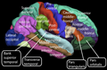
The sensory areas are the cortical areas that receive and process information from the senses. Parts of the cortex that receive sensory inputs from the thalamus are called primary sensory areas. The senses of vision, audition, and touch are served by the primary visual cortex, primary auditory cortex and primary somatosensory cortex respectively. In general, the two hemispheres receive information from the opposite (contralateral) side of the body. For example, the right primary somatosensory cortex receives information from the left limbs, and the right visual cortex receives information from the left visual field. The organization of sensory maps in the cortex reflects that of the corresponding sensing organ, in what is known as a topographic map. Neighboring points in the primary visual cortex, for example, correspond to neighboring points in the retina. This topographic map is called a retinotopic map. In the same way, there exists a tonotopic map in the primary auditory cortex and a somatotopic map in the primary sensory cortex. This last topographic map of the body onto the posterior central gyrus has been illustrated as a deformed human representation, the somatosensory homunculus, where the size of different body parts reflects the relative density of their innervation. Areas with lots of sensory innervation, such as the fingertips and the lips, require more cortical area to process finer sensation.

Motor areas

The motor areas are located in both hemispheres of the cortex. They are shaped like a pair of headphones stretching from ear to ear. The motor areas are very closely related to the control of voluntary movements, especially fine fragmented movements performed by the hand. The right half of the motor area controls the left side of the body, and vice versa.

Two areas of the cortex are commonly referred to as motor:
In addition, motor functions have been described for:
Just underneath the cerebral cortex are interconnected subcortical masses of grey matter called basal ganglia (or nuclei). The basal ganglia receive input from the substantia nigra of the midbrain and motor areas of the cerebral cortex, and send signals back to both of these locations. They are involved in motor control. They are found lateral to the thalamus. The main components of the basal ganglia are the caudate nucleus, the putamen, the globus pallidus, the substantia nigra, the nucleus accumbens, and the subthalamic nucleus. The putamen and globus pallidus are also collectively known as the lentiform nucleus, because together they form a lens-shaped body. The putamen and caudate nucleus are also collectively called the corpus striatum after their striped appearance.

Association areas

The association areas are the parts of the cerebral cortex that do not belong to the primary regions. They function to produce a meaningful perceptual experience of the world, enable us to interact effectively, and support abstract thinking and language. The parietal, temporal, and occipital lobes - all located in the posterior part of the cortex - integrate sensory information and information stored in memory. The frontal lobe or prefrontal association complex is involved in planning actions and movement, as well as abstract thought. Globally, the association areas are organized as distributed networks. Each network connects areas distributed across widely spaced regions of the cortex. Distinct networks are positioned adjacent to one another yielding a complex series of interwoven networks. The specific organization of the association networks is debated with evidence for interactions, hierarchical relationships, and competition between networks. In humans, association networks are particularly important to language function. In the past it was theorized that language abilities are localized in the left hemisphere in areas 44/45, the Broca's area, for language expression and area 22, the Wernicke's area, for language reception. However, language is no longer limited to easily identifiable areas. More recent research suggests that the processes of language expression and reception occur in areas other than just those structures around the lateral sulcus, including the frontal lobe, basal ganglia, cerebellum, and pons.

Clinical significance

There is marked cortical atrophy in Alzheimer's disease, associated with loss of gyri and sulci in the temporal lobe and parietal lobe, and parts of the frontal cortex and cingulate gyrus.

Neurodegenerative diseases such as Alzheimer's disease and Lafora disease, show as a marker, an atrophy of the grey matter of the cerebral cortex.

There are many neurodevelopmental abnormalities that can lead to a wide variety of behavioral and cognitive deficits. There are several situations in development in which both intrinsic and extrinsic factors can highly influence the course of nervous system formation. One very prominent intrinsic factor (random gene mutation) has given rise to many different classes of neurodevelopmental disorders. For example, Fragile X-Syndrome is a neurodevelopmental disease characterized by poor eye contact with others, an extreme aversion to physical/social contact, and obsessive repetition in behavioral patterns. This is an X-linked chromosomal disorder in which the FMR1 gene is found to have nearly 200 copies, instead of its intended 30. This causes the gene to become heavily methylated, which subsequently turns off expression of FMR1. Efficient functioning of this gene is known to play a role in localized protein synthesis at dendritic spines, which is essential for proper synaptogenesis and learning and memory function.

Figure idea taken from Muralidharan et al. 2013
Maternal alcohol consumption leads to death of neural crest cells which are the building blocks of certain neural tissues.

Another primary example of intrinsic neurodevelopmental deficits is Rett Syndrome, which is an X-linked single gene mutation characterized by a loss of speech and hand coordination, intellectual regression and progressive loss of motor control. This disorder is thought to arise from a mutation in the MeCP2 gene, which encodes for a transcription factor associated with chromatin remodeling. Mutations in this gene have been linked to a decreased expression of the gene that codes for BDNF (brain-derived neurotrophic factor), which is a common gene used in neurodevelopment.

Apart from intrinsic gene mutation, there are many extrinsic (environmental) factors that can greatly influence the neurodevelopmental process. One of the most commonly studied factors is the effect of maternal alcohol consumption during gestation. Infants born with defects from this factor are said to have Fetal Alcohol Spectrum Disorder (FASD). FASD is used to describe a collection of developmental disorders associated with the consumption of alcohol whilst pregnant. The spectrum includes individuals with both extreme and mild FASD which present with a variety of neurocognitive and neurobehavioral deficits. This includes deficits is sustained and focused attention, learning and memory problems, verbal processing problems, receptive language difficulties, hyperactivity, as well as a range of psychiatric impairments. Apoptotic cell death caused by an excess of glutamate activity and GABA withdrawal is thought to play a role in the loss of neuronal fibers critical for normal brain development. In addition to neuronal death, there is evidence suggesting that an improper formation of interneuronal connections and cell adhesion molecule malformation are also associated with FASD. Improper neuronal migration and synaptogenesis have been shown to occur is several cases of FASD, which could explain the extreme deficits in learning and memory.

Individuals with Fetal Alcohol Spectrum Disorder (FASD) exhibit a range of cognitive and behavioral deficits that relate heavily to neural pathologies associated with maternal alcohol consumption.

Other animals

The cerebral cortex is derived from the pallium, a layered structure found in the forebrain of all vertebrates. The basic form of the pallium is a cylindrical layer enclosing fluid-filled ventricles. Around the circumference of the cylinder are four zones, the dorsal pallium, medial pallium, ventral pallium, and lateral pallium, which are thought respectively to give rise to the neocortex, hippocampus, amygdala, and olfactory cortex.

Until recently no counterpart to the cerebral cortex had been recognized in invertebrates. However, a study published in the journal Cell in 2010, based on gene expression profiles, reported strong affinities between the cerebral cortex and the mushroom bodies of the ragworm Platynereis dumerilii. Mushroom bodies are structures in the brains of many types of worms and arthropods that are known to play important roles in learning and memory; the genetic evidence indicates a common evolutionary origin, and therefore indicates that the origins of the earliest precursors of the cerebral cortex date back to the early Precambrian era.

Additional images

Affective neuroscience

From Wikipedia, the free encyclopedia

Affective neuroscience is the study of the neural mechanisms of emotion. This interdisciplinary field combines neuroscience with the psychological study of personality, emotion, and mood. The putative existence of 'basic emotions' and their defining attributes represents a long lasting and yet unsettled issue in psychology.

Brain areas related to emotion

Emotions are thought to be related to activity in brain areas that direct our attention, motivate our behavior, and determine the significance of what is going on around us. Pioneering work by Paul Broca (1878), James Papez (1937), and Paul D. MacLean (1952) suggested that emotion is related to a group of structures in the center of the brain called the limbic system, which includes the hypothalamus, cingulate cortex, hippocampi, and other structures. Research has shown that limbic structures are directly related to emotion, but non-limbic structures have been found to be of greater emotional relevance. The following brain structures are currently thought to be involved in emotion:

Main structures of the limbic system

  • Amygdala – The amygdalae are two small, round structures located anterior to the hippocampi near the temporal poles. The amygdalae are involved in detecting and learning what parts of our surroundings are important and have emotional significance. They are critical for the production of emotion, and may be particularly so for negative emotions, especially fear. Multiple studies have shown amygdala activation when perceiving a potential threat; various circuits allow the amygdala to use related past memories to better judge the possible threat.
  • Thalamus – The thalamus is involved in relaying sensory and motor signals to the cerebral cortex, especially visual stimuli. The thalamus also plays an important role in regulating states of sleep and wakefulness.
  • Hypothalamus – The hypothalamus is involved in producing a physical output associated with an emotion as well as in reward circuits
  • Hippocampus – The hippocampus is a structure of the medial temporal lobes that is mainly involved in memory. It works to form new memories and also connecting different senses such as visual input, smell or sound to memories. The hippocampus allows memories to be stored long term and also retrieves them when necessary. It is this retrieval that is used within the amygdala to help evaluate current affective stimulus.
  • Fornix – The fornix is the main output pathway from the hippocampus to the mammillary bodies. It has been identified as a main region in controlling spatial memory functions, episodic memory and executive functions.
  • Mammillary body – Mammillary bodies are important for recollective memory.
  • Olfactory bulb – The olfactory bulbs are the first cranial nerves, located on the ventral side of the frontal lobe. They are involved in olfaction, the perception of odors.
  • Cingulate gyrus – The cingulate gyrus is located above the corpus callosum and is usually considered to be part of the limbic system. The different parts of the cingulate gyrus have different functions, and are involved with affect, visceromotor control, response selection, skeletomotor control, visuospatial processing, and in memory access. A part of the cingulate gyrus is the anterior cingulate cortex, that is thought to play a central role in attention and behaviorally demanding cognitive tasks. It may be particularly important with regard to conscious, subjective emotional awareness. This region of the brain may also play an important role in the initiation of motivated behavior. The subgenual cingulate is more active during both experimentally induced sadness and during depressive episodes.

Other brain structures related to emotion

  • Basal ganglia – Basal ganglia are groups of nuclei found on either side of the thalamus. Basal ganglia play an important role in motivation, action selection and reward learning.
  • Orbitofrontal cortex – Is a major structure involved in decision making and the influence by emotion on that decision.
  • Prefrontal cortex – The term prefrontal cortex refers to the very front of the brain, behind the forehead and above the eyes. It appears to play a critical role in the regulation of emotion and behavior by anticipating the consequences of our actions. The prefrontal cortex may play an important role in delayed gratification by maintaining emotions over time and organizing behavior toward specific goals.
  • Ventral striatum – The ventral striatum is a group of subcortical structures thought to play an important role in emotion and behavior. One part of the ventral striatum called the nucleus accumbens is thought to be involved in the experience pleasure. Individuals with addictions experience increased activity in this area when they encounter the object of their addiction.
  • Insula – The insular cortex is thought to play a critical role in the bodily experience of emotion, as it is connected to other brain structures that regulate the body’s autonomic functions (heart rate, breathing, digestion, etc.). The insula is implicated in empathy and awareness of emotion.
  • Cerebellum – Recently, there has been a considerable amount of work that describes the role of the cerebellum in emotion as well as cognition, and a "Cerebellar Cognitive Affective Syndrome" has been described. Both neuroimaging studies as well as studies following pathological lesions in the cerebellum (such as a stroke) demonstrate that the cerebellum has a significant role in emotional regulation. Lesion studies have shown that cerebellar dysfunction can attenuate the experience of positive emotions. While these same studies do not show an attenuated response to frightening stimuli, the stimuli did not recruit structures that normally would be activated (such as the amygdala). Rather, alternative limbic structures were activated, such as the ventromedial prefrontal cortex, the anterior cingulate gyrus, and the insula. This may indicate that evolutionary pressure resulted in the development of the cerebellum as a redundant fear-mediating circuit to enhance survival. It may also indicate a regulatory role for the cerebellum in the neural response to rewarding stimuli, such as money, drugs of abuse, and orgasm.

Role of the right hemisphere in emotion

The right hemisphere has been proposed over time as being directly involved in the processing of emotion. Scientific theory regarding the role of the right hemisphere has developed over time and resulted in several models of emotional functioning. C.K. Mills was one of the first researchers to propose a direct link between the right hemisphere and emotional processing, having observed decreased emotional processing in patients with lesions to the right hemisphere. Emotion was originally thought to be processed in the limbic system structures such as the hypothalamus and amygdala. As of the late 1980s to early 1990s however, neocortical structures were shown to have an involvement in emotion. These findings led to the development of the right hemisphere hypothesis and the valence hypothesis.

The right hemisphere hypothesis

The right hemisphere hypothesis asserts that the right hemisphere of the neocortical structures is specialized for the expression and perception of emotion. The Right hemisphere has been linked with mental strategies that are nonverbal, synthetic, integrative, holistic, and Gestalt which makes it ideal for processing emotion. The right hemisphere is more in touch with subcortical systems of autonomic arousal and attention as demonstrated in patients that have increased spatial neglect when damage is associated to the right brain as opposed to the left brain. Right hemisphere pathologies have also been linked with abnormal patterns of autonomic nervous system responses. These findings would help signify the relationship of the subcortical brain regions to the right hemisphere as having a strong connection.

The valence hypothesis

The valence hypothesis acknowledges the right hemisphere's role in emotion, but asserts that it is mainly focused on the processing of negative emotions whereas the left hemisphere processes positive emotions. The mode of processing of the two hemispheres has been the discussion of much debate. One version suggests the lack of a specific mode of processes, stating that the right hemisphere is solely negative emotion and the left brain is solely positive emotion. A second version suggests that there is a complex mode of processing that occurs, specifically that there is a hemispheric specialization for the expressing and experiencing of emotion, with the right hemisphere predominating in the experiencing of both positive and negative emotion. More recently, the frontal lobe has been the focus of a large amount of research, stating that the frontal lobes of both hemispheres are involved in the emotional state, while the right posterior hemisphere, the parietal and temporal lobes, is involved in the processing of emotion. Decreased right parietal lobe activity has been associated with depression and increased right parietal lobe activity with anxiety arousal. The increasing understanding of the role the different hemispheres play has led to increasingly complicated models, all based some way on the original valence model.

Relationship to cognitive neuroscience

Despite their interactions, the study of cognition has historically excluded emotion and focused on non-emotional processes (e.g., memory, attention, perception, action, problem solving and mental imagery). As a result, the study of the neural basis of non-emotional and emotional processes emerged as two separate fields: cognitive neuroscience and affective neuroscience. The distinction between non-emotional and emotional processes is now thought to be largely artificial, as the two types of processes often involve overlapping neural and mental mechanisms. Thus, when cognition is taken at its broadest definition, affective neuroscience could also be called the cognitive neuroscience of emotion.

Cognitive neuroscience tasks in affective neuroscience research

Emotion Go/No-Go

The emotion go/no-go task has been frequently used to study behavioral inhibition, particularly emotional modulation of this inhibition. A derivation of the original go/no-go paradigm, this task involves a combination of affective “go cues”, where the participant must make a motor response as quickly as possible, and affective “no-go cues,” where a response must be withheld. Because “go cues” are more common, the task is able to measure one’s ability to inhibit a response under different emotional conditions.

The task is common in tests of emotion regulation, and is often paired with neuroimaging measures to localize relevant brain function in both healthy individuals and those with affective disorders. For example, go/no-go studies converge with other methodology to implicate areas of the prefrontal cortex during inhibition of emotionally valenced stimuli.

Emotional Stroop

The emotional Stroop task, an adaptation to the original Stroop, measures attentional bias to emotional stimuli. Participants must name the ink color of presented words while ignoring the words themselves. In general, participants have more difficulty detaching attention from affectively valenced words, than neutral words. This interference from valenced words is measured by the response latency in naming the color of neutral words as compared with emotional words.

This task has been often used to test selective attention to threatening and other negatively valenced stimuli, most often in relation to psychopathology. Disorder specific attentional biases have been found for a variety of mental disorders. For example, participants with spider phobia show a bias to spider-related words but not other negatively valenced words. Similar findings have been attributed to threat words related to other anxiety disorders. However, other studies have questioned these findings. In fact, anxious participants in some studies show the Stroop interference effect for both negative and positive words, when the words are matched for emotionality. This means that the specificity effects for various disorders may be largely attributable to the semantic relation of the words to the concerns of the disorder, rather than simply the emotionality of the words.

Ekman 60 faces task

The Ekman faces task is used to measure emotion recognition of six basic emotions. Black and white photographs of 10 actors (6 male, 4 female) are presented, with each actor displaying each basic emotion. Participants are usually asked to respond quickly with the name of the displayed emotion. The task is a common tool to study deficits in emotion regulation in patients with dementia, Parkinson's, and other cognitively degenerative disorders. However, the task has also been used to analyze recognition errors in disorders such as borderline personality disorder, schizophrenia, and bipolar disorder.

Dot probe (emotion)

The emotional dot-probe paradigm is a task used to assess selective visual attention to and failure to detach attention from affective stimuli. The paradigm begins with a fixation cross at the center of a screen. An emotional stimulus and a neutral stimulus appear side by side, after which a dot appears behind either the neutral stimulus (incongruent condition) or the affective stimulus (congruent condition). Participants are asked to indicate when they see this dot, and response latency is measured. Dots that appear on the same side of the screen as the image the participant was looking at will be identified more quickly. Thus, it is possible to discern which object the participant was attending to by subtracting the reaction time to respond to congruent versus incongruent trials.

The best documented research with the dot probe paradigm involves attention to threat related stimuli, such as fearful faces, in individuals with anxiety disorders. Anxious individuals tend to respond more quickly to congruent trials, which may indicate vigilance to threat and/or failure to detach attention from threatening stimuli. A specificity effect of attention has also been noted, with individuals attending selectively to threats related to their particular disorder. For example, those with social phobia selectively attend to social threats but not physical threats. However, this specificity may be even more nuanced. Participants with obsessive-compulsive disorder symptoms initially show attentional bias to compulsive threat, but this bias is attenuated in later trials due to habituation to the threat stimuli.

Fear potentiated startle

Fear-potentiated startle (FPS) has been utilized as a psychophysiological index of fear reaction in both animals and humans. FPS is most often assessed through the magnitude of the eyeblink startle reflex, which can be measured by electromyography. This eyeblink reflex is an automatic defensive reaction to an abrupt elicitor, making it an objective indicator of fear. Typical FPS paradigms involve bursts of noise or abrupt flashes of light transmitted while an individual attends to a set of stimuli. Startle reflexes have been shown to be modulated by emotion. For example, healthy participants tend to show enhanced startle responses while viewing negatively valenced images and attenuated startle while viewing positively valenced images, as compared with neutral images.

The startle response to a particular stimulus is greater under conditions of threat. A common example given to indicate this phenomenon is that one’s startle response to a flash of light will be greater when walking in a dangerous neighborhood at night than it would under safer conditions. In laboratory studies, the threat of receiving shock is enough to potentiate startle, even without any actual shock.

Fear potentiated startle paradigms are often used to study fear learning and extinction in individuals with posttraumatic stress disorder and other anxiety disorders. In fear conditioning studies, an initially neutral stimulus is repeatedly paired with an aversive one, borrowing from classical conditioning. FPS studies have demonstrated that PTSD patients have enhanced startle responses during both danger cues and neutral/safety cues as compared with healthy participants.

Learning

There are many ways affect plays a role during learning. Recently, affective neuroscience has done much to discover this role. Deep, emotional attachment to a subject area allows a deeper understanding of the material and therefore, learning occurs and lasts. When reading, the emotions one is feeling in comparison to the emotions being portrayed in the content affects ones comprehension. Someone who is feeling sad will understand a sad passage better than someone feeling happy. Therefore, a student’s emotion plays a big role during the learning process.
Emotion can also be embodied or perceived from words read on a page or a person’s facial expression. Neuroimaging studies using fMRI have demonstrated that the same area of the brain being activated when one is feeling disgust is also activated when one observes another person feeling disgust. In a traditional learning environment, the teacher's facial expression can play a critical role in students' language acquisition. Showing a fearful facial expression when reading passages that contain fearful tones facilitates students learning of the meaning of certain vocabulary words and comprehension of the passage.

Meta-analyses

A meta-analysis is a statistical approach to synthesizing results across multiple studies. Several meta-analyses examining the brain basis of emotion have been conducted. In each meta-analysis, studies were included that investigate healthy, unmedicated adults and that used subtraction analysis to examine the areas of the brain that were more active during emotional processing than during a neutral control condition. The meta-analyses to date predominantly focus on two theoretical approaches, locationist approaches and psychological construction approaches.

It is being debated regarding the existence of the neurobiological basis of emotion. The existence of so-called 'basic emotions' and their defining attributes represents a long lasting and yet unsettled issue in psychology.[2] The available research suggests that the neurobiological existence of basic emotions is still tenable and heuristically seminal, pending some reformulation.

Locationist approaches

These approaches to emotion hypothesize that several emotion categories (including happiness, sadness, fear, anger, and disgust) are biologically basic. In this view, emotions are inherited biologically based modules that cannot be broken down into more basic psychological components. Models following a locationist approach to emotion hypothesize that all mental states belonging to a single emotional category can be consistently and specifically localized to either a distinct brain region or a defined networks of brain regions. Each basic emotion category also shares other universal characteristics: distinct facial behavior, physiology, subjective experience and accompanying thoughts and memories.

Psychological constructionist approaches

This approach to emotion hypothesizes that emotions like happiness, sadness, fear, anger and disgust (and many others) are constructed mental states that occur when many different systems in the brain work together. In this view, networks of brain regions underlie psychological operations (e.g., language, attention, etc.) that interact to produce many different kinds of emotion, perception, and cognition. One psychological operation critical for emotion is the network of brain regions that underlie valence (feeling pleasant/unpleasant) and arousal (feeling activated and energized). Emotions emerge when neural systems underlying different psychological operations interact (not just those involved in valence and arousal), producing distributed patterns of activation across the brain. Because emotions emerge from more basic components, there is heterogeneity within each emotion category; for example, a person can experience many different kinds of fear, which feel differently, and which correspond to different neural patterns in the brain. Thus, this view presents a different approach to understanding the neural bases of emotion than locationist approaches.

Phan et al. 2002

In the first neuroimaging meta-analysis of emotion, Phan et al. (2002) analyzed the results of 55 studies published in peer reviewed journal articles between January 1990 and December 2000 to determine if the emotions of fear, sadness, disgust, anger, and happiness were consistently associated with activity in specific brain regions. All studies used fMRI or PET techniques to investigate higher-order mental processing of emotion (studies of low-order sensory or motor processes were excluded). The authors’ analysis approach was to tabulate the number of studies that reported activation in specific brain regions during tasks inducing fear, sadness, disgust, anger, and happiness. For each brain region, statistical chi-squared analysis was conducted to determine if the proportion of studies reporting activation during one emotion was significantly higher than the proportion of studies reporting activation during the other emotions. Two regions showed this statistically significant pattern across studies. In the amygdala, 66% of studies inducing fear reported activity in this region, as compared to ~20% of studies inducing happiness, ~15% of studies inducing sadness (with no reported activations for anger or disgust). In the subcallosal cingulate, 46% of studies inducing sadness reported activity in this region, as compared to ~20% inducing happiness and ~20% inducing anger. This pattern of clear discriminability between emotion categories was in fact rare, with a number of other patterns occurring in limbic regions (including amydala, hippocampus, hypothalamus, and orbitofrontal cortex), paralimbic regions (including subcallosal cingulate, medial prefrontal cortex, anterior cingulate cortex, posterior cingulate cortex, insula, and temporal pole), and uni/heteromodal regions (including lateral prefrontal cortex, primary sensorimotor cortex, temporal cortex, cerebellum, and brainstem). Brain regions implicated across discrete emotion included the basal ganglia (~60% of studies inducing happiness and ~60% of studies inducing disgust reported activity in this region) and medial prefrontal cortex (happiness ~60%, anger ~55%, sadness ~40%, disgust ~40%, and fear ~30%).

Murphy et al. 2003

Murphy, et al. 2003 analyzed 106 peer reviewed journals published between January 1994 and December 2001 to examine the evidence for regional specialization of discrete emotions (fear, disgust, anger, happiness and sadness) across a larger set of studies that Phan et al. Studies included in the meta-analysis measured activity in the whole brain and regions of interest (activity in individual regions of particular interest to the study). 3-D Kolmogorov-Smirnov (KS3) statistics were used to compare rough spatial distributions of 3-D activation patterns to determine if statistically significant activations (consistently activated across studies) were specific to particular brain regions for all emotional categories. This pattern of consistently activated, regionally specific activations was identified in four brain regions: amygdala with fear, insula with disgust, globus pallidus with disgust, and lateral orbitofrontal cortex with anger. The amygdala was consistently activated in ~40% of studies inducing fear, as compared to less than 20% studies inducing happiness, sadness, or anger. The insula was consistently activated in ~ 70% of studies inducing disgust, as compared to sadness (~40%), anger (~20%), fear (~20%), and happiness (~10%). Similar to the insula, the globus pallidus was consistently activated in ~70% of studies inducing disgust, as compared to less than 25% of studies inducing sadness, fear, anger or happiness. The lateral orbitofrontal cortex was consistently activated in over 80% of studies inducing anger, as compared to fear (~30%), sadness (~20%), happiness (< 20%) and disgust (< 20%). Other regions showed different patterns of activation across categories. For example, both the dorsal medial prefrontal cortex and the rostral anterior cingulate cortex showed consistent activity across emotions (happiness ~50%, sadness ~50%, anger ~ 40%, fear ~30%, and disgust ~ 20%).

Barrett et al. 2006

Barrett, et al. 2006 examined 161 studies published between 1990-2001, subsets of which were analyzed in previous meta-analyses (Phan, et al. 2002 and Murphy et al. 2003). In this review, the authors examined the locationist hypothesis by comparing the consistency and specificity of prior meta-analytic findings specific to each hypothesized basic emotion (fear, anger, sadness, disgust, and happiness). Consistent neural patterns were defined by brain regions showing increased activity for a specific emotion (relative to a neutral control condition), regardless of the method of induction used (for example, visual vs. auditory cue). Specific neural patterns were defined as architecturally separate circuits for one emotion vs. the other emotions (for example, the fear circuit must be discriminable from the anger circuit, although both circuits may include common brain regions). In general, the results supported consistency among the findings of Phan et al. and Murphy et al., but not specificity. Consistency was determined through the comparison of chi-squared analyses that revealed whether the proportion of studies reporting activation during one emotion was significantly higher than the proportion of studies reporting activation during the other emotions. Specificity was determined through the comparison of emotion-category brain-localizations by contrasting activations in key regions that were specific to particular emotions. Increased amygdala activation during fear was the most consistently reported across induction methods (but not specific). Both meta-analyses also reported increased activations in regions of the anterior cingulate cortex during sadness, although this finding was less consistent (across induction methods) and was not specific to sadness. Both meta-analyses also found that disgust was associated with increased activity in the basal ganglia, but these findings were neither consistent nor specific. Neither consistent nor specific activity was observed across the meta-analyses for anger or for happiness. This meta-analysis additionally introduced the concept of the basic, irreducible elements of emotional life as dimensions such as approach and avoidance. This dimensional approach involved in psychological constructionist approaches is further examined in later meta-analyses of Kober et al. 2008 and Lindquist et al. 2012.

Kober et al. 2008

Instead of investigating specific emotions, Kober, et al. 2008 reviewed 162 neuroimaging studies published between 1990-2005 to determine if groups of brain regions show consistent patterns of activation during emotional experience (that is, actively experiencing an emotion first-hand) and during emotion perception (that is, perceiving a given emotion as experienced by another). This meta-analysis used multilevel kernal density analysis (MKDA) to examine fMRI and PET studies, a technique that prevents single studies from dominating the results (particularly if they report multiple nearby peaks) and that enables studies with large sample sizes (those involving more participants) to exert more influence upon the results. MKDA was used to establish a neural reference space that includes the set of regions showing consistent increases across all studies (for further discussion of MDKA see Wager et al. 2007). Next, this neural reference space was partitioned into functional groups of brain regions showing similar activation patterns across studies by first using multivariate techniques to determine co-activation patterns and then using data-reduction techniques to define the functional groupings (resulting in six groups). Consistent with a psychological construction approach to emotion, the authors discuss each functional group in terms more basic psychological operations. The first “Core Limbic” group included the left amygdala, hypothalamus, periaqueductal gray/thalamus regions, and amygdala/ventral striatum/ventral globus pallidus/thalamus regions, which the authors discuss as an integrative emotional center that plays a general role in evaluating affective significance. The second “Lateral Paralimbic” group included the ventral anterior insula/frontal operculum/right temporal pole/ posterior orbitofrontal cortex, the anterior insula/ posterior orbitofrontal cortex, the ventral anterior insula/ temporal cortex/ orbitofrontal cortex junction, the midinsula/ dorsal putamen, and the ventral striatum /mid insula/ left hippocampus, which the authors suggest plays a role in motivation, contributing to the general valuation of stimuli and particularly in reward. The third “Medial Prefrontal Cortex” group included the dorsal medial prefrontal cortex, pregenual anterior cingulate cortex, and rostral dorsal anterior cingulate cortex, which the authors discuss as playing a role in both the generation and regulation of emotion. The fourth “Cognitive/ Motor Network” group included right frontal operculum, the right interior frontal gyrus, and the pre-supplementray motor area/ left interior frontal gyrus, regions that are not specific to emotion, but instead appear to play a more general role in information processing and cognitive control. The fifth “Occipital/ Visual Association” group included areas V8 and V4 of the primary visual cortex, the medial temporal lobe, and the lateral occipital cortex, and the sixth “Medial Posterior” group included posterior cingulate cortex and area V1 of the primary visual cortex. The authors suggest that these regions play a joint role in visual processing and attention to emotional stimuli.

Vytal et al. 2010

Vytal, et al. 2010 examined 83 neuroimaging studies published between 1993-2008 to examine whether neuroimaging evidence supports the idea of biologically discrete, basic emotions (i.e. fear, anger, disgust, happiness, and sadness). Consistency analyses identified brain regions that were associated with a given emotion. Discriminability analyses identified brain regions that were significantly, differentially active when contrasting pairs of discrete emotions. This meta-analysis examined PET or fMRI studies that reported whole brain analyses identifying significant activations for at least one of the five emotions relative to a neutral or control condition. The authors used activation likelihood estimation (ALE) to perform spatially sensitive, voxel-wise (sensitive to the spatial properties of voxels) statistical comparisons across studies. This technique allows for direct statistical comparison between activation maps associated with each discrete emotion. Thus, discriminability between the five discrete emotion categories was assessed on a more precise spatial scale than what had been accomplished in prior meta-analyses. Consistency was first assessed by comparing the ALE map generated across studies for each emotion (for example, the ALE map identifying regions consistently activated by studies inducing fear) to ALE map generated by random permutations. Discriminability was then assessed by pair-wise contrasts of individual emotion ALE maps (for example, fear ALE map vs. anger ALE map; fear ALE map vs. disgust map) across all basic emotions pairings. Consistent and discriminable patterns of neural activation were observed for the five emotional categories. Happiness was consistently associated with activity in 9 regional brain clusters, the largest located in the right superior temporal gyrus. For the first time, happiness was discriminated from the other emotional categories, with the largest clusters of activity specific to happiness (vs. the other emotion categories) located in right superior temporal gyrus and left rostral anterior cingulate cortex. Sadness was consistently associated with 35 clusters (the largest activation cluster located in the left medial frontal gyrus) and was discriminated from the other emotion categories by significantly greater activity in left medial frontal gyrus, right middle temporal gyrus, and right inferior frontal gyrus. Anger was consistently associated with activity in 13 clusters (the largest of which was located in the left inferior frontal gyrus), and was discriminated from the other emotion categories by significantly greater activity in bilateral inferior frontal gyrus, and in right parahippocampal gyrus. Fear was consistently associated with 11 clusters (the largest activation cluster in the left amygdala) and was discriminated from the other emotion categories by significantly greater activity in the left amygdala and left putamen. Disgust was consistently activated with 16 clusters (the largest activation cluster in the right insula/ right inferior frontal gyrus) and was discriminated from the other emotion categories by significantly greater activity in the right putamen and the left insula.

Lindquist et al. 2012

Lindquist, et al. 2012 reviewed 91 PET and fMRI studies published between January 1990 and December 2007. The studies included in this meta-analysis used induction methods that elicit emotion experience or emotion perception of fear, sadness, disgust, anger, and happiness. The goal was to compare locationist approaches with psychological constructionist approaches to emotion. Similar to Kober et al. described above, a Multilevel Peak Kernel Density Analysis transformed the individual peak activations reported across study contrasts into a neural reference space (in other words, the set of brain regions consistently active across all study contrasts assessing emotion experience or perception). The density analysis was then used to identify voxels within the neural reference space with more consistent activations for a specific emotion category (anger, fear, happiness, sadness, and disgust) than all other emotions. Chi-squared analysis was used to create statistical maps that indicated if each previously identified and consistently active regions (those identified during density analysis) were more frequently activated in studies of each emotion category versus the average of all other emotions, regardless of activations elsewhere in the brain. Chi-squared analysis and density analysis both defined functionally consistent and selective regions, or regions which showed a relatively more consistent increase in activity for the experience or perception of one emotion category across studies in the literature. Thus, a selective region could present increased activations relatively more so to one emotion category while also having a response to multiple other emotional categories. A series of logistic regressions were then performed to identify if any of the regions that were identified as consistent and selective to an emotion category were additionally specific to a given category. Regions were defined as specific to a given emotion if they showed increased activations for only one emotional category, and never showed increased activity during instances of the other emotional categories. In other words, a region could be defined as consistent, selective and specific for e.g. fear perception if it only showed significantly greater increases in activation during the perception of fear and did not show increased activity during any other emotion categories. However, the same region would be defined as only consistent and selective (and not specific) to fear perception if it additionally displayed increased activations during anger perception. Strong support for the locationist approach was defined as evidence that basic emotion categories (anger, disgust, fear, happiness and sadness) consistently map onto areas of the brain that specifically activate in response to instances of only one emotional category. Strong support for the constructionist approach was defined as evidence that multiple psychological operations (some of which are not specific or selective to emotion) consistently occur across many brain regions and multiple emotional categories.

The results indicated that many brain regions demonstrated consistent and selective activations in the experience or perception of an emotion category (versus all the other emotion categories). Consistent with constructionist models, however, no region demonstrated functional specificity for the emotions of fear, disgust, happiness, sadness or anger. Based on the existing scientific literature, the authors proposed different roles for the brain regions that have traditionally been associated with only one emotion category. The authors propose that the amygdala, anterior insula, orbitofrontal cortex each contribute to “core affect,” which are basic feelings that are pleasant or unpleasant with some level of arousal. The amygdala, for example, appears to play a more general role in indicating if external sensory information is motivationally salient, and is particularly active when a stimulus is novel or evokes uncertainty. The anterior insula may represent core affective feelings in awareness across a number of emotion categories, driven largely by sensations originating in the body. The orbitofrontal cortex appears to function as a site for integrating sensory information from the body and sensory information from the world to guide behavior. Closely related to core affect, the authors propose that anterior cingulate and dorsolateral prefrontal cortex play vital roles in attention, with anterior cingulate supporting the use of sensory information for directing attention and motor responses during response selection and with dorsolateral prefrontal cortex supporting executive attention. In many psychological construction approaches, emotions also involve the act of interpreting one’s situation in the world relative to the internal state of the body, or what is referred to as “conceptualization.” In support of this idea, the dorsomedial prefrontal cortex and hippocampus were consistently active in this meta-analysis, regions that appear to play an important role conceptualizing during emotion, which are also involved in simulating previous experience (e.g. knowledge, memory). Language is also central to conceptualizing, and regions that support language, including ventrolateral prefrontal cortex, were also consistently active across studies of emotion experience and perception.

Mandatory Palestine

From Wikipedia, the free encyclopedia https://en.wikipedia.org/wiki/Mandatory_Palestine   Palestine 1920–...