Search This Blog

Friday, July 10, 2020

Diseconomies of scale

From Wikipedia, the free encyclopedia
 
In microeconomics, diseconomies of scale are the cost disadvantages that economic actors accrue due to an increase in organizational size or in output, resulting in production of goods and services at increased per-unit costs. The concept of diseconomies of scale is the opposite of economies of scale. In business, diseconomies of scale are the features that lead to an increase in average costs as a business grows beyond a certain size.

economies of scale
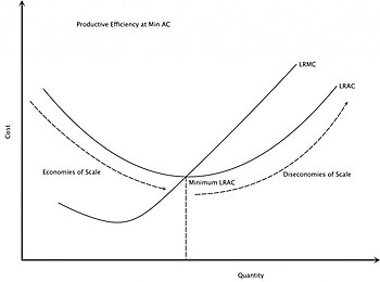
The rising part of the long-run average cost curve illustrates the effect of diseconomies of scale. The Long Run Average Cost (LRAC) curve plots the average cost of producing the lowest cost method. The Long Run Marginal Cost (LRMC) is the change in total cost attributable to a change in the output of one unit after the plant size has been adjusted to produce that rate of output at minimum LRAC.

Causes

Communication costs

Ideally, all employees of a firm would have one-on-one communication with each other so they know exactly what the other workers are doing. A firm with a single worker does not require any communication between employees. A firm with two workers requires one communication channel, directly between those two workers. A firm with three workers requires three communication channels between employees (between employees A & B, B & C, and A & C). Here is a chart of one-on-one communication channels required:

Workers Communication Channels
1 0
2 1
3 3
4 6
5 10
n

The graph of all one-on-one channels is a complete graph.

The number of one-on-one channels of communication grows more rapidly than the number of workers, thus increasing the time and costs of communication. At some point one-on-one communications between all workers becomes impractical; therefore only certain groups of employees will communicate with one another (e.g. within departments or within geographical locations). This reduces, but does not stop, the increase in unit costs; and also the organisation will incur some inefficiencies due to the reduced level of communication.

Duplication of effort

An organisation with just one person cannot have any duplication of effort between employees. If there are two employees, there could be some duplication of efforts, but this is likely to be minor, as each of the two will generally know what the other is working on. When organisations grow to thousands of workers, it is inevitable that someone, or even a team, will take on a function that is already being handled by another person or team. In colloquial terms, this is described as "one hand not knowing what the other hand is doing". General Motors, for example, developed two in-house CAD/CAM systems: CADANCE was designed by the GM Design Staff, while Fisher Graphics was created by the former Fisher Body division. These similar systems later needed to be combined into a single Corporate Graphics System, CGS, at great expense. A smaller firm would have had neither the money to allow such expensive parallel developments, nor the lack of communication and cooperation which precipitated this event. In addition to CGS, GM also used CADAM, UNIGRAPHICS, CATIA and other off-the-shelf CAD/CAM systems, thus increasing the cost of translating designs from one system to another. This endeavor eventually became so unmanageable that they acquired (and then eventually sold off) Electronic Data Systems (EDS) in an effort to control the situation. Smaller firms typically choose a single off-the-shelf CAD/CAM system, with no need to combine or translate between systems.

Office politics

"Office politics" is management behavior which a manager knows is counter to the best interest of the company, but is in his personal best interest. For example, a manager might intentionally promote an incompetent worker, knowing that the worker will never be able to compete for the manager's job. This type of behavior only makes sense in a company with multiple levels of management. The more levels there are, the more opportunity for this behavior. In a small company, such behavior could cause the company to go bankrupt, and thus cost the manager his job, so he would not make such a decision. In a large company, one manager would not have much effect on the overall health of the company, so such "office politics" are in the interest of individual managers.

Top-heavy companies

As an organisation increases in size, it becomes costly to keep control of a sprawling corporate empire, and this often results in bureaucracy as executives implement more and more levels of management. As firms increase in size, managers will initially provide a net benefit to the firm and increase productivity; however, as a firm grows and covers a larger geographical area and/or employs more people, a principal–agent problem arises, leading to lower productivity. To counter this, executives introduce standards and controls in order to maintain productivity, and this necessitates the hiring of more managers to apply these standards and controls, hence the proportion of managerial to working class begins to lean towards managerial and the company becomes "top-heavy". However, these additional managers are not providing additional output: they are spending their time implementing standards and carrying out supervision that is unnecessary in smaller firms, hence the cost-per-unit has increased.

Supply-chain disruption

Global emergencies, such as COVID-19 in 2020, can easily disrupt supply chains. This disruption has a higher chance of affecting large organizations - especially when there is only a few large suppliers. Smaller organizations with robust, local supply networks can manage supply chain shocks because any localized shock has a smaller effect on the overall ecosystem.

Other effects which reduce competitiveness of large firms

These do not always increase the cost-per-unit, but do reduce the ability of a large firm to compete.

Cannibalization

A small firm only competes with other firms, but larger firms frequently find their own products are competing with each other. A Buick was just as likely to steal customers from another GM make, such as an Oldsmobile, as it was to steal customers from other companies. This may help to explain why Oldsmobiles were discontinued after 2004. This self-competition wastes resources that should be used to compete with other firms.

Isolation of decision-makers from the results of their decisions

If a single person makes and sells donuts and decides to try jalapeƱo flavoring, they would likely know on the same day whether their decision was good or not, based on the reaction of customers. A decision-maker at a huge company that makes donuts may not know for many months if such a decision is embraced by consumers or if it is rejected, especially if their research or marketing team fails to respond in a timely manner. By that time, the decision-makers may very well have moved on to another division or company and thus see no consequence from their decision. This lack of consequences can lead to poor decisions and cause an upward-sloping average cost curve.

Slow response time

In a reverse example, the smaller firm will know immediately if people begin to request other products, and be able to respond the next day. A large company would need to do research, create an assembly line, determine which distribution chains to use, plan an advertising campaign, etc., before any changes could be made. By this time, the smaller competitors may well have grabbed that market niche.

Inertia (Unwillingness to change)

This will be defined as the "we've always done it that way, so there's no need to ever change" attitude (see appeal to tradition). An old, successful company is far more likely to have this attitude than a new, struggling one. While "change for change's sake" is counter-productive, refusal to consider change, even when indicated, is likewise toxic to a company, as changes in the industry and market conditions will inevitably demand changes in the firm in order to remain successful. An example is Polaroid Corporation's delay in moving into digital imaging, which adversely affected the company, ultimately leading to bankruptcy.

Public and government opposition

Such opposition is largely a function of the size of the firm. Behavior from Microsoft, which would have been ignored from a smaller firm, was seen as an anti-competitive and monopolistic threat, due to Microsoft's size, thus bringing about government lawsuits.

Large market share

A small company with only a 1% market share could relatively easily double market share, and hence revenues, in a year. A large company with 50% market share will find it difficult to do so.

Large market portfolio

A small investment fund can potentially yield a higher return because it can concentrate its investments in a small number of good opportunities without driving up the purchase price as they buy in, and later sell them without driving down the sale price as they sell off. Conversely, a large investment fund must spread its investments among so many securities that its results tend to track those of the market as a whole. As the size of the market controlled grows, the results will be closer to market average.

Inelasticity of supply

A company which is heavily dependent on a resource supply of a fixed or relatively-fixed size will have trouble increasing production. For instance, a timber company cannot increase production above the sustainable harvest rate of its land (although it can still increase production by acquiring more land). Similarly, service companies are limited by available labor (and thus tend to concentrate in large, densely-populated metropolitan areas); STEM (science, technology, engineering, and mathematics) professions are often-cited examples.

Reputation

Larger firms have a reputation to uphold and as a result may place more restrictions on employees, limiting their efficiency. This will be seen amplified in a regulated industry, where a company losing its license would be an extremely serious event.

Other effects related to size

Large firms also tend to be old and in mature markets. Both of these have negative implications for future growth. Old firms tend to have a large retiree base, with high associated pension and health costs, and also tend to be unionized, with associated higher labor costs and lower productivity. Mature markets tend to only offer the potential for small, incremental growth. (Everybody might go out and buy a new invention next year, but it is unlikely they will all buy cars next year, since most people already have them.)

Impact on smaller firms

While diseconomies of scale are typically associated with large mature firms, similar problems have been observed in the growth phase of small and medium-sized manufacturing companies. Mclean has observed that this can occur once the workforce exceeds around 20 employees. At this point business complexity grows more rapidly than revenue. The business experiences falling productivity, leading to rising variable costs along with rapidly rising overheads.

Solutions

Solutions to the diseconomies of scale for large firms may involve splitting the company into smaller organisations. This can either happen by default when the company is in financial difficulties, sells off its profitable divisions and shuts down the rest; or can happen proactively, if the management is willing. 

To avoid the negative effects of diseconomies of scale, a firm must stick to the lowest average output cost and try to recognise any external diseconomies of scale. Moreover, on reaching the lowest average cost, a firm must either expand to other countries to increase demand for its products, or seek new markets or produce new products that do not compete with its original products. However, neither of these actions will necessarily eliminate communications and management problems often associated with large organisations.

A systematic analysis and redesign of business processes, in order to reduce complexity, can counter diseconomies of scale. (Of course, this phase of analysis and revamping in itself can be, and usually is, a diseconomy leading to hiring of new personnel and investment in new, competing systems.) This leads to increased productivity. Improved management systems and more effective control of labor and operations can lower overhead.

Example

Returning to the example of the large donut firm, each retail location could be allowed to operate relatively autonomously from the company headquarters.

For instance, the local management may decide on the following factors instead of relying on the central management:
  1. Employee decisions such as hiring, firing, promotions and wage scales, where the local management is directly involved and likely to have better understanding of each employee. For instance, employers may choose to offer higher wages and charge higher prices if they are in an affluent area.
  2. Purchasing decisions, with each location allowed to choose its own suppliers, which may or may not be owned by the corporation (wherever they find the best quality and prices).
  3. Research and marketing decisions. Each firm may decide to develop their own recipes or utilise different signature flavour unique to their locale. For instance, when fresh apple cider is available at bargain prices from local farmers in October, they may choose to market a cinnamon donut and hot apple cider combo.
While a single, large, centrally-controlled firm may have higher ability to innovate and develop or market new products more effectively than when its resources are divided, it may lack the flexibility to offer individual customizations. Allowing the different retail locations to make decisions independent of the central management may allow them to meet local consumers' demands more efficiently. 

In addition, if the employees own a portion of the local business, employees will also have a more vested interest in its success. 

Note that all these changes will likely result in a substantial reduction in corporate headquarters staff and other support staff. For this reason, many businesses delay such a reorganization until it is too late to be effective. However, the whole company incurs reputation and legal risks arising from each unit.

Development of the cerebral cortex

From Wikipedia, the free encyclopedia
 
Corticogenesis is the process in which the cerebral cortex of the brain is formed during the development of the nervous system. The cortex is the outer layer of the brain and is composed of up to six layers. Neurons formed in the ventricular zone migrate to their final locations in one of the six layers of the cortex. The process occurs from embryonic day 10 to 17 in mice and between gestational weeks seven to 18 in humans.


Visualization of corticogenesis in the mouse. The 6 cortex layers migrate from the ventricular zone through the subplate to come to rest in the cortical plate (layers 2 through 6) or in the marginal zone (layer 1)

Cortical plates and zones

Plates

The preplate is the first stage in corticogenesis prior to the development of the cortical plate. The preplate is located between the pia and the ventricular zone. According to current knowledge, the preplate contains the first-born or pioneer neurons. These neurons are mainly thought to be Cajal-Retzius cells. The preplate also contains the predecessor to the subplate, which is sometimes referred to as a layer. As the cortical plate appears, the preplate separates into two components. The Cajal-Retzius cells go into the marginal zone, above the cortical plate, while the subplate moves to below the 6 cortical layers. It is during this transition from preplate to cortical plate when many malformations may arise.

The cortical plate is the final plate formed in corticogenesis. It includes the cortex layers two through six.

The subplate is located beneath the cortical plate. It is named for both its location relative to the cortical plate and for the time frame in which it is created. While cortical plate matures, the cells located in the subplate establish connections with neurons that have not yet moved to their destination layer within the cortical plate. Pioneer cells are also present in the subplate and work to create fibers and synapses within the plate.

Zones

The intermediate zone is located between the ventricular zone and the cortical plate. The white matter in this area is where neurons, that are created in the ventricular zone, migrate through in order to reach the cortical plate. This zone is only present during corticogenesis and eventually transforms into adult white matter.

The ventricular and subventricular zones exist below the intermediate zone and communicate to other zones through cell signalling, also creating neurons destined to migrate to other areas in the cortex.

The marginal zone, along with the cortical zone, make up the 6 layers that form the cortex. This zone is the predecessor for layer 1 of the cortex. Astrocytes form an outer limiting membrane to interact with the pia. In humans it has been found that the cells here also form a subpial layer. Cajal-Retzius cells are also present in this zone and release reelin along the radial axis, a key to proper neuronal migration during corticogenesis.

Formation of layers

The cerebral cortex is divided into layers. Each layer is formed by radial glial cells located in the ventricular zone or subventricular zone, and then migrate to their final destination.

Layer I

Layer I, the molecular layer, is the first cortical layer produced during neurogenesis at mouse E10.5 to E12.5. Of the six layers found within the neocortex, layer I is the most superficial composed of Cajal–Retzius cells and pyramidal cells. This layer is unique in the aspect that these cells migrate to the outer edge of the cortex opposed to the migration experienced by the other 5 layers. Layer one is also characterized by expression of reelin, transcription factor T-box brain 1, and cortical migratory neuronal marker.

Layers 2 and 3

The second and third layers, or the External Granular layer and External Pyramidal layer respectively, are formed around mouse E13.5 to E16. These layers are the last to form during corticogenesis and include pyramidal neurons, astrocytes, Stellates, and radial glial cells. The pyramidal and stellate neurons express SATB2 and CUX1. SATB2 and CUX1 are DNA binding proteins involved in determining the fate of cortical cells.

Layers 4, 5 and 6

The fourth, fifth and sixth layers, or the Internal Granular layer, Internal Pyramidal layer, and Polymorphic or Multiform layer respectively, are formed during mouse E11.5 to E14.5. Included in these layers are stellates, radial glia, and pyramidal neurons. Layer six is adjacent to the ventricular zone. During the production of these layers, transcription factors TBR1 and OTX1 are expressed along with CTIP2, or corticoneuronal zinc finger protein.

Neuronal migration

Neuronal migration plays significant role in corticogenesis. Throughout the process of creating the six cortical layers, all the neurons and cells migrate from the ventricular zone, through the subplate, and come to rest at their appropriate layer of the cortex. Neuronal migration is generally subdivided into radial migration, tangential migration and multipolar migration. The migration of subcortical brain functions to the cortex is known as corticalization.

Cell signaling

Appropriate formation of the cerebral cortex relies heavily on a densely intertwined network of multiple signaling pathways and distinct signaling molecules. While the majority of the process remains to be understood, some signals and pathways have been carefully unraveled in an effort to gain full knowledge of the mechanisms that control corticogenesis.

Reelin-DAB1 pathway

The Reelin-DAB1 pathway is a well-defined pathway involved in corticogenesis. Cajal-Retzius cells located in the marginal zone secrete reelin to start the cascade. Reelin is able to interact with specific neurons in the cortical plate and direct these neurons to their proper locations. It is thought that the result downstream from this signalling could influence the cytoskeleton. Reelin is secreted only by the Cajal-Retzius cells located in the marginal zone, and its receptors are confined to the cortical plate. This segregation could be used to understand the actions of Reelin.

DAB1 is a regulator protein downstream of the reelin receptors. This protein is located inside cells residing in the ventricular zone, displaying highest concentrations in migrating pyramidal cells. When either reelin or DAB1 are inactivated in mice, the resulting phenotypes are the same. In this case, the neurons are unable to migrate properly through the cortical plate. It does not affect the proliferation of neurons and in the wild does not seem to have detrimental effects on memory or learning.

Sonic hedgehog

Knocking out the Sonic hedgehog, or Shh, has been shown to severely affect corticogenesis in the genetically modified mice. The ventral and dorsal sides of the cerebrum are affected as Shh expresses the transcription factors to Nkx2 which is important in patterning the cortex. Shh is also important to corticogenesis as it affects cell proliferation and differentiation, helping neuronal progenitor cells in fate determination.

Bmp-7

Bone morphogenetic protein 7 (Bmp-7), is an important regulator in corticogenesis, though it is not understood whether it promotes or inhibits neurogenesis. Bmp-7 can be detected in the ventricular zone and is secreted into cerebrospinal fluid (CSF). The CSF is an area to promote neurogenesis and it is believed that the synergy between Bmp-7 and other regulators promote cell division along with homeostasis.

Other bone morphogenetic proteins are also known to impact corticogenesis. Bmp2, 4, 5, and 6 are expressed during the process and can compensate for one another. For example, if Bmp-4 was absent from corticogenesis, very little would change in the cortex phenotype, due to the other Bmps helping accomplish the tasks of Bmp-4. However, Bmp-7 is the only Bmp that promotes radial glia survival and therefore considered more important.

Cdk5-p35 pathway

Cdk5 has a pathway parallel to the Reelin-DAB1. This pathway affects the neuronal positioning, and results in similar malformations when absent as the Reelin or DAB1 malformations except that migration is affected at an earlier stage on the cortical plate. Cdk5/p35 pathway is also responsible for actin and microtubule dynamics involved in neuronal migration.

Cyclin-dependent kinase inhibitor 1C, or p57, also affects corticogenesis. It has been shown the p57 induces cells to exit from the cell cycle and begin differentiation, but it is dependent on Cdks. p57 is able to induce neuronal progenitor cells to start differentiating into highly specialized neurons in the cortex. However, the mechanism by which p57 is able to affect such control is not yet known.

Other signals

Besides the ones listed above, there are several more signals that affect corticogenesis. Cnr1 is a g protein receptor that is widely expressed throughout the brain, and in interneurons. In knockout mice, the cortex exhibited decreased immunoreactivity. Nrp1, Robo1, and Robo2 have also been shown to be present and important in the development of interneurons. Cdh8 is known to be expressed in the intermediate and subventricular zone, though not in specific neurons in that area, and it is suggested to regulate fiber releasing.

Disorders

Lissencephaly

Lissencephaly, or 'smooth brain', is a disorder in which the brain does not properly form the gyri and sulci as a result from neuronal migration and cortical folding. This disorder can also result in epilepsy and cognitive impariment. Type 1 lissencephaly is due to an error in migration. LISI, also known as PAFAH1B, is expressed in both dividing and migrating cells found in the brain. When LIS1 is deleted, lissencephaly occurs.

LIS1 is thought to have several important roles in the creation of the cortex. Since LIS1 is similar to the nuclear distribution protein F (nudF), they are thought to work similarly. The nud family is known to be a factor in nuclear translocation, or moving the nuclei of daughter cells after cell division has occurred. By relation, it is thought that LIS1 is a factor in neuronal migration. LIS1 is also considered to be a factor in controlling dynein, a motor protein that affects intercellular movement such as protein sorting and the process of cell division.

Another protein that contributes to a lissencephaly disorder is DCX, or Doublecortin. DCX is a microtubule associated protein that is responsible for double cortex malformations. DCX is found in the second layer of the cortex, and in fact is still present in immature neurons of an adult cortex. It is thought that DCX affects neuronal migration through affecting the microtubule dynamics. Since DCX malformations results as a similar phenotype as with LIS1 malformations, it is thought they interact with one another on a cellular level. However, it is not yet known how this occurs.

Tsc1 knockout

TSC, or tuberous sclerosis, is an autosomal dominate disorder. TSC1 or TSC2 inactivation can cause TSC and the associated tumors in the brain. When inactivation of TSC1 is present during corticogenesis, malformations of cortical tubers, or abnormal benign tissue growth, along with white matter nodes would form in mice. This replicates the effect TSC is found to have in humans afflicted with TSC. In the mice there would be a lack of GFAP in astrocytes however astrogliosis would not occur like in the human TSC.

Human Cortex Malformation (Overfolding)

The sodium channel SCN3A has been implicated in cortical malformations.

Recapitulation

Recapitulation of corticogenesis in both human and mouse embryos have been accomplished with a three dimensional culture using embryonic stem cells (ESC). Recapitulation is the theory in which an organism passes through embryonic development in stages similar to evolution of that organism. By carefully using embryo body intermediates and cultured in a serum free environment cortical progenitors form in a space and time related pattern similar to in vivo corticogenesis. Using immunocytochemical analysis on mouse neural stem cells derived from ESCs, after 6 days there was evidence of neuronal differentiation. The recapitulation ability only follows after the knowledge of spatial and temporal patterns have been identified, along with giving the knowledge that corticogenesis can occur without input from the brain.

Development of the nervous system

From Wikipedia, the free encyclopedia
 
The development of the nervous system, or neural development, or neurodevelopment, refers to the processes that generate, shape, and reshape the nervous system of animals, from the earliest stages of embryonic development to adulthood. The field of neural development draws on both neuroscience and developmental biology to describe and provide insight into the cellular and molecular mechanisms by which complex nervous systems develop, from nematodes and fruit flies to mammals.

Defects in neural development can lead to malformations such as holoprosencephaly, and a wide variety of neurological disorders including limb paresis and paralysis, balance and vision disorders, and seizures, and in humans other disorders such as Rett syndrome, Down syndrome and intellectual disability.

Overview of vertebrate brain development

Diagram of the vertebrate nervous system.

The vertebrate central nervous system (CNS) is derived from the ectoderm—the outermost germ layer of the embryo. A part of the dorsal ectoderm becomes specified to neural ectoderm – neuroectoderm that forms the neural plate along the dorsal side of the embryo. This is a part of the early patterning of the embryo (including the invertebrate embryo) that also establishes an anterior-posterior axis. The neural plate is the source of the majority of neurons and glial cells of the CNS. The neural groove forms along the long axis of the neural plate, and the neural plate folds to give rise to the neural tube. When the tube is closed at both ends it is filled with embryonic cerebrospinal fluid. As the embryo develops, the anterior part of the neural tube expands and forms three primary brain vesicles, which become the forebrain (prosencephalon), midbrain (mesencephalon), and hindbrain (rhombencephalon). These simple, early vesicles enlarge and further divide into the telencephalon (future cerebral cortex and basal ganglia), diencephalon (future thalamus and hypothalamus), mesencephalon (future colliculi), metencephalon (future pons and cerebellum), and myelencephalon (future medulla). The CSF-filled central chamber is continuous from the telencephalon to the central canal of the spinal cord, and constitutes the developing ventricular system of the CNS. Embryonic cerebrospinal fluid differs from that formed in later developmental stages, and from adult CSF; it influences the behavior of neural precursors. Because the neural tube gives rise to the brain and spinal cord any mutations at this stage in development can lead to fatal deformities like anencephaly or lifelong disabilities like spina bifida. During this time, the walls of the neural tube contain neural stem cells, which drive brain growth as they divide many times. Gradually some of the cells stop dividing and differentiate into neurons and glial cells, which are the main cellular components of the CNS. The newly generated neurons migrate to different parts of the developing brain to self-organize into different brain structures. Once the neurons have reached their regional positions, they extend axons and dendrites, which allow them to communicate with other neurons via synapses. Synaptic communication between neurons leads to the establishment of functional neural circuits that mediate sensory and motor processing, and underlie behavior.

Flowchart of human brain development.

Aspects

Some landmarks of neural development include the birth and differentiation of neurons from stem cell precursors, the migration of immature neurons from their birthplaces in the embryo to their final positions, outgrowth of axons and dendrites from neurons, guidance of the motile growth cone through the embryo towards postsynaptic partners, the generation of synapses between these axons and their postsynaptic partners, and finally the lifelong changes in synapses, which are thought to underlie learning and memory.

Typically, these neurodevelopmental processes can be broadly divided into two classes: activity-independent mechanisms and activity-dependent mechanisms. Activity-independent mechanisms are generally believed to occur as hardwired processes determined by genetic programs played out within individual neurons. These include differentiation, migration and axon guidance to their initial target areas. These processes are thought of as being independent of neural activity and sensory experience. Once axons reach their target areas, activity-dependent mechanisms come into play. Although synapse formation is an activity-independent event, modification of synapses and synapse elimination requires neural activity.

Developmental neuroscience uses a variety of animal models including the mouse Mus musculus, the fruit fly Drosophila melanogaster, the zebrafish Danio rerio, the frog Xenopus laevis, and the roundworm Caenorhabditis elegans.

Myelination, formation of the lipid myelin sheath around neuronal axons, is a process that is essential for normal brain function. The myelin sheath provides insulation for the nerve impulse when communicating between neural systems. Without it, the impulse would be disrupted and the signal would not reach its target, thus impairing normal functioning. Because so much of brain development occurs in the prenatal stage and infancy, it is crucial that myelination, along with cortical development occur properly. Magnetic resonance imaging (MRI) is a non-invasive technique used to investigate myelination and cortical maturation (the cortex is the outer layer of the brain composed of gray matter). Rather than showing the actual myelin, the MRI picks up on the myelin water fraction, a measure of myelin content. Multicomponent relaxometry (MCR) allow visualization and quantification of myelin content. MCR is also useful for tracking white matter maturation, which plays an important role in cognitive development. It has been discovered that in infancy, myelination occurs in a posterior-to-anterior pattern. Because there is little evidence of a relationship between myelination and cortical thickness, it was revealed that cortical thickness is independent of white matter. This allows various aspects of the brain to grow simultaneously, leading to a more fully developed brain.

Neural induction

During early embryonic development of the vertebrate, the dorsal ectoderm becomes specified to give rise to the epidermis and the nervous system; a part of the dorsal ectoderm becomes specified to neural ectoderm to form the neural plate which gives rise to the nervous system. The conversion of undifferentiated ectoderm to neuroectoderm requires signals from the mesoderm. At the onset of gastrulation presumptive mesodermal cells move through the dorsal blastopore lip and form a layer of mesoderm in between the endoderm and the ectoderm. Mesodermal cells migrate along the dorsal midline to give rise to the notochord that develops into the vertebral column. Neuroectoderm overlying the notochord develops into the neural plate in response to a diffusible signal produced by the notochord. The remainder of the ectoderm gives rise to the epidermis. The ability of the mesoderm to convert the overlying ectoderm into neural tissue is called neural induction.

In the early embryo, the neural plate folds outwards to form the neural groove. Beginning in the future neck region, the neural folds of this groove close to create the neural tube. The formation of the neural tube from the ectoderm is called neurulation. The ventral part of the neural tube is called the basal plate; the dorsal part is called the alar plate. The hollow interior is called the neural canal, and the open ends of the neural tube, called the neuropores, close off.

A transplanted blastopore lip can convert ectoderm into neural tissue and is said to have an inductive effect. Neural inducers are molecules that can induce the expression of neural genes in ectoderm explants without inducing mesodermal genes as well. Neural induction is often studied in Xenopus embryos since they have a simple body plan and there are good markers to distinguish between neural and non-neural tissue. Examples of neural inducers are the molecules noggin and chordin.

When embryonic ectodermal cells are cultured at low density in the absence of mesodermal cells they undergo neural differentiation (express neural genes), suggesting that neural differentiation is the default fate of ectodermal cells. In explant cultures (which allow direct cell-cell interactions) the same cells differentiate into epidermis. This is due to the action of BMP4 (a TGF-Ī² family protein) that induces ectodermal cultures to differentiate into epidermis. During neural induction, noggin and chordin are produced by the dorsal mesoderm (notochord) and diffuse into the overlying ectoderm to inhibit the activity of BMP4. This inhibition of BMP4 causes the cells to differentiate into neural cells. Inhibition of TGF-Ī² and BMP (bone morphogenetic protein) signaling can efficiently induce neural tissue from pluripotent stem cells.

Regionalization

In a later stage of development the superior part of the neural tube flexes at the level of the future midbrain—the mesencephalon, at the mesencephalic flexure or cephalic flexure. Above the mesencephalon is the prosencephalon (future forebrain) and beneath it is the rhombencephalon (future hindbrain).

The alar plate of the prosencephalon expands to form the telencephalon which gives rise to the cerebral hemispheres, whilst its basal plate becomes the diencephalon. The optical vesicle (which eventually become the optic nerve, retina and iris) forms at the basal plate of the prosencephalon.

Patterning of the nervous system

In chordates, dorsal ectoderm forms all neural tissue and the nervous system. Patterning occurs due to specific environmental conditions - different concentrations of signaling molecules

Dorsoventral axis

The ventral half of the neural plate is controlled by the notochord, which acts as the 'organiser'. The dorsal half is controlled by the ectoderm plate, which flanks either side of the neural plate.

Ectoderm follows a default pathway to become neural tissue. Evidence for this comes from single, cultured cells of ectoderm, which go on to form neural tissue. This is postulated to be because of a lack of BMPs, which are blocked by the organiser. The organiser may produce molecules such as follistatin, noggin and chordin that inhibit BMPs.

The ventral neural tube is patterned by sonic hedgehog (Shh) from the notochord, which acts as the inducing tissue. Notochord-derived Shh signals to the floor plate, and induces Shh expression in the floor plate. Floor plate-derived Shh subsequently signals to other cells in the neural tube, and is essential for proper specification of ventral neuron progenitor domains. Loss of Shh from the notochord and/or floor plate prevents proper specification of these progenitor domains. Shh binds Patched1, relieving Patched-mediated inhibition of Smoothened, leading to activation of the Gli family of transcription factors (GLI1, GLI2, and GLI3).

In this context Shh acts as a morphogen - it induces cell differentiation dependent on its concentration. At low concentrations it forms ventral interneurons, at higher concentrations it induces motor neuron development, and at highest concentrations it induces floor plate differentiation. Failure of Shh-modulated differentiation causes holoprosencephaly.

The dorsal neural tube is patterned by BMPs from the epidermal ectoderm flanking the neural plate. These induce sensory interneurons by activating Sr/Thr kinases and altering SMAD transcription factor levels.

Rostrocaudal (Anteroposterior) axis

Signals that control anteroposterior neural development include FGF and retinoic acid, which act in the hindbrain and spinal cord. The hindbrain, for example, is patterned by Hox genes, which are expressed in overlapping domains along the anteroposterior axis under the control of retinoic acid. The 3′ (3 prime end) genes in the Hox cluster are induced by retinoic acid in the hindbrain, whereas the 5′ (5 prime end) Hox genes are not induced by retinoic acid and are expressed more posteriorly in the spinal cord. Hoxb-1 is expressed in rhombomere 4 and gives rise to the facial nerve. Without this Hoxb-1 expression, a nerve similar to the trigeminal nerve arises.

Neurogenesis

Neurogenesis is the process by which neurons are generated from neural stem cells and progenitor cells. Neurons are 'post-mitotic', meaning that they will never divide again for the lifetime of the organism.

Epigenetic modifications play a key role in regulating gene expression in differentiating neural stem cells and are critical for cell fate determination in the developing and adult mammalian brain. Epigenetic modifications include DNA cytosine methylation to form 5-methylcytosine and 5-methylcytosine demethylation. DNA cytosine methylation is catalyzed by DNA methyltransferases (DNMTs). Methylcytosine demethylation is catalyzed in several sequential steps by TET enzymes that carry out oxidative reactions (e.g. 5-methylcytosine to 5-hydroxymethylcytosine) and enzymes of the DNA base excision repair (BER) pathway.

Neuronal migration

Corticogenesis: younger neurons migrate past older ones using radial glia as a scaffolding. Cajal-Retzius cells (red) release reelin (orange).
 
Neuronal migration is the method by which neurons travel from their origin or birthplace to their final position in the brain. There are several ways they can do this, e.g. by radial migration or tangential migration. This time lapse displays sequences of radial migration (also known as glial guidance) and somal translocation.

Tangential migration of interneurons from ganglionic eminence.

Radial migration

Neuronal precursor cells proliferate in the ventricular zone of the developing neocortex, where the principal neural stem cell is the radial glial cell. The first postmitotic cells must leave the stem cell niche and migrate outward to form the preplate, which is destined to become Cajal-Retzius cells and subplate neurons. These cells do so by somal translocation. Neurons migrating with this mode of locomotion are bipolar and attach the leading edge of the process to the pia. The soma is then transported to the pial surface by nucleokinesis, a process by which a microtubule "cage" around the nucleus elongates and contracts in association with the centrosome to guide the nucleus to its final destination. Radial glial cells, whose fibers serve as a scaffolding for migrating cells and a means of radial communication mediated by calcium dynamic activity, act as the main excitatory neuronal stem cell of the cerebral cortex or translocate to the cortical plate and differentiate either into astrocytes or neurons. Somal translocation can occur at any time during development.

Subsequent waves of neurons split the preplate by migrating along radial glial fibres to form the cortical plate. Each wave of migrating cells travel past their predecessors forming layers in an inside-out manner, meaning that the youngest neurons are the closest to the surface. It is estimated that glial guided migration represents 90% of migrating neurons in human and about 75% in rodents.

Tangential migration

Most interneurons migrate tangentially through multiple modes of migration to reach their appropriate location in the cortex. An example of tangential migration is the movement of interneurons from the ganglionic eminence to the cerebral cortex. One example of ongoing tangential migration in a mature organism, observed in some animals, is the rostral migratory stream connecting subventricular zone and olfactory bulb.

Axophilic migration

Many neurons migrating along the anterior-posterior axis of the body use existing axon tracts to migrate along; this is called axophilic migration. An example of this mode of migration is in GnRH-expressing neurons, which make a long journey from their birthplace in the nose, through the forebrain, and into the hypothalamus. Many of the mechanisms of this migration have been worked out, starting with the extracellular guidance cues that trigger intracellular signaling. These intracellular signals, such as calcium signaling, lead to actin  and microtubule cytoskeletal dynamics, which produce cellular forces that interact with the extracellular environment through cell adhesion proteins  to cause the movement of these cells.

Multipolar migration

There is also a method of neuronal migration called multipolar migration. This is seen in multipolar cells, which in the human, are abundantly present in the cortical intermediate zone. They do not resemble the cells migrating by locomotion or somal translocation. Instead these multipolar cells express neuronal markers and extend multiple thin processes in various directions independently of the radial glial fibers.

Neurotrophic factors

The survival of neurons is regulated by survival factors, called trophic factors. The neurotrophic hypothesis was formulated by Victor Hamburger and Rita Levi Montalcini based on studies of the developing nervous system. Victor Hamburger discovered that implanting an extra limb in the developing chick led to an increase in the number of spinal motor neurons. Initially he thought that the extra limb was inducing proliferation of motor neurons, but he and his colleagues later showed that there was a great deal of motor neuron death during normal development, and the extra limb prevented this cell death. According to the neurotrophic hypothesis, growing axons compete for limiting amounts of target-derived trophic factors and axons that fail to receive sufficient trophic support die by apoptosis. It is now clear that factors produced by a number of sources contribute to neuronal survival.
  • Nerve Growth Factor (NGF): Rita Levi Montalcini and Stanley Cohen purified the first trophic factor, Nerve Growth Factor (NGF), for which they received the Nobel Prize. There are three NGF-related trophic factors: BDNF, NT3, and NT4, which regulate survival of various neuronal populations. The Trk proteins act as receptors for NGF and related factors. Trk is a receptor tyrosine kinase. Trk dimerization and phosphorylation leads to activation of various intracellular signaling pathways including the MAP kinase, Akt, and PKC pathways.
  • CNTF: Ciliary neurotrophic factor is another protein that acts as a survival factor for motor neurons. CNTF acts via a receptor complex that includes CNTFRĪ±, GP130, and LIFRĪ². Activation of the receptor leads to phosphorylation and recruitment of the JAK kinase, which in turn phosphorylates LIFRĪ². LIFRĪ² acts as a docking site for the STAT transcription factors. JAK kinase phosphorylates STAT proteins, which dissociate from the receptor and translocate to the nucleus to regulate gene expression.
  • GDNF: Glial derived neurotrophic factor is a member of the TGFb family of proteins, and is a potent trophic factor for striatal neurons. The functional receptor is a heterodimer, composed of type 1 and type 2 receptors. Activation of the type 1 receptor leads to phosphorylation of Smad proteins, which translocate to the nucleus to activate gene expression.

Synapse formation

Neuromuscular junction

Much of our understanding of synapse formation comes from studies at the neuromuscular junction. The transmitter at this synapse is acetylcholine. The acetylcholine receptor (AchR) is present at the surface of muscle cells before synapse formation. The arrival of the nerve induces clustering of the receptors at the synapse. McMahan and Sanes showed that the synaptogenic signal is concentrated at the basal lamina. They also showed that the synaptogenic signal is produced by the nerve, and they identified the factor as Agrin. Agrin induces clustering of AchRs on the muscle surface and synapse formation is disrupted in agrin knockout mice. Agrin transduces the signal via MuSK receptor to rapsyn. Fischbach and colleagues showed that receptor subunits are selectively transcribed from nuclei next to the synaptic site. This is mediated by neuregulins.

In the mature synapse each muscle fiber is innervated by one motor neuron. However, during development many of the fibers are innervated by multiple axons. Lichtman and colleagues have studied the process of synapses elimination. This is an activity-dependent event. Partial blockage of the receptor leads to retraction of corresponding presynaptic terminals.

CNS synapses

Agrin appears not to be a central mediator of CNS synapse formation and there is active interest in identifying signals that mediate CNS synaptogenesis. Neurons in culture develop synapses that are similar to those that form in vivo, suggesting that synaptogenic signals can function properly in vitro. CNS synaptogenesis studies have focused mainly on glutamatergic synapses. Imaging experiments show that dendrites are highly dynamic during development and often initiate contact with axons. This is followed by recruitment of postsynaptic proteins to the site of contact. Stephen Smith and colleagues have shown that contact initiated by dendritic filopodia can develop into synapses.

Induction of synapse formation by glial factors: Barres and colleagues made the observation that factors in glial conditioned media induce synapse formation in retinal ganglion cell cultures. Synapse formation in the CNS is correlated with astrocyte differentiation suggesting that astrocytes might provide a synaptogenic factor. The identity of the astrocytic factors is not yet known.

Neuroligins and SynCAM as synaptogenic signals: Sudhof, Serafini, Scheiffele and colleagues have shown that neuroligins and SynCAM can act as factors that induce presynaptic differentiation. Neuroligins are concentrated at the postsynaptic site and act via neurexins concentrated in the presynaptic axons. SynCAM is a cell adhesion molecule that is present in both pre- and post-synaptic membranes.

Activity dependent mechanisms in the assembly of neural circuits

The processes of neuronal migration, differentiation and axon guidance are generally believed to be activity-independent mechanisms and rely on hard-wired genetic programs in the neurons themselves. Research findings however have implicated a role for activity-dependent mechanisms in mediating some aspects of these processes such as the rate of neuronal migration, aspects of neuronal differentiation and axon pathfinding. Activity-dependent mechanisms influence neural circuit development and are crucial for laying out early connectivity maps and the continued refinement of synapses which occurs during development. There are two distinct types of neural activity we observe in developing circuits -early spontaneous activity and sensory-evoked activity. Spontaneous activity occurs early during neural circuit development even when sensory input is absent and is observed in many systems such as the developing visual system, auditory system, motor system, hippocampus, cerebellum and neocortex.

Experimental techniques such as direct electrophysiological recording, fluorescence imaging using calcium indicators and optogenetic techniques have shed light on the nature and function of these early bursts of activity. They have distinct spatial and temporal patterns during development and their ablation during development has been known to result in deficits in network refinement in the visual system. In the immature retina, waves of spontaneous action potentials arise from the retinal ganglion cells and sweep across the retinal surface in the first few postnatal weeks. These waves are mediated by neurotransmitter acetylcholine in the initial phase and later on by glutamate. They are thought to instruct the formation of two sensory maps- the retinotopic map and eye-specific segregation. Retinotopic map refinement occurs in downstream visual targets in the brain-the superior colliculus (SC) and dorsal lateral geniculate nucleus (LGN). Pharmacological disruption and mouse models lacking the Ī²2 subunit of the nicotinic acetylcholine receptor has shown that the lack of spontaneous activity leads to marked defects in retinotopy and eye-specific segregation.

In the developing auditory system, developing cochlea generate bursts of activity which spreads across the inner hair cells and spiral ganglion neurons which relay auditory information to the brain. ATP release from supporting cells triggers action potentials in inner hair cells. In the auditory system, spontaneous activity is thought to be involved in tonotopic map formation by segregating cochlear neuron axons tuned to high and low frequencies. In the motor system, periodic bursts of spontaneous activity are driven by excitatory GABA and glutamate during the early stages and by acetylcholine and glutamate at later stages. In the developing zebrafish spinal cord, early spontaneous activity is required for the formation of increasingly synchronous alternating bursts between ipsilateral and contralateral regions of the spinal cord and for the integration of new cells into the circuit. In the cortex, early waves of activity have been observed in the cerebellum and cortical slices. Once sensory stimulus becomes available, final fine-tuning of sensory-coding maps and circuit refinement begins to rely more and more on sensory-evoked activity as demonstrated by classic experiments about the effects of sensory deprivation during critical periods.

Contemporary diffusion-weigthted MRI techniques may also uncover the macroscopic process of axonal development. The connectome can be constructed from diffusion MRI data: the vertices of the graph correspond to anatomically labelled gray matter areas, and two such vertices, say u and v, are connected by an edge if the tractography phase of the data processing finds an axonal fiber that connects the two areas, corresponding to u and v

Consensus Connectome Dynamics

Numerous braingraphs, computed from the Human Connectome Project can be downloaded from the http://braingraph.org site. The Consensus Connectome Dynamics (CCD) is a remarkable phenomenon that was discovered by continuously decreasing the minimum confidence-parameter at the graphical interface of the Budapest Reference Connectome Server. The Budapest Reference Connectome Server (http://connectome.pitgroup.org) depicts the cerebral connections of n=418 subjects with a frequency-parameter k: For any k=1,2,...,n one can view the graph of the edges that are present in at least k connectomes. If parameter k is decreased one-by-one from k=n through k=1 then more and more edges appear in the graph, since the inclusion condition is relaxed. The surprising observation is that the appearance of the edges is far from random: it resembles a growing, complex structure, like a tree or a shrub (visualized on the animation on the left).

It is hypothesized in that the growing structure copies the axonal development of the human brain: the earliest developing connections (axonal fibers) are common at most of the subjects, and the subsequently developing connections have larger and larger variance, because their variances are accumulated in the process of axonal development.

Israel and apartheid

From Wikipedia, the free encyclopedia https://en.wikipedia.org/wiki/Israel_and_apartheid A Palestinian c...