Search This Blog

Thursday, December 10, 2020

Y chromosome

From Wikipedia, the free encyclopedia
 
Human Y chromosome
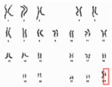
Human male karyotpe high resolution - Y chromosome cropped.png
Human Y chromosome (after G-banding)
Human male karyotpe high resolution - Chromosome Y.png
Y chromosome in human male karyogram
Features
Length (bp)57,227,415 bp
(GRCh38)
No. of genes63 (CCDS)
TypeAllosome
Centromere positionAcrocentric
(10.4 Mbp)
Complete gene lists
CCDSGene list
HGNCGene list
UniProtGene list
NCBIGene list
External map viewers
EnsemblChromosome Y
EntrezChromosome Y
NCBIChromosome Y
UCSCChromosome Y
Full DNA sequences
RefSeqNC_000024 (FASTA)
GenBankCM000686 (FASTA)

The Y chromosome is one of two sex chromosomes (allosomes) in mammals, including humans, and many other animals. The other is the X chromosome. Y is normally the sex-determining chromosome in many species, since it is the presence or absence of Y that typically determines the male or female sex of offspring produced in sexual reproduction. In mammals, the Y chromosome contains the gene SRY, which triggers male development. The DNA in the human Y chromosome is composed of about 59 million base pairs. The Y chromosome is passed only from father to son. With a 30% difference between humans and chimpanzees, the Y chromosome is one of the fastest-evolving parts of the human genome. The human Y chromosome carries an estimated 100-200 genes, with between 45 and 73 of these protein-coding. All single-copy Y-linked genes are hemizygous (present on only one chromosome) except in cases of aneuploidy such as XYY syndrome or XXYY syndrome.

Overview

Discovery

The Y chromosome was identified as a sex-determining chromosome by Nettie Stevens at Bryn Mawr College in 1905 during a study of the mealworm Tenebrio molitor. Edmund Beecher Wilson independently discovered the same mechanisms the same year. Stevens proposed that chromosomes always existed in pairs and that the Y chromosome was the pair of the X chromosome discovered in 1890 by Hermann Henking. She realized that the previous idea of Clarence Erwin McClung, that the X chromosome determines sex, was wrong and that sex determination is, in fact, due to the presence or absence of the Y chromosome. Stevens named the chromosome "Y" simply to follow on from Henking's "X" alphabetically.

The idea that the Y chromosome was named after its similarity in appearance to the letter "Y" is mistaken. All chromosomes normally appear as an amorphous blob under the microscope and only take on a well-defined shape during mitosis. This shape is vaguely X-shaped for all chromosomes. It is entirely coincidental that the Y chromosome, during mitosis, has two very short branches which can look merged under the microscope and appear as the descender of a Y-shape.

Variations

Most therian mammals have only one pair of sex chromosomes in each cell. Males have one Y chromosome and one X chromosome, while females have two X chromosomes. In mammals, the Y chromosome contains a gene, SRY, which triggers embryonic development as a male. The Y chromosomes of humans and other mammals also contain other genes needed for normal sperm production.

There are exceptions, however. Among humans, some men have two Xs and a Y ("XXY", see Klinefelter syndrome), or one X and two Ys (see XYY syndrome), and some women have three Xs or a single X instead of a double X ("X0", see Turner syndrome). There are other exceptions in which SRY is damaged (leading to an XY female), or copied to the X (leading to an XX male).

Origins and evolution

Before Y chromosome

Many ectothermic vertebrates have no sex chromosomes. If they have different sexes, sex is determined environmentally rather than genetically. For some of them, especially reptiles, sex depends on the incubation temperature. Some vertebrates are hermaphrodites, although other than a very few ray-finned fish, they are sequential (the same organism produces male or female gametes, but never both, at different points in its life), rather than simultaneous (the same organism producing both male and female gametes at the same time).

Origin

The X and Y chromosomes are thought to have evolved from a pair of identical chromosomes, termed autosomes, when an ancestral animal developed an allelic variation, a so-called "sex locus" – simply possessing this allele caused the organism to be male. The chromosome with this allele became the Y chromosome, while the other member of the pair became the X chromosome. Over time, genes that were beneficial for males and harmful to (or had no effect on) females either developed on the Y chromosome or were acquired through the process of translocation.

Until recently, the X and Y chromosomes were thought to have diverged around 300 million years ago. However, research published in 2010, and particularly research published in 2008 documenting the sequencing of the platypus genome, has suggested that the XY sex-determination system would not have been present more than 166 million years ago, at the split of the monotremes from other mammals. This re-estimation of the age of the therian XY system is based on the finding that sequences that are on the X chromosomes of marsupials and eutherian mammals are present on the autosomes of platypus and birds. The older estimate was based on erroneous reports that the platypus X chromosomes contained these sequences.

Recombination inhibition

Recombination between the X and Y chromosomes proved harmful—it resulted in males without necessary genes formerly found on the Y chromosome, and females with unnecessary or even harmful genes previously only found on the Y chromosome. As a result, genes beneficial to males accumulated near the sex-determining genes, and recombination in this region was suppressed in order to preserve this male specific region. Over time, the Y chromosome changed in such a way as to inhibit the areas around the sex determining genes from recombining at all with the X chromosome. As a result of this process, 95% of the human Y chromosome is unable to recombine. Only the tips of the Y and X chromosomes recombine. The tips of the Y chromosome that could recombine with the X chromosome are referred to as the pseudoautosomal region. The rest of the Y chromosome is passed on to the next generation intact, allowing for its use in tracking human evolution.

Degeneration

By one estimate, the human Y chromosome has lost 1,393 of its 1,438 original genes over the course of its existence, and linear extrapolation of this 1,393-gene loss over 300 million years gives a rate of genetic loss of 4.6 genes per million years. Continued loss of genes at the rate of 4.6 genes per million years would result in a Y chromosome with no functional genes – that is the Y chromosome would lose complete function – within the next 10 million years, or half that time with the current age estimate of 160 million years. Comparative genomic analysis reveals that many mammalian species are experiencing a similar loss of function in their heterozygous sex chromosome. Degeneration may simply be the fate of all non-recombining sex chromosomes, due to three common evolutionary forces: high mutation rate, inefficient selection, and genetic drift.

However, comparisons of the human and chimpanzee Y chromosomes (first published in 2005) show that the human Y chromosome has not lost any genes since the divergence of humans and chimpanzees between 6–7 million years ago, and a scientific report in 2012 stated that only one gene had been lost since humans diverged from the rhesus macaque 25 million years ago. These facts provide direct evidence that the linear extrapolation model is flawed and suggest that the current human Y chromosome is either no longer shrinking or is shrinking at a much slower rate than the 4.6 genes per million years estimated by the linear extrapolation model.

High mutation rate

The human Y chromosome is particularly exposed to high mutation rates due to the environment in which it is housed. The Y chromosome is passed exclusively through sperm, which undergo multiple cell divisions during gametogenesis. Each cellular division provides further opportunity to accumulate base pair mutations. Additionally, sperm are stored in the highly oxidative environment of the testis, which encourages further mutation. These two conditions combined put the Y chromosome at a greater opportunity of mutation than the rest of the genome. The increased mutation opportunity for the Y chromosome is reported by Graves as a factor 4.8. However, her original reference obtains this number for the relative mutation rates in male and female germ lines for the lineage leading to humans.

The observation that the Y chromosome experiences little meiotic recombination and has an accelerated rate of mutation and degradative change compared to the rest of the genome suggests an evolutionary explanation for the adaptive function of meiosis with respect to the main body of genetic information. Brandeis proposed that the basic function of meiosis (particularly meiotic recombination) is the conservation of the integrity of the genome, a proposal consistent with the idea that meiosis is an adaptation for repairing DNA damage.

Inefficient selection

Without the ability to recombine during meiosis, the Y chromosome is unable to expose individual alleles to natural selection. Deleterious alleles are allowed to "hitchhike" with beneficial neighbors, thus propagating maladapted alleles into the next generation. Conversely, advantageous alleles may be selected against if they are surrounded by harmful alleles (background selection). Due to this inability to sort through its gene content, the Y chromosome is particularly prone to the accumulation of "junk" DNA. Massive accumulations of retrotransposable elements are scattered throughout the Y. The random insertion of DNA segments often disrupts encoded gene sequences and renders them nonfunctional. However, the Y chromosome has no way of weeding out these "jumping genes". Without the ability to isolate alleles, selection cannot effectively act upon them.

A clear, quantitative indication of this inefficiency is the entropy rate of the Y chromosome. Whereas all other chromosomes in the human genome have entropy rates of 1.5–1.9 bits per nucleotide (compared to the theoretical maximum of exactly 2 for no redundancy), the Y chromosome's entropy rate is only 0.84. This means the Y chromosome has a much lower information content relative to its overall length; it is more redundant.

Genetic drift

Even if a well adapted Y chromosome manages to maintain genetic activity by avoiding mutation accumulation, there is no guarantee it will be passed down to the next generation. The population size of the Y chromosome is inherently limited to 1/4 that of autosomes: diploid organisms contain two copies of autosomal chromosomes while only half the population contains 1 Y chromosome. Thus, genetic drift is an exceptionally strong force acting upon the Y chromosome. Through sheer random assortment, an adult male may never pass on his Y chromosome if he only has female offspring. Thus, although a male may have a well adapted Y chromosome free of excessive mutation, it may never make it into the next gene pool. The repeat random loss of well-adapted Y chromosomes, coupled with the tendency of the Y chromosome to evolve to have more deleterious mutations rather than less for reasons described above, contributes to the species-wide degeneration of Y chromosomes through Muller's ratchet.

Gene conversion

As it has been already mentioned, the Y chromosome is unable to recombine during meiosis like the other human chromosomes; however, in 2003, researchers from MIT discovered a process which may slow down the process of degradation. They found that human Y chromosome is able to "recombine" with itself, using palindrome base pair sequences. Such a "recombination" is called gene conversion.

In the case of the Y chromosomes, the palindromes are not noncoding DNA; these strings of bases contain functioning genes important for male fertility. Most of the sequence pairs are greater than 99.97% identical. The extensive use of gene conversion may play a role in the ability of the Y chromosome to edit out genetic mistakes and maintain the integrity of the relatively few genes it carries. In other words, since the Y chromosome is single, it has duplicates of its genes on itself instead of having a second, homologous, chromosome. When errors occur, it can use other parts of itself as a template to correct them.

Findings were confirmed by comparing similar regions of the Y chromosome in humans to the Y chromosomes of chimpanzees, bonobos and gorillas. The comparison demonstrated that the same phenomenon of gene conversion appeared to be at work more than 5 million years ago, when humans and the non-human primates diverged from each other.

Future evolution

In the terminal stages of the degeneration of the Y chromosome, other chromosomes increasingly take over genes and functions formerly associated with it. Finally, the Y chromosome disappears entirely, and a new sex-determining system arises. Several species of rodent in the sister families Muridae and Cricetidae have reached these stages, in the following ways:

  • The Transcaucasian mole vole, Ellobius lutescens, the Zaisan mole vole, Ellobius tancrei, and the Japanese spinous country rats Tokudaia osimensis and Tokudaia tokunoshimensis, have lost the Y chromosome and SRY entirely. Tokudaia spp. have relocated some other genes ancestrally present on the Y chromosome to the X chromosome. Both sexes of Tokudaia spp. and Ellobius lutescens have an XO genotype (Turner syndrome), whereas all Ellobius tancrei possess an XX genotype. The new sex-determining system(s) for these rodents remains unclear.
  • The wood lemming Myopus schisticolor, the Arctic lemming, Dicrostonyx torquatus, and multiple species in the grass mouse genus Akodon have evolved fertile females who possess the genotype generally coding for males, XY, in addition to the ancestral XX female, through a variety of modifications to the X and Y chromosomes.
  • In the creeping vole, Microtus oregoni, the females, with just one X chromosome each, produce X gametes only, and the males, XY, produce Y gametes, or gametes devoid of any sex chromosome, through nondisjunction.

Outside of the rodents, the black muntjac, Muntiacus crinifrons, evolved new X and Y chromosomes through fusions of the ancestral sex chromosomes and autosomes.

1:1 sex ratio

Fisher's principle outlines why almost all species using sexual reproduction have a sex ratio of 1:1. W. D. Hamilton gave the following basic explanation in his 1967 paper on "Extraordinary sex ratios", given the condition that males and females cost equal amounts to produce:

  1. Suppose male births are less common than female.
  2. A newborn male then has better mating prospects than a newborn female, and therefore can expect to have more offspring.
  3. Therefore, parents genetically disposed to produce males tend to have more than average numbers of grandchildren born to them.
  4. Therefore, the genes for male-producing tendencies spread, and male births become more common.
  5. As the 1:1 sex ratio is approached, the advantage associated with producing males dies away.
  6. The same reasoning holds if females are substituted for males throughout. Therefore, 1:1 is the equilibrium ratio.

Non-therian Y chromosome

Many groups of organisms in addition to therian mammals have Y chromosomes, but these Y chromosomes do not share common ancestry with therian Y chromosomes. Such groups include monotremes, Drosophila, some other insects, some fish, some reptiles, and some plants. In Drosophila melanogaster, the Y chromosome does not trigger male development. Instead, sex is determined by the number of X chromosomes. The D. melanogaster Y chromosome does contain genes necessary for male fertility. So XXY D. melanogaster are female, and D. melanogaster with a single X (X0), are male but sterile. There are some species of Drosophila in which X0 males are both viable and fertile.

ZW chromosomes

Other organisms have mirror image sex chromosomes: where the homogeneous sex is the male, said to have two Z chromosomes, and the female is the heterogeneous sex, and said to have a Z chromosome and a W chromosome. For example, female birds, snakes, and butterflies have ZW sex chromosomes, and males have ZZ sex chromosomes.

Non-inverted Y chromosome

There are some species, such as the Japanese rice fish, in which the XY system is still developing and cross over between the X and Y is still possible. Because the male specific region is very small and contains no essential genes, it is even possible to artificially induce XX males and YY females to no ill effect.

Multiple XY pairs

Monotremes possess four or five (platypus) pairs of XY sex chromosomes, each pair consisting of sex chromosomes with homologous regions. The chromosomes of neighboring pairs are partially homologous, such that a chain is formed during mitosis. The first X chromosome in the chain is also partially homologous with the last Y chromosome, indicating that profound rearrangements, some adding new pieces from autosomes, have occurred in history.

Platypus sex chromosomes have strong sequence similarity with the avian Z chromosome, (indicating close homology), and the SRY gene so central to sex-determination in most other mammals is apparently not involved in platypus sex-determination.

Human Y chromosome

In humans, the Y chromosome spans about 58 million base pairs (the building blocks of DNA) and represents almost 2% of the total DNA in a male cell. The human Y chromosome contains over 200 genes, at least 72 of which code for proteins. Traits that are inherited via the Y chromosome are called Y-linked traits, or holandric traits (from Ancient Greek ὅλος hólos, "whole" + ἀνδρός andrós, "male").

Men can lose the Y chromosome in a subset of cells, which is called the mosaic loss of chromosome Y (LOY). This post-zygotic mutation is strongly associated with age, affecting about 15% of men 70 years of age. Smoking is another important risk factor for LOY. It has been found that men with a higher percentage of hematopoietic stem cells in blood lacking the Y chromosome (and perhaps a higher percentage of other cells lacking it) have a higher risk of certain cancers and have a shorter life expectancy. Men with LOY (which was defined as no Y in at least 18% of their hematopoietic cells) have been found to die 5.5 years earlier on average than others. This has been interpreted as a sign that the Y chromosome plays a role going beyond sex determination and reproduction (although the loss of Y may be an effect rather than a cause). Male smokers have between 1.5 and 2 times the risk of non-respiratory cancers as female smokers.

Non-combining region of Y (NRY)

The human Y chromosome is normally unable to recombine with the X chromosome, except for small pieces of pseudoautosomal regions at the telomeres (which comprise about 5% of the chromosome's length). These regions are relics of ancient homology between the X and Y chromosomes. The bulk of the Y chromosome, which does not recombine, is called the "NRY", or non-recombining region of the Y chromosome. The single-nucleotide polymorphisms (SNPs) in this region are used to trace direct paternal ancestral lines.

Genes

Gene list

In general, the human Y chromosome is extremely gene poor—it is one of the largest gene deserts in the human genome. Disregarding pseudoautosomal genes, genes encoded on the human Y chromosome include:

Y-chromosome-linked diseases

Diseases linked to the Y chromosome typically involve an aneuploidy, an atypical number of chromosomes.

Y chromosome microdeletion

Y chromosome microdeletion (YCM) is a family of genetic disorders caused by missing genes in the Y chromosome. Many affected men exhibit no symptoms and lead normal lives. However, YCM is also known to be present in a significant number of men with reduced fertility or reduced sperm count.

Defective Y chromosome

This results in the person presenting a female phenotype (i.e., is born with female-like genitalia) even though that person possesses an XY karyotype. The lack of the second X results in infertility. In other words, viewed from the opposite direction, the person goes through defeminization but fails to complete masculinization.

The cause can be seen as an incomplete Y chromosome: the usual karyotype in these cases is 45X, plus a fragment of Y. This usually results in defective testicular development, such that the infant may or may not have fully formed male genitalia internally or externally. The full range of ambiguity of structure may occur, especially if mosaicism is present. When the Y fragment is minimal and nonfunctional, the child is usually a girl with the features of Turner syndrome or mixed gonadal dysgenesis.

XXY

Klinefelter syndrome (47, XXY) is not an aneuploidy of the Y chromosome, but a condition of having an extra X chromosome, which usually results in defective postnatal testicular function. The mechanism is not fully understood; it does not seem to be due to direct interference by the extra X with expression of Y genes.

XYY

47, XYY syndrome (simply known as XYY syndrome) is caused by the presence of a single extra copy of the Y chromosome in each of a male's cells. 47, XYY males have one X chromosome and two Y chromosomes, for a total of 47 chromosomes per cell. Researchers have found that an extra copy of the Y chromosome is associated with increased stature and an increased incidence of learning problems in some boys and men, but the effects are variable, often minimal, and the vast majority do not know their karyotype.

In 1965 and 1966 Patricia Jacobs and colleagues published a chromosome survey of 315 male patients at Scotland's only special security hospital for the developmentally disabled, finding a higher than expected number of patients to have an extra Y chromosome. The authors of this study wondered "whether an extra Y chromosome predisposes its carriers to unusually aggressive behaviour", and this conjecture "framed the next fifteen years of research on the human Y chromosome".

Through studies over the next decade, this conjecture was shown to be incorrect: the elevated crime rate of XYY males is due to lower median intelligence and not increased aggression, and increased height was the only characteristic that could be reliably associated with XYY males. The "criminal karyotype" concept is therefore inaccurate.

Rare

The following Y-chromosome-linked diseases are rare, but notable because of their elucidating of the nature of the Y chromosome.

More than two Y chromosomes

Greater degrees of Y chromosome polysomy (having more than one extra copy of the Y chromosome in every cell, e.g., XYYY) are rare. The extra genetic material in these cases can lead to skeletal abnormalities, decreased IQ, and delayed development, but the severity features of these conditions are variable.

XX male syndrome

XX male syndrome occurs when there has been a recombination in the formation of the male gametes, causing the SRY portion of the Y chromosome to move to the X chromosome. When such an X chromosome contributes to the child, the development will lead to a male, because of the SRY gene.

Genetic genealogy

In human genetic genealogy (the application of genetics to traditional genealogy), use of the information contained in the Y chromosome is of particular interest because, unlike other chromosomes, the Y chromosome is passed exclusively from father to son, on the patrilineal line. Mitochondrial DNA, maternally inherited to both sons and daughters, is used in an analogous way to trace the matrilineal line.

Brain function

Research is currently investigating whether male-pattern neural development is a direct consequence of Y-chromosome-related gene expression or an indirect result of Y-chromosome-related androgenic hormone production.

Microchimerism

The presence of male chromosomes in fetal cells in the blood circulation of women was discovered in 1974.

In 1996, it was found that male fetal progenitor cells could persist postpartum in the maternal blood stream for as long as 27 years.

A 2004 study at the Fred Hutchinson Cancer Research Center, Seattle, investigated the origin of male chromosomes found in the peripheral blood of women who had not had male progeny. A total of 120 subjects (women who had never had sons) were investigated, and it was found that 21% of them had male DNA. The subjects were categorised into four groups based on their case histories:

  • Group A (8%) had had only female progeny.
  • Patients in Group B (22%) had a history of one or more miscarriages.
  • Patients Group C (57%) had their pregnancies medically terminated.
  • Group D (10%) had never been pregnant before.

The study noted that 10% of the women had never been pregnant before, raising the question of where the Y chromosomes in their blood could have come from. The study suggests that possible reasons for occurrence of male chromosome microchimerism could be one of the following:

  • miscarriages,
  • pregnancies,
  • vanished male twin,
  • possibly from sexual intercourse.

A 2012 study at the same institute has detected cells with the Y chromosome in multiple areas of the brains of deceased women.
XY sex-determination system

From Wikipedia, the free encyclopedia
 
Drosophila sex-chromosomes
 
Pollen cones of a male Ginkgo biloba tree, a dioecious species
 
Ovules of a female Ginkgo biloba

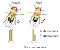
The XY sex-determination system is a sex-determination system used to classify many mammals, including humans, some insects (Drosophila), some snakes, some fish (guppies), and some plants (Ginkgo tree). In this system, the sex of an individual is determined by a pair of sex chromosomes. Females typically have two of the same kind of sex chromosome (XX), and are called the homogametic sex. Males typically have two different kinds of sex chromosomes (XY), and are called the heterogametic sex.

In humans, the presence of the Y chromosome is typically responsible for triggering male development; in the absence of the Y chromosome, the fetus will undergo female development. More specifically, it is the SRY gene located on the Y chromosome that is of importance to male differentiation. Variations to the sex gene karyotype could include rare disorders such as XX males (often due to translocation of the SRY gene to the X chromosome) or XY gonadal dysgenesis in people who are externally female (due to mutations in the SRY gene). In addition, other rare genetic variations such as Turners (XO) and Klinefelters (XXY) are seen as well. In most species in XY sex determination, an organism must have at least one X chromosome in order to survive.

The XY system contrasts in several ways with the ZW sex-determination system found in birds, some insects, many reptiles, and various other animals, in which the heterogametic sex is female. It had been thought for several decades that in all snakes sex was determined by the ZW system, but there had been observations of unexpected effects in the genetics of species in the families Boidae and Pythonidae; for example, parthenogenic reproduction produced only females rather than males, which is the opposite of what is to be expected in the ZW system. In the early years of the 21st century such observations prompted research that demonstrated that all pythons and boas so far investigated definitely have the XY system of sex determination.

A temperature-dependent sex determination system is found in some reptiles and fish.

Mechanisms

All animals have a set of DNA coding for genes present on chromosomes. In humans, most mammals, and some other species, two of the chromosomes, called the X chromosome and Y chromosome, code for sex. In these species, one or more genes are present on their Y chromosome that determine maleness. In this process, an X chromosome and a Y chromosome act to determine the sex of offspring, often due to genes located on the Y chromosome that code for maleness. Offspring have two sex chromosomes: an offspring with two X chromosomes will develop female characteristics, and an offspring with an X and a Y chromosome will develop male characteristics.

Humans

Human male XY chromosomes after G-banding

In humans, half of spermatozoa carry X chromosome and the other half Y chromosome. A single gene (SRY) present on the Y chromosome acts as a signal to set the developmental pathway towards maleness. Presence of this gene starts off the process of virilization. This and other factors result in the sex differences in humans. The cells in females, with two X chromosomes, undergo X-inactivation, in which one of the two X chromosomes is inactivated. The inactivated X chromosome remains within a cell as a Barr body.

Humans, as well as some other organisms, can have a rare chromosomal arrangement that is contrary to their phenotypic sex; for example, XX males or XY females (see androgen insensitivity syndrome). Additionally, an abnormal number of sex chromosomes (aneuploidy) may be present, such as Turner's syndrome, in which a single X chromosome is present, and Klinefelter's syndrome, in which two X chromosomes and a Y chromosome are present, XYY syndrome and XXYY syndrome. Other less common chromosomal arrangements include: triple X syndrome, 48, XXXX, and 49, XXXXX.

Other animals

In most mammals, sex is determined by presence of the Y chromosome. "Female" is the default sex, due to the absence of the Y chromosome. In the 1930s, Alfred Jost determined that the presence of testosterone was required for Wolffian duct development in the male rabbit.

SRY is a sex-determining gene on the Y chromosome in the therians (placental mammals and marsupials). Non-human mammals use several genes on the Y chromosome. Not all male-specific genes are located on the Y chromosome. Platypus, a monotreme, use five pairs of different XY chromosomes with six groups of male-linked genes, AMH being the master switch. Other species (including most Drosophila species) use the presence of two X chromosomes to determine femaleness: one X chromosome gives putative maleness, but the presence of Y chromosome genes is required for normal male development.

Other systems

Birds and many insects have a similar system of sex determination (ZW sex-determination system), in which it is the females that are heterogametic (ZW), while males are homogametic (ZZ).

Many insects of the order Hymenoptera instead have a haplo-diploid system, where the males are haploid (having just one chromosome of each type) while the females are diploid (with chromosomes appearing in pairs). Some other insects have the X0 sex-determination system, where just one chromosome type appears in pairs for the female but alone in the males, while all other chromosomes appear in pairs in both sexes.

Influences

Genetic

PBB Protein SRY image

In an interview for the Rediscovering Biology website, researcher Eric Vilain described how the paradigm changed since the discovery of the SRY gene:

For a long time we thought that SRY would activate a cascade of male genes. It turns out that the sex determination pathway is probably more complicated and SRY may in fact inhibit some anti-male genes.

The idea is instead of having a simplistic mechanism by which you have pro-male genes going all the way to make a male, in fact there is a solid balance between pro-male genes and anti-male genes and if there is a little too much of anti-male genes, there may be a female born and if there is a little too much of pro-male genes then there will be a male born.

We [are] entering this new era in molecular biology of sex determination where it's a more subtle dosage of genes, some pro-males, some pro-females, some anti-males, some anti-females that all interplay with each other rather than a simple linear pathway of genes going one after the other, which makes it very fascinating but very complicated to study.

In mammals, including humans, the SRY gene is responsible with triggering the development of non-differentiated gonads into testes, rather than ovaries. However, there are cases in which testes can develop in the absence of an SRY gene (see sex reversal). In these cases, the SOX9 gene, involved in the development of testes, can induce their development without the aid of SRY. In the absence of SRY and SOX9, no testes can develop and the path is clear for the development of ovaries. Even so, the absence of the SRY gene or the silencing of the SOX9 gene are not enough to trigger sexual differentiation of a fetus in the female direction. A recent finding suggests that ovary development and maintenance is an active process, regulated by the expression of a "pro-female" gene, FOXL2. In an interview for the TimesOnline edition, study co-author Robin Lovell-Badge explained the significance of the discovery:

We take it for granted that we maintain the sex we are born with, including whether we have testes or ovaries. But this work shows that the activity of a single gene, FOXL2, is all that prevents adult ovary cells turning into cells found in testes.

Implications

Looking into the genetic determinants of human sex can have wide-ranging consequences. Scientists have been studying different sex determination systems in fruit flies and animal models to attempt an understanding of how the genetics of sexual differentiation can influence biological processes like reproduction, ageing and disease.

Maternal

In humans and many other species of animals, the father determines the sex of the child. In the XY sex-determination system, the female-provided ovum contributes an X chromosome and the male-provided sperm contributes either an X chromosome or a Y chromosome, resulting in female (XX) or male (XY) offspring, respectively.

Hormone levels in the male parent affect the sex ratio of sperm in humans. Maternal influences also impact which sperm are more likely to achieve conception.

Human ova, like those of other mammals, are covered with a thick translucent layer called the zona pellucida, which the sperm must penetrate to fertilize the egg. Once viewed simply as an impediment to fertilization, recent research indicates the zona pellucida may instead function as a sophisticated biological security system that chemically controls the entry of the sperm into the egg and protects the fertilized egg from additional sperm.

Recent research indicates that human ova may produce a chemical which appears to attract sperm and influence their swimming motion. However, not all sperm are positively impacted; some appear to remain uninfluenced and some actually move away from the egg.

Maternal influences may also be possible that affect sex determination in such a way as to produce fraternal twins equally weighted between one male and one female.

The time at which insemination occurs during the estrus cycle has been found to affect the sex ratio of the offspring of humans, cattle, hamsters, and other mammals. Hormonal and pH conditions within the female reproductive tract vary with time, and this affects the sex ratio of the sperm that reach the egg.

Sex-specific mortality of embryos also occurs.

History

Ancient ideas on sex determination

Aristotle believed incorrectly that the sex of an infant is determined by how much heat a man's sperm had during insemination. He wrote:

...the semen of the male differs from the corresponding secretion of the female in that it contains a principle within itself of such a kind as to set up movements also in the embryo and to concoct thoroughly the ultimate nourishment, whereas the secretion of the female contains material alone. If, then, the male element prevails it draws the female element into itself, but if it is prevailed over it changes into the opposite or is destroyed.

Aristotle claimed in error that the male principle was the driver behind sex determination, such that if the male principle was insufficiently expressed during reproduction, the fetus would develop as a female.

20th century genetics

Nettie Stevens and Edmund Beecher Wilson are credited with independently discovering, in 1905, the chromosomal XY sex-determination system, i.e. the fact that males have XY sex chromosomes and females have XX sex chromosomes.

The first clues to the existence of a factor that determines the development of testis in mammals came from experiments carried out by Alfred Jost, who castrated embryonic rabbits in utero and noticed that they all developed as female.

In 1959, C. E. Ford and his team, in the wake of Jost's experiments, discovered that the Y chromosome was needed for a fetus to develop as male when they examined patients with Turner's syndrome, who grew up as phenotypic females, and found them to be X0 (hemizygous for X and no Y). At the same time, Jacob & Strong described a case of a patient with Klinefelter syndrome (XXY), which implicated the presence of a Y chromosome in development of maleness.

All these observations lead to a consensus that a dominant gene that determines testis development (TDF) must exist on the human Y chromosome. The search for this testis-determining factor (TDF) led a team of scientists in 1990 to discover a region of the Y chromosome that is necessary for the male sex determination, which was named SRY (sex-determining region of the Y chromosome).

Wednesday, December 9, 2020

Prenatal hormones and sexual orientation

From Wikipedia, the free encyclopedia

The hormonal theory of sexuality holds that, just as exposure to certain hormones plays a role in fetal sex differentiation, such exposure also influences the sexual orientation that emerges later in the adult. Prenatal hormones may be seen as the primary determinant of adult sexual orientation, or a co-factor with genes, biological factors and/or environmental and social conditions.

Sex-typed behavior

The hormonal theory of sexuality and gender identity holds that, just as exposure to certain hormones plays a role in fetal sex differentiation, such exposure also influences the sexual orientation and or gender identity that emerges later in the adult. Differences in brain structure that come about from chemical messengers and genes interacting on developing brain cells are believed to be the basis of sex differences in countless behaviors, including sexual orientation. Prenatal factors that affect or interfere with the interaction of these hormones on the developing brain can influence later sex-typed behavior in children. This hypothesis is originated from countless experimental studies in non-human mammals, yet the argument that similar effects can be seen in human neurobehavioral development is a much debated topic among scholars. Recent studies, however, have provided evidence in support of prenatal androgen exposure influencing childhood sex-typed behavior.

Fetal hormones may be seen as either the primary influence upon adult sexual orientation or as a co-factor interacting with genes and/or environmental and social conditions. However, Garcia-Falgueras and Dick Swaab disagree that social conditions influence sexual orientation to a large degree. As seen in young children as well as in vervet and rhesus monkeys, sexually differentiated behavior in toy preference is differing in males versus females, where females prefer dolls and males prefer toy balls and cars; these preferences can be seen as early as 3–8 months in humans. It is impossible to completely rule out the social environment or the child's cognitive understanding of gender when discussing sex typed play in androgen-exposed girls. Conversely, children tend towards objects which have been labelled for their own sex, or toys that they have seen members of their sex playing with previously.

An endocrinology study by Garcia-Falgueras and Swaab postulated that "In humans, the main mechanism responsible of [sic] sexual identity and orientation involves a direct effect of testosterone on the developing brain." Further, their study puts forward that intrauterine exposure to hormones is largely determinative. Sketching the argument briefly here, the authors say that sexual organs are differentiated first, and then the brain is sexually differentiated "under the influence, mainly, of sex hormones such as testosterone, estrogen and progesterone on the developing brain cells and under the presence of different genes as well ... . The changes brought about in this stage are permanent. ... Sexual differentiation of the brain is not caused by hormones alone, even though they are very important for gender identity and sexual orientation."

Organizational aspects

Fetal gonads develop primarily based on the presence or absence of androgen hormones, mainly testosterone, dihydrotestosterone (DHT) and androstenedione; production of testosterone and conversion into dihydrotestosterone during weeks 6 to 12 of pregnancy are key factors in the production of a male fetus's penis, scrotum and prostate. In a female, on the other hand, absence of these levels of androgens results in development of typically female genitals. Following this, sexual differentiation of the brain occurs; sex hormones exert organizational effects on the brain that will be activated in puberty. As a result of these two processes occurring separately, the degree of genital masculinization does not necessarily relate to the masculinization of the brain. Sex differences in the brain have been found in many structures, most notably the hypothalamus and the amygdala. However, few of these have been related to behavioral sex differences, and scientists are still working to establish firm links between early hormones, brain development and behavior. The study of the organizational theory of prenatal hormones can be difficult, as ethically researchers cannot alter hormones in a developing fetus; instead, scholars must rely on naturally occurring abnormalities of development to provide answers.

Most extensively studied in organizational effects of hormones is congenital adrenal hyperplasia (CAH). CAH is a genetic disease that results in exposure to high levels of androgens beginning early in gestation. Girls with CAH are born with masculinized genitalia, which is corrected surgically as soon as possible. CAH provides the opportunity for natural experiments, as people with CAH can be compared to people without it. However, "CAH is not a perfect experiment", since, "social responses to masculinized genitalia or factors related to the disease itself" can confound results. Nonetheless several studies have shown that CAH has a clear but not determining influence on sexual orientation; women with CAH are less likely to be exclusively heterosexual than are other women.

Since hormones alone do not determine sexual orientation and differentiation of the brain, the search for other factors that act upon sexual orientation have led genes such as the SRY and ZFY to be implicated.

Prenatal maternal stress

As of 2006 results from studies in humans had found conflicting evidence regarding the effect of prenatal exposure to hormones and psychosexual outcomes; Gooren noted in 2006 that studies in subprimate mammals are invalid measures of human sexual differentiation, as sex hormones follow a more "on-off" role in sex-typed behavior than is found in primates.

Some studies do suggest that prenatal stress significantly increases the likelihood of homosexuality or bisexuality, although varying evidence exists for which trimester is most important. Studies of endocrinology have found implications for amphetamines and thyroid-gland hormones to increase homosexuality in female offspring as well, although it has not been examined in conjunction with prenatal stress levels.

Some have postulated that postnatal (e.g., social and environmental factors) development can play a role in the sexual orientation of an individual, yet solid evidence of this has yet to be discovered. Children born through artificial insemination with donor sperm and consequently raised by lesbian couples have typically been heterosexually oriented. Summed up by Bao and Swaab, "The apparent impossibility of getting someone to change their sexual orientation ... is a major argument against the importance of the social environment in the emergence of homosexuality, as well as against the idea that homosexuality is a lifestyle choice."

Fraternal birth order

According to a multitude of studies over several decades, gay men have more older brothers on average, a phenomenon known as the fraternal birth order effect. It has been suggested that the greater the number of older male siblings the higher the level of androgen fetuses are exposed to. No evidence of birth order effects have been observed in women. The theory holds that the fraternal birth order effect is a result of a maternal immune response that is produced towards a factor of male development over several male pregnancies. Bogaert's hypothesis argues that "the target of the immune response may be male specific molecules on the surface of male fetal brain cells (e.g., including those in the anterior hypothalamus). Anti-male antibodies might bind to these molecules and thus interfere with their role in normal sexual differentiation, leading some later born males to being attracted to men as opposed to women." Garcia-Falgueras and Swaab state that "The ... fraternal birth order effect ... is putatively explained by an immunological response by the mother to a product of the Y chromosome of her sons. The chance of such an immune response to male factors would increase with every pregnancy resulting in the birth of a son."

Maternal antibodies against Y-chromosone neuroligin have been implicated in this effect, among other evidence that favours this theory. Further, while percentages of the likelihood of homosexuality have been estimated to be increased by 15–48% per older brother, these odds really account for only a few percent of the population; thus, this hypothesis cannot be universally applied to the majority of homosexual men. Most, but not all, studies have been able to reproduce the fraternal birth order effect. Some did not find any statistically significant difference in either the sibling composition or rate of older brothers of gay and straight men, including large, nationally representative studies in the US and Denmark. However, Blanchard reanalyzed Frisch's 2006 Danish study and found the birth order effect was in-fact present.

In conjunction with fraternal birth order, handedness provides further evidence of prenatal effects on sexual orientation, because handedness is regarded by many as a marker of early neurodevelopment. Other correlates to handedness (e.g., cerebral laterality, prenatal hormonal profiles, spatial ability) have been linked to sexual orientation, either empirically and/or theoretically. In right-handed individuals, the number of older brothers increased the odds of homosexual orientation, but this effect was not seen in left-handed individuals. As with other purported marks indicating higher incidence of homosexuality, however, the link with handedness remains ambiguous and several studies have been unable to replicate it.

Implicated genes in fraternal birth order

A gene of the Rh system has been discussed as a possible candidate for affecting fraternal birth order, as it has been linked to both handedness and immune system functioning. Gene variants in the Rh system are implicated in a maternal response to what is known as hemolytic disease of the newborn. Rh is a factor in blood, and in cases where the mother is absent of this (Rh-) while carrying an Rh+ fetus, an immune response may develop with deleterious effects. The Rh gene hypothesis is a strong candidate because not only does it involve the maternal immune response, but it has been implicated in handedness as well.

Variants of the androgen receptor (AR) gene have also been discussed, in that non-right-handedness in men has been linked with greater CAG repeats in the AR gene, which in turn is associated with lower testosterone. A theory that high prenatal testosterone leads to neuronal and axonal loss in the corpus callosum is supported by this hypothesis.

Male homosexuality as hypermasculine

There is evidence of a correlation between sexual orientation and traits that are determined in utero. A study by McFadden in 1998 found that auditory systems in the brain, another physical trait influenced by prenatal hormones is different in those of differing orientations; likewise the suprachiasmatic nucleus was found by Swaab and Hofman to be larger in homosexual men than in heterosexual men. The suprachiasmatic nucleus is also known to be larger in men than in women. An analysis of the hypothalamus by Swaab and Hofmann (1990;2007) found that the volume of the suprachiasmatic nucleus (SCN) in homosexual men was 1.7 times larger than a reference group of male subjects, and contained 2.1 times as many cells. During development, the volume of the SCN and the cell counts reach peak value at approximately 13 to 16 months after birth; at this age, the SCN contains the same number of cells as was found in adult male homosexuals, yet in a reference group of heterosexual males the cell numbers begin to decline to the adult value of 35% of the peak value. These results have not been replicated, however; there also has yet to be a meaningful interpretation of these results provided in the context of human sexual orientation. Some highly disputed studies suggest gay men have also been shown to have higher levels of circulating androgens and larger penises, on average, than heterosexual men.

Male homosexuality as hypomasculine

In a 1991 study, Simon LeVay demonstrated that a tiny clump of neurons of the anterior hypothalamus—which is believed to control sexual behavior and linked to prenatal hormones—known as the interstitial nuclei of the anterior was, on average, more than twice the size in heterosexual men when contrasted to homosexual men. Due to this area also being nearly twice the size in heterosexual men than in heterosexual women, the implication is that the sexual differentiation of the hypothalamus in homosexuals is in a female direction. In 2003 scientists at Oregon State University announced that they had replicated his findings in sheep.

Female homosexuality

Most empirical or theoretical research into women's sexual orientation has, historically, been guided by the idea of lesbians as essentially masculine and heterosexual women as essentially feminine. Typically, this belief is traced to the early "inversion theory" of sex researchers who state that homosexuality is a result of biological abnormalities that "invert" sexual attraction and personality. Handedness research has provided implications; because more men than women present a preference for their left hand, the higher proportion of non-right handedness that has been discovered among lesbians when compared to heterosexual women demonstrates a possible link of prenatal masculinization and sexual orientation.

Backing this up are reports that lesbians display more masculinized 2D;4D digit ratios than heterosexual women, based on data gathered from at least six different laboratories. This effect has not yet been observed between homosexual and heterosexual males. However, the validity of this measure of digit ratios remains controversial as a predictor of prenatal androgen, as many other prenatal factors may play roles in bone growth in prenatal stages of development. While many studies have found results confirming this hypothesis, others have failed to replicate these findings, leaving the validity of this measure unconfirmed.

Diethylstilbestrol (DES), a drug that has been in the past prescribed to prevent miscarriages, has also been studied in relation to women's sexual orientation. It has been observed to exert a masculinizing/

defeminizing effect on the developing brain of the fetus. When compared to controls, higher percentages of DES-exposed women (17% vs 0%) reported that they had engaged in same-sex relations; however, the great majority of DES women stated an exclusively heterosexual orientation.

Girls with congenital adrenal hyperplasia (an autosomal recessive condition which results in high androgen levels during fetal development) have more masculinized sex role identities and are more likely to have a homosexual sexual orientation as adults than controls. An alternative explanation for this effect is the fact that girls with this condition are born with masculinized external genitalia, which leads their parents to raise them in a more masculine manner, thus influencing their sexual orientation as adults. However, the degree to which the girls' genitals are masculinized does not correlate with their sexual orientation, suggesting that prenatal hormones are a stronger causal factor, not parental influence.

Together with congenital adrenal hyperplasia, DES studies have provided little support of the prenatal hormone theory of sexual orientation; they do, however, provide the framework for possible pathways to a homosexual orientation for a small number of women.

Gender dysphoria

In individuals with gender dysphoria, previously known as gender identity disorder (GID), prenatal exposure to testosterone has been hypothesized to have an effect on gender identity differentiation. The 2D;4D finger ratio, or relative lengths of the 2nd "index" and 4th "ring" fingers, has become a popular measure of prenatal androgen because of accumulated evidence suggesting the 2D;4D ratios are related to prenatal exposure to testosterone. Many children with GID differentiate a homosexual orientation during adolescence, but not all of them; adults with "early onset", or a childhood history of cross-gender behavior, often have a homosexual orientation. Adults with "late onset", or those without a childhood history of said behavior, are more likely to have a non-homosexual orientation.

Prenatal androgen exposure has been associated with an increased chance of patient-initiated gender reassignment to male after being initially raised as female in early childhood or infancy. Gooren found that organizational effects of prenatal androgens are more prevalent in gender role behavior than in gender identity, and that there are preliminary findings that suggest evidence of a male gender identity being more frequent in patients with fully male-typical prenatal androgenization.

Individuals with complete androgen insensitivity syndrome are almost always brought up as females, and the differentiation of gender identity/role is feminine. This example is important in demonstrating that chromosomes and gonads alone do not dictate gender identity and role.

Transsexualism

Because organ differentiation and brain differentiation occur at different times, in rare cases transsexualism can result. Only 23% of childhood gender problems will result in transsexuality in adulthood.

Drawing on some transsexualism cases, Garcia-Falgueras and Swaab state that "[f]rom these examples it appears that the direct action of testosterone on the developing brain in boys and the lack of such action on the developing brain in girls are crucial factors in the development of male and female gender identity and sexual orientation ... ." Countless studies have been run on peripheral levels of sex steroids in male and female homosexuals, a considerable number of which claimed to find "less 'male hormone' and/or more 'female hormone' in male homosexuals and vice versa in female homosexuals". However, these findings have been reviewed and have subsequently been dismissed by Gooren as suffering from faulty design and interpretation.

Factors implicated in the development of transsexuality include chromosomal abnormalities, polymorphisms of certain genes, and variations in aromatase (cytochrome P450 CYP19) and CYP17. Girls with congenital adrenal hyperplasia show an increase in probability of transsexuality later in life; however, this risk is still only 1–3% in CAH. Although historically abnormal sexual differentiation has pointed to androgens as a causal factor, there are codeterminants of gender identity and sexual orientation with overriding effects of androgens on the brain, in male transsexuals or homosexuals, or making androgen effects on the brain redundant, as in female transsexuals or homosexuals. These factors are currently unknown, and thus no clear cut answer for the cause of transsexualism and homosexuality exists.

Due to relatively small population sizes, generalizability of studies on transsexuality cannot be assumed.

Endocrine disruptors

Endocrine disrupting chemicals (EDCs) are chemicals that, at certain doses, can interfere with the endocrine system in mammals. Work on possible neurotoxic effects of endocrine disruptors, and their possible effects on sexual orientation when a fetus is exposed to them, is in its infancy: "we mostly know about the relationship between EDC exposure and neurobehavioral function through an examination of outcomes within a limited sphere of questions." While studies have found that xenoestrogens and xenoandrogens can alter the brain's sexual differentiation in a number of species used as animal models, from the data in hand to date, it is "misleading ...to expect EDCs to produce profiles of effects, such as sexually dimorphic behaviors, as literal copies of those produced by native hormones. Such agents are not hormones. They should not be expected to act precisely as hormones."

Causes of transsexuality

From Wikipedia, the free encyclopedia

The study of the causes of transsexuality investigates gender identity formation of transgender people, especially those who are transsexual. Transgender people have a gender identity that does not match their assigned sex, often resulting in gender dysphoria. The causes of transsexuality have been studied for decades. The most studied factors are biological, especially brain structure differences in relation to biology and sexual orientation. Environmental factors have also been proposed.

Transgender brain studies, especially those on trans women who are sexually attracted to women (gynephilic), and those on trans men who are sexually attracted to men (androphilic), are limited, as they include a small number of tested individuals. The available research indicates that the brain structure of androphilic trans women with early-onset gender dysphoria is closer to the brain structure of cisgender women's and less like cisgender men's. It also reports that both androphilic trans women and trans women with late-onset gender dysphoria who are gynephilic have different brain phenotypes, and that gynephilic trans women differ from both cisgender male and female controls in non-dimorphic brain areas. Cortical thickness, which is generally thicker in cisgender women's brains than in cisgender men's brains, may also be thicker in trans women's brains, but is present in a different location to cisgender women's brains. For trans men, research indicates that those with early-onset gender dysphoria and who are gynephilic have brains that generally correspond to their assigned sex, but that they have their own phenotype with respect to cortical thickness, subcortical structures, and white matter microstructure, especially in the right hemisphere. Hormone use can also affect transgender people's brain structure; it can cause transgender women's brains to become closer to those of cisgender women, and morphological increments observed in the brains of trans men might be due to the anabolic effects of testosterone.

Twin studies suggest that there are likely genetic causes of transsexuality, although the precise genes involved are not fully understood. One study published in the International Journal of Transgender Health found that 33% of identical twin pairs were both trans, compared to only 2.6% of non-identical twins who were raised in the same family at the same time, but were not genetically identical.

Ray Blanchard created a taxonomy of male-to-female transsexualism that proposes two distinct etiologies for androphilic and gynephilic individuals that has become controversial, supported by J. Michael Bailey, Anne Lawrence, James Cantor and others, but opposed by Charles Allen Moser, Julia Serano, and the World Professional Association for Transgender Health.

Biological factors

Genetics

A 2008 study compared 112 male-to-female transsexuals (MtFs), both androphilic and gynephilic, and who were mostly already undergoing hormone treatment, with 258 cisgender male controls. Male-to-female transsexuals were more likely than cisgender males to have a longer version of a receptor gene (longer repetitions of the gene) for the sex hormone androgen, which reduced its effectiveness at binding testosterone. The androgen receptor (NR3C4) is activated by the binding of testosterone or dihydrotestosterone, where it plays a critical role in the forming of primary and secondary male sex characteristics. The research suggests reduced androgen and androgen signaling contributes to the female gender identity of male-to-female transsexuals. The authors say that a decrease in testosterone levels in the brain during development might prevent complete masculinization of the brain in male-to-female transsexuals and thereby cause a more feminized brain and a female gender identity.

A variant genotype for a gene called CYP17, which acts on the sex hormones pregnenolone and progesterone, has been found to be linked to female-to-male (FtMs) transsexuality but not MtF transsexuality. Most notably, the FtM subjects not only had the variant genotype more frequently, but had an allele distribution equivalent to male controls, unlike the female controls. The paper concluded that the loss of a female-specific CYP17 T -34C allele distribution pattern is associated with FtM transsexuality.

Transsexuality among twins

In 2013, a twin study combined a survey of pairs of twins where one or both had undergone, or had plans and medical approval to undergo, gender transition, with a literature review of published reports of transgender twins. The study found that one third of identical twin pairs in the sample were both transgender: 13 of 39 (33%) monozygotic or identical pairs of assigned males and 8 of 35 (22.8%) pairs of assigned females. Among dizygotic or genetically non-identical twin pairs, there was only 1 of 38 (2.6%) pairs where both twins were trans. The significant percent of identical twin pairs in which both twins are trans and the virtual absence of dizygotic twins (raised in the same family at the same time) in which both were trans would provide evidence that transgender identity is significantly influenced by genetics if both sets were raised in different families.

Brain structure

General

Several studies have found a correlation between gender identity and brain structure. A first-of-its-kind study by Zhou et al. (1995) found that in a region of the brain called the bed nucleus of the stria terminalis (BSTc), a region which is known for sex and anxiety responses (and which is affected by prenatal androgens), cadavers of six persons who were described as having been male-to-female transsexual or transgender persons in life had female-normal BSTc size, similar to the study's cadavers of cisgender women. While those identified as transsexual had taken hormones, this was accounted for by including cadavers of non-transsexual male and female controls who, for a variety of medical reasons, had experienced hormone reversal. The controls still had sizes typical for their gender. No relationship to sexual orientation was found.

In a follow-up study, Kruijver et al. (2000) looked at the number of neurons in BSTc instead of volumes. They found the same results as Zhou et al. (1995), but with even more dramatic differences. One MtF subject, who had never gone on hormones, was also included and matched up with the female neuron counts nonetheless.

In 2002, a follow-up study by Chung et al. found that significant sexual dimorphism (variation between sexes) in BSTc did not become established until adulthood. Chung et al. theorized that either changes in fetal hormone levels produce changes in BSTc synaptic density, neuronal activity, or neurochemical content which later lead to size and neuron count changes in BSTc, or that the size of BSTc is affected by the generation of a gender identity inconsistent with one's assigned sex.

It has been suggested that the BSTc differences may be due to the effects of hormone replacement therapy. It has also been suggested that because pedophilic offenders have also been found to have a reduced BSTc, a feminine BSTc may be a marker for paraphilias rather than transsexuality.

In a review of the evidence in 2006, Gooren confirmed the earlier research as supporting the concept of transsexuality as a sexual differentiation disorder of the sex dimorphic brain. Dick Swaab (2004) concurs.

In 2008, a new region with properties similar to that of BSTc in regards to transsexuality was found by Garcia-Falgueras and Swaab: the interstitial nucleus of the anterior hypothalamus (INAH3), part of the hypothalamic uncinate nucleus. The same method of controlling for hormone usage was used as in Zhou et al. (1995) and Kruijver et al. (2000). The differences were even more pronounced than with BSTc; control males averaged 1.9 times the volume and 2.3 times the neurons as control females, yet regardless of hormone exposure, MtF transsexuals were within the female range and the FtM transsexual within the male range.

A 2009 MRI study by Luders et al. of 24 MtF transsexuals not yet treated with cross-sex hormones found that regional gray matter concentrations were more similar to those of cisgender men than to those of cisgender women, but there was a significantly larger volume of gray matter in the right putamen compared to cisgender men. Like earlier studies, it concluded that transsexuality was associated with a distinct cerebral pattern. (MRI allows easier study of larger brain structures, but independent nuclei are not visible due to lack of contrast between different neurological tissue types, hence other studies on e.g. BSTc were done by dissecting brains post-mortem.)

An additional feature was studied comparing 18 female-to-male transsexuals who had not yet received cross-sex hormones with 24 male and 19 female gynephilic controls, using an MRI technique called diffusion tensor imaging or DTI. DTI is a specialized technique for visualizing white matter of the brain, and white matter structure is one of the differences in neuroanatomy between men and women. The study took into account fractional anisotropy values for white matter in the medial and posterior parts of the right superior longitudinal fasciculus (SLF), the forceps minor, and the corticospinal tract. Rametti et al. (2010) discovered that, "Compared to control females, FtM showed higher FA values in posterior part of the right SLF, the forceps minor and corticospinal tract. Compared to control males, FtM showed only lower FA values in the corticospinal tract." The white matter pattern in female-to-male transsexuals was found to be shifted in the direction of biological males.

Hulshoff Pol et al. (2006) studied the gross brain volume of 8 male-to-female transsexuals and in six female-to-male transsexuals undergoing hormone treatment. They found that hormones changed the sizes of the hypothalamus in a gender consistent manner: treatment with male hormones shifted the hypothalamus towards the male direction in the same way as in male controls, and treatment with female hormones shifted the hypothalamus towards the female direction in the same way as female controls. They concluded: "The findings suggest that, throughout life, gonadal hormones remain essential for maintaining aspects of sex-specific differences in the human brain."

A 2016 review agreed with the other reviews when considering androphilic trans women and gynephilic trans men. It reported that hormone treatment may have large effects on the brain, and that cortical thickness, which is generally thicker in cisgender women's brains than in cisgender men's brains, may also be thicker in trans women's brains, but is present in a different location to cisgender women's brains. It also stated that for both trans women and trans men, "cross-sex hormone treatment affects the gross morphology as well as the white matter microstructure of the brain. Changes are to be expected when hormones reach the brain in pharmacological doses. Consequently, one cannot take hormone-treated transsexual brain patterns as evidence of the transsexual brain phenotype because the treatment alters brain morphology and obscures the pre-treatment brain pattern."

Androphilic male-to-female transsexuals

Studies have shown that androphilic male-to-female transsexuals show a shift towards the female direction in brain anatomy. In 2009, a German team of radiologists led by Gizewski compared 12 androphilic transsexuals with 12 cisgender males and 12 cisgender females. Using functional magnetic resonance imaging (fMRI), they found that when shown erotica, the cisgender men responded in several brain regions that the cisgender women did not, and that the sample of androphilic transsexuals was shifted towards the female direction in brain responses.

In another study, Rametti and colleagues used diffusion tensor imaging (DTI) to compare 18 androphilic male-to-female transsexuals with 19 gynephilic males and 19 androphilic cisgender females. The androphilic transsexuals differed from both control groups in multiple brain areas, including the superior longitudinal fasciculus, the right anterior cingulum, the right forceps minor, and the right corticospinal tract. The study authors concluded that androphilic transsexuals were halfway between the patterns exhibited by male and female controls.

A 2016 review reported that early-onset androphilic transgender women have a brain structure similar to cisgender women's and unlike cisgender men's, but that they have their own brain phenotype.

Gynephilic male-to-female transsexuals

Research on gynephilic trans women is considerably limited. While MRI taken on gynephilic male-to-female transsexuals have likewise shown differences in the brain from non-transsexuals, no feminization of the brain's structure have been identified. Neuroscientists Ivanka Savic and Stefan Arver at the Karolinska Institute used MRI to compare 24 gynephilic male-to-female transsexuals with 24 cisgender male and 24 cisgender female controls. None of the study participants were on hormone treatment. The researchers found sex-typical differentiation between the MtF transsexuals and cisgender males, and the cisgender females; but the gynephilic transsexuals "displayed also singular features and differed from both control groups by having reduced thalamus and putamen volumes and elevated GM volumes in the right insular and inferior frontal cortex and an area covering the right angular gyrus".

The researchers concluded that:

Contrary to the primary hypothesis, no sex-atypical features with signs of 'feminization' were detected in the transsexual group ... The present study does not support the dogma that [male-to-female transsexuals] have atypical sex dimorphism in the brain but confirms the previously reported sex differences. The observed differences between MtF-TR and controls raise the question as to whether gender dysphoria may be associated with changes in multiple structures and involve a network (rather than a single nodal area).

Berglund et al. (2008) tested the response of gynephilic MtF transsexuals to two steroids hypothesized to be sex pheromones: the progestin-like 4,16-androstadien-3-one (AND) and the estrogen-like 1,3,5(10),16-tetraen-3-ol (EST). Despite the difference in sexual orientation, the MtFs' hypothalamic networks activated in response to the AND pheromone, like the androphilic female control groups. Both groups experienced amygdala activation in response to EST. Gynephilic male control groups experienced hypothalamic activation in response to EST. However, the MtF subjects also experienced limited hypothalamic activation to EST. The researchers concluded that in terms of pheromone activation, MtFs occupy an intermediate position with predominantly female features. The MtF transsexual subjects had not undergone any hormonal treatment at the time of the study, according to their own declaration beforehand, and confirmed by repeated tests of hormonal levels.

A 2016 review reported that gynephilic trans women differ from both cisgender male and female controls in non-dimorphic brain areas.

Gynephilic female-to-male transsexuals

Fewer studies have been performed on the brain structure of transgender men than on transgender women. A team of neuroscientists, led by Nawata in Japan, used a technique called single-photon emission computed tomography (SPECT) to compare the regional cerebral blood flow (rCBF) of 11 gynephilic FtM transsexuals with that of 9 androphilic cis females. Although the study did not include a sample of biological males so that a conclusion of "male shift" could be made, the study did reveal that the gynephilic FtM transsexuals showed significant decrease in blood flow in the left anterior cingulate cortex and a significant increase in the right insula, two brain regions known to respond during sexual arousal.

A 2016 review reported that the brain structure of early-onset gynephilic trans men generally corresponds to their assigned sex, but that they have their own phenotype with respect to cortical thickness, subcortical structures, and white matter microstructure, especially in the right hemisphere. Morphological increments observed in the brains of trans men might be due to the anabolic effects of testosterone.

Prenatal androgen exposure

Prenatal androgen exposure, the lack thereof, or poor sensitivity to prenatal androgens are commonly cited mechanisms to explain the above discoveries. To test this, studies have examined the differences between transsexuals and cisgender individuals in digit ratio (a generally accepted marker for prenatal androgen exposure). A meta-analysis concluded that the effect sizes for this association were small or nonexistent.

Congenital adrenal hyperplasia in persons with XX sex chromosomes results in what is considered to be excess exposure to prenatal androgens, resulting in masculinization of the genitalia and, typically, controversial prenatal hormone treatment and postnatal surgical interventions. Individuals with CAH are usually raised as girls and tend to have similar cognitive abilities to the typical female, including spatial ability, verbal ability, language lateralization, handedness and aggression. Research has shown that people with CAH and XX chromosomes will be more likely to be same sex attracted, and at least 5.2% of these individuals develop serious gender dysphoria.

In males with 5-alpha-reductase deficiency, conversion of testosterone to dihydrotestosterone is disrupted, decreasing the masculinization of genitalia. Individuals with this condition are typically raised as females due to their feminine appearance at a young age. However, more than half of males with this condition raised as females become males later in their life. Scientists speculate that the definition of masculine characteristics during puberty and the increased social status afforded to men are two possible motivations for a female-to-male transition.

Psychological

Psychiatrist and sexologist David Oliver Cauldwell argued in 1947 that transsexuality was caused by multiple factors. He believed that small boys tend to admire their mothers to such a degree that they end up wanting to be like them. However, he believed that boys would lose this desire as long as his parents set limits when raising him, or he had the right genetic predispositions or a normal sexuality. In 1966, Harry Benjamin considered the causes of transsexuality to be badly understood, and argued that researchers were biased towards considering psychological causes over biological causes.

Ray Blanchard has developed a taxonomy of male-to-female transsexualism built upon the work of his colleague Kurt Freund, which assumes that trans women have one of two motivations for transition. Blanchard theorizes that "homosexual transsexuals" (a taxonomic category he uses to refer to trans women who are sexually attracted to men) transition because they are attracted to men, and characterizes them as displaying overt and obvious femininity since childhood; he characterizes "non-homosexual transsexuals" (a taxonomic category he uses to refer to trans women who are sexually attracted to women) as transitioning because they are autogynephilic (sexually aroused by the thought or image of themselves as a woman), and as being either attracted to women, attracted to both women and men, or asexual.

Autogynephilia is common among late-onset transgender women. A study on autogynephilic men found that they were more gender dysphoric than non-autogynephilic men. Michael Bailey speculated that autogynephilia may be genetic.

Blanchard's theory has gained support from J. Michael Bailey, Anne Lawrence, James Cantor, and others who argue that there are significant differences between the two groups, including sexuality, age of transition, ethnicity, IQ, fetishism, and quality of adjustment. However, the theory has been criticized in papers from Veale, Nuttbrock, Moser, and others who argue that it is poorly representative of MtF transsexuals and non-instructive, and that the experiments behind it are poorly controlled and/or contradicted by other data. Many authorities, including some supporters of the theory, criticize Blanchard's choice of wording as confusing or degrading because it focuses on trans women's assigned sex and disregards their sexual orientation identity. Lynn Conway, Andrea James, and Deidre McClosky attacked Bailey's reputation following the release of The Man Who Would Be Queen. Evolutionary biologist and trans woman Julia Serano wrote that "Blanchard's controversial theory is built upon a number of incorrect and unfounded assumptions, and there are many methodological flaws in the data he offers to support it." The World Professional Association for Transgender Health (WPATH) argued against including Blanchard's typology in the DSM, stating that there was no scientific consensus on the theory, and that there was a lack of longitudinal studies on the development of transvestic fetishism.

A 2016 review found support for the predictions of Blanchard's typology that androphilic and gynephilic trans women have different brain phenotypes. It stated that although Cantor seems to be right that Blanchard's predictions have been validated by two independent structural neuroimaging studies, there is "still only one study on nonhomosexual MtFs; to fully confirm the hypothesis, more independent studies on nonhomosexual MtFs are needed. A much better verification of the hypothesis could be supplied by a specifically designed study including homosexual and nonhomosexual MtFs." The review stated that "confirming Blanchard's prediction still needs a specifically designed comparison of homosexual MtF, homosexual male, and heterosexual male and female people."

Parenting

The failure of an attempt to raise David Reimer from infancy through adolescence as a girl after his genitals were accidentally mutilated is cited as disproving the theory that gender identity is determined solely by parenting. Between the 1960s and 2000, many other newborn and infant boys were surgically reassigned as females if they were born with malformed penises, or if they lost their penises in accidents. Many surgeons believed such males would be happier being socially and surgically reassigned female. Available evidence indicates that in such instances, parents were deeply committed to raising these children as girls and in as gender-typical a manner as possible. Six of seven cases providing orientation in adult follow-up studies identified as heterosexual males, with one retaining a female identity, but who is attracted to women. Such cases do not support the theory that parenting influences gender identity or sexual orientation of those assigned male at birth. Reimer's case is used by organizations such as the Intersex Society of North America to caution against needlessly modifying the genitals of unconsenting minors.

In 2015, the American Academy of Pediatrics released a webinar series on gender, gender identity, gender expression, transgender, etc. In the first lecture Dr. Sherer explains that parents' influence (through punishment and reward of behavior) can influence gender expression but not gender identity. She cites a Smithsonian article that shows a photo of a 3 year old President Franklin D. Roosevelt with long hair, wearing a dress. Children as old as 6 wore gender neutral clothing, consisting of white dresses, until the 1940s. In 1927, Time magazine printed a chart showing sex-appropriate colors, which consisted of pink for boys and blue for girls. Dr. Sherer argued that kids will modify their gender expression to seek reward from their parents and society but this will not affect their gender identity (their internal sense of self).

Marriage in Islam

From Wikipedia, the free encyclopedia ...