Search This Blog

Sunday, April 25, 2021

Antigen

From Wikipedia, the free encyclopedia
 
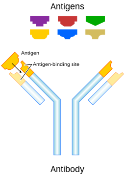
An illustration that shows how antigens induce the immune system response by interacting with an antibody that matches the molecular structure of an antigen

In immunology, an antigen (Ag) is a molecule or molecular structure, such as may be present on the outside of a pathogen, that can be bound by an antigen-specific antibody or B-cell antigen receptor. The presence of antigens in the body normally triggers an immune response. The Ag abbreviation stands for an antibody generator.

Antigens are "targeted" by antibodies. Each antibody is specifically produced by the immune system to match an antigen after cells in the immune system come into contact with it; this allows a precise identification or matching of the antigen and the initiation of an adaptive response. The antibody is said to "match" the antigen in the sense that it can bind to it due to an adaptation in a antigen-binding fragment of the antibody. In most cases, an adapted antibody can only react to and bind one specific antigen; in some instances, however, antibodies may cross-react and bind more than one antigen.

Antigens are proteins, peptides (amino acid chains) and polysaccharides (chains of monosaccharides/simple sugars) but lipids and nucleic acids become antigens only when combined with proteins and polysaccharides.

The antigen may originate from within the body ("self-protein") or from the external environment ("non-self"). The immune system identifies and attacks "non-self" external antigens and usually does not react to self-protein due to negative selection of T cells in the thymus.

Vaccines are examples of antigens in an immunogenic form, which are intentionally administered to a recipient to induce the memory function of the adaptive immune system towards antigens of the pathogen invading that recipient. Vaccines for the seasonal flu virus is a common example.

Etymology

Paul Ehrlich coined the term antibody (in German Antikörper) in his side-chain theory at the end of the 19th century. In 1899, Ladislas Deutsch (László Detre) (1874–1939) named the hypothetical substances halfway between bacterial constituents and antibodies "substances immunogenes ou antigenes" (antigenic or immunogenic substances). He originally believed those substances to be precursors of antibodies, just as zymogen is a precursor of an enzyme. But, by 1903, he understood that an antigen induces the production of immune bodies (antibodies) and wrote that the word antigen is a contraction of antisomatogen (Immunkörperbildner). The Oxford English Dictionary indicates that the logical construction should be "anti(body)-gen".

Terminology

  • Epitope – the distinct surface features of an antigen, its antigenic determinant.
    Antigenic molecules, normally "large" biological polymers, usually present surface features that can act as points of interaction for specific antibodies. Any such feature constitutes an epitope. Most antigens have the potential to be bound by multiple antibodies, each of which is specific to one of the antigen's epitopes. Using the "lock and key" metaphor, the antigen can be seen as a string of keys (epitopes) each of which matches a different lock (antibody). Different antibody idiotypes, each have distinctly formed complementarity-determining regions.
  • Allergen – A substance capable of causing an allergic reaction .The (detrimental) reaction may result after exposure via ingestion, inhalation, injection, or contact with skin.
  • Superantigen – A class of antigens that cause non-specific activation of T-cells, resulting in polyclonal T-cell activation and massive cytokine release.
  • Tolerogen – A substance that invokes a specific immune non-responsiveness due to its molecular form. If its molecular form is changed, a tolerogen can become an immunogen.
  • Immunoglobulin-binding protein – Proteins such as protein A, protein G, and protein L that are capable of binding to antibodies at positions outside of the antigen-binding site. While antigens are the "target" of antibodies, immunoglobulin-binding proteins "attack" antibodies.
  • T-dependent antigen – Antigens that require the assistance of T cells to induce the formation of specific antibodies.
  • T-independent antigen – Antigens that stimulate B cells directly.
  • Immunodominant antigens – Antigens that dominate (over all others from a pathogen) in their ability to produce an immune response. T cell responses typically are directed against a relatively few immunodominant epitopes, although in some cases (e.g., infection with the malaria pathogen Plasmodium spp.) it is dispersed over a relatively large number of parasite antigens.

Antigen-presenting cells present antigens in the form of peptides on histocompatibility molecules. The T cells selectively recognize the antigens; depending on the antigen and the type of the histocompatibility molecule, different types of T cells will be activated. For T-cell receptor (TCR) recognition, the peptide must be processed into small fragments inside the cell and presented by a major histocompatibility complex (MHC). The antigen cannot elicit the immune response without the help of an immunologic adjuvant. Similarly, the adjuvant component of vaccines plays an essential role in the activation of the innate immune system.

An immunogen is an antigen substance (or adduct) that is able to trigger a humoral (innate) or cell-mediated immune response. It first initiates an innate immune response, which then causes the activation of the adaptive immune response. An antigen binds the highly variable immunoreceptor products (B-cell receptor or T-cell receptor) once these have been generated. Immunogens are those antigens, termed immunogenic, capable of inducing an immune response.

At the molecular level, an antigen can be characterized by its ability to bind to an antibody's paratopes. Different antibodies have the potential to discriminate among specific epitopes present on the antigen surface. A hapten is a small molecule that changes the structure of an antigenic epitope. In order to induce an immune response, it needs to be attached to a large carrier molecule such as a protein (a complex of peptides). Antigens are usually carried by proteins and polysaccharides, and less frequently, lipids. This includes parts (coats, capsules, cell walls, flagella, fimbriae, and toxins) of bacteria, viruses, and other microorganisms. Lipids and nucleic acids are antigenic only when combined with proteins and polysaccharides.[citation needed] Non-microbial non-self antigens can include pollen, egg white, and proteins from transplanted tissues and organs or on the surface of transfused blood cells.

Sources

Antigens can be classified according to their source.

Exogenous antigens

Exogenous antigens are antigens that have entered the body from the outside, for example, by inhalation, ingestion or injection. The immune system's response to exogenous antigens is often subclinical. By endocytosis or phagocytosis, exogenous antigens are taken into the antigen-presenting cells (APCs) and processed into fragments. APCs then present the fragments to T helper cells (CD4+) by the use of class II histocompatibility molecules on their surface. Some T cells are specific for the peptide:MHC complex. They become activated and start to secrete cytokines, substances that activate cytotoxic T lymphocytes (CTL), antibody-secreting B cells, macrophages and other particles.

Some antigens start out as exogenous and later become endogenous (for example, intracellular viruses). Intracellular antigens can be returned to circulation upon the destruction of the infected cell.

Endogenous antigens

Endogenous antigens are generated within normal cells as a result of normal cell metabolism, or because of viral or intracellular bacterial infection. The fragments are then presented on the cell surface in the complex with MHC class I molecules. If activated cytotoxic CD8+ T cells recognize them, the T cells secrete various toxins that cause the lysis or apoptosis of the infected cell. In order to keep the cytotoxic cells from killing cells just for presenting self-proteins, the cytotoxic cells (self-reactive T cells) are deleted as a result of tolerance (negative selection). Endogenous antigens include xenogenic (heterologous), autologous and idiotypic or allogenic (homologous) antigens. Sometimes antigens are part of the host itself in an autoimmune disease.

Autoantigens

An autoantigen is usually a self-protein or protein complex (and sometimes DNA or RNA) that is recognized by the immune system of patients suffering from a specific autoimmune disease. Under normal conditions, these self-proteins should not be the target of the immune system, but in autoimmune diseases, their associated T cells are not deleted and instead attack.

Neoantigens

Neoantigens are those that are entirely absent from the normal human genome. As compared with nonmutated self-proteins, neoantigens are of relevance to tumor control, as the quality of the T cell pool that is available for these antigens is not affected by central T cell tolerance. Technology to systematically analyze T cell reactivity against neoantigens became available only recently. Neoantigens can be directly detected and quantified through a method called MANA-SRM developed by a molecular diagnostics company, Complete Omics Inc., through collaborating with a team in Johns Hopkins University School of Medicine.

Viral antigens

For virus-associated tumors, such as cervical cancer and a subset of head and neck cancers, epitopes derived from viral open reading frames contribute to the pool of neoantigens.

Tumor antigens

Tumor antigens are those antigens that are presented by MHC class I or MHC class II molecules on the surface of tumor cells. Antigens found only on such cells are called tumor-specific antigens (TSAs) and generally result from a tumor-specific mutation. More common are antigens that are presented by tumor cells and normal cells, called tumor-associated antigens (TAAs). Cytotoxic T lymphocytes that recognize these antigens may be able to destroy tumor cells.

Tumor antigens can appear on the surface of the tumor in the form of, for example, a mutated receptor, in which case they are recognized by B cells.

For human tumors without a viral etiology, novel peptides (neo-epitopes) are created by tumor-specific DNA alterations.

Process

A large fraction of human tumor mutations is effectively patient-specific. Therefore, neoantigens may also be based on individual tumor genomes. Deep-sequencing technologies can identify mutations within the protein-coding part of the genome (the exome) and predict potential neoantigens. In mice models, for all novel protein sequences, potential MHC-binding peptides were predicted. The resulting set of potential neoantigens was used to assess T cell reactivity. Exome–based analyses were exploited in a clinical setting, to assess reactivity in patients treated by either tumor-infiltrating lymphocyte (TIL) cell therapy or checkpoint blockade. Neoantigen identification was successful for multiple experimental model systems and human malignancies.

The false-negative rate of cancer exome sequencing is low—i.e.: the majority of neoantigens occur within exonic sequence with sufficient coverage. However, the vast majority of mutations within expressed genes do not produce neoantigens that are recognized by autologous T cells.

As of 2015 mass spectrometry resolution is insufficient to exclude many false positives from the pool of peptides that may be presented by MHC molecules. Instead, algorithms are used to identify the most likely candidates. These algorithms consider factors such as the likelihood of proteasomal processing, transport into the endoplasmic reticulum, affinity for the relevant MHC class I alleles and gene expression or protein translation levels.

The majority of human neoantigens identified in unbiased screens display a high predicted MHC binding affinity. Minor histocompatibility antigens, a conceptually similar antigen class are also correctly identified by MHC binding algorithms. Another potential filter examines whether the mutation is expected to improve MHC binding. The nature of the central TCR-exposed residues of MHC-bound peptides is associated with peptide immunogenicity.

Nativity

A native antigen is an antigen that is not yet processed by an APC to smaller parts. T cells cannot bind native antigens, but require that they be processed by APCs, whereas B cells can be activated by native ones.

Antigenic specificity

Antigenic specificity is the ability of the host cells to recognize an antigen specifically as a unique molecular entity and distinguish it from another with exquisite precision. Antigen specificity is due primarily to the side-chain conformations of the antigen. It is measurable and need not be linear or of a rate-limited step or equation. Both T cells and B cells are cellular components of adaptive immunity.
Transplant rejection

From Wikipedia, the free encyclopedia
 
Transplant rejection
Lung transplant rejection - high mag.jpg
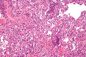
Micrograph showing lung transplant rejection. Lung biopsy. H&E stain.
SpecialtyImmunology
TreatmentImmunosuppressive drugs

Transplant rejection occurs when transplanted tissue is rejected by the recipient's immune system, which destroys the transplanted tissue. Transplant rejection can be lessened by determining the molecular similitude between donor and recipient and by use of immunosuppressant drugs after transplant.

Pretransplant rejection prevention

The first successful organ transplant, performed in 1954 by Joseph Murray, involved identical twins, and so no rejection was observed. Otherwise, the number of mismatched gene variants, namely alleles, encoding cell surface molecules called major histocompatibility complex (MHC), classes I and II, correlate with the rapidity and severity of transplant rejection. In humans MHC is also called human leukocyte antigen (HLA).

Though cytotoxic-crossmatch assay can predict rejection mediated by cellular immunity, genetic-expression tests specific to the organ type to be transplanted, for instance AlloMap Molecular Expression Testing, have a high negative predictive value. Transplanting only ABO-compatible grafts (matching blood groups between donor and recipient) helps prevent rejection mediated by humoral immunity.

ABO-incompatible transplants

Because very young children (generally under 12 months, but often as old as 24 months) do not have a well-developed immune system, it is possible for them to receive organs from otherwise incompatible donors. This is known as ABO-incompatible (ABOi) transplantation. Graft survival and patient mortality is approximately the same between ABOi and ABO-compatible (ABOc) recipients. While focus has been on infant heart transplants, the principles generally apply to other forms of solid organ transplantation.

The most important factors are that the recipient not have produced isohemagglutinins, and that they have low levels of T cell-independent antigens. UNOS regulations allow for ABOi transplantation in children under two years of age if isohemagglutinin titers are 1:4 or below, and if there is no matching ABOc recipient. Studies have shown that the period under which a recipient may undergo ABOi transplantation may be prolonged by exposure to nonself A and B antigens. Furthermore, should the recipient (for example, type B-positive with a type AB-positive graft) require eventual retransplantation, the recipient may receive a new organ of either blood type.

Limited success has been achieved in ABO-incompatible heart transplants in adults, though this requires that the adult recipients have low levels of anti-A or anti-B antibodies. Kidney transplantation is more successful, with similar long-term graft survival rates to ABOc transplants.

Immunologic mechanisms of rejection

Rejection is an adaptive immune response via cellular immunity (mediated by killer T cells inducing apoptosis of target cells) as well as humoral immunity (mediated by activated B cells secreting antibody molecules), though the action is joined by components of innate immune response (phagocytes and soluble immune proteins). Different types of transplanted tissues tend to favor different balances of rejection mechanisms.

Immunization

An animal's exposure to the antigens of a different member of the same or similar species is allostimulation, and the tissue is allogenic. Transplanted organs are often acquired from a cadaver (usually a host who had succumbed to trauma), whose tissues had already sustained ischemia or inflammation.

Dendritic cells (DCs), which are the primary antigen-presenting cells (APCs), of the donor tissue migrate to the recipient's peripheral lymphoid tissue (lymphoid follicles and lymph nodes), and present the donor's self peptides to the recipient's lymphocytes (immune cells residing in lymphoid tissues). Lymphocytes include two classes that enact adaptive immunity, also called specific immunity. Lymphocytes of specific immunity T cells—including the subclasses helper T cells and killer T cells—and B cells.

The recipient's helper T cells coordinate specific immunity directed at the donor's self peptides or at the donor's Major histocompatibility complex molecules, or at both.

Immune memory

When memory helper T cells' CD4 receptors bind to the MHC class II molecules which are expressed on the surfaces of the target cells of the graft tissue, the memory helper T cells' T cell receptors (TCRs) can recognize their target antigen that is presented by the MHC class II molecules. The memory helper T cell subsequently produces clones that, as effector cells, secrete immune signalling molecules (cytokines) in approximately the cytokine balance that had prevailed at the memory helper T cell's priming to memorize the antigen. As the priming event in this instance occurred amid inflammation, the immune memory is pro-inflammatory.

Cellular immunity

Transplant rejection is a Type IV (“delayed”) hypersensitivity reaction mediated by T cells in which the transplant recipient’s T cells become alloreactive, recognizing major histocompatibility complex (MHC) antigens on the donated organ, and promote local immune and inflammatory responses to defend against the perceived threat. The T cells can recognize the donated organ’s MHC antigens through one of two routes: a direct pathway, where the transplanted organ’s antigen-presenting cells (such as dendritic cells that contain MHC class I molecules) migrate to the recipient’s lymph nodes and activate T cells that reside there; or an indirect pathway, in which the recipient’s dendritic cells can endocytose donor proteins such as donor MHC proteins and activate T cells in secondary lymphoid tissues. Alloreactive killer T cells, also called cytotoxic T lymphocytes (CTLs), have CD8 receptors that dock to the transplanted tissue's MHC class I molecules, which display the donor's self peptides. (In the living donor, such presentation of self antigens helped maintain self tolerance.) Thereupon, the T cell receptors (TCRs) of the killer T cells recognize their matching epitope, and trigger the target cell's programmed cell death by apoptosis.

Humoral immunity

Developed through an earlier primary exposure that primed specific immunity to the nonself antigen, a transplant recipient can have specific antibody crossreacting with the donor tissue upon the transplant event, a secondary exposure. This is typical of minor blood group exposure (e.g. Kell) following allogenic blood transfusion or trauma during pregnancy. At secondary exposure, these crossreactive antibody molecules interact with aspects of innate immunity—soluble immune proteins called complement and innate immune cells called phagocytes—which inflames and destroys the transplanted tissue.

Antibody

Secreted by an activated B cell, then called plasma cell, an antibody molecule is a soluble immunoglobulin (Ig) whose basic unit is shaped like the letter Y: the two arms are the Fab regions, while the single stalk is the Fc region. Each of the two tips of Fab region is the paratope, which binds a matching molecular sequence and its 3D shape (conformation), altogether called epitope, within the target antigen.

Opsonization

The IgG's Fc region also enables opsonization by a phagocyte, a process by which the Fc receptor on the phagocyte—such as neutrophils in blood and macrophages in tissues—binds the antibody molecule's FC stalk, and the phagocyte exhibits enhanced uptake of the antigen, attached to the antibody molecule's Fab region.

Complement cascade

When the paratope of Ig class gamma (IgG) binds its matching epitope, IgG's Fc region conformationally shifts and can host a complement protein, initiating the complement cascade that terminates by punching a hole in a cell membrane. With many holes so punched, fluid rushes into the cell and ruptures it.

Cell debris can be recognized as damage associated molecular patterns (DAMPs) by pattern recognition receptors (PRRs), such as Toll-like receptors (TLRs), on membranes of phagocytes, which thereupon secrete proinflammatory cytokines, recruiting more phagocytes to traffic to the area by sensing the concentration gradient of the secreted cytokines (chemotaxis).

Tissue Mechanism
Blood Antibodies (isohaemagglutinins)
Kidney Antibodies, cell-mediated immunity (CMI)
Heart Antibodies, CMI
Skin CMI
Bonemarrow CMI
Cornea Usually accepted unless vascularised: CMI

Medical categories

Hyperacute rejection

Initiated by preexisting humoral immunity, hyperacute rejection manifests within minutes after transplant, and if tissue is left implanted brings systemic inflammatory response syndrome. Of high risk in kidney transplants is rapid clumping, namely agglutination, of red blood cells (RBCs or erythrocytes), as an antibody molecule binds multiple target cells at once.

While kidneys can routinely be obtained from human donors, most organs are in short supply leading to consideration of xenotransplants from other species. Pigs are especially likely sources for xenotransplants, chosen for the anatomical and physiological characteristics they share with humans. However, the sugar galactose-alpha-1,3-galactose (αGal) has been implicated as a major factor in hyperacute rejection in xenotransplantation. Unlike virtually all other mammals, humans and other primates do not make αGal, and in fact recognize it as an antigen. During transplantation, xenoreactive natural antibodies recognize αGal on the graft endothelium as an antigen, and the resulting complement-mediated immune response leads to a rejection of the transplant.

Acute rejection

Developing with formation of cellular immunity, acute rejection occurs to some degree in all transplants, except between identical twins, unless immunosuppression is achieved (usually through drugs). Acute rejection begins as early as one week after transplant, the risk being highest in the first three months, though it can occur months to years later. Highly vascular tissues such as kidney or liver often host the earliest signs—particularly at endothelial cells lining blood vessels—though it eventually occurs in roughly 10 to 30% of liver transplants, and 10 to 20% of kidney transplants. A single episode of acute rejection can be recognized and promptly treated, usually preventing organ failure, but recurrent episodes lead to chronic rejection. It is believed that the process of acute rejection is mediated by the cell mediated pathway, specifically by mononuclear macrophages and T-lymphocytes. Histology of acute rejection is defined by dense lymphocytic cellular infiltrate as well as vasculitis of organ donor vessels.

Chronic rejection

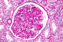
Micrograph showing a glomerulus with changes characteristic of a transplant glomerulopathy. Transplant glomerulopathy is considered a form of chronic antibody-mediated rejection. PAS stain.

The term chronic rejection initially described long-term loss of function in transplanted organs via fibrosis of the transplanted tissue's blood vessels. This is now chronic allograft vasculopathy, however, leaving chronic rejection referring to rejection due to more patent aspects of immunity.[citation needed]

Chronic rejection explains long-term morbidity in most lung-transplant recipients, the median survival roughly 4.7 years, about half the span versus other major organ transplants. In histopathology the condition is bronchiolitis obliterans, which clinically presents as progressive airflow obstruction, often involving dyspnea and coughing, and the patient eventually succumbs to pulmonary insufficiency or secondary acute infection.

Airflow obstruction not ascribable to other cause is labeled bronchiolitis obliterans syndrome (BOS), confirmed by a persistent drop—three or more weeks—in forced expiratory volume (FEV1) by at least 20%. BOS is seen in over 50% of lung-transplant recipients by 5 years, and in over 80% by ten years. First noted is infiltration by lymphocytes, followed by epithelial cell injury, then inflammatory lesions and recruitment of fibroblasts and myofibroblasts, which proliferate and secrete proteins forming scar tissue. Generally thought unpredictable, BOS progression varies widely: lung function may suddenly fall but stabilize for years, or rapidly progress to death within a few months. Risk factors include prior acute rejection episodes, gastroesophageal reflux disease, acute infections, particular age groups, HLA mis-matching, lymphocytic bronchiolitis, and graft dysfunction (e.g., airway ischemia).

Rejection due to non-adherence

One principal reason for transplant rejection is non-adherence to prescribed immunosuppressant regimens. This is particularly the case with adolescent recipients, with non-adherence rates near 50% in some instances.

Rejection detection

Diagnosis of acute rejection relies on clinical data—patient signs and symptoms but also calls on laboratory data such as blood or even tissue biopsy. The laboratory pathologist generally seeks three main histological signs: (1) infiltrating T cells, perhaps accompanied by infiltrating eosinophils, plasma cells, and neutrophils, particularly in telltale ratios, (2) structural compromise of tissue anatomy, varying by tissue type transplanted, and (3) injury to blood vessels. Tissue biopsy is restricted, however, by sampling limitations and risks/complications of the invasive procedure. Cellular magnetic resonance imaging (MRI) of immune cells radiolabeled in vivo might—similarly to Gene Expression Profiling (GEP)—offer noninvasive testing.

Rejection treatment

Hyperacute rejection manifests severely and within minutes, and so treatment is immediate: removal of the tissue. Chronic rejection is generally considered irreversible and poorly amenable to treatment—only retransplant generally indicated if feasible—though inhaled ciclosporin is being investigated to delay or prevent chronic rejection of lung transplants. Acute rejection is treated with one or several of a few strategies. Despite treatment, rejection remains a major cause of transplant failure.

Immunosuppressive therapy

A short course of high-dose corticosteroids can be applied, and repeated. Triple therapy adds a calcineurin inhibitor and an anti-proliferative agent. Where calcineurin inhibitors or steroids are contraindicated, mTOR inhibitors are used.

Immunosuppressive drugs:

Antibody-based treatments

Antibody specific to select immune components can be added to immunosuppressive therapy. The monoclonal anti-T cell antibody OKT3, once used to prevent rejection, and still occasionally used to treat severe acute rejection, has fallen into disfavor, as it commonly brings severe cytokine release syndrome and late post-transplant lymphoproliferative disorder. (OKT3 is available in the United Kingdom for named-patient use only.)

Antibody drugs:

Blood transfer

Cases refractory to immunosuppressive or antibody therapy are sometimes treated with photopheresis, or extracorporeal photoimmune therapy (ECP), to remove antibody molecules specific to the transplanted tissue.

Marrow transplant

Bone marrow transplant can replace the transplant recipient's immune system with the donor's, and the recipient accepts the new organ without rejection. The marrow's hematopoietic stem cells—the reservoir of stem cells replenishing exhausted blood cells including white blood cells forming the immune system—must be of the individual who donated the organ or of an identical twin or a clone. There is a risk of graft-versus-host disease (GVHD), however, whereby mature lymphocytes entering with marrow recognize the new host tissues as foreign and destroy them.

Gene therapy

Gene therapy is another method that can be used. In this method, the genes that cause the body to reject transplants would be deactivated. Research is still being conducted, and no gene therapies are being used to date to treat patients. Current research tends to focus on Th1 and Th17 which mediate allograft rejection via the CD4 and CD8 T cells.

Immunosuppressive drug

From Wikipedia, the free encyclopedia
 

Immunosuppressive drugs, also known as immunosuppressive agents, immunosuppressants and antirejection medications are drugs that inhibit or prevent activity of the immune system.

Classification

Immunosuppressive drugs can be classified into five groups:

Glucocorticoids

In pharmacologic (supraphysiologic) doses, glucocorticoids, such as prednisone, dexamethasone, and hydrocortisone are used to suppress various allergic, inflammatory, and autoimmune disorders. They are also administered as posttransplantory immunosuppressants to prevent the acute transplant rejection and graft-versus-host disease. Nevertheless, they do not prevent an infection and also inhibit later reparative processes.

Immunosuppressive mechanism

Glucocorticoids suppress the cell-mediated immunity. They act by inhibiting genes that code for the cytokines Interleukin 1 (IL-1), IL-2, IL-3, IL-4, IL-5, IL-6, IL-8, and TNF-alpha, the most important of which is IL-2. Smaller cytokine production reduces the T cell proliferation.

Glucocorticoids also suppress the humoral immunity, causing B cells to express smaller amounts of IL-2 and IL-2 receptors. This diminishes both B cell clone expansion and antibody synthesis.

Anti-inflammatory effects

Glucocorticoids influence all types of inflammatory events, no matter their cause. They induce the lipocortin-1 (annexin-1) synthesis, which then binds to cell membranes preventing the phospholipase A2 from coming into contact with its substrate arachidonic acid. This leads to diminished eicosanoid production. The cyclooxygenase (both COX-1 and COX-2) expression is also suppressed, potentiating the effect.

Glucocorticoids also stimulate the lipocortin-1 escaping to the extracellular space, where it binds to the leukocyte membrane receptors and inhibits various inflammatory events: epithelial adhesion, emigration, chemotaxis, phagocytosis, respiratory burst, and the release of various inflammatory mediators (lysosomal enzymes, cytokines, tissue plasminogen activator, chemokines, etc.) from neutrophils, macrophages, and mastocytes.

Cytostatics

Cytostatics inhibit cell division. In immunotherapy, they are used in smaller doses than in the treatment of malignant diseases. They affect the proliferation of both T cells and B cells. Due to their highest effectiveness, purine analogs are most frequently administered.

Alkylating agents

The alkylating agents used in immunotherapy are nitrogen mustards (cyclophosphamide), nitrosoureas, platinum compounds, and others. Cyclophosphamide (Baxter's Cytoxan) is probably the most potent immunosuppressive compound. In small doses, it is very efficient in the therapy of systemic lupus erythematosus, autoimmune hemolytic anemias, granulomatosis with polyangiitis, and other immune diseases. High doses cause pancytopenia and hemorrhagic cystitis.

Antimetabolites

Antimetabolites interfere with the synthesis of nucleic acids. These include:

Methotrexate

Methotrexate is a folic acid analogue. It binds dihydrofolate reductase and prevents synthesis of tetrahydrofolate. It is used in the treatment of autoimmune diseases (for example rheumatoid arthritis or Behcet's Disease) and in transplantations.

Azathioprine and mercaptopurine

Azathioprine (Prometheus' Imuran), is the main immunosuppressive cytotoxic substance. It is extensively used to control transplant rejection reactions. It is nonenzymatically cleaved to mercaptopurine, that acts as a purine analogue and an inhibitor of DNA synthesis. Mercaptopurine itself can also be administered directly.

By preventing the clonal expansion of lymphocytes in the induction phase of the immune response, it affects both the cell and the humoral immunity. It is also efficient in the treatment of autoimmune diseases.

Cytotoxic antibiotics

Among these, dactinomycin is the most important. It is used in kidney transplantations. Other cytotoxic antibiotics are anthracyclines, mitomycin C, bleomycin, mithramycin.

Antibodies

Antibodies are sometimes used as a quick and potent immunosuppressive therapy to prevent the acute rejection reactions as well as a targeted treatment of lymphoproliferative or autoimmune disorders (e.g., anti-CD20 monoclonals).

Polyclonal antibodies

Heterologous polyclonal antibodies are obtained from the serum of animals (e.g., rabbit, horse), and injected with the patient's thymocytes or lymphocytes. The antilymphocyte (ALG) and antithymocyte antigens (ATG) are being used. They are part of the steroid-resistant acute rejection reaction and grave aplastic anemia treatment. However, they are added primarily to other immunosuppressives to diminish their dosage and toxicity. They also allow transition to cyclosporin therapy.

Polyclonal antibodies inhibit T lymphocytes and cause their lysis, which is both complement-mediated cytolysis and cell-mediated opsonization followed by removal of reticuloendothelial cells from the circulation in the spleen and liver. In this way, polyclonal antibodies inhibit cell-mediated immune reactions, including graft rejection, delayed hypersensitivity (i.e., tuberculin skin reaction), and the graft-versus-host disease (GVHD), but influence thymus-dependent antibody production.

As of March 2005, there are two preparations available to the market: Atgam, obtained from horse serum, and Thymoglobuline, obtained from rabbit serum. Polyclonal antibodies affect all lymphocytes and cause general immunosuppression, possibly leading to post-transplant lymphoproliferative disorders (PTLD) or serious infections, especially by cytomegalovirus. To reduce these risks, treatment is provided in a hospital, where adequate isolation from infection is available. They are usually administered for five days intravenously in the appropriate quantity. Patients stay in the hospital as long as three weeks to give the immune system time to recover to a point where there is no longer a risk of serum sickness.

Because of a high immunogenicity of polyclonal antibodies, almost all patients have an acute reaction to the treatment. It is characterized by fever, rigor episodes, and even anaphylaxis. Later during the treatment, some patients develop serum sickness or immune complex glomerulonephritis. Serum sickness arises seven to fourteen days after the therapy has begun. The patient suffers from fever, joint pain, and erythema that can be soothed with the use of steroids and analgesics. Urticaria (hives) can also be present. It is possible to diminish their toxicity by using highly purified serum fractions and intravenous administration in the combination with other immunosuppressants, for example, calcineurin inhibitors, cytostatics, and corticosteroids. The most frequent combination is to use antibodies and ciclosporin simultaneously in order to prevent patients from gradually developing a strong immune response to these drugs, reducing or eliminating their effectiveness.

Monoclonal antibodies

Monoclonal antibodies are directed towards exactly defined antigens. Therefore, they cause fewer side-effects. Especially significant are the IL-2 receptor- (CD25-) and CD3-directed antibodies. They are used to prevent the rejection of transplanted organs, but also to track changes in the lymphocyte subpopulations. It is reasonable to expect similar new drugs in the future.

T-cell receptor directed antibodies

Muromonab-CD3 is a murine anti-CD3 monoclonal antibody of the IgG2a type that was previously used to prevent T-cell activation and proliferation by binding the T-cell receptor complex present on all differentiated T cells. As such it was one of the first potent immunosuppressive substances and was administered to control the steroid- and/or polyclonal antibodies-resistant acute rejection episodes. As it acts more specifically than polyclonal antibodies it was also used prophylactically in transplantations. However, muromonab-CD3 is no longer produced,[1] and this mouse monoclonal antibody has been replaced in the clinic with chimeric, humanized, or human monoclonal antibodies.

The muromonab's mechanism of action is only partially understood. It is known that the molecule binds TCR/CD3 receptor complex. In the first few administrations this binding non-specifically activates T-cells, leading to a serious syndrome 30 to 60 minutes later. It is characterized by fever, myalgia, headache, and arthralgia. Sometimes it develops in a life-threatening reaction of the cardiovascular system and the central nervous system, requiring a lengthy therapy. Past this period CD3 blocks the TCR-antigen binding and causes conformational change or the removal of the entire TCR3/CD3 complex from the T-cell surface. This lowers the number of available T-cells, perhaps by sensitizing them for the uptake by the epithelial reticular cells. The cross-binding of CD3 molecules as well activates an intracellular signal causing the T cell anergy or apoptosis, unless the cells receive another signal through a co-stimulatory molecule. CD3 antibodies shift the balance from Th1 to Th2 cells as CD3 stimulates Th1 activation.[2]

The patient may develop neutralizing antibodies reducing the effectiveness of muromonab-CD3. Muromonab-CD3 can cause excessive immunosuppression. Although CD3 antibodies act more specifically than polyclonal antibodies, they lower the cell-mediated immunity significantly, predisposing the patient to opportunistic infections and malignancies.[3]

IL-2 receptor directed antibodies

Interleukin-2 is an important immune system regulator necessary for the clone expansion and survival of activated lymphocytes T. Its effects are mediated by the trimer cell surface receptor IL-2a, consisting of the α, β, and γ chains. The IL-2a (CD25, T-cell activation antigen, TAC) is expressed only by the already-activated T lymphocytes. Therefore, it is of special significance to the selective immunosuppressive treatment, and research has been focused on the development of effective and safe anti-IL-2 antibodies. By the use of recombinant gene technology, the mouse anti-Tac antibodies have been modified, leading to the presentation of two chimeric mouse/human anti-Tac antibodies in the year 1998: basiliximab (Simulect) and daclizumab (Zenapax). These drugs act by binding the IL-2a receptor's α chain, preventing the IL-2 induced clonal expansion of activated lymphocytes and shortening their survival. They are used in the prophylaxis of the acute organ rejection after bilateral kidney transplantation, both being similarly effective and with only few side-effects.

Drugs acting on immunophilins

Ciclosporin

Like tacrolimus, ciclosporin (Novartis' Sandimmune) is a calcineurin inhibitor (CNI). It has been in use since 1983 and is one of the most widely used immunosuppressive drugs. It is a cyclic fungal peptide, composed of 11 amino acids.

Ciclosporin is thought to bind to the cytosolic protein cyclophilin (an immunophilin) of immunocompetent lymphocytes, especially T-lymphocytes. This complex of ciclosporin and cyclophilin inhibits the phosphatase calcineurin, which under normal circumstances induces the transcription of interleukin-2. The drug also inhibits lymphokine production and interleukin release, leading to a reduced function of effector T-cells.

Ciclosporin is used in the treatment of acute rejection reactions, but has been increasingly substituted with newer, and less nephrotoxic, immunosuppressants.

Calcineurin inhibitors and azathioprine have been linked with post-transplant malignancies and skin cancers in organ transplant recipients. Non-melanoma skin cancer (NMSC) after kidney transplantation is common and can result in significant morbidity and mortality. The results of several studies suggest that calcineurin inhibitors have oncogenic properties mainly linked to the production of cytokines that promote tumor growth, metastasis and angiogenesis.

This drug has been reported to reduce the frequency of regulatory T cells (T-Reg) and after converting from a CNI monotherapy to a mycophenolate monotherapy, patients were found to have increased graft success and T-Reg frequency.

Tacrolimus

Tacrolimus (trade names Prograf, Astagraf XL, Envarsus XR) is a product of the bacterium Streptomyces tsukubaensis. It is a macrolide lactone and acts by inhibiting calcineurin.

The drug is used primarily in liver and kidney transplantations, although in some clinics it is used in heart, lung, and heart/lung transplantations. It binds to the immunophilin FKBP1A, followed by the binding of the complex to calcineurin and the inhibition of its phosphatase activity. In this way, it prevents the cell from transitioning from the G0 into G1 phase of the cell cycle. Tacrolimus is more potent than ciclosporin and has less pronounced side-effects.

Sirolimus

Sirolimus (rapamycin, trade name Rapamune) is a macrolide lactone, produced by the actinomycete bacterium Streptomyces hygroscopicus. It is used to prevent rejection reactions. Although it is a structural analogue of tacrolimus, it acts somewhat differently and has different side-effects.

Contrary to ciclosporin and tacrolimus, drugs that affect the first phase of T lymphocyte activation, sirolimus affects the second phase, namely signal transduction and lymphocyte clonal proliferation. It binds to FKBP1A like tacrolimus, however the complex does not inhibit calcineurin but another protein, mTOR. Therefore, sirolimus acts synergistically with ciclosporin and, in combination with other immunosuppressants, has few side effects. Also, it indirectly inhibits several T lymphocyte-specific kinases and phosphatases, hence preventing their transition from G1 to S phase of the cell cycle. In a similar manner, Sirolimus prevents B cell differentiation into plasma cells, reducing production of IgM, IgG, and IgA antibodies.

It is also active against tumors that are PI3K/AKT/mTOR-dependent.

Everolimus

Everolimus is an analog of sirolimus and also is an mTOR inhibitor.

Zotarolimus

Zotarolimus is a semi-synthetic derivative of sirolimus used in drug-eluting stents.

Other drugs

Interferons

IFN-β suppresses the production of Th1 cytokines and the activation of monocytes. It is used to slow down the progression of multiple sclerosis. IFN-γ is able to trigger lymphocytic apoptosis.

Opioids

Prolonged use of opioids may cause immunosuppression of both innate and adaptive immunity. Decrease in proliferation as well as immune function has been observed in macrophages, as well as lymphocytes. It is thought that these effects are mediated by opioid receptors expressed on the surface of these immune cells.

TNF binding proteins

A TNF-α (tumor necrosis factor-alpha) binding protein is a monoclonal antibody or a circulating receptor such as infliximab (Remicade), etanercept (Enbrel), or adalimumab (Humira) that binds to TNF-α, preventing it from inducing the synthesis of IL-1 and IL-6 and the adhesion of lymphocyte-activating molecules. They are used in the treatment of rheumatoid arthritis, ankylosing spondylitis, Crohn's disease, and psoriasis.

These drugs may raise the risk of contracting tuberculosis or inducing a latent infection to become active. Infliximab and adalimumab have label warnings stating that patients should be evaluated for latent TB infection and treatment should be initiated prior to starting therapy with them.

TNF or the effects of TNF are also suppressed by various natural compounds, including curcumin (an ingredient in turmeric) and catechins (in green tea).

Mycophenolate

Mycophenolic acid acts as a non-competitive, selective, and reversible inhibitor of Inosine-5′-monophosphate dehydrogenase (IMPDH), which is a key enzyme in the de novo guanosine nucleotide synthesis. In contrast to other human cell types, lymphocytes B and T are very dependent on this process. Mycophenolate mofetil is used in combination with ciclosporin or tacrolimus in transplant patients.

Small biological agents

Fingolimod is a new synthetic immunosuppressant, currently in phase 3 of clinical trials. It increases the expression or changes the function of certain adhesion molecules (α4/β7 integrin) in lymphocytes, so they accumulate in the lymphatic tissue (lymphatic nodes) and their number in the circulation is diminished. In this respect, it differs from all other known immunosuppressants.

Myriocin has been reported being 10 to 100 times more potent than Ciclosporin.

Therapy

Immunosuppressive drugs are used in immunosuppressive therapy to:

Side effects

A common side-effect of many immunosuppressive drugs is immunodeficiency, because the majority of them act non-selectively, resulting in increased susceptibility to infections and decreased cancer immunosurveillance. There are also other side-effects, such as hypertension, dyslipidemia, hyperglycemia, peptic ulcers, lipodystrophy, moon face, liver and kidney injury. The immunosuppressive drugs also interact with other medicines and affect their metabolism and action. Actual or suspected immunosuppressive agents can be evaluated in terms of their effects on lymphocyte subpopulations in tissues using immunohistochemistry.

Self-image

From Wikipedia, the free encyclopedia ...