Search This Blog

Tuesday, November 30, 2021

Carbon-fiber-reinforced polymers

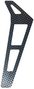
Tail of a radio-controlled helicopter, made of CFRP

Carbon Fiber-reinforced polymers (American English), carbon-fibre-reinforced polymers (Commonwealth English), or carbon-fiber-reinforced plastics, or carbon-fiber reinforced-thermoplastic (CFRP, CRP, CFRTP, also known as carbon fiber, carbon composite, or just carbon), are extremely strong and light fiber-reinforced plastics that contain carbon fibers. CFRPs can be expensive to produce, but are commonly used wherever high strength-to-weight ratio and stiffness (rigidity) are required, such as aerospace, superstructures of ships, automotive, civil engineering, sports equipment, and an increasing number of consumer and technical applications.

The binding polymer is often a thermoset resin such as epoxy, but other thermoset or thermoplastic polymers, such as polyester, vinyl ester, or nylon, are sometimes used. The properties of the final CFRP product can be affected by the type of additives introduced to the binding matrix (resin). The most common additive is silica, but other additives such as rubber and carbon nanotubes can be used.

Carbon fiber is sometimes referred to as graphite-reinforced polymer or graphite fiber-reinforced polymer (GFRP is less common, as it clashes with glass-(fiber)-reinforced polymer).

Properties

CFRP are composite materials. In this case the composite consists of two parts: a matrix and a reinforcement. In CFRP the reinforcement is carbon fiber, which provides its strength. The matrix is usually a polymer resin, such as epoxy, to bind the reinforcements together. Because CFRP consists of two distinct elements, the material properties depend on these two elements.

Reinforcement gives CFRP its strength and rigidity, measured by stress and elastic modulus respectively. Unlike isotropic materials like steel and aluminum, CFRP has directional strength properties. The properties of CFRP depend on the layouts of the carbon fiber and the proportion of the carbon fibers relative to the polymer. The two different equations governing the net elastic modulus of composite materials using the properties of the carbon fibers and the polymer matrix can also be applied to carbon fiber reinforced plastics. The following equation,

is valid for composite materials with the fibers oriented in the direction of the applied load. is the total composite modulus, and are the volume fractions of the matrix and fiber respectively in the composite, and and are the elastic moduli of the matrix and fibers respectively. The other extreme case of the elastic modulus of the composite with the fibers oriented transverse to the applied load can be found using the following equation:

The fracture toughness of carbon fiber reinforced plastics is governed by the following mechanisms: 1) debonding between the carbon fiber and polymer matrix, 2) fiber pull-out, and 3) delamination between the CFRP sheets. Typical epoxy-based CFRPs exhibit virtually no plasticity, with less than 0.5% strain to failure. Although CFRPs with epoxy have high strength and elastic modulus, the brittle fracture mechanics present unique challenges to engineers in failure detection since failure occurs catastrophically. As such, recent efforts to toughen CFRPs include modifying the existing epoxy material and finding alternative polymer matrix. One such material with high promise is PEEK, which exhibits an order of magnitude greater toughness with similar elastic modulus and tensile strength. However, PEEK is much more difficult to process and more expensive.

Despite its high initial strength-to-weight ratio, a design limitation of CFRP is its lack of a definable fatigue limit. This means, theoretically, that stress cycle failure cannot be ruled out. While steel and many other structural metals and alloys do have estimable fatigue or endurance limits, the complex failure modes of composites mean that the fatigue failure properties of CFRP are difficult to predict and design against. As a result, when using CFRP for critical cyclic-loading applications, engineers may need to design in considerable strength safety margins to provide suitable component reliability over its service life.

Environmental effects such as temperature and humidity can have profound effects on the polymer-based composites, including most CFRPs. While CFRPs demonstrate excellent corrosion resistance, the effect of moisture at wide ranges of temperatures can lead to degradation of the mechanical properties of CFRPs, particularly at the matrix-fiber interface. While the carbon fibers themselves are not affected by the moisture diffusing into the material, the moisture plasticizes the polymer matrix. This led to significant changes in properties that are dominantly influenced by the matrix in CFRPs such as compressive, interlaminar shear, and impact properties. The epoxy matrix used for engine fan blades is designed to be impervious against jet fuel, lubrication, and rain water, and external paint on the composites parts is applied to minimize damage from ultraviolet light.

Carbon fibers can cause galvanic corrosion when CRP parts are attached to aluminum or mild steel but not to stainless steel or titanium.[9]

Carbon Fiber Reinforced Plastics are very hard to machine, and causes significant tool wear. The tool wear in CFRP machining is dependent on the fiber orientation and machining condition of the cutting process. To reduce tool wear various types of coated tools are used in machining CFRP and CFRP-metal stack.

Manufacture

Carbon fiber reinforced polymer

The primary element of CFRP is a carbon filament; this is produced from a precursor polymer such as polyacrylonitrile (PAN), rayon, or petroleum pitch. For synthetic polymers such as PAN or rayon, the precursor is first spun into filament yarns, using chemical and mechanical processes to initially align the polymer chains in a way to enhance the final physical properties of the completed carbon fiber. Precursor compositions and mechanical processes used during spinning filament yarns may vary among manufacturers. After drawing or spinning, the polymer filament yarns are then heated to drive off non-carbon atoms (carbonization), producing the final carbon fiber. The carbon fibers filament yarns may be further treated to improve handling qualities, then wound on to bobbins. From these fibers, a unidirectional sheet is created. These sheets are layered onto each other in a quasi-isotropic layup, e.g. 0°, +60°, or −60° relative to each other.

From the elementary fiber, a bidirectional woven sheet can be created, i.e. a twill with a 2/2 weave. The process by which most CFRPs are made varies, depending on the piece being created, the finish (outside gloss) required, and how many of the piece will be produced. In addition, the choice of matrix can have a profound effect on the properties of the finished composite.

Many CFRP parts are created with a single layer of carbon fabric that is backed with fiberglass. A tool called a chopper gun is used to quickly create these composite parts. Once a thin shell is created out of carbon fiber, the chopper gun cuts rolls of fiberglass into short lengths and sprays resin at the same time, so that the fiberglass and resin are mixed on the spot. The resin is either external mix, wherein the hardener and resin are sprayed separately, or internal mixed, which requires cleaning after every use. Manufacturing methods may include the following:

Molding

One method of producing CFRP parts is by layering sheets of carbon fiber cloth into a mold in the shape of the final product. The alignment and weave of the cloth fibers is chosen to optimize the strength and stiffness properties of the resulting material. The mold is then filled with epoxy and is heated or air-cured. The resulting part is very corrosion-resistant, stiff, and strong for its weight. Parts used in less critical areas are manufactured by draping cloth over a mold, with epoxy either preimpregnated into the fibers (also known as pre-preg) or "painted" over it. High-performance parts using single molds are often vacuum-bagged and/or autoclave-cured, because even small air bubbles in the material will reduce strength. An alternative to the autoclave method is to use internal pressure via inflatable air bladders or EPS foam inside the non-cured laid-up carbon fiber.

Vacuum bagging

For simple pieces of which relatively few copies are needed (1–2 per day), a vacuum bag can be used. A fiberglass, carbon fiber, or aluminum mold is polished and waxed, and has a release agent applied before the fabric and resin are applied, and the vacuum is pulled and set aside to allow the piece to cure (harden). There are three ways to apply the resin to the fabric in a vacuum mold.

The first method is manual and called a wet layup, where the two-part resin is mixed and applied before being laid in the mold and placed in the bag. The other one is done by infusion, where the dry fabric and mold are placed inside the bag while the vacuum pulls the resin through a small tube into the bag, then through a tube with holes or something similar to evenly spread the resin throughout the fabric. Wire loom works perfectly for a tube that requires holes inside the bag. Both of these methods of applying resin require hand work to spread the resin evenly for a glossy finish with very small pin-holes.

A third method of constructing composite materials is known as a dry layup. Here, the carbon fiber material is already impregnated with resin (pre-preg) and is applied to the mold in a similar fashion to adhesive film. The assembly is then placed in a vacuum to cure. The dry layup method has the least amount of resin waste and can achieve lighter constructions than wet layup. Also, because larger amounts of resin are more difficult to bleed out with wet layup methods, pre-preg parts generally have fewer pinholes. Pinhole elimination with minimal resin amounts generally require the use of autoclave pressures to purge the residual gases out.

Compression molding

A quicker method uses a compression mold. This is a two-piece (male and female) mold usually made out of aluminum or steel that is pressed together with the fabric and resin between the two. The benefit is the speed of the entire process. Some car manufacturers, such as BMW, claimed to be able to cycle a new part every 80 seconds. However, this technique has a very high initial cost since the molds require CNC machining of very high precision.

Filament winding

For difficult or convoluted shapes, a filament winder can be used to make CFRP parts by winding filaments around a mandrel or a core.

Applications

Applications for CFRP include the following:

Aerospace engineering

An Airbus A350 with carbon fiber themed livery. Composite materials are used extensively throughout the A350.

The Airbus A350 XWB is built of 52% CFRP including wing spars and fuselage components, overtaking the Boeing 787 Dreamliner, for the aircraft with the highest weight ratio for CFRP, which is 50%. This was one of the first commercial aircraft to have wing spars made from composites. The Airbus A380 was one of the first commercial airliners to have a central wing-box made of CFRP; it is the first to have a smoothly contoured wing cross-section instead of the wings being partitioned span-wise into sections. This flowing, continuous cross section optimises aerodynamic efficiency. Moreover, the trailing edge, along with the rear bulkhead, empennage, and un-pressurised fuselage are made of CFRP. However, many delays have pushed order delivery dates back because of problems with the manufacture of these parts. Many aircraft that use CFRP have experienced delays with delivery dates due to the relatively new processes used to make CFRP components, whereas metallic structures have been studied and used on airframes for years, and the processes are relatively well understood. A recurrent problem is the monitoring of structural ageing, for which new methods are constantly investigated, due to the unusual multi-material and anisotropic nature of CFRP.

In 1968 a Hyfil carbon-fiber fan assembly was in service on the Rolls-Royce Conways of the Vickers VC10s operated by BOAC.

Specialist aircraft designers and manufacturers Scaled Composites have made extensive use of CFRP throughout their design range, including the first private manned spacecraft Spaceship One. CFRP is widely used in micro air vehicles (MAVs) because of its high strength to weight ratio.

Automotive engineering

Citroën SM that won 1971 Rally of Morocco with carbon fiber wheels
 
1996 McLaren F1 – first carbon fiber body shell
 
McLaren MP4 (MP4/1), first carbon fiber F1 car.

CFRPs are extensively used in high-end automobile racing. The high cost of carbon fiber is mitigated by the material's unsurpassed strength-to-weight ratio, and low weight is essential for high-performance automobile racing. Race-car manufacturers have also developed methods to give carbon fiber pieces strength in a certain direction, making it strong in a load-bearing direction, but weak in directions where little or no load would be placed on the member. Conversely, manufacturers developed omnidirectional carbon fiber weaves that apply strength in all directions. This type of carbon fiber assembly is most widely used in the "safety cell" monocoque chassis assembly of high-performance race-cars. The first carbon fiber monocoque chassis was introduced in Formula One by McLaren in the 1981 season. It was designed by John Barnard and was widely copied in the following seasons by other F1 teams due to the extra rigidity provided to the chassis of the cars.

Many supercars over the past few decades have incorporated CFRP extensively in their manufacture, using it for their monocoque chassis as well as other components. As far back as 1971, the Citroën SM offered optional lightweight carbon fiber wheels.

Use of the material has been more readily adopted by low-volume manufacturers who used it primarily for creating body-panels for some of their high-end cars due to its increased strength and decreased weight compared with the glass-reinforced polymer they used for the majority of their products.

Civil engineering

CFRP has become a notable material in structural engineering applications. Studied in an academic context as to its potential benefits in construction, it has also proved itself cost-effective in a number of field applications strengthening concrete, masonry, steel, cast iron, and timber structures. Its use in industry can be either for retrofitting to strengthen an existing structure or as an alternative reinforcing (or pre-stressing) material instead of steel from the outset of a project.

Retrofitting has become the increasingly dominant use of the material in civil engineering, and applications include increasing the load capacity of old structures (such as bridges) that were designed to tolerate far lower service loads than they are experiencing today, seismic retrofitting, and repair of damaged structures. Retrofitting is popular in many instances as the cost of replacing the deficient structure can greatly exceed the cost of strengthening using CFRP.

Applied to reinforced concrete structures for flexure, CFRP typically has a large impact on strength (doubling or more the strength of the section is not uncommon), but only a moderate increase in stiffness (perhaps a 10% increase). This is because the material used in this application is typically very strong (e.g., 3000 MPa ultimate tensile strength, more than 10 times mild steel) but not particularly stiff (150 to 250 GPa, a little less than steel, is typical). As a consequence, only small cross-sectional areas of the material are used. Small areas of very high strength but moderate stiffness material will significantly increase strength, but not stiffness.

CFRP can also be applied to enhance shear strength of reinforced concrete by wrapping fabrics or fibers around the section to be strengthened. Wrapping around sections (such as bridge or building columns) can also enhance the ductility of the section, greatly increasing the resistance to collapse under earthquake loading. Such 'seismic retrofit' is the major application in earthquake-prone areas, since it is much more economic than alternative methods.

If a column is circular (or nearly so) an increase in axial capacity is also achieved by wrapping. In this application, the confinement of the CFRP wrap enhances the compressive strength of the concrete. However, although large increases are achieved in the ultimate collapse load, the concrete will crack at only slightly enhanced load, meaning that this application is only occasionally used. Specialist ultra-high modulus CFRP (with tensile modulus of 420 GPa or more) is one of the few practical methods of strengthening cast-iron beams. In typical use, it is bonded to the tensile flange of the section, both increasing the stiffness of the section and lowering the neutral axis, thus greatly reducing the maximum tensile stress in the cast iron.

In the United States, pre-stressed concrete cylinder pipes (PCCP) account for a vast majority of water transmission mains. Due to their large diameters, failures of PCCP are usually catastrophic and affect large populations. Approximately 19,000 miles (31,000 km) of PCCP have been installed between 1940 and 2006. Corrosion in the form of hydrogen embrittlement has been blamed for the gradual deterioration of the pre-stressing wires in many PCCP lines. Over the past decade, CFRPs have been used to internally line PCCP, resulting in a fully structural strengthening system. Inside a PCCP line, the CFRP liner acts as a barrier that controls the level of strain experienced by the steel cylinder in the host pipe. The composite liner enables the steel cylinder to perform within its elastic range, to ensure the pipeline's long-term performance is maintained. CFRP liner designs are based on strain compatibility between the liner and host pipe.

CFRP is a more costly material than its counterparts in the construction industry, glass fiber-reinforced polymer (GFRP) and aramid fiber-reinforced polymer (AFRP), though CFRP is, in general, regarded as having superior properties. Much research continues to be done on using CFRP both for retrofitting and as an alternative to steel as a reinforcing or pre-stressing material. Cost remains an issue and long-term durability questions still remain. Some are concerned about the brittle nature of CFRP, in contrast to the ductility of steel. Though design codes have been drawn up by institutions such as the American Concrete Institute, there remains some hesitation among the engineering community about implementing these alternative materials. In part, this is due to a lack of standardization and the proprietary nature of the fiber and resin combinations on the market.

Carbon-fiber microelectrodes

Carbon fibers are used for fabrication of carbon-fiber microelectrodes. In this application typically a single carbon fiber with diameter of 5–7 μm is sealed in a glass capillary. At the tip the capillary is either sealed with epoxy and polished to make carbon-fiber disk microelectrode or the fiber is cut to a length of 75–150 μm to make carbon-fiber cylinder electrode. Carbon-fiber microelectrodes are used either in amperometry or fast-scan cyclic voltammetry for detection of biochemical signaling.

Sports goods

A carbon-fiber and Kevlar canoe (Placid Boatworks Rapidfire at the Adirondack Canoe Classic)

CFRP is now widely used in sports equipment such as in squash, tennis, and badminton racquets, sport kite spars, high-quality arrow shafts, hockey sticks, fishing rods, surfboards, high end swim fins, and rowing shells. Amputee athletes such as Jonnie Peacock use carbon fiber blades for running. It is used as a shank plate in some basketball sneakers to keep the foot stable, usually running the length of the shoe just above the sole and left exposed in some areas, usually in the arch.

Controversially, in 2006, cricket bats with a thin carbon-fiber layer on the back were introduced and used in competitive matches by high-profile players including Ricky Ponting and Michael Hussey. The carbon fiber was claimed to merely increase the durability of the bats, but it was banned from all first-class matches by the ICC in 2007.

A CFRP bicycle frame weighs less than one of steel, aluminum, or titanium having the same strength. The type and orientation of the carbon-fiber weave can be designed to maximize stiffness in required directions. Frames can be tuned to address different riding styles: sprint events require stiffer frames while endurance events may require more flexible frames for rider comfort over longer periods. The variety of shapes it can be built into has further increased stiffness and also allowed aerodynamic tube sections. CFRP forks including suspension fork crowns and steerers, handlebars, seatposts, and crank arms are becoming more common on medium as well as higher-priced bicycles. CFRP rims remain expensive but their stability compared to aluminium reduces the need to re-true a wheel and the reduced mass reduces the moment of inertia of the wheel. CFRP spokes are rare and most carbon wheelsets retain traditional stainless steel spokes. CFRP also appears increasingly in other components such as derailleur parts, brake and shifter levers and bodies, cassette sprocket carriers, suspension linkages, disc brake rotors, pedals, shoe soles, and saddle rails. Although strong and light, impact, over-torquing, or improper installation of CFRP components has resulted in cracking and failures, which may be difficult or impossible to repair.

Other applications

The fire resistance of polymers and thermo-set composites is significantly improved if a thin layer of carbon fibers is moulded near the surface because a dense, compact layer of carbon fibers efficiently reflects heat.

CFRP is being used in an increasing number of high-end products that require stiffness and low weight, these include:

  • Musical instruments, including violin bows; guitar picks, necks (carbon fiber rods), and pick-guards; drum shells; bagpipe chanters; and entire musical instruments such as Luis and Clark's carbon fiber cellos, violas, and violins; and Blackbird Guitars' acoustic guitars and ukuleles; also audio components such as turntables and loudspeakers.
  • Firearms use it to replace certain metal, wood, and fiberglass components but many of the internal parts are still limited to metal alloys as current reinforced plastics are unsuitable.
  • High-performance drone bodies and other radio-controlled vehicle and aircraft components such as helicopter rotor blades.
  • Lightweight poles such as: tripod legs, tent poles, fishing rods, billiards cues, walking sticks, and high-reach poles such as for window cleaning.
  • Dentistry, carbon fiber posts are used in restoring root canal treated teeth.
  • Railed train bogies for passenger service. This reduces the weight by up to 50% compared to metal bogies, which contributes to energy savings.
  • Laptop shells and other high performance cases.
  • Carbon woven fabrics.
  • Archery, carbon fiber arrows and bolts, stock, and rail.
  • As a filament for the 3D fused deposition modeling printing process, carbon fiber-reinforced plastic (polyamide-carbon filament) is used for the production of sturdy but lightweight tools and parts due to its high strength and tear length.
  • District heating pipe rehabilitation, using CIPP method.

Disposal and recycling

CFRPs have a long service lifetime when protected from the sun. When it is time to decommission CFRPs, they cannot be melted down in air like many metals. When free of vinyl (PVC or polyvinyl chloride) and other halogenated polymers, CFRPs can be thermally decomposed via thermal depolymerization in an oxygen-free environment. This can be accomplished in a refinery in a one-step process. Capture and reuse of the carbon and monomers is then possible. CFRPs can also be milled or shredded at low temperature to reclaim the carbon fiber; however, this process shortens the fibers dramatically. Just as with downcycled paper, the shortened fibers cause the recycled material to be weaker than the original material. There are still many industrial applications that do not need the strength of full-length carbon fiber reinforcement. For example, chopped reclaimed carbon fiber can be used in consumer electronics, such as laptops. It provides excellent reinforcement of the polymers used even if it lacks the strength-to-weight ratio of an aerospace component.

Carbon nanotube reinforced polymer (CNRP)

In 2009, Zyvex Technologies introduced carbon nanotube-reinforced epoxy and carbon pre-pregs. Carbon nanotube reinforced polymer (CNRP) is several times stronger and tougher than CFRP and is used in the Lockheed Martin F-35 Lightning II as a structural material for aircraft.
 

Gold nanoparticles in chemotherapy

From Wikipedia, the free encyclopedia
 
Gold nanoparticles

Gold nanoparticles in chemotherapy and radiotherapy is the use of colloidal gold in therapeutic treatments, often for cancer or arthritis. Gold nanoparticle technology shows promise in the advancement of cancer treatments. Some of the properties that gold nanoparticles possess, such as small size, non-toxicity and non-immunogenicity make these molecules useful candidates for targeted drug delivery systems. With tumor-targeting delivery vectors becoming smaller, the ability to by-pass the natural barriers and obstacles of the body becomes more probable. To increase specificity and likelihood of drug delivery, tumor specific ligands may be grafted onto the particles along with the chemotherapeutic drug molecules, to allow these molecules to circulate throughout the tumor without being redistributed into the body.

Physical properties

Solutions of gold nanoparticles of various sizes. The size difference causes the difference in colors.

Size

Gold nanoparticles range in size depending on which therapy they are being used for. In photothermal cancer therapy, many gold nanoparticle molecules are used in each test and they must all be uniform in size. Including PEG coating, the nanoparticles measured to be ~130 nm in diameter. Gold nanoparticles that act as drug delivery systems in conjugation with chemotherapeutic drugs typically range in size from 10 to 100 nm.

Surface area plays a very important role in drug delivery and per mg of gold, as diameters decrease, the surface areas needed to transport drugs increase to the point where a single 1mL volume of 1.8 nm spherical gold nanoparticles have the same surface area as a cell phone.

Drug vectorization requires greater specificity, and are synthesized within the single digit measurements ranging from 3-7 nm.

Antibacterial treatments are testing different sizes for cell type targeting; 10, 20 and 40 nm.

Color

Due to the ability to tune the size and absorption of AuNPs, these molecules can vary in the colors they emit. Colors of AuNP solutions typically range from vibrant red to pale blue. These colors play a necessary role in the synthesis of AuNPs as indicators of reduction. The color of AuNPs can be modified by the application of pressure which reddish the nanoparticles.

Synthesis

Other synthesis may include cell type targeting. A tumor consists of a multitude of cell types, and thus targeting a single type of cell is ineffective and potentially dangerous. At most, this type of targeting would only have a minor effect on killing the tumor. Tumors are constantly changing and thus phenotype targeting is rendered useless. Two main problems persist: how to get to the target and how to destroy a variety of cells.

Treatments

Photothermal cancer therapy

A direct method of accessing and destroying tumour cells can be accomplished by photothermal cancer therapy or photodynamic therapy (PDT). This procedure is known to treat small tumours that are difficult to access and avoids the drawbacks (adverse effects) of conventional methods, including the unnecessary destruction of healthy tissues. The cells are destroyed by exposure to light, rupturing membranes causing the release of digestive enzymes. AuNPs have high absorption cross sections requiring only minimal input of irradiation energy. Human breast carcinoma cells infused with metal nanoparticles in vitro have been shown to have an increase in morbidity with exposure to near infrared (NIR). Short term exposure in vivo (4–6 minutes) to NIR had undergone the same effect. Hirsch et al observed that extreme heating in tumours would cause irreversible tissue damage including coagulation, cell shrinkage and loss of nuclear straining. Results of their in vivo nanoshell therapy of mice revealed penetration of the tumor ~5mm.The metal particles were tuned to high absorption and scattering, resulting in effective conversion of light into heat covering a large surface area. The El-Sayed group studied AuNP effects in vitro and in vivo. They determined that the NIR wavelengths were converted into heat on the picosecond timescale, allowing for short exposure of CW to minimize possible exposure to healthy cells. In vitro, photothermal therapy was used in oral epithelial cell lines, (HSC 313 and HOC 3 Clone 8) and one benign epithelial cell line (HaCaT). El-Sayed et al found that the malignant cells that had undergone incubation in AuNPs conjugated with anti-epithelial growth factor receptor (EGFR) required half the energy to destroy a cell than a benign cell. Their material included gold coated silica nanoshells that could selectively absorb NIR waves. The particles were tuned by varying the thickness of the Au shell and changing the size of the silica core. In exposing these particles to NIR, the efficacy of Au was measured through the decrease of EFGR in oral squamous carcinoma cells. There are various biotechnological advances for in vivo delivery of drugs. To effectively target the malignant cells, the AuNPs were conjugated by polyethylene glycol, a process known as PEGylation. This masks the foreign particles from the immune system such that it arrives at its destination and increases circulation time in the system. Antibody conjugation lines the surface of the nanoparticle with cell markers to limit spread only to malignant cells. In vivo testing of mice that developed murine colon carcinoma tumour cells. They were injected with the solution of AuNPs that were allowed to spread after 6 hours. Surrounding cells were swabbed with PEG and exposed to laser treatment for detection of abnormal heating indicating areas where Au nanoshells may have gathered. The injected area was also swabbed with PEG to maximize light penetration.

Despite the unquestionable success of gold nanorods or nanoshells as photothermal agents in preclinical research, they have yet to obtain the approval for clinical use because their size is above the renal excretion threshold. In 2019, the first NIR-absorbing plasmonic ultrasmall-in-nano architecture has been reported, and jointly combine: (i) an efficient photothermal conversion suitable for multiple hyperthermia treatments, and (ii) renal excretion of the building blocks after the therapeutic action.

Radiofrequency therapy

X-ray radiography procedures involves the diagnosis of cancer cells through the process of image acquisition. These techniques rely on the absorption of x-rays on the exposed tissue in order to improve image quality. In certain radiological procedures such as Radiofrequency therapy, a contrast agent is injected into the targeted cancer tissue and result in increased x-ray attenuation.

Radiofrequency therapy treatment involves the destruction of tumor cancer tissue cells through the differential heating of cancer tissue by radio-frequency diathermy. This differential heating is a result of the blood supply in the body carrying away the heat and cooling the heated tissue.

Gold nanoparticles are excellent absorbers of x-rays, due to its high atomic number of 197Au. This allows for a higher mass of the element, providing for a greater area of x-ray absorption. By acting as a contrast agent and injected into cancerous tumor cells, it would result in a higher dose of the cancerous tissue being exposed during radiotherapy treatment. Additionally gold nanoparticles are more efficiently removed from cells of healthy tissue, in comparison with cancer cells - a feature that makes them a promising radiosensitizers

Angiogenesis therapy

Angiogenesis is a process involving the formation of new blood vessels from pre-existing vessels. It involves the degradation of the extracellular matrix, activation, migration, proliferation, and differentiation of endothelial cells into vessels. It is said to play a large part in the growth and spread of cancer cells.

The process of angiogenesis involves the use of both promoters and inhibitors, balancing the process by only forming new blood vessels when needed. Examples of promoters include Vascular Endothelial Growth Factor (VEGF) and fibroblast growth factor (FGF) Examples of inhibitors include Vascular Endothelial Growth Factor Receptor 1, etc.

Tumor progression occurs as a result of the transition from a tumor in the dormant proliferation stage to the active stage as a result of oxygen and nutrients. This active stage leads to a state of cellular hypoxia, which causes an increased regulation of pro-angiogenesis proteins such as VEGF. This results in the spreading of inflammatory proteins and cancer cells alongside the newly created blood vessels.

AuNPs have the ability to inhibit angiogenesis by directly coordinating to heparin binding growth factors. They inhibit phosphorylation of proteins responsible for angiogenesis in a dose dependent matter. At concentrations 335-670 nM, almost complete inhibition of phosphorylation was observed. As a consequence of angiogenesis, rheumatoid arthritis has been found to develop due to the greater ability to spread inflammatory proteins. Through the inhibition of angiogenesis, the reduction of rheumatoid arthritis is prevalent. In addition, angiogenic inhibitors have a critical limitation due to the instability of biological conditions and high dosage required. To counter this, an emerging strategy for the development of therapies targeting tumor-associated angiogenesis through the use of nanotechnology and anti-angiogenic agents was developed, known as anti-angiogenic therapy. This approach solved the limitation instability by speeding up the delivery of angiogenesis inhibitors.

Gold nanoparticles display anti-angiogenic properties by inhibiting the function of pro-angiogenic heparin-binding growth factors (HG – GFs), with prime examples being the vascular endothelial growth factor 165 (VEGF165) and the basic fibroblast growth factor (bFGF) - both of which are pro-angiogenic promoters. Studies by Rochelle R. Arvizo, et al. have shown that the use of AuNPs of various size and surface charge plays an important role in its inhibitory effects.

In today’s biological fields, the use of nanotechnology has allowed for the indirect use of AuNPs to deliver DNA to mammalian cells; thereby reducing tumor agents and increasing efficiency of electron transfer by modulating the activity of glucose oxidase. Current ongoing research by the Mayo Clinic laboratories includes the examination of AuNPs as messengers to deliver reagents capable of manipulating the angiogenic response in vivo.

Current angiogenic inhibitors used today which are approved by the USFDA to treat cancer is Ayastin, Nexavar, Sutent and Affinitor.

Anti-bacterial therapy

Gold nanoparticles are used as bacteria targeting particles in antibacterial therapy. The therapy targets bacteria with light absorbing gold nanoparticles (10 nm, 20 nm, 40 nm) conjugated with specific antibodies, thus selectively kill bacteria using laser.

Studies has shown the effectiveness of this method on killing Staphylococcus aureus, which is significant human pathogen responsible for a wide range of diseases such as skin and wound infections, toxic shock syndrome, septic arthritis, endocarditis, and osteomyelitis. In this system, the bacteria damage is caused by inducing strong laser which leads to overheating effects accompanied by the bubble-formation phenomena around clustered gold nanoparticles.

The selective targeting of S. aureus was performed using a monoclonal antibody to one of the major surface-clustered proteins, protein A (spa), which is linked to the peptidoglycan portion of the cell wall. Monoclonal antibodies ensure the targeting of the specific cell, which is essential to this mechanism. Killing efficiency depends on local overheating effects accompanied by the bubble-formation phenomena, the bubble formation would enhance the PT killing effect.Better heating efficiency results from an enhanced ability to confine the nanosecond laser-pulse within the nanocluster’s size. Overlapping of bubbles from different nanoparticles within the nanoclusters decreases the bubble-formation threshold. An increase in the cluster’s average local absorption and its potential redshifting (from 525 nm for a single gold spherical nanoparticle to 700–800 nm for nanoclusters) in response to plasmon-plasmon resonance.

Drug vectorization

Another way in which AuNPs can be used in cancer therapy is as agents for targeted drug delivery. Research shows that AuNPs can be easily functionalized and conjugated with a variety of molecules, including chemotherapeutic drugs such as Doxorubicin. One major complication with the current methods of treating cancer with chemotherapy is that treatment is not optimized to specifically target cancer cells and the widespread distribution of chemotherapeutic drugs throughout the body can cause harmful side effects such as naseua, hair loss, and cardiotoxicity. Since many of the characteristics of AuNPs allow them to target cancer cells specifically and accumulate within tumor cells, these molecules can act as tumor-targeting drug delivery systems. Once within the tumor microenvironment, these complexes dissociate and release the chemotherapeutic, allowing the drug to take effect and eventually cause apoptosis.

Gold nanoparticles have their advantages in drug vectorization. They can pack several different sizes and types of dendrimers and several different types of ligands in order to effectively treat different types of cancers. For example, research shows that 80~90% of breast cancer’s tumor cells have estrogen receptors and 60~70% of prostate cancer’s tumor cells have androgen receptors. These significant amount of hormone receptors play a role in intermolecular actions. This role is now used by targeting and therapeutic ligands on gold nanoparticles to target tissue-selective anti-tumor drug delivery. In order to have multiple targeting and therapeutic ligands bind with gold nanoparticles, the gold nanoparticles must first undergo polymer stabilization. Then, anti-estrogen molecules with thiolated PEG are bound to gold nanoparticles via Au-S bonds, forming thiolate protected gold nanoparticles.

PEGylated gold nanoparticles

Docetaxel is packed into PEGylated gold nanoparticles Docetaxel is an anti-mitotic chemotherapy medicine which showing great performance in clinical trial. Docetaxel was approved by FDA, to treat several different kinds of cancer. i.e. breast cancer(include locally advanced or metastatic).

Market approval

A Pilot Study of AuroLase™ Therapy (gold nano shells) in refractory and/or recurrent tumors of the head and neck was completed in 2009 and two trials are currently using AuroLase™ therapy for the treatment of primary/metastatic lung cancer and for prostate cancer. Other gold nanoparticles on the market are mostly for synthesis of nanoparticle complexes in research. Nanocomposix specializes in the production of various sizes of nanoparticles, controlled by varying the concentrations of reducing reagent and HAuCl4.

Sigma Aldrich offers six different sizes of spherical gold nanoparticles and have developed gold nanourchins for similar usage. The surface causes a red shift in the surface plasmon peak as compared to spherical gold nanoaprticles.

Nanopartz offers gold nanoparticles and gold nanorods for preclinical in vivo therapeutics that have been used extensively in preclinical therapeutics including photothermal hyperthermia and chemotherapeutic drug delivery. The pilot study using the Ntracker  gold nanorods was completed in 2012 and was used on seven canines with varying degrees of solid cancer tumors. The results showed significant loading of the gold nanorods after intravenous injection into the cancer tumors and significant heating of the tumors from an external laser.

Adverse effects and limitations

Shape

Depending on the shape of the molecule, the absorbance will vary, i.e. spherical particles will absorb wavelengths in the NIR region with a relatively low absorbance compared to long rods. Chan et al observed that 50 nm spherical nanoparticles were taken up more efficiently than both larger and smaller particles of the same shape. In regards to size, the spheres were taken up more efficiently than the rods. Ability of greater uptake of nanoshells into the cell will localize in the perinuclear membrane and accumulate to deliver toxic effects.

Charge

Electrostatic interactions were also investigated by Rotello et al by conjugating AuNPs with anionic and cationic functional groups. Their results showed that toxicity was more established in AuNPs conjugated with cationic functional groups as a consequence of electrostatic interactions with the anionic cell membrane.

Concentration

The concentrations of gold nanoparticles in biological systems for practical usage range from 1-100 nanoparticles per cell. High concentrations may lead to adverse effects for cell structure and function, which may not appear non-toxic in assays but preparation of the particles have been found to produce abnormal effects in the cell. If large concentrations quickly clear the blood vessels, the nanoshells may accumulate in major organs (mainly the liver and spleen). Residual concentrations of these particles were also found in kidneys, lungs, muscle, brain, and bone of mice after 28 days. The concentration of the solution injected intravenously 2.4*1011 nanoshells/mL. Even without complete clearance from the system, the nanoshells did not cause any physiological complications in the mice. Su et al observed a correlation with the concentration of Au3Cu and cell damage. Cells were incubated in concentrations of 0.001 and 200 mg mL−1 Au3Cu. They concluded a 15% cell viability and dose dependent cell damage. Reduction in cell viability was detected in vivo experiments; also related to dosage. Cytotoxicity is not a major concern in the usage of AuNPs, as they localize in the vesicles and cytoplasm as opposed to the nucleus. Thus, no complications spawned due to their aggregation in these parts of the cell.

Heating

Two key factors to consider when irradiating gold nanoparticles in cancer cells are the lattice cooling rate and lattice heat content. The lattice cooling rate is how fast heat in the particle is distributed to its surroundings. If the cooling rate for a particle is too low, the lattice heat content can be increased with moderate energy radiation (40 µJ/fs with 100-fs laser at 800 nm) to the point where gold nanorods can be melted to create spherical nanoparticles which become photothermally inactive. This decomposition has been shown using gold nanorods coated with phosphatidylcholine ligands in HeLa cells using a pulsed laser and were no longer useful for treatment due to their low NIR radiation absorbance. High energy laser pulses have also been shown to fragment nanorods into smaller particles. While these structural changes induced by laser pulses could be used to deactivate the photothermal effects of these particles after treatment, the resulting spherical particles or other particle fragments could lead to complications during or after treatment when gold nanoparticles are used for clinical treatment and imaging of cancer cells.

A limitation of photothermal chemotherapy using gold nanoparticles involves the choice of laser when conducting treatment. Pulsed lasers offer very selective treatment of cancer cells within a small, localized area, but can lead to potential destruction of particles and have a low heating efficiency due to heat lost during the single pulse excitation. Continuous wave lasers have a higher heating efficiency and work better in heating larger areas with lower risk of destroying the nanoparticles being heated. However, treatment with continuous wave lasers are much longer compared to treatment with a pulsed laser. A limitation of photothermal therapy with respect to the laser used is the depth of the tumor being treated. Most lasers used to induce tumor ablation using gold nanoparticles can only reach several centimeters into soft tissue, making it impossible to reach tumors farther in the body. Finding a way to carry out therapy in cells farther into the body without damaging surrounding cells is essential to making this technique viable as a cancer treatment in the future.

Toxicity

Toxic precursors

Studies in human leukemia cells revealed that prolonged exposure in AuNPs did not harm the cells, even at ~100 μM of Au. Rather they reduced the amount of reactive oxygen species in the cell. However, precursors to AuNP synthesis (CTAB and HAuCl4) were found to be toxic at small concentrations (10 μM); free CTAB especially. Studies in HeLa cells by Niidome et al further support this statement by examining the correlation with the removal of excess CTAB and cell viability rose to 90%.

Toxicity of nanoparticles in vivo and in vitro

After using nanoparticles for photothermal therapy, it has been shown in vitro that high concentrations of reactive oxygen species (ROS) are formed within the treated cancer cells. While these species are not of concern to the dead cancer cells, they can cause oxidative stress in surrounding healthy cells if enough ROS are created leading to healthy cell death. This oxidative stress can be passivated using polymers as reducing agents (after degradation of the nanoparticle) and damage from ROS can be reduced using targeted uptake of the nanoparticles to the cancer cells. The mechanism for the oxidative stress caused by nanoparticles in the body is still the subject of study and provides a possible limitation when using gold nanoparticles with radiation within the body.

While there are many in vitro studies of gold nanoparticles used for chemotherapy, in vivo studies are both rare and often report conflicting results. For example, one in vivo study has shown that 13-nm gold nanoparticles circulated in the bloodstream often “accumulate in the liver and spleen and…have long blood circulation times." Also, nanoparticles from 8 to 37 nanometers have been shown to cause abnormal symptoms leading to death in mice due to medical complications in the spleen, liver, and lungs. Yet, other studies have shown that 20 nm gold nanoparticles can pass into the retina without causing any cytotoxic effects and nanoparticles of 13 nm diameter were not toxic in the body. Many argue that these results differ due to different concentrations on nanoparticles used for these experiments and requires further research.

Biosafety and biokinetics investigations on biodegradable ultrasmall-in-nano architectures have demonstrated that gold nanoparticles are able to avoid metal accumulation in organisms through escaping by the renal pathway.

Part of the issue with these studies is the lack of reliable methods for determining the uptake of gold nanoparticles in vivo without examining the tumor site post-mortem. Gold nanoparticle uptake in cells is often carried out by examining the organs of injected mice post-mortem. This technique cannot be replicated during clinical trials, so new methods need to be developed to determine the uptake of cells to avoid higher concentrations of gold nanoparticles in the body leading to toxic effects. One recently suggested method to counter this limitation is radiolabeling. The uptake of thiolated gold nanoparticles has recently been monitored using 111In-labeled polymer shells that surround the gold nanoparticle and shows a possible way around this problem, but these polymer shells can be removed from the particle making a more stable labeling system required for these kinds of studies.

Other uses

The ligand used to decrease aggregation of gold nanorods.

Gold nanoparticles may be used in an indirectly therapeutic way. The issue of angiogenesis describes the formation of new blood vessels, which not only increased spread of cancerous cells, but may proliferate the spread of proteins responsible for rheumatoid arthritis. As AuNPs reduce angiogenesis, rheumatoid arthritis is reduced as a result. Chamberland et al studied the use of anti-TNF conjugated gold nanorods (AuNRs) ex vivo in rat tail joints to reduce the effect of rheumatoid arthritis. They observed the effects of the drug delivery system via PAT technology. The properties of the AuNRs found to be the most efficient had measurements of 45 x 15 nm with an absorption peak of 660 nm. This tuning allowed for better contrast between the targeted areas and intra-articular tissue. Thus, the etanercept conjugated AuNRs were seen to increase the light sensitivity. The imaging technique provides greater opportunities for sensitive in vivo drug tracking in biothechnology.

HIV

Several valences of AuNPs were found to inhibit HIV fusion. 2-nm AuNP-mercaptobenzoic acid were conjugated to a derivative of a known CCR5 antagonist, which is a small molecule that antagonize CCR5 receptor, and CCR5 is commonly used by HIV to enter the cell. The CCR5 antagonist would bind to CCR5, leaving no spots for HIV to bind. This will ultimately lead to an effect that restrict HIV infection.

Hepatitis B

Prepared AuNPs-Hepatitis B virus (HBV) DNA gene probes could be used to detect HBV DNA directly. The detection-visualized fluorescence-based method is highly sensitive, simple, low cost, which could potentially apply to multi-gene detection chips. The probe used here is essentially a biosensor, to specifically detect a certain material.

Tuberculosis

A successful application of the AuNP-nanoprobe colorimetric method to clinical diagnosis reported by Baptista et al. was the sensitive detection in clinical samples of Mycobacterium tuberculosis, the cause of human tuberculosis.

Colloidal gold

From Wikipedia, the free encyclopedia
 
Suspensions of gold nanoparticles of various sizes. The size difference causes the difference in colors.

Colloidal gold is a sol or colloidal suspension of nanoparticles of gold in a fluid, usually water. The colloid is usually either an intense red colour (for spherical particles less than 100 nm) or blue/purple (for larger spherical particles or nanorods). Due to their optical, electronic, and molecular-recognition properties, gold nanoparticles are the subject of substantial research, with many potential or promised applications in a wide variety of areas, including electron microscopy, electronics, nanotechnology, materials science, and biomedicine.

The properties of colloidal gold nanoparticles, and thus their potential applications, depend strongly upon their size and shape. For example, rodlike particles have both a transverse and longitudinal absorption peak, and anisotropy of the shape affects their self-assembly.

History

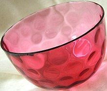
This cranberry glass bowl was made by adding a gold salt (probably gold chloride) to molten glass.

Used since ancient times as a method of staining glass colloidal gold was used in the 4th-century Lycurgus Cup, which changes color depending on the location of light source.

During the Middle Ages, soluble gold, a solution containing gold salt, had a reputation for its curative property for various diseases. In 1618, Francis Anthony, a philosopher and member of the medical profession, published a book called Panacea Aurea, sive tractatus duo de ipsius Auro Potabili (Latin: gold potion, or two treatments of potable gold). The book introduces information on the formation of colloidal gold and its medical uses. About half a century later, English botanist Nicholas Culpepper published book in 1656, Treatise of Aurum Potabile, solely discussing the medical uses of colloidal gold.

In 1676, Johann Kunckel, a German chemist, published a book on the manufacture of stained glass. In his book Valuable Observations or Remarks About the Fixed and Volatile Salts-Auro and Argento Potabile, Spiritu Mundi and the Like, Kunckel assumed that the pink color of Aurum Potabile came from small particles of metallic gold, not visible to human eyes. In 1842, John Herschel invented a photographic process called chrysotype (from the Greek χρῡσός meaning "gold") that used colloidal gold to record images on paper.

Modern scientific evaluation of colloidal gold did not begin until Michael Faraday's work in the 1850s. In 1856, in a basement laboratory of Royal Institution, Faraday accidentally created a ruby red solution while mounting pieces of gold leaf onto microscope slides. Since he was already interested in the properties of light and matter, Faraday further investigated the optical properties of the colloidal gold. He prepared the first pure sample of colloidal gold, which he called 'activated gold', in 1857. He used phosphorus to reduce a solution of gold chloride. The colloidal gold Faraday made 150 years ago is still optically active. For a long time, the composition of the 'ruby' gold was unclear. Several chemists suspected it to be a gold tin compound, due to its preparation. Faraday recognized that the color was actually due to the miniature size of the gold particles. He noted the light scattering properties of suspended gold microparticles, which is now called Faraday-Tyndall effect.

In 1898, Richard Adolf Zsigmondy prepared the first colloidal gold in diluted solution. Apart from Zsigmondy, Theodor Svedberg, who invented ultracentrifugation, and Gustav Mie, who provided the theory for scattering and absorption by spherical particles, were also interested in the synthesis and properties of colloidal gold.

With advances in various analytical technologies in the 20th century, studies on gold nanoparticles has accelerated. Advanced microscopy methods, such as atomic force microscopy and electron microscopy, have contributed the most to nanoparticle research. Due to their comparably easy synthesis and high stability, various gold particles have been studied for their practical uses. Different types of gold nanoparticle are already used in many industries, such as electronics.

Physical properties

Optical

The variation of scattering cross section of 100 nm-radius gold nanoparticle vs. the wavelength

Colloidal gold has been used by artists for centuries because of the nanoparticle’s interactions with visible light. Gold nanoparticles absorb and scatter light resulting in colours ranging from vibrant reds (smaller particles) to blues to black and finally to clear and colorless (larger particles), depending on particle size, shape, local refractive index, and aggregation state. These colors occur because of a phenomenon called localized surface plasmon resonance (LSPR), in which conduction electrons on the surface of the nanoparticle oscillate in resonance with incident light.

Effect of size

As a general rule, the wavelength of light absorbed increases as a function of increasing nano particle size. For example, pseudo-spherical gold nanoparticles with diameters ~ 30 nm have a peak LSPR absorption at ~530 nm.

Effect of local refractive index

Changes in the apparent color of a gold nanoparticle solution can also be caused by the environment in which the colloidal gold is suspended. The optical properties of gold nanoparticles depends on the refractive index near the nanoparticle surface, therefore both the molecules directly attached to the nanoparticle surface (i.e. nanoparticle ligands) and/or the nanoparticle solvent both may influence observed optical features. As the refractive index near the gold surface increases, the NP LSPR will shift to longer wavelengths In addition to solvent environment, the extinction peak can be tuned by coating the nanoparticles with non-conducting shells such as silica, bio molecules, or aluminium oxide.

Effect of aggregation

When gold nano particles aggregate, the optical properties of the particle change, because the effective particle size, shape, and dielectric environment all change.

Medical research

Electron microscopy

Colloidal gold and various derivatives have long been among the most widely used labels for antigens in biological electron microscopy. Colloidal gold particles can be attached to many traditional biological probes such as antibodies, lectins, superantigens, glycans, nucleic acids, and receptors. Particles of different sizes are easily distinguishable in electron micrographs, allowing simultaneous multiple-labelling experiments.

In addition to biological probes, gold nanoparticles can be transferred to various mineral substrates, such as mica, single crystal silicon, and atomically flat gold(III), to be observed under atomic force microscopy (AFM).

Drug delivery system

Gold nanoparticles can be used to optimize the biodistribution of drugs to diseased organs, tissues or cells, in order to improve and target drug delivery. Nanoparticle-mediated drug delivery is feasible only if the drug distribution is otherwise inadequate. These cases include drug targeting of unstable (proteins, siRNA, DNA), delivery to the difficult sites (brain, retina, tumors, intracellular organelles) and drugs with serious side effects (e.g. anti-cancer agents). The performance of the nanoparticles depends on the size and surface functionalities in the particles. Also, the drug release and particle disintegration can vary depending on the system (e.g. biodegradable polymers sensitive to pH). An optimal nanodrug delivery system ensures that the active drug is available at the site of action for the correct time and duration, and their concentration should be above the minimal effective concentration (MEC) and below the minimal toxic concentration (MTC).

Gold nanoparticles are being investigated as carriers for drugs such as Paclitaxel. The administration of hydrophobic drugs require molecular encapsulation and it is found that nanosized particles are particularly efficient in evading the reticuloendothelial system.

Tumor detection

In cancer research, colloidal gold can be used to target tumors and provide detection using SERS (surface enhanced Raman spectroscopy) in vivo. These gold nanoparticles are surrounded with Raman reporters, which provide light emission that is over 200 times brighter than quantum dots. It was found that the Raman reporters were stabilized when the nanoparticles were encapsulated with a thiol-modified polyethylene glycol coat. This allows for compatibility and circulation in vivo. To specifically target tumor cells, the polyethylenegylated gold particles are conjugated with an antibody (or an antibody fragment such as scFv), against, e.g. epidermal growth factor receptor, which is sometimes overexpressed in cells of certain cancer types. Using SERS, these pegylated gold nanoparticles can then detect the location of the tumor.

Gold nanoparticles accumulate in tumors, due to the leakiness of tumor vasculature, and can be used as contrast agents for enhanced imaging in a time-resolved optical tomography system using short-pulse lasers for skin cancer detection in mouse model. It is found that intravenously administrated spherical gold nanoparticles broadened the temporal profile of reflected optical signals and enhanced the contrast between surrounding normal tissue and tumors.

Tumor targeting via multifunctional nanocarriers. Cancer cells reduce adhesion to neighboring cells and migrate into the vasculature-rich stroma. Once at the vasculature, cells can freely enter the bloodstream. Once the tumor is directly connected to the main blood circulation system, multifunctional nanocarriers can interact directly with cancer cells and effectively target tumors.

Gene therapy

Gold nanoparticles have shown potential as intracellular delivery vehicles for siRNA oligonucleotides with maximal therapeutic impact.

Multifunctional siRNA-gold nanoparticles with several biomolecules: PEG, cell penetration and cell adhesion peptides and siRNA. Two different approaches were employed to conjugate the siRNA to the gold nanoparticle: (1) Covalent approach: use of thiolated siRNA for gold-thiol binding to the nanoparticle; (2) Ionic approach: interaction of the negatively charged siRNA to the modified surface of the AuNP through ionic interactions.

Gold nanoparticles show potential as intracellular delivery vehicles for antisense oligonucleotides (single and double stranded DNA) by providing protection against intracellular nucleases and ease of functionalization for selective targeting.

Photothermal agents

Gold nanorods are being investigated as photothermal agents for in-vivo applications. Gold nanorods are rod-shaped gold nanoparticles whose aspect ratios tune the surface plasmon resonance (SPR) band from the visible to near-infrared wavelength. The total extinction of light at the SPR is made up of both absorption and scattering. For the smaller axial diameter nanorods (~10 nm), absorption dominates, whereas for the larger axial diameter nanorods (>35 nm) scattering can dominate. As a consequence, for in-vivo studies, small diameter gold nanorods are being used as photothermal converters of near-infrared light due to their high absorption cross-sections. Since near-infrared light transmits readily through human skin and tissue, these nanorods can be used as ablation components for cancer, and other targets. When coated with polymers, gold nanorods have been observed to circulate in-vivo with half-lives longer than 6 hours, bodily residence times around 72 hours, and little to no uptake in any internal organs except the liver.

Despite the unquestionable success of gold nanorods as photothermal agents in preclinical research, they have yet to obtain the approval for clinical use because the size is above the renal excretion threshold. In 2019, the first NIR-absorbing plasmonic ultrasmall-in-nano architecture has been reported, and jointly combine: (i) a suitable photothermal conversion for hyperthermia treatments, (ii) the possibility of multiple photothermal treatments and (iii) renal excretion of the building blocks after the therapeutic action.

Radiotherapy dose enhancer

Considerable interest has been shown in the use of gold and other heavy-atom-containing nanoparticles to enhance the dose delivered to tumors. Since the gold nanoparticles are taken up by the tumors more than the nearby healthy tissue, the dose is selectively enhanced. The biological effectiveness of this type of therapy seems to be due to the local deposition of the radiation dose near the nanoparticles. This mechanism is the same as occurs in heavy ion therapy.

Detection of toxic gas

Researchers have developed simple inexpensive methods for on-site detection of hydrogen sulfide H
2
S
present in air based on the antiaggregation of gold nanoparticles (AuNPs). Dissolving H
2
S
into a weak alkaline buffer solution leads to the formation of HS-, which can stabilize AuNPs and ensure they maintain their red color allowing for visual detection of toxic levels of H
2
S
.

Gold nanoparticle based biosensor

Gold nanoparticles are incorporated into biosensors to enhance its stability, sensitivity, and selectivity. Nanoparticle properties such as small size, high surface-to-volume ratio, and high surface energy allow immobilization of large range of biomolecules. Gold nanoparticle, in particular, could also act as "electron wire" to transport electrons and its amplification effect on electromagnetic light allows it to function as signal amplifiers. Main types of gold nanoparticle based biosensors are optical and electrochemical biosensor.

Optical biosensor

Gold nanoparticle-based (Au-NP) biosensor for Glutathione (GSH). The AuNPs are functionalised with a chemical group that binds to GSH and makes the NPs partially collapse, and thus change colour. The exact amount of GSH can be derived via UV-vis spectroscopy through a calibration curve.

Gold nanoparticles improve the sensitivity of optical sensor by response to the change in local refractive index. The angle of the incidence light for surface plasmon resonance, an interaction between light wave and conducting electrons in metal, changes when other substances are bounded to the metal surface. Because gold is very sensitive to its surroundings' dielectric constant, binding of an analyte would significantly shift gold nanoparticle's SPR and therefore allow more sensitive detection. Gold nanoparticle could also amplify the SPR signal. When the plasmon wave pass through the gold nanoparticle, the charge density in the wave and the electron I the gold interacted and resulted in higher energy response, so called electron coupling. Since the analyte and bio-receptor now bind to the gold, it increases the apparent mass of the analyte and therefore amplified the signal. These properties had been used to build DNA sensor with 1000-fold sensitive than without the Au NP. Humidity sensor was also built by altering the atom interspacing between molecules with humidity change, the interspacing change would also result in a change of the Au NP's LSPR.

Electrochemical biosensor

Electrochemical sensor convert biological information into electrical signals that could be detected. The conductivity and biocompatibility of Au NP allow it to act as "electron wire". It transfers electron between the electrode and the active site of the enzyme. It could be accomplished in two ways: attach the Au NP to either the enzyme or the electrode. GNP-glucose oxidase monolayer electrode was constructed use these two methods. The Au NP allowed more freedom in the enzyme's orientation and therefore more sensitive and stable detection. Au NP also acts as immobilization platform for the enzyme. Most biomolecules denatures or lose its activity when interacted with the electrode. The biocompatibility and high surface energy of Au allow it to bind to a large amount of protein without altering its activity and results in a more sensitive sensor. Moreover, Au NP also catalyzes biological reactions. Gold nanoparticle under 2 nm has shown catalytic activity to the oxidation of styrene.

Immunological biosensor

Gold nanoparticles have been coated with peptides and glycans for use in immunological detection methods. The possibility to use glyconanoparticles in ELISA was unexpected, but the method seems to have a high sensitivity and thus offers potential for development of specific assays for diagnostic identification of antibodies in patient sera.

Thin films

Gold nanoparticles capped with organic ligands, such as alkanethiol molecules, can self-assemble into large monolayers (>cm). The particles are first prepared in organic solvent, such as chloroform or toluene, and are then spread into monolayers either on a liquid surface or on a solid substrate. Such interfacial thin films of nanoparticles have close relationship with Langmuir-Blodgett monolayers made from surfactants.

The mechanical properties of nanoparticle monolayers have been studied extensively. For 5 nm spheres capped with dodecanethiol, the Young's modulus of the monolayer is on the order of GPa. The mechanics of the membranes are guided by strong interactions between ligand shells on adjacent particles. Upon fracture, the films crack perpendicular to the direction of strain at a fracture stress of 11 2.6 MPa, comparable to that of cross-linked polymer films. Free-standing nanoparticle membranes exhibit bending rigidity on the order of 10 eV, higher than what is predicted in theory for continuum plates of the same thickness, due to nonlocal microstructural constraints such as nonlocal coupling of particle rotational degrees of freedom. On the other hand, resistance to bending is found to be greatly reduced in nanoparticle monolayers that are supported at the air/water interface, possibly due to screening of ligand interactions in a wet environment.

Surface chemistry

In many different types of colloidal gold syntheses, the interface of the nanoparticles can display widely different character – ranging from an interface similar to a self-assembled monolayer to a disordered boundary with no repeating patterns. Beyond the Au-Ligand interface, conjugation of the interfacial ligands with various functional moieties (from small organic molecules to polymers to DNA to RNA) afford colloidal gold much of its vast functionality.

Ligand exchange/functionalization

After initial nanoparticle synthesis, colloidal gold ligands are often exchanged with new ligands designed for specific applications. For example, Au NPs produced via the Turkevich-style (or Citrate Reduction) method are readily reacted via ligand exchange reactions, due to the relatively weak binding between the carboxyl groups and the surfaces of the NPs. This ligand exchange can produce conjugation with a number of biomolecules from DNA to RNA to proteins to polymers (such as PEG) to increase biocompatibility and functionality. For example, ligands have been shown to enhance catalytic activity by mediating interactions between adsorbates and the active gold surfaces for specific oxygenation reactions. Ligand exchange can also be used to promote phase transfer of the colloidal particles. Ligand exchange is also possible with alkane thiol-arrested NPs produced from the Brust-type synthesis method, although higher temperatures are needed to promote the rate of the ligand detachment. An alternative method for further functionalization is achieved through the conjugation of the ligands with other molecules, though this method can cause the colloidal stability of the Au NPs to breakdown.

Ligand removal

In many cases, as in various high-temperature catalytic applications of Au, the removal of the capping ligands produces more desirable physicochemical properties. The removal of ligands from colloidal gold while maintaining a relatively constant number of Au atoms per Au NP can be difficult due to the tendency for these bare clusters to aggregate. The removal of ligands is partially achievable by simply washing away all excess capping ligands, though this method is ineffective in removing all capping ligand. More often ligand removal achieved under high temperature or light ablation followed by washing. Alternatively, the ligands can be electrochemically etched off.

Surface structure and chemical environment

The precise structure of the ligands on the surface of colloidal gold NPs impact the properties of the colloidal gold particles. Binding conformations and surface packing of the capping ligands at the surface of the colloidal gold NPs tend to differ greatly from bulk surface model adsorption, largely due to the high curvature observed at the nanoparticle surfaces. Thiolate-gold interfaces at the nanoscale have been well-studied and the thiolate ligands are observed to pull Au atoms off of the surface of the particles to for “staple” motifs that have significant Thiyl-Au(0) character. The citrate-gold surface, on the other hand, is relatively less-studied due to the vast number of binding conformations of the citrate to the curved gold surfaces. A study performed in 2014 identified that the most-preferred binding of the citrate involves two carboxylic acids and the hydroxyl group of the citrate binds three surface metal atoms.

Health and safety

As gold nanoparticles (AuNPs) are further investigated for targeted drug delivery in humans, their toxicity needs to be considered. For the most part, it is suggested that AuNPs are biocompatible,[citation needed] but the concentrations at which they become toxic needs to be determined, and if those concentrations fall within the range of used concentrations. Toxicity can be tested in vitro and in vivo. In vitro toxicity results can vary depending on the type of the cellular growth media with different protein compositions, the method used to determine cellular toxicity (cell health, cell stress, how many cells are taken into a cell), and the capping ligands in solution. In vivo assessments can determine the general health of an organism (abnormal behavior, weight loss, average life span) as well as tissue specific toxicology (kidney, liver, blood) and inflammation and oxidative responses. In vitro experiments are more popular than in vivo experiments because in vitro experiments are more simplistic to perform than in vivo experiments.

Toxicity and hazards in synthesis

While AuNPs themselves appear to have low or negligible toxicity, and the literature shows that the toxicity has much more to do with the ligands rather than the particles themselves, the synthesis of them involves chemicals that are hazardous. Sodium borohydride, a harsh reagent, is used to reduce the gold ions to gold metal. The gold ions usually come from chloroauric acid, a potent acid. Because of the high toxicity and hazard of reagents used to synthesize AuNPs, the need for more “green” methods of synthesis arose.

Toxicity due to capping ligands

Some of the capping ligands associated with AuNPs can be toxic while others are nontoxic. In gold nanorods (AuNRs), it has been shown that a strong cytotoxicity was associated with CTAB-stabilized AuNRs at low concentration, but it is thought that free CTAB was the culprit in toxicity. Modifications that overcoat these AuNRs reduces this toxicity in human colon cancer cells (HT-29) by preventing CTAB molecules from desorbing from the AuNRs back into the solution. Ligand toxicity can also be seen in AuNPs. Compared to the 90% toxicity of HAuCl4 at the same concentration, AuNPs with carboxylate termini were shown to be non-toxic. Large AuNPs conjugated with biotin, cysteine, citrate, and glucose were not toxic in human leukemia cells (K562) for concentrations up to 0.25 M. Also, citrate-capped gold nanospheres (AuNSs) have been proven to be compatible with human blood and did not cause platelet aggregation or an immune response. However, citrate-capped gold nanoparticles sizes 8-37 nm were found to be lethally toxic for mice, causing shorter lifespans, severe sickness, loss of appetite and weight, hair discoloration, and damage to the liver, spleen, and lungs; gold nanoparticles accumulated in the spleen and liver after traveling a section of the immune system. There are mixed-views for polyethylene glycol (PEG)-modified AuNPs. These AuNPs were found to be toxic in mouse liver by injection, causing cell death and minor inflammation. However, AuNPs conjugated with PEG copolymers showed negligible toxicity towards human colon cells (Caco-2). AuNP toxicity also depends on the overall charge of the ligands. In certain doses, AuNSs that have positively-charged ligands are toxic in monkey kidney cells (Cos-1), human red blood cells, and E. coli because of the AuNSs interaction with the negatively-charged cell membrane; AuNSs with negatively-charged ligands have been found to be nontoxic in these species. In addition to the previously mentioned in vivo and in vitro experiments, other similar experiments have been performed. Alkylthiolate-AuNPs with trimethlyammonium ligand termini mediate the translocation of DNA across mammalian cell membranes in vitro at a high level, which is detrimental to these cells. Corneal haze in rabbits have been healed in vivo by using polyethylemnimine-capped gold nanoparticles that were transfected with a gene that promotes wound healing and inhibits corneal fibrosis.

Toxicity due to size of nanoparticles

Toxicity in certain systems can also be dependent on the size of the nanoparticle. AuNSs size 1.4 nm were found to be toxic in human skin cancer cells (SK-Mel-28), human cervical cancer cells (HeLa), mouse fibroblast cells (L929), and mouse macrophages (J774A.1), while 0.8, 1.2, and 1.8 nm sized AuNSs were less toxic by a six-fold amount and 15 nm AuNSs were nontoxic. There is some evidence for AuNP buildup after injection in in vivo studies, but this is very size dependent. 1.8 nm AuNPs were found to be almost totally trapped in the lungs of rats. Different sized AuNPs were found to buildup in the blood, brain, stomach, pancreas, kidneys, liver, and spleen.

Biosafety and biokinetics investigations on biodegradable ultrasmall-in-nano architectures have demonstrated that gold nanoparticles are able to avoid metal accumulation in organisms through escaping by the renal pathway.

Synthesis

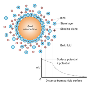
Potential difference as a function of distance from particle surface.

Generally, gold nanoparticles are produced in a liquid ("liquid chemical methods") by reduction of chloroauric acid (H[AuCl
4
]
). To prevent the particles from aggregating, stabilizing agents are added. Citrate acts both as the reducing agent and colloidal stabilizer.

They can be functionalized with various organic ligands to create organic-inorganic hybrids with advanced functionality.

Turkevich method

This simple method was pioneered by J. Turkevich et al. in 1951 and refined by G. Frens in the 1970s. It produces modestly monodisperse spherical gold nanoparticles of around 10–20 nm in diameter. Larger particles can be produced, but at the cost of monodispersity and shape. In this method, hot chloroauric acid is treated with sodium citrate solution, producing colloidal gold. The Turkevich reaction proceeds via formation of transient gold nanowires. These gold nanowires are responsible for the dark appearance of the reaction solution before it turns ruby-red.

Capping agents

A capping agent is used during nanoparticle synthesis to inhibit particle growth and aggregation. The chemical blocks or reduces reactivity at the periphery of the particle—a good capping agent has a high affinity for the new nuclei. Citrate ions or tannic acid function both as a reducing agent and a capping agent. Less sodium citrate results in larger particles.

Brust-Schiffrin method

This method was discovered by Brust and Schiffrin in the early 1990s, and can be used to produce gold nanoparticles in organic liquids that are normally not miscible with water (like toluene). It involves the reaction of a chlorauric acid solution with tetraoctylammonium bromide (TOAB) solution in toluene and sodium borohydride as an anti-coagulant and a reducing agent, respectively.

Here, the gold nanoparticles will be around 5–6 nm. NaBH4 is the reducing agent, and TOAB is both the phase transfer catalyst and the stabilizing agent.

TOAB does not bind to the gold nanoparticles particularly strongly, so the solution will aggregate gradually over the course of approximately two weeks. To prevent this, one can add a stronger binding agent, like a thiol (in particular, alkanethiols), which will bind to gold, producing a near-permanent solution. Alkanethiol protected gold nanoparticles can be precipitated and then redissolved. Thiols are better binding agents because there is a strong affinity for the gold-sulfur bonds that form when the two substances react with each other. Tetra-dodecanthiol is a commonly used strong binding agent to synthesize smaller particles. Some of the phase transfer agent may remain bound to the purified nanoparticles, this may affect physical properties such as solubility. In order to remove as much of this agent as possible, the nanoparticles must be further purified by soxhlet extraction.

Perrault method

This approach, discovered by Perrault and Chan in 2009, uses hydroquinone to reduce HAuCl4 in an aqueous solution that contains 15 nm gold nanoparticle seeds. This seed-based method of synthesis is similar to that used in photographic film development, in which silver grains within the film grow through addition of reduced silver onto their surface. Likewise, gold nanoparticles can act in conjunction with hydroquinone to catalyze reduction of ionic gold onto their surface. The presence of a stabilizer such as citrate results in controlled deposition of gold atoms onto the particles, and growth. Typically, the nanoparticle seeds are produced using the citrate method. The hydroquinone method complements that of Frens, as it extends the range of monodispersed spherical particle sizes that can be produced. Whereas the Frens method is ideal for particles of 12–20 nm, the hydroquinone method can produce particles of at least 30–300 nm.

Martin method

This simple method, discovered by Martin and Eah in 2010, generates nearly monodisperse "naked" gold nanoparticles in water. Precisely controlling the reduction stoichiometry by adjusting the ratio of NaBH4-NaOH ions to HAuCl4-HCl ions within the "sweet zone," along with heating, enables reproducible diameter tuning between 3–6 nm. The aqueous particles are colloidally stable due to their high charge from the excess ions in solution. These particles can be coated with various hydrophilic functionalities, or mixed with hydrophobic ligands for applications in non-polar solvents. In non-polar solvents the nanoparticles remain highly charged, and self-assemble on liquid droplets to form 2D monolayer films of monodisperse nanoparticles.

Nanotech studies

Bacillus licheniformis can be used in synthesis of gold nanocubes with sizes between 10 and 100 nanometres. Gold nanoparticles are usually synthesized at high temperatures in organic solvents or using toxic reagents. The bacteria produce them in much milder conditions.

Navarro et al. method

For particles larger than 30 nm, control of particle size with a low polydispersity of spherical gold nanoparticles remains challenging. In order to provide maximum control on the NP structure, Navarro and co-workers used a modified Turkevitch-Frens procedure using sodium acetylacetonate as the reducing agent and sodium citrate as the stabilizer.

Sonolysis

Another method for the experimental generation of gold particles is by sonolysis. The first method of this type was invented by Baigent and Müller. This work pioneered the use of ultrasound to provide the energy for the processes involved and allowed the creation of gold particles with a diameter of under 10 nm. In another method using ultrasound, the reaction of an aqueous solution of HAuCl4 with glucose, the reducing agents are hydroxyl radicals and sugar pyrolysis radicals (forming at the interfacial region between the collapsing cavities and the bulk water) and the morphology obtained is that of nanoribbons with width 30–50 nm and length of several micrometers. These ribbons are very flexible and can bend with angles larger than 90°. When glucose is replaced by cyclodextrin (a glucose oligomer), only spherical gold particles are obtained, suggesting that glucose is essential in directing the morphology toward a ribbon.

Block copolymer-mediated method

An economical, environmentally benign and fast synthesis methodology for gold nanoparticles using block copolymer has been developed by Sakai et al. In this synthesis methodology, block copolymer plays the dual role of a reducing agent as well as a stabilizing agent. The formation of gold nanoparticles comprises three main steps: reduction of gold salt ion by block copolymers in the solution and formation of gold clusters, adsorption of block copolymers on gold clusters and further reduction of gold salt ions on the surfaces of these gold clusters for the growth of gold particles in steps, and finally its stabilization by block copolymers. But this method usually has a limited-yield (nanoparticle concentration), which does not increase with the increase in the gold salt concentration. Ray et al. improved this synthesis method by enhancing the nanoparticle yield by manyfold at ambient temperature.

Platinum nanoparticle

From Wikipedia, the free encyclopedia https://en.wikipedia.org/wiki/Platinum_nanoparticle     Platinum nanoparticles are usually in...