Search This Blog

Sunday, April 29, 2018

Neuroscience of sex differences

From Wikipedia, the free encyclopedia

Neuroscience of sex differences is the study of the characteristics of the brain that separate the male brain and the female brain. Psychological sex differences are thought by some to reflect the interaction of genes, hormones and social learning on brain development throughout the lifespan.

Some evidence from brain morphology and function studies indicates that male and female brains cannot always be assumed to be identical from either a structural or functional perspective, and some brain structures are sexually dimorphic.[1][2]

Experts note that neural sexual dimorphisms in humans exist only as averages, with overlapping variabilities,[3] and that it is unknown to what extent each is influenced by genetics or environment, even in adulthood.[4][5]

History

Ideas of differences in the male and female brain circulated during the time of ancient Greek philosophers around 850 B.C. Aristotle claimed that males did not "receive their soul" until 40 days post-gestation and females did not until 80 days. In 1854, Emil Huschke discovered that "the frontal lobe in the male is all of 1% larger than that of the female."[6] As the 19th century progressed, scientists began researching sexual dimorphisms in the brain significantly more.[7] Until around 21 years ago, scientists knew of several structural sexual dimorphisms of the brain, but they did not think that gender had any impact on how the human brain performs daily tasks. Through molecular, animal, and neuroimaging studies, a great deal of information regarding the differences between male and female brains and how much they differ in regards to both structure and function has been uncovered.[8]

Evolutionary explanations

Sexual selection

It is thought that male and female differences in learning ability have contributed to sexual selection and mate preference throughout evolution. The hippocampus has even been found to exhibit seasonal activity in some mammals where it is active during breeding periods but inactive during hibernation; this is because spatial learning is more present during the breeding season.[9]

Females show enhanced information recall compared to males. This may be due to the fact that females have a more intricate evaluation of risk-scenario contemplation, based on a prefrontal cortical control of the amygdala. For example, the ability to recall information better than males most likely originated from sexual selective pressures on females during competition with other females in mate selection. Recognition of social cues was an advantageous characteristic because it ultimately maximized offspring and was therefore selected for during evolution.[1]

Oxytocin is a hormone that induces contraction of the uterus and lactation in mammals. It is also a characteristic hormone of nursing mothers. Studies have found that oxytocin improves spatial memory. Through activation of the MAP kinase pathway, oxytocin plays a role in the enhancement of long-term synaptic plasticity, which is a change in strength between two neurons over a synapse that lasts for minutes or longer, and long-term memory. This hormone may have helped mothers remember the location of distant food sources so they could better nurture their offspring.[1]

Male vs. female brain anatomy

Hemisphere differences

A popular theory regarding language functions suggests that women use both hemispheres more equally, whereas men are more strongly lateralized to the left hemisphere.[10] This theory found initial support in a high-profile study of 19 men and 19 women, which found stronger lateralization in men during one of the three language tasks assessed.[11] In 2008, some researchers concluded that further studies have failed to replicate this finding, and a meta-analysis of 29 studies comparing language lateralization in males and females found no overall difference.[12] However, in 2013, researchers at the Perelman School of Medicine at the University of Pennsylvania mapped notable differences in male and female neural wiring. The study used diffusion tensor imaging of 949 individuals aged 8–22 years, and concluded that in all supratentorial regions of the brain inter-hemispheric connectivity was greater in women's and girls' brains, whereas intra-hemispheric connectivity was greater in the brains of men and boys. The effect was reversed in cerebellar connections.[13] The detected differences in neural connectivity were negligible up to the age of 13, but became much more prominent in the 14 to 17-year-olds.[13] In terms of the potential effect on behaviour, the authors concluded, "Overall, the results suggest that male brains are structured to facilitate connectivity between perception and coordinated action, whereas female brains are designed to facilitate communication between analytical and intuitive processing modes".[13]

Amygdala

image of Amygdala
The amygdala (red) in a human brain.

According to some researchers, "the research on sex differences in the amygdala has produced conflicting results".[14] After correcting for the overall difference in brain volume between men and women, a 2016 meta-analysis indicated that the amygdala is not significantly larger in either sex.[15] Some studies, however, report increased amygdala activity during the processing of affective scenes in men relative to women (Schienle et al., 2005; Goldstein et al., 2010), and meta-analysis supports this view, showing larger effect sizes in studies of affective processing including only men compared with those including only women (Sergerie et al., 2008). However several studies using similar stimuli have reported a larger amygdala response in women (Klein et al., 2003; McClure et al., 2004; Hofer et al., 2006; Domes et al., 2010), and others have reported no sex difference at all (Wrase et al., 2003; Caseras et al., 2007; Aleman and Swart, 2008). A possible explanation for these inconsistent results is that sex differences in amygdala response are valence-dependent. Furthermore, according to other researchers,[16] "Correlation analyses revealed that gray matter thickness in left ventromedial PFC was inversely correlated with task-related activation in the amygdala. These data add support to a general role of the ventromedial PFC in regulating activity of the amygdala."

Research has been done on post-traumatic stress disorder (PTSD), an anxiety disorder found in both sexes, which is particularly common in war veterans, assault victims and women who have experienced abuse. Emotional memory encoding varies in the amygdala on the right and left and occurs equally for both genders: the right triggers unpleasant and fear-related memories, both declarative (conscious) and episodic (nonconcious).[17]

Amygdala volume correlates positively with fearfulness in girls but not in boys.[18]

Hippocampus

Several studies have indicated that the hippocampi of men and women differ anatomically, neurochemically, and also in degree of long-term potentiation.[19] Such evidence indicates that sex may influence the role of the hippocampus in learning. By contrast, a 2016 meta-analysis indicated that the hippocampus does not differ in volume between men and women.[20] One experiment examined the effects of stress on Pavlovian conditioning performance in both sexes and found that males' performance under stress was enhanced while female performance was impaired. Activation of the hippocampus is more dominant on the left side of hippocampus in females, while it is more dominant on the right side in males. This in turn influences cognitive reasoning; women use more verbal strategies than men when performing a task that requires cognitive thinking.[21] The hippocampus's relationship with other structures in the brain influences learning and has been found to be sexually dimorphic as well.[1]

Estradiol has been found to influence hippocampal development. Studies have shown endogenous neurogenesis, or the internally-driven formation of new neurons, to be higher in the male hippocampus than in that of the female. However, following exogenous estradiol injections, the number of new neurons in the hippocampi of females reached levels equivalent to those found in the hippocampi of males. Conversely, injecting estradiol in males did not increase neurogenesis in males. However, antagonizing endogenous estradiol in males reduced neurogenesis, but did not change the number of new neurons in females. Thus, hippocampal sex differences appear to be mediated in part through endogenous estradiol levels.[22]

Frontal lobe

The ventromedial prefrontal cortex (VMPC) plays a key role in social emotional processing. In accordance with the sexual dimorphism of the amygdala, the right VMPC is more dominant in an active limbic system for males while the left is more dominant in females. These differences carry out to a behavioral level. For example, Koscik et al. wrote:
"A man with a unilateral right VMPC lesion, who was well educated and had worked successfully as a minister, was entirely unable to return to any form of gainful employment after his brain damage. He requires supervision for daily tasks and demonstrates severe disturbances in behavior and emotional regulation, including impulsivity and poor judgment. By contrast, a man with a unilateral left VMPC lesion was able to return to his job at a grain elevator and remains successfully employed there. He is remarkably free of disturbances to his social life and emotional functioning."[23]

Orbital prefrontal cortex

Positron emission tomography studies have shown that men and women ranging from the ages of 19 to 32 years old metabolize glucose at significantly different rates in the orbital prefrontal cortex. Infant males who exhibited lesions on their orbital prefrontal cortex struggled with object reversal experiments, but females exhibiting such lesions did not have impaired performance in object reversal.[24]

Other regions and not region-specific

There are sex differences in locus coeruleus dendritic structure that allow for an increased reception and processing of limbic information in females compared to males.[18]

Aggressive and defiant behavior is also associated with decreased right anterior cingulate cortex (ACC) volume in boys.[18]

According to the neuroscience journal review series Progress in Brain Research, it has been found that males have larger and longer planum temporale and Sylvian fissure while females have significantly larger proportionate volumes to total brain volume in the superior temporal cortex, Broca's area, the hippocampus and the caudate.[25] The midsagittal and fiber numbers in the anterior commissure that connect the temporal poles and mass intermedia that connects the thalami is also larger in females.[25]

The journal review also found that the male brain volume was slightly larger than the female brain volume, a difference is often attributed to the larger average body and skull sizes of males. In female brains, however, greater cortical thickness, cortical complexity and cortical surface area are observed after adjusting for brain volume differences.[25] Given that cortical complexity and cortical features are positively correlated with intelligence, researchers postulated that these differences in cortical complexity may have been an evolutionary adaptation to the difference in brain volume of males and females.[25]

White/grey matter

Global and regional grey matter (GM) differs in men and women. Women have larger left orbitofrontal GM volumes and overall cortical thickness than men.[26] Behavioral implications of the greater volume have not yet been discovered. Women have a higher percentage of GM, whereas men have a higher percentage of white matter (WM) and of CSF (cerebrospinal fluid). In men the percentage of GM was higher in the left hemisphere, the percentage of WM was symmetric, and the percentage of CSF was higher in the right. Women showed no asymmetries. Both GM and WM volumes correlated moderately with global, verbal, and spatial performance across groups. However, the regression of cognitive performance and WM volume was significantly steeper in women.[27]

In a 2013 meta-analysis, researchers found on average males had larger grey matter volume in bilateral amygdalae, hippocampi, anterior parahippocampal gyri, posterior cingulate gyri, precuneus, putamen and temporal poles, areas in the left posterior and anterior cingulate gyri, and areas in the cerebellum bilateral VIIb, VIIIa and Crus I lobes, left VI and right Crus II lobes.[2] On the other hand, females on average had larger grey matter volume at the right frontal pole, inferior and middle frontal gyri, pars triangularis, planum temporale/parietal operculum, anterior cingulate gyrus, insular cortex, and Heschl's gyrus; bilateral thalami and precuneus; the left parahippocampal gyrus and lateral occipital cortex (superior division).[2] The meta-analysis found larger volumes in females were most pronounced in areas in the right hemisphere related to language in addition to several limbic structures such as the right insular cortex and anterior cingulate gyrus.[2]

Amber Ruigrok's 2013 meta-analysis also found greater grey matter density in the average male left amygdala, hippocampus, insula, pallidum, putamen, claustrum and right cerebellum.[2] The meta-analysis also found greater grey matter density in the average female left frontal pole[2]

Brain networks

A 2014 meta-analysis by researcher Ashley C.Hill found that although men and women commonly used the same brain networks for working memory, specific regions were gender specific.[28] For example, both men and women's active working memory networks composed of bilateral middle frontal gyri, left cingulate gyrus, right precuneus, left inferior and superior parietal lobes, right claustrum, and left middle temporal gyrus but women also tended have consistent activity in the limbic regions such as the anterior cingulate, bilateral amygdala and right hippocampus while men tended to have a distributed networks spread out among the cerebellum, portions of the superior parietal lobe, the left insula and bilateral thalamus.[28] In the work of[29] the authors have computed structural connectomes of 96 subjects of the Human Connectome Project, and they have proven that in numerous graph-theoretical parameters, the structural connectome of women are significantly better connected than the connectome of men. For example, women's connectome has more edges, higher minimum bipartition width, larger eigengap, greater minimum vertex cover than that of men. The minimum bipartition width (or the minimum balanced cut (graph theory)) is well-known measure of quality of computer multistage interconnection networks, it describes the possible bottlenecks in network communication: The higher this value is, the better is the network. The larger eigengap shows that the female connectome is better expander graph than the connectome of males. The better expanding property, the higher minimum bipartition width and the greater minimum vertex cover show deep advantages in network connectivity in the case of female braingraph.

Brain differences between homo- and heterosexuals

Brain wiring comparisons of homosexuals and persons of the opposite sex show that homosexuals may be born with a predisposition to be homosexual. Research at the Stockholm Brain Institute in Sweden found that homosexual men and heterosexual women have similar brain characteristics. Specifically, these similarities are in the overall size of the brain and the activity of the amygdala. The same is for heterosexual men and homosexual women. Molecular biologist at the National Institutes of Health, Dean Hamer, says, "this is from a series of observations showing there's a biological reason for sexual orientation".[30]

Ivanka Savic – Berglund conducted a study in which MRIs were used to measure the volume and shapes of the brain. She also used PET scans to view blood flow to the amygdala. Savic – Berglund found that in homosexual men and heterosexual women, the blood flowed to areas involved in fear and anxiety, whereas in heterosexual men and homosexual women, it tended to flow to pockets linked to aggression. When looking at hemisphere differences, the right hemisphere was found to be slightly larger than the left in heterosexual men and homosexual women, whereas those of homosexual men and heterosexual women were more symmetrical.[31]

Research has indicated that the corpus callosum is larger in homosexual men than in heterosexual men. This is significant because the corpus callosum is a structure that is developed early. In the Journal Science Simon LeVay showed that the third interstitial nucleus of the hypothalamus has neurons that are packed more together in homosexual men than in heterosexual men.[32] Connections from the amygdala to other parts of the brain are similar between homosexuals and persons of the opposite gender as shown through PET and MRI scans. For example, in homosexual men and heterosexual women, there were more connections from the left amygdala. In homosexual women and heterosexual men, there were more connections from the right amygdala. LeVay's results were not replicated in other studies. A 2001 study that attempted to replicate the findings concluded that "Although there was a trend for INAH3 to occupy a smaller volume in homosexual men than in heterosexual men, there was no difference in the number of neurons within the nucleus based on sexual orientation."[33]

Neurochemical differences

Hormones

Steroid hormones have several effects on brain development as well as maintenance of homeostasis throughout adulthood. One effect they exhibit is on the hypothalamus, where they increase synapse formation.[34] Estrogen receptors have been found in the hypothalamus, pituitary gland, hippocampus, and frontal cortex, indicating the estrogen plays a role in brain development. Gonadal hormone receptors have also been found in the basal forebrain nuclei.[35]

Estrogen and the female brain

Estradiol influences cognitive function, specifically by enhancing learning and memory in a dose-sensitive manner. Too much estrogen can have negative effects by weakening performance of learned tasks as well as hindering performance of memory tasks; this can result in females exhibiting poorer performance of such tasks when compared to males.[36]

It has been suggested that during development, estrogen can exhibit both feminizing and defeminizing effects on the human brain; high levels of estrogen induce male neural traits to develop while moderate levels induce female traits. In females, defeminizing effects are resisted because of the presence of α-fetoprotein (AFP), a carrier protein proposed to transport estrogen into brain cells, allowing the female brain to properly develop. The role of AFP is significant at crucial stages of development, however. Prenatally, AFP blocks estrogen. Postnatally, AFP decreases to ineffective levels; therefore, it is probable that estrogen exhibits its effects on female brain development postnatally.[37]

Ovariectomies, surgeries inducing menopause, or natural menopause cause fluctuating and decreased estrogen levels in women. This in turn can "attenuate the effects" of endogenous opioid peptides.  Opioid peptides are known to play a role in emotion and motivation. β-endorphin (β-EP), an endogenous opioid peptide, content has been found to decrease (in varying amounts/brain region), post ovariectomy, in female rats within the hypothalamus, hippocampus, and pituitary gland. Such a change in β-EP levels could be the cause of mood swings, behavioral disturbances, and hot flashes in post menopausal women.[35]

Testosterone and the male brain

Testosterone has been found to play a big role during development but may have independent effects on sexually dimorphic brain regions in adulthood. Studies have shown that the medial amygdala of male hamsters exhibits lateralization and sexual dimorphism prior to puberty. Furthermore, organization of this structure during development is influenced by the presence of androgens and testosterone. This is evident when comparing medial amygdala volume of male and female rats, adult male brains have a medial amygdala of greater volume than do adult female brains which is partially due to androgen circulation.[38] It also heavily influences male development; a study found that perinatal females introduced to elevated testosterone levels exhibited male behavior patterns. In the absence of testosterone, female behavior is retained.[34] Testosterone's influence on the brain is caused by organizational developmental effects. It has been shown to influence proaptotic proteins so that they increase neuronal cell death in certain brain regions. Another way testosterone affects brain development is by aiding in the construction of the "limbic hypothalamic neural networks".[34]

Similar to how estrogen enhances memory and learning in women, testosterone has been found to enhance memory recall in men. In a study testing a correlation between memory recall and testosterone levels in men, "fMRI analysis revealed that higher testosterone levels were related to increased brain activation in the amygdala during encoding of neutral pictures".[39]

Oxytocin and Vasopressin

Oxytocin is positively correlated with maternal behaviours, social recognition, social contact, sexual behaviour and pair bonding. Oxytocin appears at higher levels in women than in men.[40] Vasopressin on the other hand is more present in men and mediates sexual behavior, aggression and other social functions.[40][41]

Neurotransmitters

Whole level 5-HT serotonin levels are higher in women versus men while men synthesize serotonin significantly faster than women. Healthy women also have higher 5-HT transport availability in the diencephalon and brainstem areas of the brain.[42] Dopamine function is also increased in women especially dopamine transporter which regulates the availability of receptors. Women before the onset of menopause synthesize higher levels of striatal presynaptic dopamine than age-matched men.[42] Other neurotransmitters like μ-opioids show significantly higher binding potential in the cerebellum, amygdala and the thalamus for women than it does so for men.[43] Women are also more dependent on norepinephrine in the formation of long term emotional memories than men are.[43]

Male vs. female brain functionality

Neural masculinization is a developmental process where different sex hormones assist in the expression of male behavior.[44]

Stress

image of stress regions in brain
Regions of the brain associated with stress and fear.

Stress has been found to induce an increase in serotonin, norepinephrine, and dopamine levels within the basolateral amygdala of male rats, but not within that of female rats. Furthermore, object recognition is impaired in males as a result of short term stress exposure. Neurochemical levels in the brain can change under the influence of stress exposure, particularly in regions associated with spatial and non-spatial memory, such as the prefrontal cortex and the hippocampus. Dopamine metabolite levels decrease post stress in male rats' brains, specifically within the CA1 region of the hippocampus.[45]

In female rats, both short term (1 hour) and long term (21 days) stress has been found to actually enhance spatial memory. Under stress, male rats exhibit deleterious effects on spatial memory, however female rats show a degree of resistance to this phenomenon. Stressed female rats' norepinephrine (NE) levels go up by about 50% in their prefrontal cortex while that of male rats goes down 50%.[45]

Cognitive tasks

It was once thought that sex differences in cognitive task and problem solving did not occur until puberty. However, new evidence now suggests that cognitive and skill differences are present earlier in development. For example, researchers have found that three- and four-year-old boys were better at targeting and at mentally rotating figures within a clock face than girls of the same age were. Prepubescent girls, however, excelled at recalling lists of words. These sex differences in cognition correspond to patterns of ability rather than overall intelligence. Laboratory settings are used to systematically study the sexual dimorphism in problem solving task performed by adults.[46]

On average, males excel relative to females at certain spatial tasks. Specifically, males have an advantage in tests that require the mental rotation or manipulation of an object.[47] In a computer simulation of a maze task, males completed the task faster and with fewer errors than their female counterparts. Additionally, males have displayed higher accuracy in tests of targeted motor skills, such as guiding projectiles.[46] Males are also faster on reaction time and finger tapping tests.[48]

On average, females excel relative to males on tests that measure recollection. They have an advantage on processing speed involving letters, digits and rapid naming tasks.[48] Females tend to have better object location memory and verbal memory.[49] They also perform better at verbal learning.[50] Females have better performance at matching items and precision tasks, such as placing pegs into designated holes. In maze and path completion tasks, males learn the goal route in fewer trials than females, but females remember more of the landmarks presented. This shows that females use landmarks in everyday situations to orient themselves more than males. Females are better at remembering whether objects had switched places or not.[46]

Studies using the Iowa gambling task, or Iowa Card Task, have examined cognitive reasoning and decision-making in males and females. A study in which participants of various age groups who were asked to perform the Iowa Card Task produced data showing that males and females differ in their decision making processes on the neurological level. The study suggests that decision-making in females may be guided by avoidance of negativity while decision making in males is mainly guided by assessing the long term outcome of a situation. They also found that males outperformed females in the Iowa Card Task, but there was a negative correlation between elevated testosterone levels and performance in the card task which indicates gonadal hormones influence decision-making.[24]

Methane from tundra, ocean floor didn't spike during previous natural warming period

Date:                August 23, 2017
Source:            Oregon State University
Original page:  https://www.sciencedaily.com/releases/2017/08/170823131234.htm
 
Scientists concerned that global warming may release huge stores of methane from reservoirs beneath Arctic tundra and deposits of marine hydrates -- a theory known as the "clathrate gun" hypothesis -- have turned to geologic history to search for evidence of significant methane release during past warming events.

A new study published this week in the journal Nature suggests, however, that the last ice age transition to a warmer climate some 11,500 years ago did not include massive methane flux from marine sediments or the tundra. Instead, the likely source of rising levels of atmospheric methane was from tropical wetlands, authors of the new study say.

While this certainly is good news, the study also points at a larger role of humans in the recent methane rise, noted Edward Brook, an Oregon State University paleoclimatologist and co-author on the study

"Our findings show that natural geologic emissions of methane -- for example, leakage from oil seeps or gas deposits in the ground -- are much smaller than previously thought," Brook said. "That means that a greater percentage of the methane in the atmosphere today is due to human activities, including oil drilling, and the extraction and transport of natural gas."

The study suggests that human emissions of geologic methane may be as much as 25 percent higher than previous estimates. Although not as abundant as carbon dioxide, methane is a much more powerful greenhouse gas and therefore the rising levels are an important contributor to global warming.

"This means we have even more potential to fight global warming by curbing methane emissions from our fossil fuel use," said Vasilii Petrenko, an associate professor of earth and environmental sciences at the University of Rochester, and lead author on the study.

Anthropogenic methane emissions are the second largest contributor to global warming after carbon dioxide, but there has been uncertainty as to the source of that methane and whether it has changed over time, Brook noted. The new study sheds light on the issue by analyzing levels of atmospheric methane from the last deglaciation in air bubbles that have been trapped in pristine ice cores from Antarctica's Taylor Glacier.

The researchers were able to estimate the magnitude of methane emissions from roughly 11,500 years ago by measuring radioactive carbon isotopes in methane, (carbon-14, also known as 14C or radiocarbon), which decay fairly rapidly. Methane released from those marine hydrates and permafrost is old enough that any 14C originally present has now decayed away.

They found that amount of methane from ancient "14C-free sources" was very low -- less than 10 percent of the total methane -- during the entire range of sampling, from 11,800 to 11,300 years ago.

"A lot of people have painted the Arctic as a methane time bomb," Brook said, "but this shows that it may be more stable than we thought. Past performance isn't always a predictor of the future, but it is a good analog. We should be more concerned about anthropogenic sources of methane into the atmosphere, which continue to increase."

The levels of 14C in the ice cores suggest that the increase in methane during the last deglaciation had another source -- likely from tropical wetlands, said Christo Buizert, an Oregon State University researcher and co-author on the paper.

"Methane is not stored in the tropics for long periods of time, but produced every day by microbial activity in wetlands," Buizert said. "We know from other studies that rainfall increased in the tropics during the last warming period, and that likely created wetlands that produced the increase in methane during the last warming period."

Atmospheric methane has increased from 750 parts per billion in the year 1750 to more than 1,800 parts per billion today -- mostly from anthropogenic sources, especially leakage from fossil fuel production, the creation of rice paddies, and cattle ranching, the researchers say.

"All of the natural gas that we mine is very old and leaking inevitably occurs during that process," Brook said. "Natural gas is considered a cleaner energy source than coal, but it can be a significant problem depending on how much of the methane is leaking out."

The key to documenting the source of atmospheric methane is the pristine ice cores of Taylor Glacier in Antarctica, where dry, windy conditions have allowed this ancient ice to be slowly brought to the surface. One reason scientists had yet to pin down the sources of methane during the last ice age is that the amount of 14C is so small, it takes enormous amounts of ice to get enough air to measure the isotope.

In fact, it takes some 2,000 pounds of ice, running a melting instrument over three days, to get enough air to produce one sample of measurable 14C. Drilling down in the center of the ice sheet to find that much ice from the end of the last ice age would be prohibitively costly and labor-intensive, but the unique conditions at Taylor Glacier -- pushing that old ice toward the surface -- made it possible.

Story Source:
Materials provided by Oregon State University. Note: Content may be edited for style and length.

Journal Reference:
  1. Vasilii V. Petrenko, Andrew M. Smith, Hinrich Schaefer, Katja Riedel, Edward Brook, Daniel Baggenstos, Christina Harth, Quan Hua, Christo Buizert, Adrian Schilt, Xavier Fain, Logan Mitchell, Thomas Bauska, Anais Orsi, Ray F. Weiss, Jeffrey P. Severinghaus. Minimal geological methane emissions during the Younger Dryas–Preboreal abrupt warming event. Nature, 2017; 548 (7668): 443 DOI: 10.1038/nature23316

Amygdala

From Wikipedia, the free encyclopedia
Amygdala
Amyg.png
Location of the amygdalae in the human brain
Amigdale1.jpg
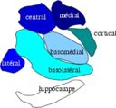
Subdivisions of the amygdala
Details
Identifiers
Latin corpus amygdaloideum
MeSH D000679
NeuroNames 237
NeuroLex ID birnlex_1241
TA A14.1.09.402
FMA 61841


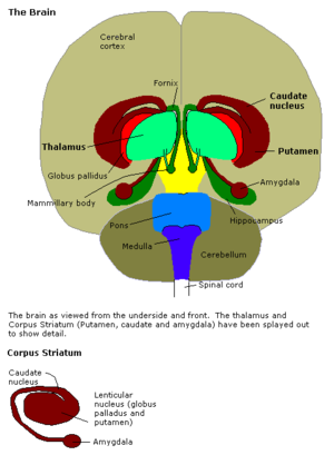
Human brain in the coronal orientation. Amygdalae are shown in dark red.

The amygdala (/əˈmɪɡdələ/; plural: amygdalae; also corpus amygdaloideum; Latin from Greek, ἀμυγδαλή, amygdalē, 'almond', 'tonsil'[1]) is one of two almond-shaped groups of nuclei located deep and medially within the temporal lobes of the brain in complex vertebrates, including humans.[2] Shown in research to perform a primary role in the processing of memory, decision-making and emotional responses, the amygdalae are considered part of the limbic system.[3]

Structure


Subdivisions of the mouse amygdala

The regions described as amygdala nuclei encompass several structures with distinct connectional and functional characteristics in humans and other animals.[4] Among these nuclei are the basolateral complex, the cortical nucleus, the medial nucleus, the central nucleus, and the intercalated cell clusters. The basolateral complex can be further subdivided into the lateral, the basal, and the accessory basal nuclei.[3][5][6]

MRI coronal view of the amygdala
MRI coronal view of the right amygdala

Anatomically, the amygdala,[7] and more particularly its central and medial nuclei,[8] have sometimes been classified as a part of the basal ganglia.

Hemispheric specializations

There are functional differences between the right and left amygdala. In one study, electrical stimulations of the right amygdala induced negative emotions, especially fear and sadness. In contrast, stimulation of the left amygdala was able to induce either pleasant (happiness) or unpleasant (fear, anxiety, sadness) emotions.[9] Other evidence suggests that the left amygdala plays a role in the brain's reward system.[10]

Each side holds a specific function in how we perceive and process emotion. The right and left portions of the amygdala have independent memory systems, but work together to store, encode, and interpret emotion.

The right hemisphere is associated with negative emotion. It plays a role in the expression of fear and in the processing of fear-inducing stimuli. Fear conditioning, which is when a neutral stimulus acquires aversive properties, occurs within the right hemisphere. When an individual is presented with a conditioned, aversive stimulus, it is processed within the right amygdala, producing an unpleasant or fearful response. This emotional response conditions the individual to avoid fear-inducing stimuli.

The right hemisphere is also linked to declarative memory, which consists of facts and information from previously experienced events and must be consciously recalled. It also plays a significant role in the retention of episodic memory. Episodic memory consists of the autobiographical aspects of memory, permitting you to recall your personal emotional and sensory experience of an event. This type of memory does not require conscious recall. The right amygdala plays a role in the association of time and places with emotional properties.[11]

Development

There is considerable growth within the first few years of structural development in both male and female amygdalae.[12] Within this early period, female limbic structures grow at a more rapid pace than the male ones. Amongst female subjects, the amygdala reaches its full growth potential approximately 1.5 years before the peak of male development. The structural development of the male amygdala occurs over a longer period than in women. Despite the early development of female amygdalae, they reach their growth potential sooner than males, whose amygdalae continue to develop. The larger relative size of the male amygdala may be attributed to this extended developmental period.

In addition to longer periods of development, other neurological and hormonal factors may contribute to sex-specific developmental differences. The amygdala is rich in androgen receptors – nuclear receptors that bind to testosterone. Androgen receptors play a role in the DNA binding that regulates gene expression. Though testosterone is present within the female hormonal systems, women have lower levels of testosterone than men. The abundance of testosterone in the male hormonal system may contribute to development. In addition, the grey matter volume on the amygdala is predicted by testosterone levels, which may also contribute to the increased mass of the male amygdala.

In addition to sex differences, there are observable developmental differences between the right and left amygdala in both males and females. The left amygdala reaches its developmental peak approximately 1.5–2 years prior to the right amygdala. Despite the early growth of the left amygdala, the right increases in volume for a longer period of time. The right amygdala is associated with response to fearful stimuli as well as face recognition. It is inferred that the early development of the left amygdala functions to provide infants the ability to detect danger.[12] In childhood, the amygdala is found to react differently to same-sex versus opposite-sex individuals. This reactivity decreases until a person enters adolescence, where it increases dramatically at puberty.[13]

Sex distinction

The amygdala is one of the best-understood brain regions with regard to differences between the sexes. The amygdala is larger in males than females in children ages 7–11,[14] in adult humans,[15] and in adult rats.[16]

In addition to size, other functional and structural differences between male and female amygdalae have been observed. Subjects' amygdala activation was observed when watching a horror film and subliminal stimuli. The results of the study showed a different lateralization of the amygdala in men and women. Enhanced memory for the film was related to enhanced activity of the left, but not the right, amygdala in women, whereas it was related to enhanced activity of the right, but not the left, amygdala in men.[17] One study found evidence that on average, women tend to retain stronger memories for emotional events than men.[18]

The right amygdala is also linked with taking action as well as being linked to negative emotions,[19] which may help explain why males tend to respond to emotionally stressful stimuli physically. The left amygdala allows for the recall of details, but it also results in more thought rather than action in response to emotionally stressful stimuli, which may explain the absence of physical response in women.

Function

Connections

The amygdala sends projections to the hypothalamus, the dorsomedial thalamus, the thalamic reticular nucleus, the nuclei of the trigeminal nerve and the facial nerve, the ventral tegmental area, the locus coeruleus, and the laterodorsal tegmental nucleus.[5]


Coronal section of brain through intermediate mass of third ventricle. Amygdala is shown in purple.

The medial nucleus is involved in the sense of smell and pheromone-processing. It receives input from the olfactory bulb and olfactory cortex.[20] The lateral amygdalae, which send impulses to the rest of the basolateral complexes and to the centromedial nuclei, receive input from the sensory systems. The centromedial nuclei are the main outputs for the basolateral complexes, and are involved in emotional arousal in rats and cats.[5][6][21]

Emotional learning

In complex vertebrates, including humans, the amygdalae perform primary roles in the formation and storage of memories associated with emotional events. Research indicates that, during fear conditioning, sensory stimuli reach the basolateral complexes of the amygdalae, particularly the lateral nuclei, where they form associations with memories of the stimuli. The association between stimuli and the aversive events they predict may be mediated by long-term potentiation,[22][23] a sustained enhancement of signaling between affected neurons.[24] There have been studies that show that damage to the amygdala can interfere with memory that is strengthened by emotion. One study examined a patient with bilateral degeneration of the amygdala. He was told a violent story accompanied by matching pictures and was observed based on how much he could recall from the story. The patient had less recollection of the story than patients with functional amygdala, showing that the amygdala has a strong connection with emotional learning.[25]

Emotional memories are thought to be stored in synapses throughout the brain. Fear memories, for example, are considered to be stored in the neuronal connections from the lateral nuclei to the central nucleus of the amygdalae and the bed nuclei of the stria terminalis (part of the extended amygdala). Of course, these connections are not the sole site of fear memories given that the nuclei of the amygdala receive and send information to other brain regions that are important for memory such as the hippocampus. Some sensory neurons project their axon terminals to the central nucleus.[26] The central nuclei are involved in the genesis of many fear responses such as defensive behavior (freezing or escape responses), autonomic nervous system responses (changes in blood pressure and heart rate/tachycardia), neuroendocrine responses (stress-hormone release), etc. Damage to the amygdalae impairs both the acquisition and expression of Pavlovian fear conditioning, a form of classical conditioning of emotional responses.[24]

The amygdalae are also involved in appetitive (positive) conditioning. It seems that distinct neurons respond to positive and negative stimuli, but there is no clustering of these distinct neurons into clear anatomical nuclei.[27][28] However, lesions of the central nucleus in the amygdala have been shown to reduce appetitive learning in rats. Lesions of the basolateral regions do not exhibit the same effect.[29] Research like this indicates that different nuclei within the amygdala have different functions in appetitive conditioning.[30][31] Nevertheless, researchers found an example of appetitive emotional learning showing an important role for the basolateral amygdala: The naïve female mice are innately attracted to non-volatile pheromones contained in male-soiled bedding, but not by the male-derived volatiles, become attractive if associated with non-volatile attractive pheromones, which act as unconditioned stimulus in a case of Pavlovian associative learning.[32] In the vomeronasal, olfactory and emotional systems, Fos protein show that non-volatile pheromones stimulate the vomeronasal system, whereas air-borne volatiles activate only the olfactory system. Thus, the acquired preference for male-derived volatiles reveals an olfactory-vomeronasal associative learning. Moreover, the reward system is differentially activated by the primary pheromones and secondarily attractive odorants. Exploring the primary attractive pheromone activates the basolateral amygdala and the shell of nucleus accumbens but neither the ventral tegmental area nor the orbitofrontal cortex. In contrast, exploring the secondarily attractive male-derived odorants involves activation of a circuit that includes the basolateral amygdala, prefrontal cortex and ventral tegmental area. Therefore, the basolateral amygdala stands out as the key center for vomeronasal-olfactory associative learning.[33]

Memory modulation

The amygdala is also involved in the modulation of memory consolidation. Following any learning event, the long-term memory for the event is not formed instantaneously. Rather, information regarding the event is slowly assimilated into long-term (potentially lifelong) storage over time, possibly via long-term potentiation. Recent studies suggest that the amygdala regulates memory consolidation in other brain regions. Also, fear conditioning, a type of memory that is impaired following amygdala damage, is mediated in part by long-term potentiation.[22][23]

During the consolidation period, the memory can be modulated. In particular, it appears that emotional arousal following the learning event influences the strength of the subsequent memory for that event. Greater emotional arousal following a learning event enhances a person's retention of that event. Experiments have shown that administration of stress hormones to mice immediately after they learn something enhances their retention when they are tested two days later.[34]

The amygdala, especially the basolateral nuclei, are involved in mediating the effects of emotional arousal on the strength of the memory for the event, as shown by many laboratories including that of James McGaugh. These laboratories have trained animals on a variety of learning tasks and found that drugs injected into the amygdala after training affect the animals' subsequent retention of the task. These tasks include basic classical conditioning tasks such as inhibitory avoidance, where a rat learns to associate a mild footshock with a particular compartment of an apparatus, and more complex tasks such as spatial or cued water maze, where a rat learns to swim to a platform to escape the water. If a drug that activates the amygdalae is injected into the amygdalae, the animals had better memory for the training in the task.[35] If a drug that inactivates the amygdalae is injected, the animals had impaired memory for the task.

Buddhist monks who do compassion meditation have been shown to modulate their amygdala, along with their temporoparietal junction and insula, during their practice.[36] In an fMRI study, more intensive insula activity was found in expert meditators than in novices.[37] Increased activity in the amygdala following compassion-oriented meditation may contribute to social connectedness.[38]

Amygdala activity at the time of encoding information correlates with retention for that information. However, this correlation depends on the relative "emotionalness" of the information. More emotionally arousing information increases amygdalar activity, and that activity correlates with retention. Amygdala neurons show various types of oscillation during emotional arousal, such as theta activity. These synchronized neuronal events could promote synaptic plasticity (which is involved in memory retention) by increasing interactions between neocortical storage sites and temporal lobe structures involved in declarative memory.[39]


Research using Rorschach test blot 03 finds that the number of unique responses to this random figure links to larger sized amygdalae. The researchers note, "Since previous reports have indicated that unique responses were observed at higher frequency in the artistic population than in the nonartistic normal population, this positive correlation suggests that amygdalar enlargement in the normal population might be related to creative mental activity."[40]

Neuropsychological correlates of amygdala activity

Early research on primates provided explanations as to the functions of the amygdala, as well as a basis for further research. As early as 1888, rhesus monkeys with a lesioned temporal cortex (including the amygdala) were observed to have significant social and emotional deficits.[41]  Heinrich Klüver and Paul Bucy later expanded upon this same observation by showing that large lesions to the anterior temporal lobe produced noticeable changes, including overreaction to all objects, hypoemotionality, loss of fear, hypersexuality, and hyperorality, a condition in which inappropriate objects are placed in the mouth. Some monkeys also displayed an inability to recognize familiar objects and would approach animate and inanimate objects indiscriminately, exhibiting a loss of fear towards the experimenters. This behavioral disorder was later named Klüver-Bucy syndrome accordingly,[42] and later research proved it was specifically due to amygdala lesions. Monkey mothers who had amygdala damage showed a reduction in maternal behaviors towards their infants, often physically abusing or neglecting them.[43] In 1981, researchers found that selective radio frequency lesions of the whole amygdala caused Klüver-Bucy syndrome.[44]

With advances in neuroimaging technology such as MRI, neuroscientists have made significant findings concerning the amygdala in the human brain. A variety of data shows the amygdala has a substantial role in mental states, and is related to many psychological disorders. Some studies have shown children with anxiety disorders tend to have a smaller left amygdala. In the majority of the cases, there was an association between an increase in the size of the left amygdala with the use of SSRIs (antidepressant medication) or psychotherapy. The left amygdala has been linked to social anxiety, obsessive and compulsive disorders, and post traumatic stress, as well as more broadly to separation and general anxiety.[45] In a 2003 study, subjects with borderline personality disorder showed significantly greater left amygdala activity than normal control subjects. Some borderline patients even had difficulties classifying neutral faces or saw them as threatening.[46] Individuals with psychopathy show reduced autonomic responses to instructed fear cues than otherwise healthy individuals.[47] In 2006, researchers observed hyperactivity in the amygdala when patients were shown threatening faces or confronted with frightening situations. Patients with severe social phobia showed a correlation with increased response in the amygdala.[48] Similarly, depressed patients showed exaggerated left amygdala activity when interpreting emotions for all faces, and especially for fearful faces. Interestingly, this hyperactivity was normalized when patients were administered antidepressant medication.[49] By contrast, the amygdala has been observed to respond differently in people with bipolar disorder. A 2003 study found that adult and adolescent bipolar patients tended to have considerably smaller amygdala volumes and somewhat smaller hippocampal volumes.[50] Many studies have focused on the connections between the amygdala and autism.[51]

Studies in 2004 and 2006 showed that normal subjects exposed to images of frightened faces or faces of people from another race will show increased activity of the amygdala, even if that exposure is subliminal.[52][53] However, the amygdala is not necessary for the processing of fear-related stimuli, since persons in whom it is bilaterally damaged show rapid reactions to fearful faces, even in the absence of a functional amygdala.[54]

Recent research suggests that parasites, in particular toxoplasma, form cysts in the brain of rats, often taking up residence in the amygdala. This may provide clues as to how specific parasites may contribute to the development of disorders, including paranoia.[55]

Future studies have been proposed to address the role of the amygdala in positive emotions, and the ways in which the amygdala networks with other brain regions.[56]

Sexual orientation

Recent studies have suggested possible correlations between brain structure, including differences in hemispheric ratios and connection patterns in the amygdala, and sexual orientation. Homosexual men tend to exhibit more feminine patterns in the amygdala than heterosexual males do, just as homosexual females tend to show more masculine patterns in the amygdala than heterosexual women do. It was observed that amygdala connections were more widespread from the left amygdala in homosexual males, as is also found in heterosexual females. Amygdala connections were more widespread from the right amygdala in homosexual females, as in heterosexual males.[57][58]

Social interaction

Amygdala volume correlates positively with both the size (the number of contacts a person has) and the complexity (the number of different groups to which a person belongs) of social networks.[59][60] Individuals with larger amygdalae had larger and more complex social networks. They were also better able to make accurate social judgments about other persons' faces.[61] The amygdala's role in the analysis of social situations stems specifically from its ability to identify and process changes in facial features. It does not, however, process the direction of the gaze of the person being perceived.[62][63]

The amygdala is also thought to be a determinant of the level of a person's emotional intelligence. It is particularly hypothesized that larger amygdalae allow for greater emotional intelligence, enabling greater societal integration and cooperation with others.[64]

The amygdala processes reactions to violations concerning personal space. These reactions are absent in persons in whom the amygdala is damaged bilaterally.[65] Furthermore, the amygdala is found to be activated in fMRI when people observe that others are physically close to them, such as when a person being scanned knows that an experimenter is standing immediately next to the scanner, versus standing at a distance.[65]

Aggression

Animal studies have shown that stimulating the amygdala appears to increase both sexual and aggressive behavior. Likewise, studies using brain lesions have shown that harm to the amygdala may produce the opposite effect. Thus, it appears that this part of the brain may play a role in the display and modulation of aggression.[66]

Fear

There are cases of human patients with focal bilateral amygdala lesions, due to the rare genetic condition Urbach-Wiethe disease.[67][68] Such patients fail to exhibit fear-related behaviors, leading one, Patient S.M., to be dubbed the "woman with no fear". This finding reinforces the conclusion that the amygdala "plays a pivotal role in triggering a state of fear".[69]

Alcoholism and binge drinking

The amygdala appears to play a role in binge drinking, being damaged by repeated episodes of intoxication and withdrawal.[70] Alcoholism is associated with dampened activation in brain networks responsible for emotional processing[clarification needed], including the amygdala.[71] Protein kinase C-epsilon in the amygdala is important for regulating behavioral responses to morphine, ethanol, and controlling anxiety-like behavior. The protein is involved in controlling the function of other proteins and plays a role in development of the ability to consume a large amount of ethanol.[72][73]

Anxiety

There may also be a link between the amygdala and anxiety.[74] In particular, there is a higher prevalence of females that are affected by anxiety disorders. In an experiment, degu pups were removed from their mother but allowed to hear her call. In response, the males produced increased serotonin receptors in the amygdala but females lost them. This led to the males being less affected by the stressful situation.

The clusters of the amygdala are activated when an individual expresses feelings of fear or aggression. This occurs because the amygdala is the primary structure of the brain responsible for fight or flight response. Anxiety and panic attacks can occur when the amygdala senses environmental stressors that stimulate fight or flight response.

The amygdala is directly associated with conditioned fear. Conditioned fear is the framework used to explain the behavior produced when an originally neutral stimulus is consistently paired with a stimulus that evokes fear. The amygdala represents a core fear system in the human body, which is involved in the expression of conditioned fear. Fear is measured by changes in autonomic activity including increased heart rate, increased blood pressure, as well as in simple reflexes such as flinching or blinking.

The central nucleus of the amygdala has direct correlations to the hypothalamus and brainstem – areas directly related to fear and anxiety. This connection is evident from studies of animals that have undergone amygdalae removal. Such studies suggest that animals lacking an amygdala have less fear expression and indulge in non-species-like behavior. Many projection areas of the amygdala are critically involved in specific signs that are used to measure fear and anxiety.

Mammals have very similar ways of processing and responding to danger. Scientists have observed similar areas in the brain – specifically in the amygdala – lighting up or becoming more active when a mammal is threatened or beginning to experience anxiety. Similar parts of the brain are activated when rodents and when humans observe a dangerous situation, the amygdala playing a crucial role in this assessment. By observing the amygdala’s functions, people can determine why one rodent may be much more anxious than another. There is a direct relationship between the activation of the amygdala and the level of anxiety the subject feels.

Feelings of anxiety start with a catalyst – an environmental stimulus that provokes stress. This can include various smells, sights, and internal feelings that result in anxiety. The amygdala reacts to this stimuli by preparing to either stand and fight or to turn and run. This response is triggered by the release of adrenaline into the bloodstream. Consequently, blood sugar rises, becoming immediately available to the muscles for quick energy. Shaking may occur in an attempt to return blood to the rest of the body. A better understanding of the amygdala and its various functions may lead to a new way of treating clinical anxiety.[75]

Posttraumatic stress disorder

There seems to be a connection with the amygdalae and how the brain processes posttraumatic stress disorder. Multiple studies have found that the amygdalae may be responsible for the emotional reactions of PTSD patients. One study in particular found that when PTSD patients are shown pictures of faces with fearful expressions, their amygdalae tended to have a higher activation than someone without PTSD.[76]

Bipolar disorder

Amygdala dysfunction during face emotion processing is well-documented in bipolar disorder. Individuals with bipolar disorder showed greater amygdala activity (especially the amygdala/medial-prefrontal-cortex circuit).[77] [78]

Political orientation

Amygdala size has been correlated with cognitive styles with regard to political thinking. A study found that "greater liberalism was associated with increased gray matter volume in the anterior cingulate cortex, whereas greater conservatism was associated with increased volume of the right amygdala." These findings suggest that the volume of the amygdala and anterior cingulate gyrus may be associated with an individual's ability to tolerate uncertainty and conflict.[79]

United States labor law

From Wikipedia, the free encyclopedia https://en.wikipedia.org/wiki/Uni...