Search This Blog

Friday, October 26, 2018

Development of the nervous system


From Wikipedia, the free encyclopedia


Development of the nervous system refers to the processes that generate, shape, and reshape the nervous system of animals, from the earliest stages of embryogenesis to adulthood. The field of neural development draws on both neuroscience and developmental biology to describe and provide insight into the cellular and molecular mechanisms by which complex nervous systems develop, from the nematode and fruit fly to mammals. Defects in neural development can lead to malformations and a wide variety of sensory, motor, and cognitive impairments, including holoprosencephaly and other neurological disorders such as Rett syndrome, Down syndrome and intellectual disability.

Overview of brain development

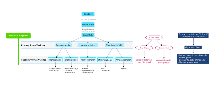
The mammalian central nervous system (CNS) is derived from the ectoderm—the outermost tissue layer—of the embryo. In the third week of human development the neuroectoderm appears and forms the neural plate along the dorsal side of the embryo. The neural plate is the source of the majority of neurons and glial cells of the CNS. A groove forms along the long axis of the neural plate and, by week four of development, the neural plate wraps in on itself to give rise to the neural tube, which is filled with cerebrospinal fluid (CSF). As the embryo develops, the anterior part of the neural tube forms a series of bulges called vesicles, which become the primary anatomical regions of the brain: the forebrain (prosencephalon), midbrain (mesencephalon), and hindbrain (rhombencephalon). These simple, early vesicles enlarge and further divide into the telencephalon (future cerebral cortex and basal ganglia), diencephalon (future thalamus and hypothalamus), mesencephalon (future colliculi), metencephalon (future pons and cerebellum), and myelencephalon (future medulla). The CSF-filled central chamber is continuous from the telencephalon to the spinal cord, and constitutes the developing ventricular system of the CNS. Because the neural tube gives rise to the brain and spinal cord any mutations at this stage in development can lead to lethal deformities like anencephaly or lifelong disabilities like spina bifida. During this time, the walls of the neural tube contain neural stem cells, which drive brain growth as they divide many times. Gradually some of the cells stop dividing and differentiate into neurons and glial cells, which are the main cellular components of the CNS. The newly generated neurons migrate to different parts of the developing brain to self-organize into different brain structures. Once the neurons have reached their regional positions, they extend axons and dendrites, which allow them to communicate with other neurons via synapses. Synaptic communication between neurons leads to the establishment of functional neural circuits that mediate sensory and motor processing, and underlie behavior.
Flowchart of human brain development

Aspects

Some landmarks of neural development include the birth and differentiation of neurons from stem cell precursors, the migration of immature neurons from their birthplaces in the embryo to their final positions, outgrowth of axons and dendrites from neurons, guidance of the motile growth cone through the embryo towards postsynaptic partners, the generation of synapses between these axons and their postsynaptic partners, and finally the lifelong changes in synapses, which are thought to underlie learning and memory.

Typically, these neurodevelopmental processes can be broadly divided into two classes: activity-independent mechanisms and activity-dependent mechanisms. Activity-independent mechanisms are generally believed to occur as hardwired processes determined by genetic programs played out within individual neurons. These include differentiation, migration and axon guidance to their initial target areas. These processes are thought of as being independent of neural activity and sensory experience. Once axons reach their target areas, activity-dependent mechanisms come into play. Although synapse formation is an activity-independent event, modification of synapses and synapse elimination requires neural activity.

Developmental neuroscience uses a variety of animal models including mice Mus musculus, the fruit fly Drosophila melanogaster, the zebrafish Danio rerio, Xenopus laevis tadpoles and the worm Caenorhabditis elegans, among others.

Myelination, formation of the lipid myelin bilayer around neuronal axons, is a process that is essential for normal brain function. The myelin sheath provides insulation for the nerve impulse when communicating between neural systems. Without it, the impulse would be disrupted and the signal would not reach its target, thus impairing normal functioning. Because so much of brain development occurs in the prenatal stage and infancy, it is crucial that myelination, along with cortical development occur properly. Magnetic resonance imaging (MRI) is a non-invasive technique used to investigate myelination and cortical maturation (the cortex is the outer layer of the brain composed of gray matter). Rather than showing the actual myelin, the MRI picks up on the myelin water fraction (MWF), a measure of myelin content. Multicomponent relaxometry (MCR) allow visualization and quantification of myelin content. MCR is also useful for tracking white matter maturation, which plays an important role in cognitive development. It has been discovered that in infancy, myelination occurs in a posterior-to-anterior pattern. Because there is little evidence of a relationship between myelination and cortical thickness, it was revealed that cortical thickness is independent of white matter MWF. This allows various aspects of the brain to grow simultaneously, leading to a more fully developed brain.

Neural induction

During early embryonic development the ectoderm becomes specified to give rise to the epidermis (skin) and the neural plate. The conversion of undifferentiated ectoderm to neuro-ectoderm requires signals from the mesoderm. At the onset of gastrulation presumptive mesodermal cells move through the dorsal blastopore lip and form a layer in between the endoderm and the ectoderm. These mesodermal cells that migrate along the dorsal midline give rise to a structure called the notochord. Ectodermal cells overlying the notochord develop into the neural plate in response to a diffusible signal produced by the notochord. The remainder of the ectoderm gives rise to the epidermis (skin). The ability of the mesoderm to convert the overlying ectoderm into neural tissue is called neural induction.

The neural plate folds outwards during the third week of gestation to form the neural groove. Beginning in the future neck region, the neural folds of this groove close to create the neural tube. The formation of the neural tube from the ectoderm is called neurulation. The ventral part of the neural tube is called the basal plate; the dorsal part is called the alar plate. The hollow interior is called the neural canal. By the end of the fourth week of gestation, the open ends of the neural tube, called the neuropores, close off.

A transplanted blastopore lip can convert ectoderm into neural tissue and is said to have an inductive effect. Neural inducers are molecules that can induce the expression of neural genes in ectoderm explants without inducing mesodermal genes as well. Neural induction is often studied in xenopus embryos since they have a simple body pattern and there are good markers to distinguish between neural and non-neural tissue. Examples of neural inducers are the molecules noggin and chordin.

When embryonic ectodermal cells are cultured at low density in the absence of mesodermal cells they undergo neural differentiation (express neural genes), suggesting that neural differentiation is the default fate of ectodermal cells. In explant cultures (which allow direct cell-cell interactions) the same cells differentiate into epidermis. This is due to the action of BMP4 (a TGF-β family protein) that induces ectodermal cultures to differentiate into epidermis. During neural induction, noggin and chordin are produced by the dorsal mesoderm (notochord) and diffuse into the overlying ectoderm to inhibit the activity of BMP4. This inhibition of BMP4 causes the cells to differentiate into neural cells. Inhibition of TGF-β and BMP (bone morphogenetic protein) signaling can efficiently induce neural tissue from human pluripotent stem cells, a model of early human development.

Regionalization

Late in the fourth week, the superior part of the neural tube flexes at the level of the future midbrain—the mesencephalon. Above the mesencephalon is the prosencephalon (future forebrain) and beneath it is the rhombencephalon (future hindbrain).

The optical vesicle (which eventually become the optic nerve, retina and iris) forms at the basal plate of the prosencephalon. The alar plate of the prosencephalon expands to form the cerebral hemispheres (the telencephalon) whilst its basal plate becomes the diencephalon. Finally, the optic vesicle grows to form an optic outgrowth.

Patterning of the nervous system

In chordates, dorsal ectoderm forms all neural tissue and the nervous system. Patterning occurs due to specific environmental conditions - different concentrations of signaling molecules

Dorsoventral axis

The ventral half of the neural plate is controlled by the notochord, which acts as the 'organiser'. The dorsal half is controlled by the ectoderm plate, which flanks either side of the neural plate.

Ectoderm follows a default pathway to become neural tissue. Evidence for this comes from single, cultured cells of ectoderm, which go on to form neural tissue. This is postulated to be because of a lack of BMPs, which are blocked by the organiser. The organiser may produce molecules such as follistatin, noggin and chordin that inhibit BMPs.

The ventral neural tube is patterned by sonic hedgehog (Shh) from the notochord, which acts as the inducing tissue. Notochord-derived Shh signals to the floor plate, and induces Shh expression in the floor plate. Floor plate-derived Shh subsequently signals to other cells in the neural tube, and is essential for proper specification of ventral neuron progenitor domains. Loss of Shh from the notochord and/or floor plate prevents proper specification of these progenitor domains. Shh binds Patched1, relieving Patched-mediated inhibition of Smoothened, leading to activation of Gli family of transcription factors (Gli1, Gli2, and Gli3) transcription factors.

In this context Shh acts as a morphogen - it induces cell differentiation dependent on its concentration. At low concentrations it forms ventral interneurones, at higher concentrations it induces motor neuron development, and at highest concentrations it induces floor plate differentiation. Failure of Shh-modulated differentiation causes holoprosencephaly.

The dorsal neural tube is patterned by BMPs from the epidermal ectoderm flanking the neural plate. These induce sensory interneurones by activating Sr/Thr kinases and altering SMAD transcription factor levels.

Rostrocaudal (Anteroposterior) axis

Signals that control anteroposterior neural development include FGF and retinoic acid, which act in the hindbrain and spinal cord. The hindbrain, for example, is patterned by Hox genes, which are expressed in overlapping domains along the anteroposterior axis under the control of retinoic acid. The 3' genes in the Hox cluster are induced by retinoic acid in the hindbrain, whereas the 5' Hox genes are not induced by retinoic acid and are expressed more posteriorly in the spinal cord. Hoxb-1 is expressed in rhombomere 4 and gives rise to the facial nerve. Without this Hoxb-1 expression, a nerve similar to the trigeminal nerve arises.

Neurogenesis

Neurogenesis is the process by which neurons are generated from neural stem cells and progenitor cells. Neurons are 'post-mitotic', meaning that they will never divide again for the lifetime of the organism.

Neuronal migration

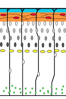
Corticogenesis: younger neurons migrate past older ones using radial glia as a scaffolding. Cajal-Retzius cells (red) release reelin (orange)

Neuronal migration is the method by which neurons travel from their origin or birthplace to their final position in the brain. There are several ways they can do this, e.g. by radial migration or tangential migration. This time lapse displays sequences of radial migration (also known as glial guidance) and somal translocation.

Tangential migration of interneurons from ganglionic eminence

Radial migration

Neuronal precursor cells proliferate in the ventricular zone of the developing neocortex, where the principal neural stem cell is the radial glial cell. The first postmitotic cells must leave the stem cell niche and migrate outward to form the preplate, which is destined to become Cajal-Retzius cells and subplate neurons. These cells do so by somal translocation. Neurons migrating with this mode of locomotion are bipolar and attach the leading edge of the process to the pia. The soma is then transported to the pial surface by nucleokinesis, a process by which a microtubule "cage" around the nucleus elongates and contracts in association with the centrosome to guide the nucleus to its final destination. Radial glial cells, whose fibers serve as a scaffolding for migrating cells and a means of radial communication mediated by calcium dynamic activity, act as the main excitatory neuronal stem cell of the cerebral cortex or translocate to the cortical plate and differentiate either into astrocytes or neurons. Somal translocation can occur at any time during development.

Subsequent waves of neurons split the preplate by migrating along radial glial fibres to form the cortical plate. Each wave of migrating cells travel past their predecessors forming layers in an inside-out manner, meaning that the youngest neurons are the closest to the surface. It is estimated that glial guided migration represents 90% of migrating neurons in human and about 75% in rodents.

Tangential migration

Most interneurons migrate tangentially through multiple modes of migration to reach their appropriate location in the cortex. An example of tangential migration is the movement of interneurons from the ganglionic eminence to the cerebral cortex. One example of ongoing tangential migration in a mature organism, observed in some animals, is the rostral migratory stream connecting subventricular zone and olfactory bulb.

Axophilic migration

Many neurons migrating along the anterior-posterior axis of the body use existing axon tracts to migrate along; this is called axophilic migration. An example of this mode of migration is in GnRH-expressing neurons, which make a long journey from their birthplace in the nose, through the forebrain, and into the hypothalamus. Many of the mechanisms of this migration have been worked out, starting with the extracellular guidance cues that trigger intracellular signaling. These intracellular signals, such as calcium signaling, lead to actin  and microtubule cytoskeletal dynamics, which produce cellular forces that interact with the extracellular environment through cell adhesion proteins to cause the movement of these cells.

Other modes of migration

There is also a method of neuronal migration called multipolar migration. This is seen in multipolar cells, which are abundantly present in the cortical intermediate zone. They do not resemble the cells migrating by locomotion or somal translocation. Instead these multipolar cells express neuronal markers and extend multiple thin processes in various directions independently of the radial glial fibers.

Neurotrophic factors

The survival of neurons is regulated by survival factors, called trophic factors. The neurotrophic hypothesis was formulated by Victor Hamburger and Rita Levi Montalcini based on studies of the developing nervous system. Victor Hamburger discovered that implanting an extra limb in the developing chick led to an increase in the number of spinal motor neurons. Initially he thought that the extra limb was inducing proliferation of motor neurons, but he and his colleagues later showed that there was a great deal of motor neuron death during normal development, and the extra limb prevented this cell death. According to the neurotrophic hypothesis, growing axons compete for limiting amounts of target-derived trophic factors and axons that fail to receive sufficient trophic support die by apoptosis. It is now clear that factors produced by a number of sources contribute to neuronal survival.
  • Nerve Growth Factor (NGF): Rita Levi Montalcini and Stanley Cohen purified the first trophic factor, Nerve Growth Factor (NGF), for which they received the Nobel Prize. There are three NGF-related trophic factors: BDNF, NT3, and NT4, which regulate survival of various neuronal populations. The Trk proteins act as receptors for NGF and related factors. Trk is a receptor tyrosine kinase. Trk dimerization and phosphorylation leads to activation of various intracellular signaling pathways including the MAP kinase, Akt, and PKC pathways.
  • CNTF: Ciliary neurotrophic factor is another protein that acts as a survival factor for motor neurons. CNTF acts via a receptor complex that includes CNTFRα, GP130, and LIFRβ. Activation of the receptor leads to phosphorylation and recruitment of the JAK kinase, which in turn phosphorylates LIFRβ. LIFRβ acts as a docking site for the STAT transcription factors. JAK kinase phosphorylates STAT proteins, which dissociate from the receptor and translocate to the nucleus to regulate gene expression.
  • GDNF: Glial derived neurotrophic factor is a member of the TGFb family of proteins, and is a potent trophic factor for striatal neurons. The functional receptor is a heterodimer, composed of type 1 and type 2 receptors. Activation of the type 1 receptor leads to phosphorylation of Smad proteins, which translocate to the nucleus to activate gene expression.

Synapse formation

Neuromuscular junction

Much of our understanding of synapse formation comes from studies at the neuromuscular junction. The transmitter at this synapse is acetylcholine. The acetylcholine receptor (AchR) is present at the surface of muscle cells before synapse formation. The arrival of the nerve induces clustering of the receptors at the synapse. McMahan and Sanes showed that the synaptogenic signal is concentrated at the basal lamina. They also showed that the synaptogenic signal is produced by the nerve, and they identified the factor as Agrin. Agrin induces clustering of AchRs on the muscle surface and synapse formation is disrupted in agrin knockout mice. Agrin transduces the signal via MuSK receptor to rapsyn. Fischbach and colleagues showed that receptor subunits are selectively transcribed from nuclei next to the synaptic site. This is mediated by neuregulins.

In the mature synapse each muscle fiber is innervated by one motor neuron. However, during development many of the fibers are innervated by multiple axons. Lichtman and colleagues have studied the process of synapses elimination. This is an activity-dependent event. Partial blockage of the receptor leads to retraction of corresponding presynaptic terminals.

CNS synapses

Agrin appears not to be a central mediator of CNS synapse formation and there is active interest in identifying signals that mediate CNS synaptogenesis. Neurons in culture develop synapses that are similar to those that form in vivo, suggesting that synaptogenic signals can function properly in vitro. CNS synaptogenesis studies have focused mainly on glutamatergic synapses. Imaging experiments show that dendrites are highly dynamic during development and often initiate contact with axons. This is followed by recruitment of postsynaptic proteins to the site of contact. Stephen Smith and colleagues have shown that contact initiated by dendritic filopodia can develop into synapses.
Induction of synapse formation by glial factors: Barres and colleagues made the observation that factors in glial conditioned media induce synapse formation in retinal ganglion cell cultures. Synapse formation in the CNS is correlated with astrocyte differentiation suggesting that astrocytes might provide a synaptogenic factor. The identity of the astrocytic factors is not yet known.

Neuroligins and SynCAM as synaptogenic signals: Sudhof, Serafini, Scheiffele and colleagues have shown that neuroligins and SynCAM can act as factors that induce presynaptic differentiation. Neuroligins are concentrated at the postsynaptic site and act via neurexins concentrated in the presynaptic axons. SynCAM is a cell adhesion molecule that is present in both pre- and post-synaptic membranes.

Activity dependent mechanisms in the assembly of neural circuits

The processes of neuronal migration, differentiation and axon guidance are generally believed to be activity-independent mechanisms and rely on hard-wired genetic programs in the neurons themselves. New research findings however have implicated a role for activity-dependent mechanisms in mediating some aspects of the aforementioned processes such as the rate of neuronal migration, aspects of neuronal differentiation and axon pathfinding. Activity-dependent mechanisms influence neural circuit development and are crucial for laying out early connectivity maps and the continued refinement of synapses which occurs during development. There are two distinct types of neural activity we observe in developing circuits -early spontaneous activity and sensory-evoked activity. Spontaneous activity occurs early during neural circuit development even when sensory input is absent and is observed in many systems such as the developing visual system, auditory system, motor system, hippocampus, cerebellum, and neocortex.

Experimental techniques such as direct electrophysiological recording, fluorescence imaging using calcium indicators and optogenetic techniques have shed light on the nature and function of these early bursts of activity. They have distinct spatial and temporal patterns during development and their ablation during development has been known to result in deficits in network refinement in the visual system. In the immature retina, waves of spontaneous action potentials arise from the retinal ganglion cells and sweep across the retinal surface in the first few postnatal weeks. These waves are mediated by neurotransmitter acetylcholine in the initial phase and later on by glutamate. They are thought to instruct the formation of two sensory maps- the retinotopic map and eye-specific segregation. Retinotopic map refinement occurs in downstream visual targets in the brain-the superior colliculus (SC) and dorsal lateral geniculate nucleus (LGN). Pharmacological disruption and mouse models lacking the β2 subunit of the nicotinic acetylcholine receptor has shown that the lack of spontaneous activity leads to marked defects in retinotopy and eye-specific segregation.

In the developing auditory system, developing cochlea generate bursts of activity which spreads across the inner hair cells and spiral ganglion neurons which relay auditory information to the brain. ATP release from supporting cells triggers action potentials in inner hair cells. In the auditory system, spontaneous activity is thought to be involved in tonotopic map formation by segregating cochlear neuron axons tuned to high and low frequencies. In the motor system, periodic bursts of spontaneous activity are driven by excitatory GABA and glutamate during the early stages and by acetylcholine and glutamate at later stages. In the developing zebrafish spinal cord, early spontaneous activity is required for the formation of increasingly synchronous alternating bursts between ipsilateral and contralateral regions of the spinal cord and for the integration of new cells into the circuit. In the cortex, early waves of activity have been observed in the cerebellum and cortical slices. Once sensory stimulus becomes available, final fine-tuning of sensory-coding maps and circuit refinement begins to rely more and more on sensory-evoked activity as demonstrated by classic experiments about the effects of sensory deprivation during critical periods.

Contemporary diffusion-weigthted MRI techniques may also uncover the macroscopic process of axonal development. The connectome can be constructed from diffusion MRI data: the vertices of the graph correspond to anatomically labelled gray matter areas, and two such vertices, say u and v, are connected by an edge if the tractography phase of the data processing finds an axonal fiber that connects the two areas, corresponding to u and v.
Consensus Connectome Dynamics

Numerous braingraphs, computed from the Human Connectome Project can be downloaded from the http://braingraph.org site. The Consensus Connectome Dynamics (CCD) is a remarkable phenomenon that was discovered by continuously decreasing the minimum confidence-parameter at the graphical interface of the Budapest Reference Connectome Server. The Budapest Reference Connectome Server (http://connectome.pitgroup.org) depicts the cerebral connections of n=418 subjects with a frequency-parameter k: For any k=1,2,...,n one can view the graph of the edges that are present in at least k connectomes. If parameter k is decreased one-by-one from k=n through k=1 then more and more edges appear in the graph, since the inclusion condition is relaxed. The surprising observation is that the appearance of the edges is far from random: it resembles a growing, complex structure, like a tree or a shrub (visualized on the animation on the left).

It is hypothesized in  that the growing structure copies the axonal development of the human brain: the earliest developing connections (axonal fibers) are common at most of the subjects, and the subsequently developing connections have larger and larger variance, because their variances are accumulated in the process of axonal development.
Synapse elimination

Several motorneurons compete for each neuromuscular junction, but only one survives until adulthood. Competition in vitro has been shown to involve a limited neurotrophic substance that is released, or that neural activity infers advantage to strong post-synaptic connections by giving resistance to a toxin also released upon nerve stimulation. In vivo, it is suggested that muscle fibres select the strongest neuron through a retrograde signal.

Adult neurogenesis

Contrary to popular belief, neurogenesis also occurs in specific parts of the adult brain.

Interoception

From Wikipedia, the free encyclopedia

Interoception is involved in many different physiological
systems like the cardiorespiratory system, gastrointestinal
system, nociceptive system, endocrine and immune systems

Interoception is contemporarily defined as the sense of the internal state of the body. It encompasses the brain’s process of integrating signals relayed from the body into specific subregions—like the brainstem, thalamus, insula, somatosensory, and anterior cingulate cortex—allowing for a nuanced representation of the physiological state of the body. This is important for maintaining homeostatic conditions in the body and, potentially, aiding in self-awareness.

Interoceptive signals are projected to the brain via a diversity of neural pathways that allow for the sensory processing and prediction of internal bodily states. Misrepresentations of internal states, or a disconnect between the body’s signals and the brain’s interpretation and prediction of those signals, have been suggested to underlie some mental disorders such as anxiety, depression, panic disorder, anorexia nervosa, bulimia nervosa, posttraumatic stress disorder (PTSD), obsessive compulsive disorder (OCD), autism spectrum disorders, somatic symptom disorder, and illness anxiety disorder.

The contemporary definition of interoception is not synonymous with the term “visceroception”. Visceroception refers to the perception of bodily signals arising specifically from the viscera: the heart, lungs, stomach, and bladder, along with other internal organs in the trunk of the body. This does not include organs like the brain and skin. Interoception encompasses visceral signaling, but more broadly relates to all physiological tissues that relay a signal to the central nervous system about the current state of the body. Interoceptive signals are transmitted to the brain via multiple pathways including (1) the lamina I spinothalamic pathway, (2) the classical viscerosensory pathway, (3) the vagus nerve and glossopharyngeal nerve, (4) chemosensory pathways in the blood, and (5) somatosensory pathways from the skin.

Interoceptive signals arise from many different physiological systems of the body. The most commonly studied system is cardiovascular interoception which is typically measured by directing attention towards the sensation of the heartbeat during various tasks. Other physiological systems integral to interoceptive processing include the respiratory system, gastrointestinal and genitourinary systems, nociceptive system, thermoregulatory system, endocrine and immune systems. Soft cutaneous touch is another sensory signal often included within the interoceptive processing system.

History and etymology

Early to mid-1900s

Interoception received increased visibility in the 21st century, but the concept was originally introduced by the Nobel Laureate Sir Charles S. Sherrington in 1906. Sherrington referred to interoception (although never stating the term, using “interoceptive” instead) as a way to describe receptors in the body based on their location and function. Here, interoception was confined to the viscera, excluding all receptors and information from the body that would have been considered “exteroceptive” or “proprioceptive.” In Sherrington’s model, exteroceptive receptors received information from outward stimuli, like light, touch, sound, and odor. He classified temperature and nociception as exteroceptive sensations as well; however, these have now been regarded as having interoceptive qualities. He further divided the internal milieu of the body by its somatic and autonomic functions. Here, proprioceptors were localized to skeletal tissue, which control voluntary movement. Interoceptors, a term which has lost prevalence in modern literature, were thus confined to visceral involuntary smooth muscle (e.g. surrounding blood vessels).

Many experiments were conducted in the 1950s and 1960s regarding interoceptive processing. However, research did not start immediately after Sherrington’s discussion of interoceptors because a book by John Newport Langley was published stating that the autonomic nervous system only used efferent (brain-to-body) signaling to implement its functions. This narrowed perspective halted research on interoceptive receptors for many years. Once it became apparent that interoceptive receptors are present in many tissues of the body other researchers began to investigate afferent body-to-brain signals, mainly by conducting animal experiments to see if interoceptive conditioning was possible. Using principles of Pavlovian conditioning, different physiological systems in dogs were perturbed to elicit a conditioned response to food. For example, in one experiment, dogs’ pelvises were distended using infusions of solution when food was presented to them. After rounds of pairing the two, salivation occurred without presenting food once the pelvis was distended. Interoceptive conditioning studies like this illustrated that interoceptive sensations may be important for learned behavior and emotion.

Mid-1900s to 2000

This graph shows the number of articles that reference the term “interoception” specifically from 1905 to 2015. A clear increase
in the number of publications per year on the topic is seen in the
21st century

The late 1950s and the 1960s saw an increased interest in interoception based on the number of publications released during that time period that referenced the term. This increase has been referred to as the “biofeedback blip,” where researchers examined the ability of a person to gain control over autonomic functions as a method of treatment for varying conditions.

Interoception did not gain widespread popularity within the scientific community until the mid- to late-twentieth century. Some researchers chose to use the term visceroceptor and interoceptor interchangeably, in line with Sherrington’s definition of the term; some combined proprioceptive and visceroceptive information into one category - interoception - based on physiological data about the lack of differences in nerve impulses, and some proposed that interoception is composed of more than just endogenous (internal) stimuli. There is ongoing debate about which sensory signals could or should be classified as "interoceptive."

During the 1980s, psychophysiologists began to examine cardiovascular interoception extensively, introducing several different tasks for studying heartbeat perception: heartbeat counting, heartbeat tapping, and heartbeat detection. Psychiatrists were also beginning to look at the effects of pharmacological stimulation on panic disorder symptoms as well. This led to a growing interest in interoception during this time period, including the development of theoretical models on the integration of interoceptive information within the body over time.

2000 and on

The twenty-first century has seen an exponential increase in publications on the topic of interoception, and a recognition of the multifaceted nature of this concept. With a renewed interest in research, different ideas about interoception have emerged. One definition widens the concept to encompass “the skin and all that is underneath the skin” and the perception and function of bodily activity to more fully understand psychosomatic processes. In a similar vein, neuroanatomists hoping to find the anatomical basis of interoceptive functioning have stated the existence of a homeostatic pathway from the body to the brain that represents “the physiological status of all tissues in the body” and that this mapping onto the brain provides an individual with subjective feeling states which are critical for human emotion and self-awareness.

For example, interoception is the fundament of the modern view on allostasis and allostatic load. The regulatory model of allostasis claims that the brain’s primary role as an organ is the predictive regulation of internal sensations. Predictive regulation refers to the brain’s ability to anticipate needs and prepare to fulfill them before they arise. Therefore, in this model, the brain is responsible for efficient regulation of its internal milieu.

Interoception is sometimes generally referred to as “the perception of internal body states” although there are many interoceptive processes in the body which are not perceived. Importantly, interoception is made possible through a process of “integrating the information coming from inside the body into the central nervous system.” This definition deviates from Sherrington’s original proposition, but exemplifies the dynamic and widening breadth of interoception as a concept in modern literature.

Facets of interoception

This table defines many of the different facets or components of interoception

Although interoception as a term has more recently gained increased popularity, different aspects of it have been studied since the 1950s. These include the features of attention, detection, magnitude, discrimination, accuracy, sensibility, and self-report. Despite not using the word “interoception” specifically, many publications in the physiology and medical fields have focused on understanding interoceptive information processing in different organ systems. Attention describes the ability to observe sensations within the body, it can be directed voluntarily in a "top down" manner or it can be attracted involuntarily in a "bottom up" manner. Detection reflects the presence or absence of a conscious report of interoceptive stimuli, like a heartbeat or growling stomach. Magnitude is the intensity of the stimulus, or how strongly the stimuli is felt. Discrimination describes the ability to localize interoceptive stimuli in the body to specific organs and differentiate them from other bodily stimuli that also occur, like distinguishing between a heart which is beating hard from an upset stomach. Accuracy (or sensitivity) refers to how precisely and correctly an individual can monitor specific interoceptive processes. Self-report is itself multifaceted. It describes the ability to reflect on interoceptive experiences occurring over different periods of time, make judgments about them, and describe them. Brain-body interactions can also be studied using neuroimaging techniques to map functional interactions between brain and peripheral signals. Although all of these components of interoception have been studied since the mid-twentieth century, they have not been brought together under the umbrella-term “interoception” until more recently. The term "interoceptive awareness" is also frequently used to encompass any (or all) of the different interoception features that are accessible to conscious self-report. This multifaceted approach offers a unified way of looking at interoceptive functioning and its different features, it clarifies the definition of interoception itself, and it informs structured ways of assessing interoceptive experiences in an individual.

This graph shows the number of articles published on
interoception vs. articles published on interoceptive facets
without directly referencing the term from 1905-2015. The
timeline starts one year before the publication of Charles
Sherrington’s book, “The integrative action of the nervous
system,” which introduced “interoception"

Interoceptive physiology

Cardiovascular system

This table illustrates the most common tasks that are used to assess cardiovascular interoception, broken down by the corresponding facets that they test. The exemplars at the bottom refer to specific studies or researchers who developed the task.

Cardiac interoception has been widely studied as a method of evaluating interoceptive sensation. This is done using different tasks including heartbeat counting, heartbeat tapping, heartbeat detection and heartbeat attention tasks. Heartbeat counting tasks ask participants to count the number of felt heartbeats during short time periods. Their reported count is then compared with the actual count obtained with an electrocardiogram. This measures the participant’s attention to his or her own heartbeat, the accuracy with which that is perceived, and the ability of the participant to report that measurement; however, results can be influenced by the participant’s preexisting knowledge of his or her heart rate and an insensitivity to heart rate change. Heartbeat detection tasks work by providing a participant with a musical tone which is played simultaneously or non-simultaneously with one’s heartbeat, asking the participant to report whether it is simultaneous or not with the tones. Heartbeat detection is commonly used because of its ability discern an individual’s performance above chance levels, so-called "good detectors." However, such detection rates among participants for this task are usually only 35%. It also measures the participant’s attention, detection, discrimination, accuracy and self-report of the interoceptive process. Heartbeat attention tasks are the most minimalistic, and involve simply the top-down direction of attention towards an interoceptive sensation such as the heartbeat, breath, or stomach. Most perceptions of heartbeat sensations usually occur during a time of homeostatic perturbation, such as when the state of the body changes from external or internal influences such as physical exertion or elevated arousal states (e.g., riding a roller coaster, watching a scary movie, public speaking anxiety, or having a panic attack). For this reason, cardiac interoception is also sometimes studied by inducing perturbations of bodily state. This can be done pharmacologically using adrenaline-like drugs, such as isoproterenol, which mimics activation of the sympathetic nervous system, resulting in increased heart rate and respiration rate, similar to the “fight-or-flight” response. This approach provides a physiological basis for understanding psychiatric and neurological disorders that are characterized by heightened sympathetic nervous system activity.

Respiratory and chemoreceptive system

Respiratory perception can differ from other interoceptive physiological symptoms because of an individual’s ability to exert voluntary control over the system with controlled breathing or breathing exercises. This system is often measured using restrictive breathing loads and/or CO2 inhalation, which are designed to mimic labored breathing sensations. Dyspnea, or difficulty breathing, is a commonly felt sensation associated with panic attacks; however, due to the voluntary control of breathing, this domain of interoception usually requires implementation of much more elaborate experimental controls to quantify in comparison to cardiac interoception.

Gastrointestinal and genitourinary systems

Common interoceptive sensations related to the gastrointestinal and genitourinary systems are hunger and fullness. These are homeostatic signals that tell an individual when to eat and when to stop eating. The dorsal mid-insula appears to be integral in taste processing during gastrointestinal interoceptive attention tasks. Rectal and bladder distensions are used as a method to perturb the homeostatic environment of the gastrointestinal and genitourinary systems, using placement of balloon catheters which can be inflated to achieve different stimulus intensities. Associative fear learning paradigms have been used to study how innocuous signals might lead to abnormal states of gastrointestinal hypersensitivity and anxiety. Biofeedback therapy has been used for individuals with impaired gastrointestinal interoception, showing positive outcomes for some patients.

Nociceptive system

Nociception refers to the receiving and processing of pain inducing stimuli by the central nervous system. Functional brain imaging studies during painful stimulation of the skin with heated probes, during mechanical compression, and electric shock have suggested that the insular cortex is prominently activated during pain processing. Thus while pain was once thought of as an exteroceptive sensation, based on functional imaging and anatomical evidence it is now understood that it has an interoceptive component.

Thermoregulatory system

Temperature and pain are thought to be represented as “feelings” of coolness/warmness and pleasantness/unpleasantness in the brain. These sensory and affective characteristics of thermoregulation may motivate certain behavioral responses depending on the state of the body (for example, moving away from a source of heat to a cooler space). Such perturbations in the internal homeostatic environment of an organism are thought to be key aspects of a motivational process giving rise to emotional states, and have been proposed to be represented principally by the insular cortex as feelings. These feelings then influence drives when the anterior cingulate cortex is activated.

Endocrine and immune systems

The endocrine and immune systems are necessary body systems that aid in allostasis and homeostatic control. Imbalances in these systems, along with other genetic and social factors, may be linked to interoceptive dysregulation in depression. These increased allostatic changes may cause a hyperawareness of interoceptive signaling and a hypo-awareness of exteroceptive signaling in depression patients.

Soft touch

Soft touch refers to the stimulation of slow, unmyelinated C Tactile afferents. This is accompanied by a sense of pleasantness, and has been likened to other interoceptive modalities like thermoregulation and nociception because of the similarities in anatomical function. Soft touch activates the insula rather than the somatosensory cortex, indicating that it has an affective importance absent in Aβ fibers. Since soft touch utilizes a separate pathway, it may have a social relevancy, allowing the body to separate the “noise” of outward stimuli from stimuli that evokes an affective feeling.

Neuroanatomical pathways

Multiple neural pathways relay information integral to interoceptive processing from the body to the brain. these include the lamina I spinothalamic pathway, the visceroceptive pathway, and the somatosensory pathway.

Lamina I spinothalamic pathway

The lamina I spinothalamic pathway is commonly known for carrying information to the brain about temperature and pain, but it has been suggested to more broadly relay all information about the homeostatic condition of the body:

Visceroceptive pathway

The visceroceptive pathway relays information about visceral organs to the brain:

Somatosensory pathway

The somatosensory pathway relays information about proprioception and discriminative touch to the brain through different receptors in the skin:

Cortical processing of interoception

Thalamus

The thalamus receives signals from sympathetic and parasympathetic afferents during interoceptive processing. The ventromedial posterior nucleus (VMpo) is a subregion of the thalamus which receives sympathetic information from lamina I spinothalamic neurons. The human VMpo is much larger than that of primates and sub-primates and is important for processing of nociceptive, thermoregulatory, and visceral sensations. The ventromedial basal nucleus (VMb) receives parasympathetic information from visceral and gustatory systems.

Insular cortex


This image divides the insula into its anterior, mid, and
posterior regions, with each being denoted by different
colors.

The insula is critically involved in the processing, integration, and cortical representation of visceral and interoceptive information. Lamina I spinothalamic and vagal afferents project via the brainstem and thalamus to the posterior and mid dorsal insula respectively. From there, information travels to the posterior and mid-insula, which combines visceral and somatosensory information. The insula is also activated during a variety of exteroceptive and affective tasks. The insula is considered to be a "hub" region because it has an extremely high number of connections with other brain areas, suggesting it may be important for an integration of lower-level physiological information and salience.

The insula connects to many regions in the brain and is highly
involved in many homeostatic functions.

Anterior insular cortex

The anterior insular cortex (AIC) is involved in the representation of “cognitive feelings” which arise from the moment-to-moment integration of homeostatic information from the body. These feelings engender self-awareness by creating a sentient being (someone able to feel and perceive) aware of bodily and cognitive processing.

Cytoarchitecture and granulation

The insular cortex differs cytoarchitecturally based on its
anterior, mid, and posterior regions. The posterior insular
cortex is granular, the mid insular cortex is dysgranular (or
slightly granulated) and the anterior insular cortex has no
granulation whatsoever.

The insula contains three major subregions defined by the presence or absence of a granule cell layer: granular, dysgranular (slightly granulated) agranular. Each of these portions of the insular cortex are important for different levels of functional connectivity. Information from the thalamus is projected to all three regions. Those with increased granulation are considered to be capable of receiving sensory input.

Anterior cingulate cortex

The anterior cingulate cortex (ACC) plays a significant role in motivation and the creation of emotion. An emotion can be seen as comprising both a feeling and a motivation based on that feeling. According to one view, the “feeling” is represented in the insula, while the “motivation” is represented in the ACC. Many interoceptive tasks activate the insula and ACC together, specifically tasks that elicit strong aversive feeling states like pain.

Somatosensory cortex

The sensory motor cortex provides an alternative pathway for sensing interoceptive stimuli. Although not following the conventional pathway for interoceptive awareness, skin afferents which project to the primary and secondary somatosensory cortices provide the brain with information regarding bodily information. This area of the brain is commonly engaged by gastrointestinal distension and nociceptive stimulation, but it likely plays a role in representing other interoceptive sensations as well. In one study, a patient with bilateral insula and ACC damage was given isoproterenol as a method of exciting the cardiovascular system. Despite damage to putative interoceptive areas of the brain, the patient was able to perceive his heartbeat with similar accuracy compared to healthy individuals; however, once lidocaine was applied to the patient’s chest over the region of maximum cardiac sensation and the test was run again, the patient did not sense any change in heartbeat whatsoever. This suggested that somatosensory information from afferents innervating the skin outside of the heart may provide information to the brain about the heart’s pounding through the somatosensory cortex.

Interoception and emotion

The relationship between interoception and emotional experience is an intimate one. In the late 19th century, Charles Darwin noted and discussed the involvement of sensations from the viscera by describing similarities between humans and animals reactions to fear in his book, The Expression of Emotions in Man and Animals. Later, William James and Carl Lange developed the James-Lange theory of emotion, which states that bodily sensations provide the critical basis for emotional experience. The somatic marker hypothesis, proposed by Antonio Damasio, expands upon the James-Lange theory and posits that decisions and the ensuing behaviors are optimally guided by physiological patterns of interoceptive and emotional information. Ensuing models focusing on the neurobiology of feelings states emphasized that the brain’s mapping of different physiological body states are the critical ingredients for emotional experience and consciousness. In another model, A.D. Craig argues that the intertwining of interoceptive and homeostatic processes is responsible for initiating and maintaining motivational states and engendering human self-awareness.

Interoception and mental health

Disturbances of interoception occur prominently and frequently in psychiatric disorders. These symptom fluctuations are often observed during the most severe expression of dysfunction, and they figure prominently in diagnostic classification of several psychiatric disorders. A few typical examples are reviewed next.

Panic disorder

Palpitations and dyspnea are hallmarks of panic attacks. Studies have shown that panic disorder patients report a heightened experience of interoceptive sensations, but these studies have failed to clarify whether this is simply due to their systematic bias toward describing such feelings. However, other studies have shown that panic disorder patients feel heartbeat sensations more intensely when the state of the body is perturbed by pharmacological agents, suggesting they exhibit heightened sensitivity to experiencing interoceptive sensations.

Generalized anxiety disorder

Patients with generalized anxiety disorder (GAD) frequently report being bothered by interoceptive feelings of muscle tension, headaches, fatigue, gastrointestinal complaints, and pain.

Posttraumatic stress disorder

Functional neuroimaging studies have shown that posttraumatic stress disorder (PTSD) patients exhibit a decreased activation in the right anterior insula, a region of the brain that is largely responsible for identifying the mismatch between cognitive and interoceptive states. Further, because PTSD patients have shown decreased activation within many nodes of the lamina I homeostatic pathway—a pathway through which the thalamus sends interoceptive information to the anterior insula and anterior cingulate—it has been suggested that PTSD patients experience reduced interoceptive awareness.

Anxiety disorders

The broad consensus of studies investigating the link between interoceptive awareness and anxiety disorders is that people with anxiety disorders experience heightened awareness of and accuracy in identifying interoceptive processes. Functional imaging studies provide evidence that people with anxiety disorders experience heightened interoceptive accuracy, suggested by hyperactivation in the anterior cingulate cortex—a region of the brain associated with interoception—in several different kinds of anxiety disorders. The insula has been suggested to be abnormal in a large scale study across anxiety disorders in general. Other studies have found that interoceptive accuracy is increased in these patients, as evidenced by their superior ability in heartbeat detection tasks in comparison to healthy controls.

Anorexia nervosa

Anorexia nervosa (AN) has been associated with interoceptive disturbances. Patients with AN often develop insensitivity to interoceptive cues of hunger, and yet are highly anxious and report disturbed interoceptive experiences, both inside and out. While AN patients concentrate on distorted perceptions of their body exterior in fear of weight gain, they also report altered physical states within their bodies, such as indistinct feelings of fullness, or an inability to distinguish emotional states from bodily sensations in general (called alexithymia).

Bulimia nervosa

Studies suggest that patients acutely suffering from bulimia nervosa (BN) exhibit heightened interoceptive accuracy, but that some patients who have recovered from the disorder show reduced interoceptive awareness under resting physiological conditions. Further, BN patients consistently report reduced sensitivity to many other kinds of internal and external sensations, exhibiting increased thresholds to heat pain compared to healthy subjects and an increased gastric capacity. Neuroimaging literature suggests a pattern of heightened interoceptive awareness in patients with BN based on increased activity and volume in the insula and anterior cingulate cortex—regions associated with interoception and taste processing—when looking at food.

Major depressive disorder

Major depressive disorder (MDD) has been theoretically linked to interoceptive dysfunction. Studies have shown that women with MDD are less accurate on heartbeat counting tasks than are men with MDD and that, in general, patients with MDD are less accurate at counting heartbeat than are patients with panic or anxiety disorders. However, patients with MDD do not always exhibit reduced cardiac interoceptive accuracy; depressed patients experiencing high levels of anxiety will actually be more accurate on heartbeat detection tasks than depressed patients with lower levels of anxiety.

Somatic symptom disorders

Patients with somatic symptom disorders score lower on heartbeat detection tasks than healthy controls, suggesting that interoceptive accuracy is poor in psychosomatic disorders. It has also been found that patients with psychosomatic disorders who are anxious or stressed report physical symptom discomfort at lower heart rates during exercise treadmill tests, implying poorer interoceptive distress tolerance in somatic symptom disorders with comorbid psychiatric conditions.

Obsessive compulsive disorder

Results from a study investigating the relationship between obsessive compulsive disorder (OCD) and internal body signals found that patients with OCD were more accurate on a heartbeat perception task than healthy controls and anxiety patients heightened interoceptive awareness.

Autism spectrum disorder

Patients with autism spectrum disorder (ASD) may have poorer interoceptive awareness than [control] subjects. It is hypothesized that this decrease in interoceptive accuracy is due to alexithymia, which is often associated with ASD. However, it has also been found that children with ASD actually show greater interoceptive sensitivity than [control subjects] when measured over a long period of time. Further investigation into the relationship between interoception and ASD is needed in order to fully understand the interoceptive aspect of the disorder.

Current theories of interoceptive processing

Embodied predictive interoception coding (EPIC)

The EPIC model proposes a method of understanding the brain’s response to stimuli contrary to the classic "stimulus-response" model. The classical view of information processing is that when a peripheral stimulus provided information to the central nervous system, it was processed in the brain, and a response was elicited. The EPIC model deviates from this and proposes that the brain is involved in a process of active inference, that is, assiduously making predictions about situations based on previous experiences. These predictions, when coupled with incoming sensory signals, allow the brain to compute a prediction error. Interoceptive prediction errors signal the occurrence of discrepancies within the body, which the brain attempts to minimize. This can be done by 1) modifying the predictions through brain-related pathways, 2) altering the body position/location in order to better align incoming sensory signals with the prediction, or 3) altering the brain’s method of receiving incoming stimuli. Interoceptive prediction error signals are a key component of many theories of interoceptive dysfunction in physical and mental health.

Research and treatments

As attention on interoception increases among the scientific community, new research methods and treatment tactics are beginning to emerge. Ongoing research in interoception has shown the importance of perturbing interoceptive systems. This allows researchers the ability to document the effects of non-baseline states, which occur during times of panic or anxiety. It also provides the participant the ability to gauge the intensity of sensations within the body. This can be done through pharmacological interventions, balloon distensions, or respiratory breathing loads depending on the interoceptive system of interest.

An open float pool

Another research method used to study interoception is specialized flotation environments. Floating removes external stimuli so that individuals can more easily focus on the interoceptive sensations within their bodies. One idea with floating is that over many float sessions, patients with different kinds of disorders may learn to become more attuned or tolerant of their interoceptive sensations not only in the float tank but also in their everyday lives.

Whole body hyperthermia may provide a new treatment technique for major depressive disorder. It is thought that reducing one of the bodily symptoms of depression, which is increased inflammation, using whole body hyperthermia will also reduce depressive feelings represented in the brain. In theory, these techniques will help patients better attune themselves to their interoceptive sensations, allowing them a better understanding of what occurs in their bodies.

Acupuncture is an alternative treatment type for many people suffering from anxiety and depression. Typically, it is self-prescribed by patients; however, results are inconclusive on its ability to manage symptoms of depression. Recently, massage therapy has been shown to have the ability to reduce symptoms of generalized anxiety disorder.

Meditation and mindfulness have been looked into as possible techniques to enhance interoceptive awareness based on their tendency to redirect focus within oneself. However, studies show that even though meditation and mindfulness practices promote attention to interoceptive sensations, they do not clearly increase interoceptive awareness in specific domains such as breath or body [null monitoring.]

Although a universal definition of interoception has not been reached, research on interoception and psychiatric disorders has shown a link between interoceptive processing and mental disorders. It has been proposed that exposure therapy, commonly used among anxiety disorders, may provide a basis for a model of interoceptive exposure therapy that could be incorporated into treatment plans of different psychiatric disorders. One proposal states that multiple interoceptive challenges assessing different physiological systems could provide diagnosticians with the ability to create an “interoceptive profile” for a specific individual, creating a patient-specific treatment plan.

Functional programming

From Wikipedia, the free encyclopedia https://en.wikipedia.org/wiki/Functional_programming In computer sc...