Search This Blog

Tuesday, February 12, 2019

History of biochemistry

From Wikipedia, the free encyclopedia

The history of biochemistry can be said to have started with the ancient Greeks who were interested in the composition and processes of life, although biochemistry as a specific scientific discipline has its beginning around the early 19th century. Some argued that the beginning of biochemistry may have been the discovery of the first enzyme, diastase (today called amylase), in 1833 by Anselme Payen, while others considered Eduard Buchner's first demonstration of a complex biochemical process alcoholic fermentation in cell-free extracts to be the birth of biochemistry. Some might also point to the influential work of Justus von Liebig from 1842, Animal chemistry, or, Organic chemistry in its applications to physiology and pathology, which presented a chemical theory of metabolism, or even earlier to the 18th century studies on fermentation and respiration by Antoine Lavoisier.

The term “biochemistry” itself is derived from the combining form bio-, meaning "life", and chemistry. The word is first recorded in English in 1848, while in 1877, Felix Hoppe-Seyler used the term (Biochemie in German) in the foreword to the first issue of Zeitschrift für Physiologische Chemie (Journal of Physiological Chemistry) as a synonym for physiological chemistry and argued for the setting up of institutes dedicate to its studies. Nevertheless, several sources cite German chemist Carl Neuberg as having coined the term for the new discipline in 1903, and some credit it to Franz Hofmeister.

The subject of study in biochemistry is the chemical processes in living organisms, and its history involves the discovery and understanding of the complex components of life and the elucidation of pathways of biochemical processes. Much of biochemistry deals with the structures and functions of cellular components such as proteins, carbohydrates, lipids, nucleic acids and other biomolecules; their metabolic pathways and flow of chemical energy through metabolism; how biological molecules give rise to the processes that occur within living cells; it also focuses on the biochemical processes involved in the control of information flow through biochemical signalling, and how they relate to the functioning of whole organisms. Over the last 40 years the field has had success in explaining living processes such that now almost all areas of the life sciences from botany to medicine are engaged in biochemical research.

Among the vast number of different biomolecules, many are complex and large molecules (called polymers), which are composed of similar repeating subunits (called monomers). Each class of polymeric biomolecule has a different set of subunit types. For example, a protein is a polymer whose subunits are selected from a set of twenty or more amino acids, carbohydrates are formed from sugars known as monosaccharides, oligosaccharides, and polysaccharides, lipids are formed from fatty acids and glycerols, and nucleic acids are formed from nucleotides. Biochemistry studies the chemical properties of important biological molecules, like proteins, and in particular the chemistry of enzyme-catalyzed reactions. The biochemistry of cell metabolism and the endocrine system has been extensively described. Other areas of biochemistry include the genetic code (DNA, RNA), protein synthesis, cell membrane transport, and signal transduction.

Proto-biochemistry

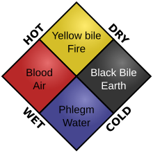
The four humors. In this diagram, each kind of food would result in a different physiological result. For example, cold and dry food would produce black bile.
 
In a sense, the study of biochemistry can be considered to have started in ancient times, for example when biology first began to interest society—as the ancient Chinese developed a system of medicine based on yin and yang, and also the five phases, which both resulted from alchemical and biological interests. Its beginning in the ancient Indian culture was linked to an interest in medicine, as they developed the concept of three humors that were similar to the Greek's four humours. They also delved into the interest of bodies being composed of tissues. The ancient Greeks conception of "biochemistry" was linked with their ideas on matter and disease, where good health was thought to come from a balance of the four elements and four humors in the human body. As in the majority of early sciences, the Islamic world contributed significantly to early biological advancements as well as alchemical advancements; especially with the introduction of clinical trials and clinical pharmacology presented in Avicenna's The Canon of Medicine. On the side of chemistry, early advancements were heavily attributed to exploration of alchemical interests but also included: metallurgy, the scientific method, and early theories of atomism. In more recent times, the study of chemistry was marked by milestones such as the development of Mendeleev's periodic table, Dalton's atomic model, and the conservation of mass theory. This last mention has the most importance of the three due to the fact that this law intertwines chemistry with thermodynamics in an intercalated manner.

Enzymes

As early as the late 18th century and early 19th century, the digestion of meat by stomach secretions and the conversion of starch to sugars by plant extracts and saliva were known. However, the mechanism by which this occurred had not been identified.

In the 19th century, when studying the fermentation of sugar to alcohol by yeast, Louis Pasteur concluded that this fermentation was catalyzed by a vital force contained within the yeast cells called ferments, which he thought functioned only within living organisms. He wrote that "alcoholic fermentation is an act correlated with the life and organization of the yeast cells, not with the death or putrefaction of the cells."

Anselme Payen discovered in 1833 the first enzyme who called diastase and in 1878 German physiologist Wilhelm Kühne (1837–1900) coined the term enzyme, which comes from Greek ενζυμον "in leaven", to describe this process. The word enzyme was used later to refer to nonliving substances such as pepsin, and the word ferment used to refer to chemical activity produced by living organisms.
In 1897 Eduard Buchner began to study the ability of yeast extracts to ferment sugar despite the absence of living yeast cells. In a series of experiments at the University of Berlin, he found that the sugar was fermented even when there were no living yeast cells in the mixture. He named the enzyme that brought about the fermentation of sucrose "zymase". In 1907 he received the Nobel Prize in Chemistry "for his biochemical research and his discovery of cell-free fermentation". Following Buchner's example; enzymes are usually named according to the reaction they carry out. Typically the suffix -ase is added to the name of the substrate (e.g., lactase is the enzyme that cleaves lactose) or the type of reaction (e.g., DNA polymerase forms DNA polymers). 

Shown above is the restriction endonuclease EcoR1 in its 3D computer generated form.
 
Having shown that enzymes could function outside a living cell, the next step was to determine their biochemical nature. Many early workers noted that enzymatic activity was associated with proteins, but several scientists (such as Nobel laureate Richard Willstätter) argued that proteins were merely carriers for the true enzymes and that proteins per se were incapable of catalysis. However, in 1926, James B. Sumner showed that the enzyme urease was a pure protein and crystallized it; Sumner did likewise for the enzyme catalase in 1937. The conclusion that pure proteins can be enzymes was definitively proved by Northrop and Stanley, who worked on the digestive enzymes pepsin (1930), trypsin and chymotrypsin. These three scientists were awarded the 1946 Nobel Prize in Chemistry.

This discovery, that enzymes could be crystallized, meant that scientists eventually could solve their structures by x-ray crystallography. This was first done for lysozyme, an enzyme found in tears, saliva and egg whites that digests the coating of some bacteria; the structure was solved by a group led by David Chilton Phillips and published in 1965. This high-resolution structure of lysozyme marked the beginning of the field of structural biology and the effort to understand how enzymes work at an atomic level of detail.

Metabolism

Early metabolic interest

Santorio Santorio in his steelyard balance, from Ars de statica medecina, first published 1614
 
The term metabolism is derived from the Greek Μεταβολισμός – Metabolismos for "change", or "overthrow". The history of the scientific study of metabolism spans 800 years. The earliest of all metabolic studies began during the early thirteenth century (1213-1288) by a Muslim scholar from Damascus named Ibn al-Nafis. al-Nafis stated in his most well-known work Theologus Autodidactus that "that body and all its parts are in a continuous state of dissolution and nourishment, so they are inevitably undergoing permanent change." Although al-Nafis was the first documented physician to have an interest in biochemical concepts, the first controlled experiments in human metabolism were published by Santorio Santorio in 1614 in his book Ars de statica medecina. This book describes how he weighed himself before and after eating, sleeping, working, sex, fasting, drinking, and excreting. He found that most of the food he took in was lost through what he called "insensible perspiration".

Metabolism: 20th century - present

One of the most prolific of these modern biochemists was Hans Krebs who made huge contributions to the study of metabolism. Krebs was a student of extremely important Otto Warburg, and wrote a biography of Warburg by that title in which he presents Warburg as being educated to do for biological chemistry what Fischer did for organic chemistry. Which he did. Krebs discovered the urea cycle and later, working with Hans Kornberg, the citric acid cycle and the glyoxylate cycle. These discoveries led to Krebs being awarded the Nobel Prize in physiology in 1953, which was shared with the German biochemist Fritz Albert Lipmann who also codiscovered the essential cofactor coenzyme A.

Glucose absorption

In 1960, the biochemist Robert K. Crane revealed his discovery of the sodium-glucose cotransport as the mechanism for intestinal glucose absorption. This was the very first proposal of a coupling between the fluxes of an ion and a substrate that has been seen as sparking a revolution in biology. This discovery, however, would not have been possible if it were not for the discovery of the molecule glucose's structure and chemical makeup. These discoveries are largely attributed to the German chemist Emil Fischer who received the Nobel Prize in chemistry nearly 60 years earlier.

Glycolysis

Shown here is a step-wise depiction of glycolysis along with the required enzymes.
 
Since metabolism focuses on the breaking down (catabolic processes) of molecules and the building of larger molecules from these particles (anabolic processes), the use of glucose and its involvement in the formation of adenosine triphosphate (ATP) is fundamental to this understanding. The most frequent type of glycolysis found in the body is the type that follows the Embden-Meyerhof-Parnas (EMP) Pathway, which was discovered by Gustav Embden, Otto Meyerhof, and Jakob Karol Parnas. These three men discovered that glycolysis is a strongly determinant process for the efficiency and production of the human body. The significance of the pathway shown in the adjacent image is that by identifying the individual steps in this process doctors and researchers are able to pinpoint sites of metabolic malfunctions such as pyruvate kinase deficiency that can lead to severe anemia. This is most important because cells, and therefore organisms, are not capable of surviving without proper functioning metabolic pathways.

Instrumental advancements (20th century)

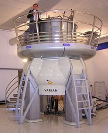
This is an example of a very large NMR instrument known as the HWB-NMR with a 21.2T (Tesla) magnet.
 
Since then, biochemistry has advanced, especially since the mid-20th century, with the development of new techniques such as chromatography, X-ray diffraction, NMR spectroscopy, radioisotopic labelling, electron microscopy and molecular dynamics simulations. These techniques allowed for the discovery and detailed analysis of many molecules and metabolic pathways of the cell, such as glycolysis and the Krebs cycle (citric acid cycle). The example of an NMR instrument shows that some of these instruments, such as the HWB-NMR, can be very large in size and can cost anywhere from a few hundred dollars to millions of dollars ($16 million for the one shown here).

Polymerase chain reaction

Shown above is a model of a thermo cycler that is currently being used in polymerase chain reaction.
 
Polymerase chain reaction (PCR) is the primary gene amplification technique that has revolutionized modern biochemistry. Polymerase chain reaction was developed by Kary Mullis in 1983. There are four steps to a proper polymerase chain reaction: 1) denaturation 2) extension 3) insertion (of gene to be expressed) and finally 4) amplification of the inserted gene. These steps with simple illustrative examples of this process can be seen in the image below and to the right of this section. This technique allows for the copy of a single gene to be amplified into hundreds or even millions of copies and has become a cornerstone in the protocol for any biochemist that wishes to work with bacteria and gene expression. PCR is not only used for gene expression research but is also capable of aiding laboratories in diagnosing certain diseases such a lymphomas, some types of leukemia, and other malignant diseases that can sometimes puzzle doctors. Without polymerase chain reaction development, there are many advancements in the field of bacterial study and protein expression study that would not have come to fruition. The development of the theory and process of polymerase chain reaction is essential but the invention of the thermal cycler is equally as important because the process would not be possible without this instrument. This is yet another testament to the fact that the advancement of technology is just as crucial to sciences such as biochemistry as is the painstaking research that leads to the development of theoretical concepts.
Nicotinamide adenine dinucleotide

From Wikipedia, the free encyclopedia

Nicotinamide adenine dinucleotide
Skeletal formula of the oxidized form
Ball-and-stick model of the oxidized form
Names
Other names
Diphosphopyridine nucleotide (DPN+), Coenzyme I
Identifiers
3D model (JSmol)
ChEBI
ChEMBL
ChemSpider
DrugBank
ECHA InfoCard 100.000.169
KEGG
PubChem CID
RTECS number UU3450000
UNII
Properties
C21H27N7O14P2
Molar mass 663.43 g/mol
Appearance White powder
Melting point 160 °C (320 °F; 433 K)
Hazards
Main hazards Not hazardous
NFPA 704
Flammability code 1: Must be pre-heated before ignition can occur. Flash point over 93 °C (200 °F). E.g., canola oilHealth code 1: Exposure would cause irritation but only minor residual injury. E.g., turpentineReactivity code 0: Normally stable, even under fire exposure conditions, and is not reactive with water. E.g., liquid nitrogenSpecial hazards (white): no codeNFPA 704 four-colored diamond
1
1
0
Except where otherwise noted, data are given for materials in their standard state (at 25 °C [77 °F], 100 kPa).

Nicotinamide adenine dinucleotide (NAD) is a cofactor found in all living cells. The compound is called a dinucleotide because it consists of two nucleotides joined through their phosphate groups. One nucleotide contains an adenine nucleobase and the other nicotinamide. Nicotinamide adenine dinucleotide exists in two forms: an oxidized and reduced form abbreviated as NAD+ and NADH respectively.

In metabolism, nicotinamide adenine dinucleotide is involved in redox reactions, carrying electrons from one reaction to another. The cofactor is, therefore, found in two forms in cells: NAD+ is an oxidizing agent – it accepts electrons from other molecules and becomes reduced. This reaction forms NADH, which can then be used as a reducing agent to donate electrons. These electron transfer reactions are the main function of NAD. However, it is also used in other cellular processes, most notably a substrate of enzymes that add or remove chemical groups from proteins, in posttranslational modifications. Because of the importance of these functions, the enzymes involved in NAD metabolism are targets for drug discovery.

In organisms, NAD can be synthesized from simple building-blocks (de novo) from the amino acids tryptophan or aspartic acid. In an alternative fashion, more complex components of the coenzymes are taken up from food as niacin. Similar compounds are released by reactions that break down the structure of NAD. These preformed components then pass through a salvage pathway that recycles them back into the active form. Some NAD is converted into nicotinamide adenine dinucleotide phosphate (NADP); the chemistry of this related coenzyme is similar to that of NAD, but it has different roles in metabolism.

Although NAD+ is written with a superscript plus sign because of the formal charge on a particular nitrogen atom, at physiological pH for the most part it is actually a singly charged anion (charge of minus 1), while NADH is a doubly charged anion, because of the two bridging phosphate groups.

Physical and chemical properties

Nicotinamide adenine dinucleotide, like all dinucleotides, consists of two nucleosides joined by a pair of bridging phosphate groups. The nucleosides each contain a ribose ring, one with adenine attached to the first carbon atom (the 1' position) and the other with nicotinamide at this position. The nicotinamide moiety can be attached in two orientations to this anomeric carbon atom. Because of these two possible structures, the compound exists as two diastereomers. It is the β-nicotinamide diastereomer of NAD+ that is found in organisms. These nucleotides are joined together by a bridge of two phosphate groups through the 5' carbons.

The redox reactions of nicotinamide adenine dinucleotide.
 
In metabolism, the compound accepts or donates electrons in redox reactions. Such reactions (summarized in formula below) involve the removal of two hydrogen atoms from the reactant (R), in the form of a hydride ion (H), and a proton (H+). The proton is released into solution, while the reductant RH2 is oxidized and NAD+ reduced to NADH by transfer of the hydride to the nicotinamide ring.
RH2 + NAD+ → NADH + H+ + R;
From the hydride electron pair, one electron is transferred to the positively charged nitrogen of the nicotinamide ring of NAD+, and the second hydrogen atom transferred to the C4 carbon atom opposite this nitrogen. The midpoint potential of the NAD+/NADH redox pair is −0.32 volts, which makes NADH a strong reducing agent. The reaction is easily reversible, when NADH reduces another molecule and is re-oxidized to NAD+. This means the coenzyme can continuously cycle between the NAD+ and NADH forms without being consumed.

In appearance, all forms of this coenzyme are white amorphous powders that are hygroscopic and highly water-soluble. The solids are stable if stored dry and in the dark. Solutions of NAD+ are colorless and stable for about a week at 4 °C and neutral pH, but decompose rapidly in acids or alkalis. Upon decomposition, they form products that are enzyme inhibitors.

UV absorption spectra of NAD+ and NADH.

Both NAD+ and NADH strongly absorb ultraviolet light because of the adenine. For example, peak absorption of NAD+ is at a wavelength of 259 nanometers (nm), with an extinction coefficient of 16,900 M−1cm−1. NADH also absorbs at higher wavelengths, with a second peak in UV absorption at 339 nm with an extinction coefficient of 6,220 M−1cm−1. This difference in the ultraviolet absorption spectra between the oxidized and reduced forms of the coenzymes at higher wavelengths makes it simple to measure the conversion of one to another in enzyme assays – by measuring the amount of UV absorption at 340 nm using a spectrophotometer.

NAD+ and NADH also differ in their fluorescence. NADH in solution has an emission peak at 460 nm and a fluorescence lifetime of 0.4 nanoseconds, while the oxidized form of the coenzyme does not fluoresce. The properties of the fluorescence signal changes when NADH binds to proteins, so these changes can be used to measure dissociation constants, which are useful in the study of enzyme kinetics. These changes in fluorescence are also used to measure changes in the redox state of living cells, through fluorescence microscopy.

Concentration and state in cells

In rat liver, the total amount of NAD+ and NADH is approximately 1 μmole per gram of wet weight, about 10 times the concentration of NADP+ and NADPH in the same cells. The actual concentration of NAD+ in cell cytosol is harder to measure, with recent estimates in animal cells ranging around 0.3 mM, and approximately 1.0 to 2.0 mM in yeast. However, more than 80% of NADH fluorescence in mitochondria is from bound form, so the concentration in solution is much lower.

Data for other compartments in the cell are limited, although in the mitochondrion the concentration of NAD+ is similar to that in the cytosol. This NAD+ is carried into the mitochondrion by a specific membrane transport protein, since the coenzyme cannot diffuse across membranes.

The balance between the oxidized and reduced forms of nicotinamide adenine dinucleotide is called the NAD+/NADH ratio. This ratio is an important component of what is called the redox state of a cell, a measurement that reflects both the metabolic activities and the health of cells. The effects of the NAD+/NADH ratio are complex, controlling the activity of several key enzymes, including glyceraldehyde 3-phosphate dehydrogenase and pyruvate dehydrogenase. In healthy mammalian tissues, estimates of the ratio between free NAD+ and NADH in the cytoplasm typically lie around 700; the ratio is thus favourable for oxidative reactions. The ratio of total NAD+/NADH is much lower, with estimates ranging from 3–10 in mammals. In contrast, the NADP+/NADPH ratio is normally about 0.005, so NADPH is the dominant form of this coenzyme. These different ratios are key to the different metabolic roles of NADH and NADPH.

Biosynthesis

NAD+ is synthesized through two metabolic pathways. It is produced either in a de novo pathway from amino acids or in salvage pathways by recycling preformed components such as nicotinamide back to NAD+.

De novo production

Some metabolic pathways that synthesize and consume NAD+ in vertebrates. The abbreviations are defined in the text.
 
Most organisms synthesize NAD+ from simple components. The specific set of reactions differs among organisms, but a common feature is the generation of quinolinic acid (QA) from an amino acid—either tryptophan (Trp) in animals and some bacteria, or aspartic acid (Asp) in some bacteria and plants. The quinolinic acid is converted to nicotinic acid mononucleotide (NaMN) by transfer of a phosphoribose moiety. An adenylate moiety is then transferred to form nicotinic acid adenine dinucleotide (NaAD). Finally, the nicotinic acid moiety in NaAD is amidated to a nicotinamide (Nam) moiety, forming nicotinamide adenine dinucleotide.

In a further step, some NAD+ is converted into NADP+ by NAD+ kinase, which phosphorylates NAD+. In most organisms, this enzyme uses ATP as the source of the phosphate group, although several bacteria such as Mycobacterium tuberculosis and a hyperthermophilic archaeon Pyrococcus horikoshii, use inorganic polyphosphate as an alternative phosphoryl donor.

Salvage pathways use three precursors for NAD+.

Salvage pathways

Besides assembling NAD+ de novo from simple amino acid precursors, cells also salvage preformed compounds containing a pyridine base. The three vitamin precursors used in these salvage metabolic pathways are nicotinic acid (NA), nicotinamide (Nam) and nicotinamide riboside (NR). These compounds can be taken up from the diet and are termed vitamin B3 or niacin. However, these compounds are also produced within cells and by digestion of cellular NAD+. Some of the enzymes involved in these salvage pathways appear to be concentrated in the cell nucleus, which may compensate for the high level of reactions that consume NAD+ in this organelle. There are some reports that mammalian cells can take up extracellular NAD+ from their surroundings, and both nicotinamide and nicotinamide riboside can be absorbed from the gut.

Despite the presence of the de novo pathway, the salvage reactions are essential in humans; a lack of niacin in the diet causes the vitamin deficiency disease pellagra. This high requirement for NAD+ results from the constant consumption of the coenzyme in reactions such as posttranslational modifications, since the cycling of NAD+ between oxidized and reduced forms in redox reactions does not change the overall levels of the coenzyme.

The salvage pathways used in microorganisms differ from those of mammals. Some pathogens, such as the yeast Candida glabrata and the bacterium Haemophilus influenzae are NAD+ auxotrophs – they cannot synthesize NAD+ – but possess salvage pathways and thus are dependent on external sources of NAD+ or its precursors. Even more surprising is the intracellular pathogen Chlamydia trachomatis, which lacks recognizable candidates for any genes involved in the biosynthesis or salvage of both NAD+ and NADP+, and must acquire these coenzymes from its host.

Functions

Rossmann fold in part of the lactate dehydrogenase of Cryptosporidium parvum, showing NAD+ in red, beta sheets in yellow, and alpha helices in purple.
 
Nicotinamide adenine dinucleotide has several essential roles in metabolism. It acts as a coenzyme in redox reactions, as a donor of ADP-ribose moieties in ADP-ribosylation reactions, as a precursor of the second messenger molecule cyclic ADP-ribose, as well as acting as a substrate for bacterial DNA ligases and a group of enzymes called sirtuins that use NAD+ to remove acetyl groups from proteins. In addition to these metabolic functions, NAD+ emerges as an adenine nucleotide that can be released from cells spontaneously and by regulated mechanisms, and can therefore have important extracellular roles.

Oxidoreductase binding of NAD

The main role of NAD+ in metabolism is the transfer of electrons from one molecule to another. Reactions of this type are catalyzed by a large group of enzymes called oxidoreductases. The correct names for these enzymes contain the names of both their substrates: for example NADH-ubiquinone oxidoreductase catalyzes the oxidation of NADH by coenzyme Q. However, these enzymes are also referred to as dehydrogenases or reductases, with NADH-ubiquinone oxidoreductase commonly being called NADH dehydrogenase or sometimes coenzyme Q reductase.

There are many different superfamilies of enzymes that bind NAD+ / NADH. One of the most common superfamilies include a structural motif known as the Rossmann fold. The motif is named after Michael Rossmann who was the first scientist to notice how common this structure is within nucleotide-binding proteins.

An example of a NAD-binding bacterial enzyme involved in amino acid metabolism that does not have Rossmann fold: 

In this diagram, the hydride acceptor C4 carbon is shown at the top. When the nicotinamide ring lies in the plane of the page with the carboxy-amide to the right, as shown, the hydride donor lies either "above" or "below" the plane of the page. If "above" hydride transfer is class A, if "below" hydride transfer is class B.
 
When bound in the active site of an oxidoreductase, the nicotinamide ring of the coenzyme is positioned so that it can accept a hydride from the other substrate. Depending on the enzyme, the hydride donor is positioned either "above" or "below" the plane of the planar C4 carbon, as defined in the figure. Class A oxidoreductases transfer the atom from above; class B enzymes transfer it from below. Since the C4 carbon that accepts the hydrogen is prochiral, this can be exploited in enzyme kinetics to give information about the enzyme's mechanism. This is done by mixing an enzyme with a substrate that has deuterium atoms substituted for the hydrogens, so the enzyme will reduce NAD+ by transferring deuterium rather than hydrogen. In this case, an enzyme can produce one of two stereoisomers of NADH.

Despite the similarity in how proteins bind the two coenzymes, enzymes almost always show a high level of specificity for either NAD+ or NADP+. This specificity reflects the distinct metabolic roles of the respective coenzymes, and is the result of distinct sets of amino acid residues in the two types of coenzyme-binding pocket. For instance, in the active site of NADP-dependent enzymes, an ionic bond is formed between a basic amino acid side-chain and the acidic phosphate group of NADP+. On the converse, in NAD-dependent enzymes the charge in this pocket is reversed, preventing NADP+ from binding. However, there are a few exceptions to this general rule, and enzymes such as aldose reductase, glucose-6-phosphate dehydrogenase, and methylenetetrahydrofolate reductase can use both coenzymes in some species.

Role in redox metabolism

A simplified outline of redox metabolism, showing how NAD+ and NADH link the citric acid cycle and oxidative phosphorylation.

The redox reactions catalyzed by oxidoreductases are vital in all parts of metabolism, but one particularly important area where these reactions occur is in the release of energy from nutrients. Here, reduced compounds such as glucose and fatty acids are oxidized, thereby releasing energy. This energy is transferred to NAD+ by reduction to NADH, as part of beta oxidation, glycolysis, and the citric acid cycle. In eukaryotes the electrons carried by the NADH that is produced in the cytoplasm are transferred into the mitochondrion (to reduce mitochondrial NAD+) by mitochondrial shuttles, such as the malate-aspartate shuttle. The mitochondrial NADH is then oxidized in turn by the electron transport chain, which pumps protons across a membrane and generates ATP through oxidative phosphorylation. These shuttle systems also have the same transport function in chloroplasts.

Since both the oxidized and reduced forms of nicotinamide adenine dinucleotide are used in these linked sets of reactions, the cell maintains significant concentrations of both NAD+ and NADH, with the high NAD+/NADH ratio allowing this coenzyme to act as both an oxidizing and a reducing agent. In contrast, the main function of NADPH is as a reducing agent in anabolism, with this coenzyme being involved in pathways such as fatty acid synthesis and photosynthesis. Since NADPH is needed to drive redox reactions as a strong reducing agent, the NADP+/NADPH ratio is kept very low.

Although it is important in catabolism, NADH is also used in anabolic reactions, such as gluconeogenesis. This need for NADH in anabolism poses a problem for prokaryotes growing on nutrients that release only a small amount of energy. For example, nitrifying bacteria such as Nitrobacter oxidize nitrite to nitrate, which releases sufficient energy to pump protons and generate ATP, but not enough to produce NADH directly. As NADH is still needed for anabolic reactions, these bacteria use a nitrite oxidoreductase to produce enough proton-motive force to run part of the electron transport chain in reverse, generating NADH.

Non-redox roles

The coenzyme NAD+ is also consumed in ADP-ribose transfer reactions. For example, enzymes called ADP-ribosyltransferases add the ADP-ribose moiety of this molecule to proteins, in a posttranslational modification called ADP-ribosylation. ADP-ribosylation involves either the addition of a single ADP-ribose moiety, in mono-ADP-ribosylation, or the transferral of ADP-ribose to proteins in long branched chains, which is called poly(ADP-ribosyl)ation. Mono-ADP-ribosylation was first identified as the mechanism of a group of bacterial toxins, notably cholera toxin, but it is also involved in normal cell signaling. Poly(ADP-ribosyl)ation is carried out by the poly(ADP-ribose) polymerases. The poly(ADP-ribose) structure is involved in the regulation of several cellular events and is most important in the cell nucleus, in processes such as DNA repair and telomere maintenance. In addition to these functions within the cell, a group of extracellular ADP-ribosyltransferases has recently been discovered, but their functions remain obscure. NAD+ may also be added onto cellular RNA as a 5'-terminal modification.

The structure of cyclic ADP-ribose.
 
Another function of this coenzyme in cell signaling is as a precursor of cyclic ADP-ribose, which is produced from NAD+ by ADP-ribosyl cyclases, as part of a second messenger system. This molecule acts in calcium signaling by releasing calcium from intracellular stores. It does this by binding to and opening a class of calcium channels called ryanodine receptors, which are located in the membranes of organelles, such as the endoplasmic reticulum.

NAD+ is also consumed by sirtuins, which are NAD-dependent deacetylases, such as Sir2. These enzymes act by transferring an acetyl group from their substrate protein to the ADP-ribose moiety of NAD+; this cleaves the coenzyme and releases nicotinamide and O-acetyl-ADP-ribose. The sirtuins mainly seem to be involved in regulating transcription through deacetylating histones and altering nucleosome structure. However, non-histone proteins can be deacetylated by sirtuins as well. These activities of sirtuins are particularly interesting because of their importance in the regulation of aging.

Other NAD-dependent enzymes include bacterial DNA ligases, which join two DNA ends by using NAD+ as a substrate to donate an adenosine monophosphate (AMP) moiety to the 5' phosphate of one DNA end. This intermediate is then attacked by the 3' hydroxyl group of the other DNA end, forming a new phosphodiester bond. This contrasts with eukaryotic DNA ligases, which use ATP to form the DNA-AMP intermediate.

Extracellular actions of NAD+

In recent years, NAD+ has also been recognized as an extracellular signaling molecule involved in cell-to-cell communication. NAD+ is released from neurons in blood vessels, urinary bladder, large intestine, from neurosecretory cells, and from brain synaptosomes, and is proposed to be a novel neurotransmitter that transmits information from nerves to effector cells in smooth muscle organs. Further studies are needed to determine the underlying mechanisms of its extracellular actions and their importance for human health and diseases.

Research

The enzymes that make and use NAD+ and NADH are important in both pharmacology and the research into future treatments for disease. Drug design and drug development exploits NAD+ in three ways: as a direct target of drugs, by designing enzyme inhibitors or activators based on its structure that change the activity of NAD-dependent enzymes, and by trying to inhibit NAD+ biosynthesis.

It has been studied for its potential use in the therapy of neurodegenerative diseases such as Alzheimer's and Parkinson's disease. A placebo-controlled clinical trial in people with Parkinson's failed to show any effect.

NAD+ is also a direct target of the drug isoniazid, which is used in the treatment of tuberculosis, an infection caused by Mycobacterium tuberculosis. Isoniazid is a prodrug and once it has entered the bacteria, it is activated by a peroxidase enzyme, which oxidizes the compound into a free radical form. This radical then reacts with NADH, to produce adducts that are very potent inhibitors of the enzymes enoyl-acyl carrier protein reductase, and dihydrofolate reductase.

Since a large number of oxidoreductases use NAD+ and NADH as substrates, and bind them using a highly conserved structural motif, the idea that inhibitors based on NAD+ could be specific to one enzyme is surprising. However, this can be possible: for example, inhibitors based on the compounds mycophenolic acid and tiazofurin inhibit IMP dehydrogenase at the NAD+ binding site. Because of the importance of this enzyme in purine metabolism, these compounds may be useful as anti-cancer, anti-viral, or immunosuppressive drugs. Other drugs are not enzyme inhibitors, but instead activate enzymes involved in NAD+ metabolism. Sirtuins are a particularly interesting target for such drugs, since activation of these NAD-dependent deacetylases extends lifespan in some animal models. Compounds such as resveratrol increase the activity of these enzymes, which may be important in their ability to delay aging in both vertebrate, and invertebrate model organisms. In one experiment, mice given NAD for one week had improved nuclear-mitochrondrial communication.

Because of the differences in the metabolic pathways of NAD+ biosynthesis between organisms, such as between bacteria and humans, this area of metabolism is a promising area for the development of new antibiotics. For example, the enzyme nicotinamidase, which converts nicotinamide to nicotinic acid, is a target for drug design, as this enzyme is absent in humans but present in yeast and bacteria.

In bacteriology, NAD, sometimes referred to factor V, is used a supplement to culture media for some fastidious bacteria.

History

Arthur Harden, co-discoverer of NAD.

The coenzyme NAD+ was first discovered by the British biochemists Arthur Harden and William John Young in 1906. They noticed that adding boiled and filtered yeast extract greatly accelerated alcoholic fermentation in unboiled yeast extracts. They called the unidentified factor responsible for this effect a coferment. Through a long and difficult purification from yeast extracts, this heat-stable factor was identified as a nucleotide sugar phosphate by Hans von Euler-Chelpin. In 1936, the German scientist Otto Heinrich Warburg showed the function of the nucleotide coenzyme in hydride transfer and identified the nicotinamide portion as the site of redox reactions.

Vitamin precursors of NAD+ were first identified in 1938, when Conrad Elvehjem showed that liver has an "anti-black tongue" activity in the form of nicotinamide. Then, in 1939, he provided the first strong evidence that niacin is used to synthesize NAD+. In the early 1940s, Arthur Kornberg was the first to detect an enzyme in the biosynthetic pathway. In 1949, the American biochemists Morris Friedkin and Albert L. Lehninger proved that NADH linked metabolic pathways such as the citric acid cycle with the synthesis of ATP in oxidative phosphorylation. In 1958, Jack Preiss and Philip Handler discovered the intermediates and enzymes involved in the biosynthesis of NAD+; salvage synthesis from nicotinic acid is termed the Preiss-Handler pathway. In 2004, Charles Brenner and co-workers uncovered the nicotinamide riboside kinase pathway to NAD+. Nicotinamide riboside (NR) is currently manufactured by ChromaDex under the brand name Tru Niagen.

The non-redox roles of NAD(P) were discovered later. The first to be identified was the use of NAD+ as the ADP-ribose donor in ADP-ribosylation reactions, observed in the early 1960s. Studies in the 1980s and 1990s revealed the activities of NAD+ and NADP+ metabolites in cell signaling – such as the action of cyclic ADP-ribose, which was discovered in 1987. The metabolism of NAD+ remained an area of intense research into the 21st century, with interest heightened after the discovery of the NAD+-dependent protein deacetylases called sirtuins in 2000, by Shin-ichiro Imai and coworkers.

Politics of Europe

From Wikipedia, the free encyclopedia ...