Typical eukaryotic cell
Cellular respiration is a set of metabolic reactions and processes that take place in the cells of organisms to convert biochemical energy from nutrients into adenosine triphosphate (ATP), and then release waste products. The reactions involved in respiration are catabolic reactions,
which break large molecules into smaller ones, releasing energy in the
process, as weak so-called "high-energy" bonds are replaced by stronger
bonds in the products. Respiration is one of the key ways a cell
releases chemical energy to fuel cellular activity. Cellular respiration
is considered an exothermic redox reaction
which releases heat. The overall reaction occurs in a series of
biochemical steps, most of which are redox reactions themselves.
Although cellular respiration is technically a combustion reaction,
it clearly does not resemble one when it occurs in a living cell
because of the slow release of energy from the series of reactions.
Nutrients that are commonly used by animal and plant cells in respiration include sugar, amino acids and fatty acids, and the most common oxidizing agent (electron acceptor) is molecular oxygen (O2).
The chemical energy stored in ATP (its third phosphate group is weakly
bonded to the rest of the molecule and is cheaply broken allowing
stronger bonds to form, thereby transferring energy for use by the cell)
can then be used to drive processes requiring energy, including biosynthesis, locomotion or transportation of molecules across cell membranes.
Aerobic respiration
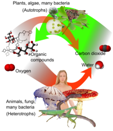
Aerobic respiration (red arrows)
is the main means by which both fungi and animals utilize chemical
energy in the form of organic compounds that were previously created
through photosynthesis (green arrow).
Aerobic respiration requires oxygen (O2) in order to create ATP. Although carbohydrates, fats, and proteins are consumed as reactants, it is the preferred method of pyruvate breakdown in glycolysis and requires that pyruvate enter the mitochondria in order to be fully oxidized by the Krebs cycle.
The products of this process are carbon dioxide and water, but the
energy transferred is used to break bonds in ADP as the third phosphate
group is added to form ATP (adenosine triphosphate), by substrate-level phosphorylation, NADH and FADH2
| Simplified reaction: | C6H12O6 (s) + 6 O2 (g) → 6 CO2 (g) + 6 H2O (l) + heat |
| ΔG = −2880 kJ per mol of C6H12O6 |
The negative ΔG indicates that the reaction can occur spontaneously.
The potential of NADH and FADH2 is converted to more ATP through an electron transport chain with oxygen as the "terminal electron acceptor". Most of the ATP produced by aerobic cellular respiration is made by oxidative phosphorylation. This works by the energy released in the consumption of pyruvate being used to create a chemiosmotic potential by pumping protons across a membrane. This potential is then used to drive ATP synthase and produce ATP from ADP
and a phosphate group. Biology textbooks often state that 38 ATP
molecules can be made per oxidised glucose molecule during cellular
respiration (2 from glycolysis, 2 from the Krebs cycle, and about 34
from the electron transport system).
However, this maximum yield is never quite reached because of losses
due to leaky membranes as well as the cost of moving pyruvate and ADP
into the mitochondrial matrix, and current estimates range around 29 to
30 ATP per glucose.
Aerobic metabolism is up to 15 times more efficient than
anaerobic metabolism (which yields 2 molecules ATP per 1 molecule
glucose). However some anaerobic organisms, such as methanogens are able to continue with anaerobic respiration,
yielding more ATP by using other inorganic molecules (not oxygen) as
final electron acceptors in the electron transport chain. They share
the initial pathway of glycolysis
but aerobic metabolism continues with the Krebs cycle and oxidative
phosphorylation. The post-glycolytic reactions take place in the
mitochondria in eukaryotic cells, and in the cytoplasm in prokaryotic cells.
Glycolysis
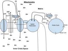
Out of the cytoplasm it goes into the Krebs cycle with the acetyl CoA. It then mixes with CO2
and makes 2 ATP, NADH, and FADH. From there the NADH and FADH go into
the NADH reductase, which produces the enzyme. The NADH pulls the
enzyme's electrons to send through the electron transport chain. The
electron transport chain pulls H+ ions through the chain. From the electron transport chain, the released hydrogen ions make ADP for an end result of 32 ATP. O2
attracts itself to the left over electron to make water. Lastly, ATP
leaves through the ATP channel and out of the mitochondria.
Glycolysis is a metabolic pathway that takes place in the cytosol
of cells in all living organisms. This pathway can function with or
without the presence of oxygen. In humans, aerobic conditions produce pyruvate and anaerobic conditions produce lactate. In aerobic conditions, the process converts one molecule of glucose into two molecules of pyruvate (pyruvic acid), generating energy in the form of two net molecules of ATP. Four molecules of ATP per glucose are actually produced, however, two are consumed as part of the preparatory phase. The initial phosphorylation of glucose is required to increase the reactivity (decrease its stability) in order for the molecule to be cleaved into two pyruvate molecules by the enzyme aldolase. During the pay-off phase of glycolysis, four phosphate groups are transferred to ADP by substrate-level phosphorylation to make four ATP, and two NADH are produced when the pyruvate are oxidized. The overall reaction can be expressed this way:
Glucose + 2 NAD+ + 2 Pi + 2 ADP → 2 pyruvate + 2 NADH + 2 ATP + 2 H+ + 2 H2O + heat
Starting with glucose, 1 ATP is used to donate a phosphate to
glucose to produce glucose 6-phosphate. Glycogen can be converted into
glucose 6-phosphate as well with the help of glycogen phosphorylase.
During energy metabolism, glucose 6-phosphate becomes fructose
6-phosphate. An additional ATP is used to phosphorylate fructose
6-phosphate into fructose 1,6-disphosphate by the help of
phosphofructokinase. Fructose 1,6-diphosphate then splits into two
phosphorylated molecules with three carbon chains which later degrades
into pyruvate.
Glycolysis can be literally translated as "sugar splitting".
Oxidative decarboxylation of pyruvate
Pyruvate is oxidized to acetyl-CoA and CO2 by the pyruvate dehydrogenase complex (PDC). The PDC contains multiple copies of three enzymes and is located in the mitochondria
of eukaryotic cells and in the cytosol of prokaryotes. In the
conversion of pyruvate to acetyl-CoA, one molecule of NADH and one
molecule of CO2 is formed.
Citric acid cycle
This is also called the Krebs cycle or the tricarboxylic acid cycle. When oxygen is present, acetyl-CoA is produced from the pyruvate molecules created from glycolysis. Once acetyl-CoA is formed, aerobic or anaerobic respiration can occur.
When oxygen is present, the mitochondria will undergo aerobic
respiration which leads to the Krebs cycle. However, if oxygen is not
present, fermentation of the pyruvate molecule will occur. In the
presence of oxygen, when acetyl-CoA is produced, the molecule then
enters the citric acid cycle (Krebs cycle) inside the mitochondrial matrix, and is oxidized to CO2 while at the same time reducing NAD to NADH. NADH can be used by the electron transport chain to create further ATP
as part of oxidative phosphorylation. To fully oxidize the equivalent
of one glucose molecule, two acetyl-CoA must be metabolized by the Krebs
cycle. Two waste products, H2O and CO2, are created during this cycle.
The citric acid cycle is an 8-step process involving 18 different enzymes and co-enzymes.
During the cycle, acetyl-CoA (2 carbons) + oxaloacetate (4 carbons)
yields citrate (6 carbons), which is rearranged to a more reactive form
called isocitrate (6 carbons). Isocitrate is modified to become
α-ketoglutarate (5 carbons), succinyl-CoA, succinate, fumarate, malate,
and, finally, oxaloacetate.
The net gain of high-energy compounds from one cycle is 3 NADH, 1 FADH2,
and 1 GTP; the GTP may subsequently be used to produce ATP. Thus, the
total yield from 1 glucose molecule (2 pyruvate molecules) is 6 NADH, 2
FADH2, and 2 ATP.
Oxidative phosphorylation
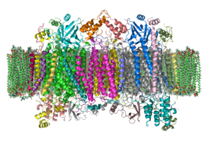
In eukaryotes, oxidative phosphorylation occurs in the mitochondrial cristae. It comprises the electron transport chain that establishes a proton gradient
(chemiosmotic potential) across the boundary of inner membrane by
oxidizing the NADH produced from the Krebs cycle. ATP is synthesized by
the ATP synthase enzyme when the chemiosmotic gradient is used to drive
the phosphorylation of ADP. The electrons are finally transferred to
exogenous oxygen and, with the addition of two protons, water is formed.
Efficiency of ATP production
The table below describes the reactions involved when one glucose
molecule is fully oxidized into carbon dioxide. It is assumed that all
the reduced coenzymes are oxidized by the electron transport chain and used for oxidative phosphorylation.
| Step | coenzyme yield | ATP yield | Source of ATP |
|---|---|---|---|
| Glycolysis preparatory phase |
|
−2 | Phosphorylation of glucose and fructose 6-phosphate uses two ATP from the cytoplasm. |
| Glycolysis pay-off phase |
|
4 | Substrate-level phosphorylation |
| 2 NADH | 3 or 5 | Oxidative phosphorylation : Each NADH produces net 1.5 ATP (instead of usual 2.5) due to NADH transport over the mitochondrial membrane | |
| Oxidative decarboxylation of pyruvate | 2 NADH | 5 | Oxidative phosphorylation |
| Krebs cycle |
|
2 | Substrate-level phosphorylation |
| 6 NADH | 15 | Oxidative phosphorylation | |
| 2 FADH2 | 3 | Oxidative phosphorylation | |
| Total yield | 30 or 32 ATP | From the complete oxidation of one glucose molecule to carbon dioxide and oxidation of all the reduced coenzymes. | |
Although there is a theoretical yield of 38 ATP molecules per glucose
during cellular respiration, such conditions are generally not realized
because of losses such as the cost of moving pyruvate (from
glycolysis), phosphate, and ADP (substrates for ATP synthesis) into the
mitochondria. All are actively transported using carriers that utilize
the stored energy in the proton electrochemical gradient.
- Pyruvate is taken up by a specific, low Km transporter to bring it into the mitochondrial matrix for oxidation by the pyruvate dehydrogenase complex.
- The phosphate carrier (PiC) mediates the electroneutral exchange (antiport) of phosphate (H2PO4−; Pi) for OH− or symport of phosphate and protons (H+) across the inner membrane, and the driving force for moving phosphate ions into the mitochondria is the proton motive force.
- The ATP-ADP translocase (also called adenine nucleotide translocase, ANT) is an antiporter and exchanges ADP and ATP across the inner membrane. The driving force is due to the ATP (−4) having a more negative charge than the ADP (−3), and thus it dissipates some of the electrical component of the proton electrochemical gradient.
The outcome of these transport processes using the proton electrochemical gradient is that more than 3 H+
are needed to make 1 ATP. Obviously this reduces the theoretical
efficiency of the whole process and the likely maximum is closer to
28–30 ATP molecules. In practice the efficiency may be even lower because the inner membrane of the mitochondria is slightly leaky to protons. Other factors may also dissipate the proton gradient creating an apparently leaky mitochondria. An uncoupling protein known as thermogenin
is expressed in some cell types and is a channel that can transport
protons. When this protein is active in the inner membrane it short
circuits the coupling between the electron transport chain and ATP synthesis. The potential energy from the proton gradient is not used to make ATP but generates heat. This is particularly important in brown fat thermogenesis of newborn and hibernating mammals.
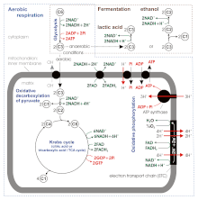
Stoichiometry of aerobic respiration and most known fermentation types in eucaryotic cell. [6] Numbers in circles indicate counts of carbon atoms in molecules, C6 is glucose C6H12O6, C1 carbon dioxide CO2. Mitochondrial outer membrane is omitted.
According to some of newer sources the ATP yield during aerobic
respiration is not 36–38, but only about 30–32 ATP molecules / 1
molecule of glucose , because:
- ATP : NADH+H+ and ATP : FADH2 ratios during the oxidative phosphorylation appear to be not 3 and 2, but 2.5 and 1.5 respectively. Unlike in the substrate-level phosphorylation, the stoichiometry here is difficult to establish.
- ATP synthase produces 1 ATP / 3 H+. However the exchange of matrix ATP for cytosolic ADP and Pi (antiport with OH− or symport with H+) mediated by ATP–ADP translocase and phosphate carrier consumes 1 H+ / 1 ATP as a result of regeneration of the transmembrane potential changed during this transfer, so the net ratio is 1 ATP : 4 H+.
- The mitochondrial electron transport chain proton pump transfers across the inner membrane 10 H+ / 1 NADH+H+ (4 + 2 + 4) or 6 H+ / 1 FADH2 (2 + 4).
- So the final stoichiometry is
- 1 NADH+H+ : 10 H+ : 10/4 ATP = 1 NADH+H+ : 2.5 ATP
- 1 FADH2 : 6 H+ : 6/4 ATP = 1 FADH2 : 1.5 ATP
- ATP : NADH+H+ coming from glycolysis ratio during the oxidative phosphorylation is
- 1.5, as for FADH2, if hydrogen atoms (2H++2e−) are transferred from cytosolic NADH+H+ to mitochondrial FAD by the glycerol phosphate shuttle located in the inner mitochondrial membrane.
- 2.5 in case of malate-aspartate shuttle transferring hydrogen atoms from cytosolic NADH+H+ to mitochondrial NAD+
So finally we have, per molecule of glucose
- Substrate-level phosphorylation: 2 ATP from glycolysis + 2 ATP (directly GTP) from Krebs cycle
- Oxidative phosphorylation
- 2 NADH+H+ from glycolysis: 2 × 1.5 ATP (if glycerol phosphate shuttle transfers hydrogen atoms) or 2 × 2.5 ATP (malate-aspartate shuttle)
- 2 NADH+H+ from the oxidative decarboxylation of pyruvate and 6 from Krebs cycle: 8 × 2.5 ATP
- 2 FADH2 from the Krebs cycle: 2 × 1.5 ATP
Altogether this gives 4 + 3 (or 5) + 20 + 3 = 30 (or 32) ATP per molecule of glucose
The total ATP yield in ethanol or lactic acid fermentation is only 2 molecules coming from glycolysis, because pyruvate is not transferred to the mitochondrion and finally oxidized to the carbon dioxide (CO2), but reduced to ethanol or lactic acid in the cytoplasm.
Fermentation
Without oxygen, pyruvate (pyruvic acid)
is not metabolized by cellular respiration but undergoes a process of
fermentation. The pyruvate is not transported into the mitochondrion,
but remains in the cytoplasm, where it is converted to waste products
that may be removed from the cell. This serves the purpose of oxidizing
the electron carriers so that they can perform glycolysis again and
removing the excess pyruvate. Fermentation oxidizes NADH to NAD+ so it
can be re-used in glycolysis. In the absence of oxygen, fermentation
prevents the buildup of NADH in the cytoplasm and provides NAD+ for
glycolysis. This waste product varies depending on the organism. In
skeletal muscles, the waste product is lactic acid. This type of fermentation is called lactic acid fermentation.
In strenuous exercise, when energy demands exceed energy supply, the
respiratory chain cannot process all of the hydrogen atoms joined by
NADH. During anaerobic glycolysis, NAD+ regenerates when pairs of
hydrogen combine with pyruvate to form lactate. Lactate formation is
catalyzed by lactate dehydrogenase in a reversible reaction. Lactate can
also be used as an indirect precursor for liver glycogen. During
recovery, when oxygen becomes available, NAD+ attaches to hydrogen from
lactate to form ATP. In yeast, the waste products are ethanol and carbon dioxide. This type of fermentation is known as alcoholic or ethanol fermentation. The ATP generated in this process is made by substrate-level phosphorylation, which does not require oxygen.
Fermentation is less efficient at using the energy from glucose:
only 2 ATP are produced per glucose, compared to the 38 ATP per glucose
nominally produced by aerobic respiration. This is because the waste products of fermentation still contain chemical potential energy that can be released by oxidation. Ethanol,
for example, can be burned in an internal combustion engine like
gasoline. Glycolytic ATP, however, is created more quickly. For
prokaryotes to continue a rapid growth rate when they are shifted from
an aerobic environment to an anaerobic environment, they must increase
the rate of the glycolytic reactions. For multicellular organisms,
during short bursts of strenuous activity, muscle cells use fermentation
to supplement the ATP production from the slower aerobic respiration,
so fermentation may be used by a cell even before the oxygen levels are
depleted, as is the case in sports that do not require athletes to pace
themselves, such as sprinting.
Anaerobic respiration
Cellular respiration is the process by which biological fuels are
oxidised in the presence of an inorganic electron acceptor (such as
oxygen) to produce large amounts of energy, to drive the bulk production
of ATP.
Anaerobic respiration is used by some microorganisms in
which neither oxygen (aerobic respiration) nor pyruvate derivatives
(fermentation) is the final electron acceptor. Rather, an inorganic
acceptor such as sulfate or nitrate is used.
Such organisms are typically found in unusual places such as underwater caves or near hydrothermal vents at the bottom of the ocean.


