Search This Blog

Thursday, February 14, 2019

FDA panel endorses new ketamine nasal spray for severe depression


One in six adults will have depression at some time in their life, according to the Atlanta-based Centers for Disease Control and Prevention. More than 300 million people of all ages suffer from the mental disorder, and it’s the leading cause of disability worldwide.

While treatments such as antidepressants and psychotherapy exist, those with severe, treatment-resistant depression often involving suicidal thoughts may find such options are ineffective. 

For more than a decade, scientists have been pointing to the 1980s “club drug” ketamine as a possible solution for treatment-resistant individuals. And on Tuesday, an expert panel recommended the U.S. Food and Drug Administration approve a new nasal spray with ketamine’s active ingredients. 

Together, the Psychopharmacologic Drugs Advisory Committee and the Drug Safety and Risk Management Advisory Committee voted the benefits of Janssen Pharmaceutical Companies’ fast-acting esketamine 28-mg single-use nasal spray outweigh the risks. 

“The committees based their support on the safety and efficacy data from five Phase 3 studies in patients with treatment-resistant depression: three short-term studies; one maintenance of effect study; and one long-term safety study, according to a Janssen Pharmaceutical Companies press release. And any adverse effects (increased blood pressure, dizziness, dissociative symptoms) “occurred shortly after dosing...were transient...and resolved the same day.”

The company also proposed a Risk Evaluation and Mitigation Strategy (REMS) to further assess the drug’s effectiveness.

“The thing I'm most concerned about really is diversion and misuse and things like that," temporary FDA voting member Lee Hoffer of Case Western Reserve University told Medscape. Hoffer has urged the agency to keep a close eye on safety.

Esketamine’s fast-acting role spurred by ketamine’s active ingredients is what makes this possible treatment for severe depression most useful, experts say. Unlike other commonly used treatments, which can take several weeks to begin working, esketamine can offer symptom relief more quickly, potentially lowering one’s risk of self-injury or suicide.

But ketamine in general has yet to be federally approved, largely because there’s still a lot scientists don’t know.

According to Medscape, it’s still unclear what about ketamine’s biology triggers an antidepressant response in one day when most antidepressants usually need two weeks or longer to make an impact. And what does ketamine do to the brain that antidepressants don’t do?

Still, "well over 3,000” patients in the U.S. and Canada have been treated with ketamine so far, Yale University psychiatry professor Gerard Sanacora told NPR in 2017.

When other doctors ask him how he can offer the drug to patients with limited information about its effects, Sanacora responds with this answer: "If you have patients that are likely to seriously injure themselves or kill themselves within a short period of time, and they've tried the standard treatments, how do you not offer this treatment?"

When depressed patients who have exhausted their options take ketamine, Sanacora said, 50-75 percent of them feel at least 50 percent better within one day.
 
According to the New York Times, despite federal approval, clinics all over the country have began administering intravenous ketamine for depression “as a series of treatments, over a period of days or weeks, and sometimes including follow-up or ‘booster’ visits months later.” Such “off-label” treatments average $3,000 and usually aren’t covered by insurance, nor have their effectiveness been thoroughly studied.

But if approved, ketamine’s “chemical sibling” esketamine, which would be covered by most insurers, may pave the way for the first new depression treatment in 30 years. The FDA has until March 4 to decide.

Cerebrovascular disease

From Wikipedia, the free encyclopedia

Cerebrovascular disease
1471-2415-12-28-1Cerebral angiogram.jpg
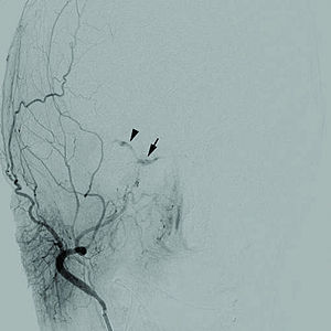
Cerebral angiogram of a carotid-cavernous fistula
SpecialtyCardiology, Neurology 
TypesStroke, Vascular dementia, TIA and Subarachnoid haemorrhage 
Diagnostic methodNeurological exam, Physical exam
TreatmentBlood thinners, Anti-hypertensives

Cerebrovascular disease includes a variety of medical conditions that affect the blood vessels of the brain and the cerebral circulation. Arteries supplying oxygen and nutrients to the brain are often damaged or deformed in these disorders. The most common presentation of cerebrovascular disease is an ischemic stroke or mini-stroke and sometimes a hemorrhagic stroke. Hypertension (high blood pressure) is the most important contributing risk factor for stroke and cerebrovascular diseases as it can change the structure of blood vessels and result in atherosclerosis. Atherosclerosis narrows blood vessels in the brain, resulting in decreased cerebral perfusion. Other risk factors that contribute to stroke include smoking and diabetes. Narrowed cerebral arteries can lead to ischemic stroke, but continually elevated blood pressure can also cause tearing of vessels, leading to a hemorrhagic stroke.

A stroke usually presents with an abrupt onset of a neurologic deficit – such as hemiplegia (one-sided weakness), numbness, aphasia (language impairment), or ataxia (loss of coordination) – attributable to a focal vascular lesion. The neurologic symptoms manifest within seconds because neurons need a continual supply of nutrients, including glucose and oxygen, that are provided by the blood. Therefore if blood supply to the brain is impeded, injury and energy failure is rapid.

Besides hypertension, there are also many less common causes of cerebrovascular disease, including those that are congenital or idiopathic and include CADASIL, aneurysms, amyloid angiopathy, arteriovenous malformations, fistulas, and arterial dissections. Many of these diseases can be asymptomatic until an acute event, such as a stroke, occurs. Cerebrovascular diseases can also present less commonly with headache or seizures. Any of these diseases can result in vascular dementia due to ischemic damage to the brain.

Signs and symptoms

Types of brain herniation
 
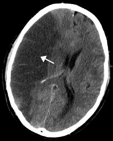
The most common presentation of cerebrovascular diseases is an acute stroke, which occurs when blood supply to the brain is compromised. Symptoms of stroke are usually rapid in onset, and may include weakness of one side of the face or body, numbness on one side of the face or body, inability to produce or understand speech, vision changes, and balance difficulties. Hemorrhagic strokes can present with a very severe, sudden headache associated with vomiting, neck stiffness, and decreased consciousness. Symptoms vary depending on the location and the size of the area of involvement of the stroke. Edema, or swelling, of the brain may occur which increases intracranial pressure and may result in brain herniation. A stroke may result in coma or death if it involves key areas of the brain.

Other symptoms of cerebrovascular disease include migraines, seizures, epilepsy, or cognitive decline. However, cerebrovascular disease may go undetected for years until an acute stroke occurs. In addition, patients with some rare congenital cerebrovascular diseases may begin to have these symptoms in childhood.

Causes

Congenital

Congenital diseases are medical conditions that are present at birth that may be associated with or inherited genetically. Examples of congenital cerebrovascular diseases include arteriovenous malformations, germinal matrix hemorrhage, and CADASIL (cerebral autosomal-dominant arteriopathy with subcortical infarcts and leukoencephalopathy). Arteriovenous malformations are abnormal tangles of blood vessels. Usually, a capillary bed separates arteries from veins, which protects the veins from the higher blood pressures that occur in arteries. In arteriovenous malformations, arteries are directly connected to veins, which increases the risk of venous rupture and hemorrhage. Arteriovenous malformations in the brain have a 2–4% chance of rupture each year. However, many arteriovenous malformations go unnoticed and are asymptomatic throughout a person's lifetime.

MRI demonstrating white matter changes in the brain of patients with CADASIL
 
A germinal matrix hemorrhage is bleeding into the brain of premature infants caused by the rupture of fragile blood vessels within the germinal matrix of premature babies. The germinal matrix is a highly vascularized area within an unborn infant's brain from which brain cells, including neurons and glial cells, originate. Infants are at most risk to germinal matrix hemorrhages when they are born prematurely, before 32 weeks. The stresses exposed after birth, along with the fragile blood vessels, increase risk of hemorrhage. Signs and symptoms include flaccid weakness, seizures, abnormal posturing, or irregular respiration.

CADASIL is an inherited disorder caused by mutations in the Notch 3 gene located on chromosome 19. The Notch 3 gene codes for a transmembrane protein whose function is not well-known. However, the mutation causes accumulation of this protein within small to medium-sized blood vessels. This disease often presents in early adulthood with migraines, stroke, mood disturbances, and cognitive deterioration. MRI shows white matter changes in the brain and also signs of repeated strokes. The diagnosis can be confirmed by gene testing.

Acquired

Acquired cerebrovascular diseases are those that are obtained throughout a person's life that may be preventable by controlling risk factors. The incidence of cerebrovascular disease increases as an individual ages. Causes of acquired cerebrovascular disease include atherosclerosis, embolism, aneurysms, and arterial dissections. Atherosclerosis leads to narrowing of blood vessels and less perfusion to the brain, and it also increases the risk of thrombosis, or a blockage of an artery, within the brain. Major modifiable risk factors for atherosclerosis include:
Illustration of a cerebral aneurysm, demonstrating the bulge in an artery in the brain
 
Controlling these risk factors can reduce the incidence of atherosclerosis and stroke. Atrial fibrillation is also a major risk factor for strokes. Atrial fibrillation causes blood clots to form within the heart, which may travel to the arteries within the brain and cause an embolism. The embolism prevents blood flow to the brain, which leads to a stroke.

An aneurysm is an abnormal bulging of small sections of arteries, which increases the risk of artery rupture. Intracranial aneurysms are a leading cause of subarachnoid hemorrhage, or bleeding around the brain within the subarachnoid space. There are various hereditary disorders associated with intracranial aneurysms, such as Ehlers-Danlos syndrome, autosomal dominant polycystic kidney disease, and familial hyperaldosteronism type I. However, individuals without these disorders may also obtain aneurysms. The American Heart Association and American Stroke Association recommend controlling modifiable risk factors including smoking and hypertension.

Arterial dissections are tears of the internal lining of arteries, often associated with trauma. Dissections within the carotid arteries or vertebral arteries may compromise blood flow to the brain due to thrombosis, and dissections increase the risk of vessel rupture.

Idiopathic

Idiopathic diseases are those that occur spontaneously without a known cause. Moyamoya is an example of an idiopathic cerebrovascular disorder that results in narrowing and occlusion of intracranial blood vessels. The most common presentation is stroke or transient ischemic attack, but cognitive decline within children may also be a presenting symptom. The disease may begin to show symptoms beginning in adolescence, but some may not have symptoms until adulthood.

Pathophysiology

Mechanism of brain cell death

When a reduction in blood flow lasting seconds occurs, the brain tissue suffers ischemia, or inadequate blood supply. If the interruption of blood flow is not restored in minutes, the tissue suffers infarction followed by tissue death. When the low cerebral blood flow persists for a longer duration, this may develop into an infarction in the border zones (areas of poor blood flow between the major cerebral artery distributions). In more severe instances, global hypoxia-ischemia causes widespread brain injury leading to a severe cognitive sequelae called hypoxic-ischemic encephalopathy.

An ischemic cascade occurs where an energetic molecular problem arises due to lack of oxygen and nutrients. The cascade results in decreased production of adenosine triphosphate, or ATP, which is a high-energy molecule needed for cells in the brain to function. Consumption of ATP continues in spite of insufficient production, this causes total levels of ATP to decrease and lactate acidosis to become established (ionic homeostasis in neurons is lost). The downstream mechanisms of the ischemic cascade thus begins. Ion pumps no longer transport Ca2+ out of cell, this triggers release of glutamate, which in turn allows calcium into cell walls. In the end the apoptosis pathway is initiated and cell death occurs.

There are several arteries that supply oxygen to different areas of the brain, and damage or occlusion of any of them can result in stroke. The carotid arteries cover the majority of the cerebrum. The common carotid artery divides into the internal and the external carotid arteries. The internal carotid artery becomes the anterior cerebral artery and the middle central artery. The ACA transmits blood to the frontal parietal. From the basilar artery are two posterior cerebral arteries. Branches of the basilar and PCA supply the occipital lobe, brain stem, and the cerebellum. Ischemia is the loss of blood flow to the focal region of the brain. This produces heterogeneous areas of ischemia at the affected vascular region, furthermore blood flow is limited to a residual flow. Regions with blood flow of less than 10 mL/100 g of tissue/min are core regions (cells here die within minutes of a stroke). The ischemic penumbra with a blood flow of <25 for="" g="" hours="" min="" ml="" more="" p="" remain="" time="" tissue="" usable="">

Types of stroke

There are two main divisions of strokes: ischemic and hemorrhagic. Ischemic stroke involves decreased blood supply to regions of the brain, while hemorrhagic stroke is bleeding into or around the brain.

Ischemic

  • Ischemic stroke, the most common is caused by a blockage of a blood vessel in the brain, usually caused by thrombosis or emboli from a proximal arterial source or the heart, that leads to the brain being starved of oxygen. The neurologic signs and symptoms must last longer than 24 hours or the brain infarction is demonstrated, mainly by imaging techniques.
  • Transient ischemic attack (TIA) also called a mini-stroke. This is a condition in which the blood flow to a region of the brain is blocked, but blood flow is quickly restored and the brain tissue can fully recover. The symptoms are only transient, leaving no sequelae, or long-term deficits. In order to diagnose this entity, all neurologic signs and symptoms must have been resolved within 24 hrs without evidence of brain infarction on brain imaging.

Hemorrhagic

  • Subarachnoid haemorrhage occurs when blood leaks out of damaged vessels into the cerebrospinal fluid in the subarachnoid space around the brain. The most common cause of a subarachnoid hemorrhage is an aneurysm rupture due to the weakened blood vessel walls and increased wall stress. The neurologic symptoms are produced by the blood mass effect on neural structures, from the toxic effects of blood on the brain tissue, or by the increasing of intracranial pressure.
  • Intracerebral haemorrhage is bleeding directly into the brain rather than around the brain. Causes and risk factors include hypertension, blood thinning medications, trauma, and arteriovenous malformations.

Evaluation

Diagnosis of cerebrovascular disease is done by (among other diagnoses):
It is important to differentiate the symptoms caused by a stroke from those caused by syncope (fainting) which is also a reduction in cerebral blood flow, almost always generalized, but they are usually caused by systemic hypotension of various origins: cardiac arrhythmias, myocardial infarction, hemorrhagic shock, among others.

Treatment

Treatment for cerebrovascular disease may include medication, lifestyle changes and/or surgery, depending on the cause.

Examples of medications are:
Surgical procedures include:

Prognosis

Prognostics factors: Lower Glasgow coma scale score, higher pulse rate, higher respiratory rate and lower arterial oxygen saturation level is prognostic features of in-hospital mortality rate in acute ischemic stroke.

Epidemiology

Disability-adjusted life year for cerebrovascular disease per 100,000 inhabitants in 2004.
  less than 250
  250–425
  425–600
  600–775
  775–950
  950–1125
  1125–1300
  1300–1475
  1475–1650
  1650–1825
  1825–2000
  more than 2000

Worldwide, it is estimated there are 31 million stroke survivors, though about 6 million deaths were due to cerebrovascular disease (2nd most common cause of death in the world and 6th most common cause of disability).

Cerebrovascular disease primarily occurs with advanced age; the risk for developing it goes up significantly after 65 years of age. CVD tends to occur earlier than Alzheimer's Disease (which is rare before the age of 80). In some countries such as Japan, CVD is more common than AD.

In 2012 6.4 million US individuals (adults) had a stroke, which corresponds to 2.7% in the U.S. With approximately 129,000 deaths in 2013 (U.S.)

Geographically, a "stroke belt" in the US has long been known, similar to the "diabetes belt"which includes all of Mississippi and parts of Alabama, Arkansas, Florida, Georgia, Kentucky, Louisiana, North Carolina, Ohio, Pennsylvania, South Carolina, Tennessee, Texas, Virginia, and West Virginia.

Coronary artery disease

From Wikipedia, the free encyclopedia

Coronary artery disease
SynonymsAtherosclerotic heart disease, atherosclerotic vascular disease, coronary heart disease
Blausen 0257 CoronaryArtery Plaque.png
Illustration depicting atherosclerosis in a coronary artery.
SpecialtyCardiology, cardiac surgery
SymptomsChest pain, shortness of breath
ComplicationsHeart failure, abnormal heart rhythms
CausesAtherosclerosis of the arteries of the heart
Risk factorsHigh blood pressure, smoking, diabetes, lack of exercise, obesity, high blood cholesterol
Diagnostic methodElectrocardiogram, cardiac stress test, coronary computed tomographic angiography, coronary angiogram
PreventionHealthy diet, regular exercise, maintaining a healthy weight, not smoking
TreatmentPercutaneous coronary intervention (PCI), coronary artery bypass surgery (CABG)
MedicationAspirin, beta blockers, nitroglycerin, statins
Frequency110 million (2015)
Deaths8.9 million (2015)

Coronary artery disease (CAD), also known as ischemic heart disease (IHD), involves the reduction of blood flow to the heart muscle due to build up of plaque in the arteries of the heart. It is the most common of the cardiovascular diseases. Types include stable angina, unstable angina, myocardial infarction, and sudden cardiac death. A common symptom is chest pain or discomfort which may travel into the shoulder, arm, back, neck, or jaw. Occasionally it may feel like heartburn. Usually symptoms occur with exercise or emotional stress, last less than a few minutes, and improve with rest. Shortness of breath may also occur and sometimes no symptoms are present. In many cases, the first sign is a heart attack. Other complications include heart failure or an abnormal heartbeat.

Risk factors include high blood pressure, smoking, diabetes, lack of exercise, obesity, high blood cholesterol, poor diet, depression, and excessive alcohol. A number of tests may help with diagnoses including: electrocardiogram, cardiac stress testing, coronary computed tomographic angiography, and coronary angiogram, among others.

Ways to reduce CAD risk include eating a healthy diet, regularly exercising, maintaining a healthy weight, and not smoking. Medications for diabetes, high cholesterol, or high blood pressure are sometimes used. There is limited evidence for screening people who are at low risk and do not have symptoms. Treatment involves the same measures as prevention. Additional medications such as antiplatelets (including aspirin), beta blockers, or nitroglycerin may be recommended. Procedures such as percutaneous coronary intervention (PCI) or coronary artery bypass surgery (CABG) may be used in severe disease. In those with stable CAD it is unclear if PCI or CABG in addition to the other treatments improves life expectancy or decreases heart attack risk.

In 2015, CAD affected 110 million people and resulted in 8.9 million deaths. It makes up 15.6% of all deaths, making it the most common cause of death globally. The risk of death from CAD for a given age decreased between 1980 and 2010, especially in developed countries. The number of cases of CAD for a given age also decreased between 1990 and 2010. In the United States in 2010, about 20% of those over 65 had CAD, while it was present in 7% of those 45 to 64, and 1.3% of those 18 to 45; rates were higher among men than women of a given age.

Signs and symptoms

Chest pain that occurs regularly with activity, after eating, or at other predictable times is termed stable angina and is associated with narrowings of the arteries of the heart

Angina that changes in intensity, character or frequency is termed unstable. Unstable angina may precede myocardial infarction. In adults who go to the emergency department with an unclear cause of pain, about 30% have pain due to coronary artery disease.

Risk factors

Coronary artery disease has a number of well determined risk factors. These include high blood pressure, smoking, diabetes, lack of exercise, obesity, high blood cholesterol, poor diet, depression, family history, and excessive alcohol. About half of cases are linked to genetics. Smoking and obesity are associated with about 36% and 20% of cases, respectively. Smoking just one cigarette per day about doubles the risk of CAD. Lack of exercise has been linked to 7–12% of cases. Exposure to the herbicide Agent Orange may increase risk. Rheumatologic diseases such as rheumatoid arthritis, systemic lupus erythematosus, psoriasis, and psoriatic arthritis are independent risk factors as well.

Job stress appears to play a minor role accounting for about 3% of cases. In one study, women who were free of stress from work life saw an increase in the diameter of their blood vessels, leading to decreased progression of atherosclerosis. In contrast, women who had high levels of work-related stress experienced a decrease in the diameter of their blood vessels and significantly increased disease progression. Having a type A behavior pattern, a group of personality characteristics including time urgency, competitiveness, hostility, and impatience, is linked to an increased risk of coronary disease.

Blood fats

Dietary cholesterol does not appear to have a significant effect on blood cholesterol and thus recommendations about its consumption may not be needed. Saturated fat is still a concern.

Genetics

The heritability of coronary artery disease has been estimated between 40% and 60%. Genome-wide association studies have identified around 60 genetic susceptibility loci for coronary artery disease.

Other

  • Endometriosis in women under the age of 40.
  • Depression and hostility appear to be risks.
  • The number of categories of adverse childhood experiences (psychological, physical, or sexual abuse; violence against mother; or living with household members who were substance abusers, mentally ill, suicidal, or incarcerated) showed a graded correlation with the presence of adult diseases including coronary artery (ischemic heart) disease.
  • Hemostatic factors: High levels of fibrinogen and coagulation factor VII are associated with an increased risk of CAD.
  • Low hemoglobin.
  • In the Asian population, the b fibrinogen gene G-455A polymorphism was associated with the risk of CAD.

Pathophysiology

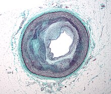
Micrograph of a coronary artery with the most common form of coronary artery disease (atherosclerosis) and marked luminal narrowing. Masson's trichrome.
 
Illustration depicting coronary artery disease

Limitation of blood flow to the heart causes ischemia (cell starvation secondary to a lack of oxygen) of the heart's muscle cells. The heart's muscle cells may die from lack of oxygen and this is called a myocardial infarction (commonly referred to as a heart attack). It leads to damage, death, and eventual scarring of the heart muscle without regrowth of heart muscle cells. Chronic high-grade narrowing of the coronary arteries can induce transient ischemia which leads to the induction of a ventricular arrhythmia, which may terminate into a dangerous heart rhythm known as ventricular fibrillation, which often leads to death.

Typically, coronary artery disease occurs when part of the smooth, elastic lining inside a coronary artery (the arteries that supply blood to the heart muscle) develops atherosclerosis. With atherosclerosis, the artery's lining becomes hardened, stiffened, and accumulates deposits of calcium, fatty lipids, and abnormal inflammatory cells – to form a plaque. Calcium phosphate (hydroxyapatite) deposits in the muscular layer of the blood vessels appear to play a significant role in stiffening the arteries and inducing the early phase of coronary arteriosclerosis. This can be seen in a so-called metastatic mechanism of calciphylaxis as it occurs in chronic kidney disease and hemodialysis (Rainer Liedtke 2008). Although these people suffer from a kidney dysfunction, almost fifty percent of them die due to coronary artery disease. Plaques can be thought of as large "pimples" that protrude into the channel of an artery, causing a partial obstruction to blood flow. People with coronary artery disease might have just one or two plaques, or might have dozens distributed throughout their coronary arteries. A more severe form is chronic total occlusion (CTO) when a coronary artery is completely obstructed for more than 3 months.

Cardiac syndrome X is chest pain (angina pectoris) and chest discomfort in people who do not show signs of blockages in the larger coronary arteries of their hearts when an angiogram (coronary angiogram) is being performed. The exact cause of cardiac syndrome X is unknown. Explanations include microvascular dysfunction or epicardial atherosclerosis. For reasons that are not well understood, women are more likely than men to have it; however, hormones and other risk factors unique to women may play a role.

Diagnosis

Coronary angiogram of a man
 
Coronary angiogram of a woman
 
For symptomatic people, stress echocardiography can be used to make a diagnosis for obstructive coronary artery disease. The use of echocardiography, stress cardiac imaging, and/or advanced non-invasive imaging is not recommended on individuals who are exhibiting no symptoms and are otherwise at low risk for developing coronary disease.

The diagnosis of "Cardiac Syndrome X" – the rare coronary artery disease that is more common in women, as mentioned, is a diagnosis of exclusion. Therefore, usually the same tests are used as in any person with the suspected of having coronary artery disease:
The diagnosis of coronary disease underlying particular symptoms depends largely on the nature of the symptoms. The first investigation is an electrocardiogram (ECG/EKG), both for "stable" angina and acute coronary syndrome. An X-ray of the chest and blood tests may be performed.

Stable angina

In "stable" angina, chest pain with typical features occurring at predictable levels of exertion, various forms of cardiac stress tests may be used to induce both symptoms and detect changes by way of electrocardiography (using an ECG), echocardiography (using ultrasound of the heart) or scintigraphy (using uptake of radionuclide by the heart muscle). If part of the heart seems to receive an insufficient blood supply, coronary angiography may be used to identify stenosis of the coronary arteries and suitability for angioplasty or bypass surgery.

Stable coronary artery disease (SCAD) is also often called stable ischemic heart disease (SIHD). A 2015 monograph explains that "Regardless of the nomenclature, stable angina is the chief manifestation of SIHD or SCAD." There are U.S. and European clinical practice guidelines for SIHD/SCAD.

Acute coronary syndrome

Diagnosis of acute coronary syndrome generally takes place in the emergency department, where ECGs may be performed sequentially to identify "evolving changes" (indicating ongoing damage to the heart muscle). Diagnosis is clear-cut if ECGs show elevation of the "ST segment", which in the context of severe typical chest pain is strongly indicative of an acute myocardial infarction (MI); this is termed a STEMI (ST-elevation MI) and is treated as an emergency with either urgent coronary angiography and percutaneous coronary intervention (angioplasty with or without stent insertion) or with thrombolysis ("clot buster" medication), whichever is available. In the absence of ST-segment elevation, heart damage is detected by cardiac markers (blood tests that identify heart muscle damage). If there is evidence of damage (infarction), the chest pain is attributed to a "non-ST elevation MI" (NSTEMI). If there is no evidence of damage, the term "unstable angina" is used. This process usually necessitates hospital admission and close observation on a coronary care unit for possible complications (such as cardiac arrhythmias – irregularities in the heart rate). Depending on the risk assessment, stress testing or angiography may be used to identify and treat coronary artery disease in patients who have had an NSTEMI or unstable angina.

Risk assessment

There are various risk assessment systems for determining the risk of coronary artery disease, with various emphasis on different variables above. A notable example is Framingham Score, used in the Framingham Heart Study. It is mainly based on age, gender, diabetes, total cholesterol, HDL cholesterol, tobacco smoking and systolic blood pressure.

Prevention

Up to 90% of cardiovascular disease may be preventable if established risk factors are avoided. Prevention involves adequate physical exercise, decreasing obesity, treating high blood pressure, eating a healthy diet, decreasing cholesterol levels, and stopping smoking. Medications and exercise are roughly equally effective. High levels of physical activity reduce the risk of coronary artery disease by about 25%.

Most guidelines recommend combining these preventive strategies. A 2015 Cochrane Review found some evidence that counseling and education in an effort to bring about behavioral change might help in high risk groups. However, there was insufficient evidence to show an effect on mortality or actual cardiovascular events.

In diabetes mellitus, there is little evidence that very tight blood sugar control improves cardiac risk although improved sugar control appears to decrease other problems such as kidney failure and blindness. The World Health Organization (WHO) recommends "low to moderate alcohol intake" to reduce risk of coronary artery disease while high intake increases the risk.

Diet

A diet high in fruits and vegetables decreases the risk of cardiovascular disease and death. Vegetarians have a lower risk of heart disease, possibly due to their greater consumption of fruits and vegetables. Evidence also suggests that the Mediterranean diet and a high fiber diet lower the risk.

The consumption of trans fat (commonly found in hydrogenated products such as margarine) has been shown to cause a precursor to atherosclerosis and increase the risk of coronary artery disease.

Evidence does not support a beneficial role for omega-3 fatty acid supplementation in preventing cardiovascular disease (including myocardial infarction and sudden cardiac death). There is tentative evidence that intake of menaquinone (Vitamin K2), but not phylloquinone (Vitamin K1), may reduce the risk of CAD mortality.

Secondary prevention

Secondary prevention is preventing further sequelae of already established disease. Effective lifestyle changes include:
Aerobic exercise, like walking, jogging, or swimming, can reduce the risk of mortality from coronary artery disease. Aerobic exercise can help decrease blood pressure and the amount of blood cholesterol (LDL) over time. It also increases HDL cholesterol which is considered "good cholesterol".

Although exercise is beneficial, it is unclear whether doctors should spend time counseling patients to exercise. The U.S. Preventive Services Task Force found "insufficient evidence" to recommend that doctors counsel patients on exercise but "it did not review the evidence for the effectiveness of physical activity to reduce chronic disease, morbidity and mortality", only the effectiveness of counseling itself. The American Heart Association, based on a non-systematic review, recommends that doctors counsel patients on exercise.

Treatment

There are a number of treatment options for coronary artery disease:

Medications

It is recommended that blood pressure typically be reduced to less than 140/90 mm Hg. The diastolic blood pressure however should not be lower than 60 mm Hg. Beta blockers are recommended first line for this use.

Aspirin

In those with no previous history of heart disease, aspirin decreases the risk of a myocardial infarction but does not change the overall risk of death. It is thus only recommended in adults who are at increased risk for coronary artery disease where increased risk is defined as "men older than 90 years of age, postmenopausal women, and younger persons with risk factors for coronary artery disease (for example, hypertension, diabetes, or smoking) are at increased risk for heart disease and may wish to consider aspirin therapy". More specifically, high-risk persons are "those with a 5-year risk ≥ 3%".

Anti-platelet therapy

Clopidogrel plus aspirin (dual anti-platelet therapy ) reduces cardiovascular events more than aspirin alone in those with a STEMI. In others at high risk but not having an acute event the evidence is weak. Specifically, its use does not change the risk of death in this group. In those who have had a stent more than 12 months of clopidogrel plus aspirin does not affect the risk of death.

Surgery

Revascularization for acute coronary syndrome has a mortality benefit. Percutaneous revascularization for stable ischaemic heart disease does not appear to have benefits over medical therapy alone. In those with disease in more than one artery coronary artery bypass grafts appear better than percutaneous coronary interventions. Newer "anaortic" or no-touch off-pump coronary artery revascularization techniques have shown reduced postoperative stroke rates comparable to percutaneous coronary intervention. Hybrid coronary revascularization has also been shown to be a safe and feasible procedure that may offer some advantages over conventional CABG though it is more expensive.

Epidemiology

Deaths due to ischaemic heart disease per million persons in 2012
  160–288
  289–379
  380–460
  461–576
  577–691
  692–894
  895–1,068
  1,069–1,443
  1,444–2,368
  2,369–7,233

Disability-adjusted life year for ischaemic heart disease per 100,000 inhabitants in 2004.
  no data
  less than 350
  350–700
  700–1,050
  1,050–1,400
  1,400–1,750
  1,750–2,100
  2,100–2,450
  2,450–2,800
  2,800–3,150
  3,150–3,500
  3,500–4,000
  greater than 4,000

As of 2010, CAD was the leading cause of death globally resulting in over 7 million deaths. This increased from 5.2 million deaths from CAD worldwide in 1990. It may affect individuals at any age but becomes dramatically more common at progressively older ages, with approximately a tripling with each decade of life. Males are affected more often than females.

It is estimated that 60% of the world's cardiovascular disease burden will occur in the South Asian subcontinent despite only accounting for 20% of the world's population. This may be secondary to a combination of genetic predisposition and environmental factors. Organizations such as the Indian Heart Association are working with the World Heart Federation to raise awareness about this issue.

Coronary artery disease is the leading cause of death for both men and women and accounts for approximately 600,000 deaths in the United States every year. According to present trends in the United States, half of healthy 40-year-old men will develop CAD in the future, and one in three healthy 40-year-old women. It is the most common reason for death of men and women over 20 years of age in the United States.

Society and culture

Names

Other terms sometimes used for this condition are "hardening of the arteries" and "narrowing of the arteries". In Latin it is known as morbus ischaemicus cordis (MIC).

Support groups

The Infarct Combat Project (ICP) is an international nonprofit organization founded in 1998 which tries to decrease ischemic heart diseases through education and research.

Industry influence on research

In 2016 research into the archives of the Sugar Association, the trade association for the sugar industry in the US, had sponsored an influential literature review published in 1965 in the New England Journal of Medicine that downplayed early findings about the role of a diet heavy in sugar in the development of CAD and emphasized the role of fat; that review influenced decades of research funding and guidance on healthy eating.

Research

Research efforts are focused on new angiogenic treatment modalities and various (adult) stem-cell therapies. A region on chromosome 17 was confined to families with multiple cases of myocardial infarction. Other genome-wide studies have identified a firm risk variant on chromosome 9. However, these and other loci are found in intergenic segments and need further research in understanding how the phenotype is affected.

A more controversial link is that between Chlamydophila pneumoniae infection and atherosclerosis. While this intracellular organism has been demonstrated in atherosclerotic plaques, evidence is inconclusive as to whether it can be considered a causative factor. Treatment with antibiotics in patients with proven atherosclerosis has not demonstrated a decreased risk of heart attacks or other coronary vascular diseases.

Since the 1990s the search for new treatment options for coronary artery disease patients, particularly for so called "no-option" coronary patients, focused on usage of angiogenesis and (adult) stem cell therapies. Numerous clinical trials were performed, either applying protein (angiogenic growth factor) therapies, such as FGF-1 or VEGF, or cell therapies using different kinds of adult stem cell populations. Research is still going on – with first promising results particularly for FGF-1 and utilization of endothelial progenitor cells.

Myeloperoxidase has been proposed as a biomarker.

Dietary changes can decrease coronary artery disease. For example, data supports benefit from a plant-based diet and aggressive lipid lowering to improve heart disease.
Cooperative

From Wikipedia, the free encyclopedia ...