| Leprosy | |
|---|---|
| Other names | Hansen's disease (HD) |
 | |
| A 24-year-old man with leprosy (1886) | |
| Pronunciation | |
| Specialty | Infectious disease |
| Symptoms | Decreased ability to feel pain |
| Causes | Mycobacterium leprae or Mycobacterium lepromatosis |
| Risk factors | Close contact with a case of leprosy, living in poverty |
| Treatment | Multidrug therapy |
| Medication | Rifampicin, dapsone, clofazimine |
| Frequency | 514,000 (2015) |
Leprosy, also known as Hansen's disease (HD), is a long-term infection by the bacteria Mycobacterium leprae or Mycobacterium lepromatosis. Initially, a person who is infected does not have symptoms and typically remains this way for 5 to 20 years. Symptoms that develop include granulomas of the nerves, respiratory tract, skin, and eyes. This may result in a lack of ability to feel pain, which can lead to the loss of parts of extremities due to repeated injuries or infection due to unnoticed wounds. Weakness and poor eyesight may also be present.
Leprosy is spread between people, although extensive contact is necessary. Spread is thought to occur through a cough or contact with fluid from the nose of a person infected by leprosy. It is not spread during pregnancy to the unborn children or through sexual contact. Leprosy occurs more commonly among those living in poverty. Genetic factors also play a role in susceptibility. The two main types of disease - paucibacillary and multibacillary - differ in the number of bacteria present. A person with paucibacillary disease has five or fewer poorly pigmented numb skin patches while a person with multibacillary disease has more than five. The diagnosis is confirmed by finding acid-fast bacilli in a biopsy of the skin or by detecting the bacteria's DNA using polymerase chain reaction.
Leprosy is curable with multidrug therapy. Treatment of paucibacillary leprosy is with the medications dapsone, rifampicin, and clofazimine for six months. Treatment for multibacillary uses the same medications for 12 months. A number of other antibiotics may also be used. These treatments are provided free of charge by the World Health Organization. At the end of 2016, there were 173,000 leprosy cases globally, down from some 5.2 million in the 1980s. The number of new cases in 2016 was 216,000. Most new cases occur in 16 countries, with India accounting for more than half. In the past 20 years, 16 million people worldwide have been cured of leprosy. About 200 cases are reported per year in the United States.
Leprosy has affected humanity for thousands of years. The disease takes its name from the Greek word λέπρᾱ (léprā), from λεπῐ́ς (lepís; "scale"), while the term "Hansen's disease" is named after the Norwegian physician Gerhard Armauer Hansen. Separating people by placing them in leper colonies still occurs in places such as India, China, and Africa. However, most colonies have closed, since leprosy is not very contagious. Social stigma has been associated with leprosy for much of history, which continues to be a barrier to self-reporting and early treatment. Some consider the word "leper" offensive, preferring the phrase "person affected with leprosy". It is classified as a neglected tropical disease. World Leprosy Day was started in 1954 to draw awareness to those affected by leprosy.
Signs and symptoms
The
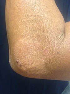
first noticeable sign of leprosy is often the development of pale or
pinkish patches of skin that may be insensitive to temperature or pain. This is sometimes accompanied or preceded by nerve problems including numbness or tenderness in the hands or feet.
Secondary infections, in turn, can result in tissue loss, causing
fingers and toes to become shortened and deformed, as cartilage is
absorbed into the body.
Approximately 30% of those affected experience nerve damage, and
the nerve damage sustained is irreversible, even with treatment of the
infection. Damage to nerves may cause sensation abnormalities, which may lead to infection, ulceration, and joint deformity.
 Paucibacillary leprosy (PB): Pale skin patch with loss of sensation
 Skin lesions on the thigh of a person with leprosy
 Hands deformed by leprosy
.jpg)


Cause
M. leprae and M. lepromatosis

M. leprae, one of the causative agents of leprosy: As an acid-fast bacterium, M. leprae appears red when a Ziehl-Neelsen stain is used.
M. leprae and M. lepromatosis are the causative agents of leprosy. M. lepromatosis is a relatively newly identified mycobacterium isolated from a fatal case of diffuse lepromatous leprosy in 2008. M. lepromatosis is indistinguishable clinically from M. leprae.
An intracellular, acid-fast bacterium, M. leprae is aerobic and rod-shaped, and is surrounded by the waxy cell membrane coating characteristic of the genus Mycobacterium.
Due to extensive loss of genes necessary for independent growth, M. leprae and M. lepromatosis are obligate intracellular pathogens, and unculturable in the laboratory, a factor that leads to difficulty in definitively identifying the organism under a strict interpretation of Koch's postulates. The use of nonculture-based techniques such as molecular genetics has allowed for alternative establishment of causation.
While the causative organisms have to date been impossible to culture in vitro, it has been possible to grow them in animals such as mice and armadillos.
Naturally occurring infection also has been reported in nonhuman primates, including the African chimpanzee, sooty mangabey, and cynomolgus macaque, as well as in
armadillos and red squirrels. Multilocus sequence typing of the armadillo M. leprae strains suggests that they were of human origin for at most a few hundred years.
Thus, armadillos likely first acquired the organism incidentally from
early American explorers. This incidental transmission was sustained in
the armadillo population, and it is now transmitted back to humans,
making leprosy a zoonotic disease.
Red squirrels (Sciurus vulgaris)—a threatened species—in England were found to have leprosy in November 2016.
It has been suggested that the trade in red squirrel fur, highly prized
in the medieval period and intensively traded, may have been
responsible for the leprosy epidemic in medieval Europe. Within Great
Britain, widespread leprosy is found early in East Anglia, to which many
of the squirrel furs were traded, and the strain is the same as that
found in modern red squirrels on Brownsea Island. However, no squirrel cases have spread to a human for hundreds of years.
Risk factors
The
greatest risk factor for developing leprosy is contact with another
person infected by leprosy. Contacts of people with leprosy are five to
eight times more likely to develop leprosy than members of the general
population. Leprosy also occurs more commonly among those living in poverty. Not all people who are infected with M. leprae develop symptoms.
Other risk factors are poorly understood. Conditions that reduce
immune function, such as malnutrition, other illnesses, or host genetic
differences, may increase the risk of developing leprosy. Infection with HIV does not appear to increase the risk of developing leprosy.
Transmission
Transmission of leprosy occurs during close contact with those who are infected. Transmission is proposed to be by nasal droplets, but many questions remain about its mode of transmission and epidemiology.
Leprosy is not known to be either sexually transmitted or highly
infectious. People are generally no longer infectious after the first
month of standard multidrug therapy.
Leprosy may also be transmitted to humans by armadillos, although the mechanism is not fully understood.
Two exit routes of M. leprae from the human body often
described are the skin and the nasal mucosa, although their relative
importance is not clear. Lepromatous cases show large numbers of
organisms deep in the dermis, but whether they reach the skin surface in sufficient numbers is doubtful.
The skin and the upper respiratory tract are the most likely
entry route. While older research dealt with the skin route, recent
research has increasingly favored the respiratory route. Experimental transmission of leprosy through aerosols containing M. leprae in immunosuppressed mice was accomplished, suggesting a similar possibility in humans.
Genetics
Several genes have been associated with a susceptibility to leprosy.
Often, the immune system is able to eliminate leprosy during the early
infection stage before severe symptoms develop. A defect in cell-mediated immunity may cause susceptibility to leprosy. The region of DNA responsible for this variability is also involved in Parkinson's disease, giving rise to current speculation that the two disorders may be linked in some way at the biochemical level.
Some evidence indicates not all people who are infected with M. leprae
develop leprosy, and genetic factors have long been thought to play a
role, due to the observation of clustering of leprosy around certain
families, and the failure to understand why certain individuals develop
lepromatous leprosy while others develop other types of leprosy.
Mechanism
How the infection produces the symptoms of the disease is not known.
Diagnosis
In countries where people are frequently infected, a person is considered to have leprosy if they have one these signs:
- Skin lesion consistent with leprosy and with definite sensory loss
- Positive skin smears
Skin lesions can be single or many, and usually hypopigmented, although sometimes reddish or copper-colored. The lesions may be macules (flat), papules (raised), or nodular.
The sensory loss at the skin lesion is important because this feature
can help differentiate it from other causes of skin lesions such as tinea versicolor.
Thickened nerves are associated with leprosy and can be accompanied by
loss of sensation or muscle weakness. However, without the
characteristic skin lesion and sensory loss, muscle weakness is not
considered a reliable sign of leprosy.
In some cases, acid-fast leprosy bacilli in skin smears are considered diagnostic; however, the diagnosis is clinical.
In countries or areas where the disease is uncommon, such as the
United States, diagnosis of leprosy is often delayed because healthcare
providers are unaware of leprosy and its symptoms. Early diagnosis and
treatment prevent nerve involvement, the hallmark of leprosy, and the
disability it causes.
Many kinds of leprosy are known, but some symptoms are common to
them, including runny nose, dry scalp, eye problems, skin lesions,
muscle weakness, reddish skin, smooth, shiny, diffuse thickening of
facial skin, ear, and hand, loss of sensation in fingers and toes,
thickening of peripheral nerves, and flat nose due to destruction of
nasal cartilage. Also, phonation and resonation of sound occur during
speech. Often, atrophy of the testes with resulting impotence occurs.
There is no recommended test to diagnose latent leprosy in
asymptomatic contacts. However few people with latent leprosy went on to
develop a positive test.
Classification
Several different approaches for classifying leprosy exist, but parallels exist.
- The World Health Organization system distinguishes "paucibacillary" and "multibacillary" based upon the proliferation of bacteria.("pauci-" refers to a low quantity.)
- The Ridley-Jopling scale provides five gradations.
- The ICD-10, though developed by the WHO, uses Ridley-Jopling and not the WHO system. It also adds an indeterminate ("I") entry.
- In MeSH, three groupings are used.
| WHO | Ridley-Jopling | ICD-10 | MeSH | Description | Lepromin test |
|---|---|---|---|---|---|
| Paucibacillary | tuberculoid ("TT"), borderline tuberculoid ("BT") |
A30.1, A30.2 | Tuberculoid | It is characterized by one or more hypopigmented skin macules and patches where skin sensations are lost because of damaged peripheral nerves that have been attacked by the human host's immune cells. | Positive |
| Multibacillary | midborderline or borderline ("BB") |
A30.3 | Borderline | Borderline leprosy is of intermediate severity and is the most common form. Skin lesions resemble tuberculoid leprosy, but are more numerous and irregular; large patches may affect a whole limb, and peripheral nerve involvement with weakness and loss of sensation is common. This type is unstable and may become more like lepromatous leprosy or may undergo a reversal reaction, becoming more like the tuberculoid form. |
|
| Multibacillary | borderline lepromatous ("BL"), and lepromatous ("LL") |
A30.4, A30.5 | Lepromatous | It is associated with symmetric skin lesions, nodules, plaques, thickened dermis, and frequent involvement of the nasal mucosa resulting in nasal congestion and nose bleeds, but, typically, detectable nerve damage is late. Loss of eyebrows and lashes can be seen in advanced disease. | Negative |
A difference in immune response to the tuberculoid and lepromatous forms is seen.
Leprosy may also be divided into:
This disease may also occur with only neural involvement, without skin lesions.
Prevention
Early
detection of the disease is important, since physical and neurological
damage may be irreversible even if cured. Medications can decrease the
risk of those living with people with leprosy from acquiring the disease
and likely those with whom people with leprosy come into contact
outside the home. The WHO recommends that preventative medicine is given to people who are in close contact with someone who has leprosy.
The suggested preventative treatment is a single dose of rifampicin
(SDR) in adults and children over 2 years old who do not already have
leprosy or tuberculosis.
Preventative treatment is associated with a 57% reduction in infections
within 2 years and a 30% reduction in infections within 6 years.
The Bacillus Calmette–Guérin (BCG) vaccine offers a variable amount of protection against leprosy in addition to its target of tuberculosis.
It appears to be 26 to 41% effective (based on controlled trials) and
about 60% effective based on observational studies with two doses
possibly working better than one.
The WHO concluded in 2018 that the BCG vaccine at birth reduces leprosy
risk and is recommended in countries with high incidence of TB and
leprosy. Development of a more effective vaccine is ongoing.
Treatment

MDT antileprosy drugs: standard regimens from 2010
Anti-leprosy medication
A number of leprostatic agents are available for treatment. A 3-drug regimen of rifampicin, dapsone and clofazimine is recommended for all people with leprosy, for 6 months for paucibacillary leprosy and 12 months for multibacillary leprosy.
Multidrug therapy (MDT) remains highly effective, and people are no longer infectious after the first monthly dose. It is safe and easy to use under field conditions due to its presentation in calendar blister packs. Post-treatment relapse rates remain low.
Resistance has been reported in several countries, although the number
of cases is small. A person with rifampicin-resistant leprosy should be
treated with two second line drugs. Evidence on the potential benefits
and harms of alternative regimens for drug-resistant leprosy is not yet
available.
Skin changes
For people with nerve damage, protective footwear may help prevent ulcers and secondary infection. Canvas shoes may be better than PVC-boots. There may be no difference between double rocker shoes and below-knee plaster.
Topical ketanserin seems to have a better effect on ulcer healing than clioquinol cream or zinc paste, but the evidence for this is weak. Likewise, topical phenytoin has shown more efficacy than saline dressings.
Epidemiology

World distribution of leprosy, 2003

Disability-adjusted life year for leprosy per 100,000 inhabitants in 2004
no data
<1 .5="" span="">
1.5–3
3–4.5
4.5–6
6–7.5
7.5–9
9–10.5
10.5–12
12–13.5
13.5–15
15–20
>20
In 2016, there were 216,108 new leprosy cases registered, corresponding to a global detection rate of 0.29 per 10,000 people. As of 2015, 14 countries reported contain 94% of new leprosy cases. India had the greatest number of new cases (60%), followed by Brazil (13%) and Indonesia (8%).
Although the number of cases worldwide continues to fall, pockets of
high prevalence remain in certain areas such as Brazil, South Asia
(India, Nepal, Bhutan), some parts of Africa (Tanzania, Madagascar,
Mozambique), and the western Pacific. About 150 to 250 cases are
diagnosed in the United States each year.
In the United States there are about 5,000 people who no longer have
leprosy but have long-term complications of disease and continue to
receive care.
In the 1960s, there were tens of millions of leprosy cases. A
series of national (the International Federation of Anti-Leprosy
Associations) and international (the WHO's "Global Strategy for Reducing
Disease Burden Due to Leprosy") initiatives have reduced the total
number and the number of new cases of the disease.
Disease burden
Although
the number of new leprosy cases occurring each year is important as a
measure of transmission, it is difficult to measure due to leprosy's
long incubation period, delays in diagnosis after onset of the disease,
and the lack of laboratory tools to detect it in the very early stages.
Instead, the registered prevalence
is used. Registered prevalence is a useful proxy indicator of the
disease burden, as it reflects the number of active leprosy cases
diagnosed with the disease and receiving treatment with MDT at a given
point in time. The prevalence rate is defined as the number of cases
registered for MDT treatment among the population in which the cases
have occurred, again at a given point in time.
New case detection is another indicator of the disease that is
usually reported by countries on an annual basis. It includes cases
diagnosed with the onset of disease in the year in question (true
incidence) and a large proportion of cases with onset in previous years
(termed a backlog prevalence of undetected cases).
Endemic countries also report the number of new cases with
established disabilities at the time of detection, as an indicator of
the backlog prevalence. Determination of the time of onset of the
disease is, in general, unreliable, is very labor-intensive, and is
seldom done in recording these statistics.
History

G. H. A. Hansen, discoverer of M. leprae
Using comparative genomics, in 2005, geneticists traced the origins
and worldwide distribution of leprosy from East Africa or the Near East
along human migration routes. They found four strains of M. leprae
with specific regional locations. Strain 1 occurs predominantly in
Asia, the Pacific region, and East Africa; strain 4, in West Africa and
the Caribbean; strain 3 in Europe, North Africa, and the Americas; and
strain 2 only in Ethiopia, Malawi, Nepal/north India, and New Caledonia.
On the basis of this, they offer a map of the dissemination of
leprosy in the world. This confirms the spread of the disease along the
migration, colonisation, and slave trade routes taken from East Africa
to India, West Africa to the New World, and from Africa into Europe and
vice versa.
The oldest skeletal evidence for the disease date from 2000 BCE,
as found in human remains from the archaeological sites of Balathal in
India and Harappa in Pakistan.
Although retrospectively identifying descriptions of leprosy-like
symptoms is difficult, what appears to be leprosy was discussed by Hippocrates in 460 BC. In 1846, Francis Adams produced The Seven Books of Paulus Aegineta
which included a commentary on all medical and surgical knowledge and
descriptions and remedies to do with leprosy from the Romans, Greeks,
and Arabs.
Interpretations of the presence of leprosy have been made on the
basis of descriptions in ancient Indian (Atharva Veda and Kausika
Sutra), Greek, and Middle Eastern documentary sources that describe skin
afflictions.
Leprosy probably did not exist in Greece or the Middle East before Common Era. It did not exist in the Americas before colonization by modern Europeans.
Skeletal remains from the second millennium BC, discovered in
2009, represent the oldest documented evidence for leprosy. Located at
Balathal, in Rajasthan, northwest India, the discoverers suggest that if
the disease did migrate from Africa to India, during the third
millennium BC "at a time when there was substantial interaction among
the Indus Civilization, Mesopotamia, and Egypt, there needs to be
additional skeletal and molecular evidence of leprosy in India and
Africa so as to confirm the African origin of the disease."
A proven human case was verified by DNA taken from the shrouded remains
of a man discovered in a tomb next to the Old City of Jerusalem dated
by radiocarbon methods to 1–50 AD.

Distribution of leprosy around the world in 1891
However, a study published in 2018 found the oldest strains of
leprosy in remains from Europe, the oldest strain being from Great
Chesterford and dating back to 415 to 545 AD. These findings suggest a
different path for the spread of leprosy where it may have originated in
Western Eurasia. This study also indicates that there were more strains
in Europe at the time than previously determined.
The causative agent of leprosy, M. leprae, was discovered by G. H. Armauer Hansen in Norway in 1873, making it the first bacterium to be identified as causing disease in humans.
The first effective treatment (promin) became available in the 1940s. In the 1950s, dapsone
was introduced. The search for further effective antileprosy drugs led
to the use of clofazimine and rifampicin in the 1960s and 1970s.
Later, Indian scientist Shantaram Yawalkar and his colleagues
formulated a combined therapy using rifampicin and dapsone, intended to
mitigate bacterial resistance. Multi-drug therapy (MDT) combining all three drugs was first recommended by the WHO in 1981. These three antileprosy drugs are still used in the standard MDT regimens.
Leprosy was once believed to be highly contagious and was treated with mercury—as was syphilis, which was first described in 1530. Many early cases thought to be leprosy could actually have been syphilis.
Resistance has developed to initial treatment. Until the
introduction of MDT in the early 1980s, leprosy could not be diagnosed
and treated successfully within the community.
Japan
still has sanatoriums (although Japan's sanatoriums no longer have
active leprosy cases, nor are survivors held in them by law).
The importance of the nasal mucosa in the transmission of M leprae was recognized as early as 1898 by Schäffer, in particular, that of the ulcerated mucosa.
Society and culture

Two lepers denied entrance to town, 14th century
The word "leprosy" comes from the greek word "λέπος (lépos) – skin" and "λεπερός (leperós) – scaly man.
India
British India
enacted the Leprosy Act of 1898 which institutionalized those affected
and segregated them by sex to prevent reproduction. The Act was
difficult to enforce but was repealed in 1983 only after MDT therapy had
become widely available. In 1983, the National Leprosy Elimination
Programme, previously the National Leprosy Control Programme, changed
its methods from surveillance to the treatment of people with leprosy.
India still accounts for over half of the global disease burden.
Treatment cost
Between 1995 and 1999, the WHO, with the aid of the Nippon Foundation,
supplied all endemic countries with free MDT in blister packs,
channeled through ministries of health. This free provision was extended
in 2000 and again in 2005, 2010 and 2015 with donations by the MDT
manufacturer Novartis
through the WHO. In the latest agreement signed between the company and
the WHO in October 2015, the provision of free MDT by the WHO to all
endemic countries will run until the end of 2020. At the national level,
nongovernment organizations
affiliated with the national program will continue to be provided with
an appropriate free supply of this WHO-supplied MDT by the government.
Historical texts
Written
accounts of leprosy date back thousands of years. Various skin diseases
translated as leprosy appear in the ancient Indian text, the Atharava Veda, as early as 2000 BC. Another Indian text, the Manusmriti
(1500 BC), prohibited contact with those infected with the disease and
made marriage to a person infected with leprosy punishable.
Biblically speaking, the Hebraic root tsara or tsaraath
(צָרַע,—tsaw-rah'—to be struck with leprosy, to be leprous) and the
Greek (λεπρός—lepros), are of broader classification than the more
narrow use of the term related to Hansen's Disease. Any progressive
skin disease (a whitening or splotchy bleaching of skin, raised
manifestations of scales, scabs, infections, rashes, etc.…) as well as
generalized molds and surface discoloration of any clothing, leather,
and/or discoloration on walls surfaces throughout homes all came under
the "law of leprosy" (Leviticus 14:54–57). Ancient sources also such as the Talmud (Sifra 63) make clear that tzaraath refers to various types of lesions or stains associated with ritual impurity
and occurring on cloth, leather, or houses, as well as skin. It may
sometimes be a symptom of the disease described in this article but has
many other causes, as well. The New Testament describes instances of Jesus healing people with leprosy Luke 17:11, although the precise relationship between this, tzaraath, and Hansen's disease is not established.
The biblical perception that people with leprosy were unclean may be connected to a passage from Leviticus
13: 44–46, among others. Judeo–Christian belief, for some, held that
leprosy was of moral consequence, and, as in many societies, early Christians
believed that those affected by leprosy were being punished by God for
sinful behavior. Moral associations have persisted throughout history.
Pope Gregory the Great (540–604) and Isidor of Seville (560–636) considered people with the disease to be heretics.
Middle Ages

Medieval leper bell
It is believed that a rise in leprosy in Europe occurred in the
Middle Ages based on the increased number of hospitals created to treat
people with leprosy in the 12th and 13th centuries. France alone had nearly 2,000 leprosariums during this period.
The social perception in medieval communities was generally one
of fear, and those people infected with the disease were thought to be
unclean, untrustworthy, and morally corrupt.
People with leprosy were also often required to wear clothing that
identified them as such or carry a bell announcing their presence.
Segregation from mainstream society was common. The third Lateran Council of 1179 and a 1346 edict by King Edward
expelled lepers from city limits. Because of the moral stigma of the
disease, methods of treatment were both physical and spiritual, and
leprosariums were established under the purview of the church.
19th century
Norway
Norway
was the location of a progressive stance on leprosy tracking and
treatment and played an influential role in European understanding of
the disease. In 1832, Dr. JJ Hjort conducted the first leprosy survey,
thus establishing a basis for epidemiological surveys. Subsequent
surveys resulted in the establishment of a national leprosy registry to
study the causes of leprosy and for tracking of the rate of infection.
Early leprosy research throughout Europe was conducted by Norwegian scientists Daniel Cornelius Danielssen and Carl Wilhelm Boeck.
Their work resulted in the establishment of the National Leprosy
Research and Treatment Center. Danielssen and Boeck believed the cause
of leprosy transmission was hereditary. This stance was influential in
advocating for the isolation of those infected by sex to prevent
reproduction.
Colonialism and imperialism

Father Damien on his deathbed in 1889
Though leprosy in Europe was again on the decline by the 1860s,
Western countries embraced isolation treatment out of fear of the spread
of disease from developing countries, minimal understanding of
bacteriology, lack of diagnostic ability or knowledge of how contagious
the disease was, and missionary activity.
Growing imperialism and pressures of the industrial revolution
resulted in a Western presence in countries where leprosy was endemic,
namely the British presence in India. Isolation treatment methods were
observed by Surgeon-Mayor Henry Vandyke Carter
of the British Colony in India while visiting Norway, and these methods
were applied in India with the financial and logistical assistance of
religious missionaries. Colonial and religious influence and associated
stigma continued to be a major factor in the treatment and public
perception of leprosy in endemic developing countries until the
mid-twentieth century.
Stigma
Despite effective treatment and education efforts, leprosy stigma
continues to be problematic in developing countries where the disease is
common. Leprosy is most common amongst impoverished or marginalized
populations where social stigma is likely to be compounded by other
social inequities. Fears of ostracism, loss of employment, or expulsion
from family and society may contribute to a delayed diagnosis and
treatment.
Folk beliefs, lack of education, and religious connotations of
the disease continue to influence social perceptions of those afflicted
in many parts of the world. In Brazil, for example, folklore holds that
leprosy is transmitted by dogs, it is a disease associated with sexual
promiscuity, and is sometimes thought to be punishment for sins or moral
transgressions.
Socioeconomic factors also have a direct impact. Lower-class domestic
workers who are often employed by those in a higher socioeconomic class
may find their employment in jeopardy as physical manifestations of the
disease become apparent. Skin discoloration and darker pigmentation
resulting from the disease also have social repercussions.
In extreme cases in northern India, leprosy is equated with an
"untouchable" status that "often persists long after (individuals with
leprosy) have been cured of the disease, creating lifelong prospects of
divorce, eviction, loss of employment, and ostracism from family and
social networks."
 Leprosy in Tahiti, circa 1895
 A 26-year-old woman with leprous lesions
 A 13-year-old boy with severe leprosy


Programs and treatment
The
WHO states that diagnosis and treatment with MDT are easy and
effective, and a 45% decline in disease burden has occurred since MDT
has become more widely available. The organization emphasizes the
importance of fully integrating leprosy treatment into public health
services, effective diagnosis and treatment, and access to information.
In some instances in India, community-based rehabilitation is
embraced by local governments and NGOs alike. Often, the identity
cultivated by a community environment is preferable to reintegration,
and models of self-management and collective agency independent of NGOs
and government support have been desirable and successful.
Notable cases
- Saint Damien DeVeuster, a Roman Catholic priest from Belgium, himself eventually contracting leprosy, ministered to lepers who had been placed under a government-sanctioned medical quarantine on the island of Molokaʻi in the Kingdom of Hawaiʻi.
- Baldwin IV of Jerusalem was a Christian king of Latin Jerusalem afflicted with leprosy.
- King Henry IV of England (reigned 1399 to 1413) possibly had leprosy.
- Vietnamese poet Hàn Mặc Tử
- Ōtani Yoshitsugu, a Japanese daimyō
- Forough Farrokhzad made a 22-minute documentary about a leprosy colony in Iran in 1962 called The House Is Black. The film humanizes the people affected and opens by saying that "there is no shortage of ugliness in the world, but by closing our eyes on ugliness, we will intensify it."
Research directions
More
research in ulcer prevention and treatment in leprosy is needed to
better guide management of skin changes caused by leprosy-induced nerve
damage.
Other animals
Wild nine-banded armadillos (Dayspus novemcinctus) in south central United States often carry Mycobacterium leprae.
This is believed to be because armadillos have such a low body
temperature. Leprosy lesions appear mainly in cooler body regions such
as the skin and mucous membranes of the upper respiratory tract. Because of armadillos' armor, skin lesions are hard to see. Abrasions around the eyes, nose and feet are the most common signs. Infected armadillos make up a large reservoir of M. leprae
and may be a source of infection for some humans in the United States
or other locations in the armadillos' home range. In armadillo leprosy,
lesions did not persist at the site of entry in animals, M. leprae multiplied in macrophages at the site of inoculation and lymph nodes.

.jpg)


