Search This Blog

Saturday, May 18, 2019

Nicotinic acetylcholine receptor

From Wikipedia, the free encyclopedia

Nicotinic acetylcholine receptors, or nAChRs, are receptor polypeptides that respond to the neurotransmitter acetylcholine. Nicotinic receptors also respond to drugs, including the nicotinic receptor agonist nicotine. They are found in the central and peripheral nervous system, muscle, and many other tissues of many organisms, including humans. At the neuromuscular junction they are the primary receptor in muscle for motor nerve-muscle communication that controls muscle contraction. In the peripheral nervous system: (1) they transmit outgoing signals from the presynaptic to the postsynaptic cells within the sympathetic and parasympathetic nervous system, and (2) they are the receptors found on skeletal muscle that receive acetylcholine released to signal for muscular contraction. In the immune system, nAChRs regulate inflammatory processes and signal through distinct intracellular pathways. In insects, the cholinergic system is limited to the central nervous system.

The nicotinic receptors are considered cholinergic receptors, since they respond to acetylcholine. Nicotinic receptors get their name from nicotine, which does not stimulate the muscarinic acetylcholine receptor, but instead selectively binds to the nicotinic receptor. The muscarinic acetylcholine receptor likewise gets its name from a chemical that selectively attaches to that receptor — muscarine. Acetylcholine itself binds to both muscarinic and nicotinic acetylcholine receptors. 

As ionotropic receptors, nAChRs are directly linked to ion channels. New evidence suggests that these receptors can also use second messengers (as metabotropic receptors do) in some cases. Nicotinic acetylcholine receptors are the best-studied of the ionotropic receptors.

Since nicotinic receptors help transmit outgoing signals for the sympathetic and parasympathetic systems, nicotinic receptor antagonists such as hexamethonium interfere with the transmission of these signals. Thus, for example, nicotinic receptor antagonists interfere with the baroreflex that normally corrects changes in blood pressure by sympathetic and parasympathetic stimulation of the heart.

Structure

Nicotinic receptor structure
 
Nicotinic receptors, with a molecular mass of 290 kDa, are made up of five subunits, arranged symmetrically around a central pore. Each subunit comprises four transmembrane domains with both the N- and C-terminus located extracellularly. They possess similarities with GABAA receptors, glycine receptors, and the type 3 serotonin receptors (which are all ionotropic receptors), or the signature Cys-loop proteins.

In vertebrates, nicotinic receptors are broadly classified into two subtypes based on their primary sites of expression: muscle-type nicotinic receptors and neuronal-type nicotinic receptors. In the muscle-type receptors, found at the neuromuscular junction, receptors are either the embryonic form, composed of α1, β1, γ, and δ subunits in a 2:1:1:1 ratio, or the adult form composed of α1, β1, δ, and ε subunits in a 2:1:1:1 ratio. The neuronal subtypes are various homomeric (all one type of subunit) or heteromeric (at least one α and one β) combinations of twelve different nicotinic receptor subunits: α2−α10 and β2−β4. Examples of the neuronal subtypes include: (α4)3(β2)2, (α4)2(β2)3, (α3)2(β4)3, α4α6β3(β2)2, (α7)5, and many others. In both muscle-type and neuronal-type receptors, the subunits are very similar to one another, especially in the hydrophobic regions. 

A number of electron microscopy and x-ray crystallography studies have provided very high resolution structural information for muscle and neuronal nAChRs and their binding domains.

Binding to the receptor

As with all ligand-gated ion channels, opening of the nAChR channel pore requires the binding of a chemical messenger. Several different terms are used to refer to the molecules that bind receptors, such as ligand, agonist, or transmitter. As well as the endogenous agonist acetylcholine, agonists of the nAChR include nicotine, epibatidine, and choline. Nicotinic antagonists that block the receptor include mecamylamine, dihydro-β-erythroidine, and hexamethonium

In muscle-type nAChRs, the acetylcholine binding sites are located at the α and either ε or δ subunits interface. In neuronal nAChRs, the binding site is located at the interface of an α and a β subunit or between two α subunits in the case of α7 receptors. The binding site is located in the extracellular domain near the N terminus. When an agonist binds to the site, all present subunits undergo a conformational change and the channel is opened and a pore with a diameter of about 0.65 nm opens.

Opening the channel

Nicotinic AChRs may exist in different interconvertible conformational states. Binding of an agonist stabilises the open and desensitised states. In normal physiological conditions, the receptor needs exactly two molecules of ACh to open. Opening of the channel allows positively charged ions to move across it; in particular, sodium enters the cell and potassium exits. The net flow of positively charged ions is inward. 

The nAChR is a non-selective cation channel, meaning that several different positively charged ions can cross through. It is permeable to Na+ and K+, with some subunit combinations that are also permeable to Ca2+. The amount of sodium and potassium the channels allow through their pores (their conductance) varies from 50–110 pS, with the conductance depending on the specific subunit composition as well as the permeant ion.

Many neuronal nAChRs can affect the release of other neurotransmitters. The channel usually opens rapidly and tends to remain open until the agonist diffuses away, which usually takes about 1 millisecond. However, AChRs can spontaneously open with no ligands bound or can spontaneously close with ligands bound, and mutations in the channel can shift the likelihood of either event. Therefore, ACh binding changes the probability of pore opening, which increases as more ACh binds. 

The nAChR is unable to bind ACh when bound to any of the snake venom α-neurotoxins. These α-neurotoxins antagonistically bind tightly and noncovalently to nAChRs of skeletal muscles and in neurons, thereby blocking the action of ACh at the postsynaptic membrane, inhibiting ion flow and leading to paralysis and death. The nAChR contains two binding sites for snake venom neurotoxins. Progress towards discovering the dynamics of binding action of these sites has proved difficult, although recent studies using normal mode dynamics have aided in predicting the nature of both the binding mechanisms of snake toxins and of ACh to nAChRs. These studies have shown that a twist-like motion caused by ACh binding is likely responsible for pore opening, and that one or two molecules of α-bungarotoxin (or other long-chain α-neurotoxin) suffice to halt this motion. The toxins seem to lock together neighboring receptor subunits, inhibiting the twist and therefore, the opening motion.

Effects

The activation of receptors by nicotine modifies the state of neurons through two main mechanisms. On one hand, the movement of cations causes a depolarization of the plasma membrane (which results in an excitatory postsynaptic potential in neurons) leading to the activation of voltage-gated ion channels. On the other hand, the entry of calcium acts, either directly or indirectly, on different intracellular cascades. This leads, for example, to the regulation of the activity of some genes or the release of neurotransmitters.

Receptor regulation

Receptor desensitisation

Ligand-bound desensitisation of receptors was first characterised by Katz and Thesleff in the nicotinic acetylcholine receptor.

Prolonged or repeated exposure to a stimulus often results in decreased responsiveness of that receptor toward a stimulus, termed desensitisation. nAChR function can be modulated by phosphorylation by the activation of second messenger-dependent protein kinases. PKA and PKC, as well as tyrosine kinases, have been shown to phosphorylate the nAChR resulting in its desensitisation. It has been reported that, after prolonged receptor exposure to the agonist, the agonist itself causes an agonist-induced conformational change in the receptor, resulting in receptor desensitisation. Desensitised receptors can revert to a prolonged open state when an agonist is bound in the presence of a positive allosteric modulator, for example PNU-120596. Also, there is evidence that indicates specific chaperone molecules have regulatory effects on these receptors.

Roles

The subunits of the nicotinic receptors belong to a multigene family (16 members in humans) and the assembly of combinations of subunits results in a large number of different receptors. These receptors, with highly variable kinetic, electrophysiological and pharmacological properties, respond to nicotine differently, at very different effective concentrations. This functional diversity allows them to take part in two major types of neurotransmission. Classical synaptic transmission (wiring transmission) involves the release of high concentrations of neurotransmitter, acting on immediately neighboring receptors. In contrast, paracrine transmission (volume transmission) involves neurotransmitters released by synaptic boutons, which then diffuse through the extra-cellular medium until they reach their receptors, which may be distant. Nicotinic receptors can also be found in different synaptic locations; for example the muscle nicotinic receptor always functions post-synaptically. The neuronal forms of the receptor can be found both post-synaptically (involved in classical neurotransmission) and pre-synaptically where they can influence the release of multiple neurotransmitters.

Subunits

17 vertebrate nAChR subunits have been identified, which are divided into muscle-type and neuronal-type subunits. However, although an α8 subunit/gene is present in avian species such as the chicken, it is not present in human or mammalian species.

The nAChR subunits have been divided into 4 subfamilies (I-IV) based on similarities in protein sequence. In addition, subfamily III has been further divided into 3 types. 

Neuronal-type Muscle-type
I II III IV
α9, α10 α7, α8 1 2 3 α1, β1, δ, γ, ε
α2, α3, α4, α6 β2, β4 β3, α5

Neuromuscular junction

From Wikipedia, the free encyclopedia

At the neuromuscular junction, the nerve fiber is able to transmit a signal to the muscle fiber by releasing ACh (and other substances), causing muscle contraction.
 
Muscles will contract or relax when they receive signals from the nervous system. The neuromuscular junction is the site of the signal exchange. The steps of this process in vertebrates occur as follows:(1) The action potential reaches the axon terminal. (2) Voltage-dependent calcium gates open, allowing calcium to enter the axon terminal. (3) Neurotransmitter vesicles fuse with the presynaptic membrane and ACh is released into the synaptic cleft via exocytosis. (4) ACh binds to postsynaptic receptors on the sarcolemma. (5) This binding causes ion channels to open and allows sodium and other cations to flow across the membrane into the muscle cell. (6) The flow of sodium ions across the membrane into and potassium ions out of the muscle cell generates an action potential which travels to the myofibril and results in muscle contraction.Labels:A: Motor Neuron AxonB: Axon TerminalC. Synaptic CleftD. Muscle CellE. Part of a Myofibril
 
Neuromuscular junctions
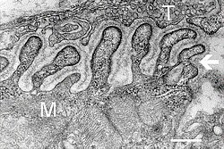
Electron micrograph of neuromuscular junction (cross-section).jpg
Electron micrograph showing a cross section through the neuromuscular junction. T is the axon terminal, M is the muscle fiber. The arrow shows junctional folds with basal lamina. Active zones are visible on the tips between the folds. Scale is 0.3 µm. Source: NIMH
Synapse diag4.png
Detailed view of a neuromuscular junction:
Details
Identifiers
Latinsynapssis neuromuscularis; junctio neuromuscularis
MeSHD009469
THH2.00.06.1.02001
FMA61803

A neuromuscular junction (or myoneural junction) is a chemical synapse formed by the contact between a motor neuron and a muscle fiber. It is at the neuromuscular junction that a motor neuron is able to transmit a signal to the muscle fiber, causing muscle contraction

Muscles require innervation to function—and even just to maintain muscle tone, avoiding atrophy. Synaptic transmission at the neuromuscular junction begins when an action potential reaches the presynaptic terminal of a motor neuron, which activates voltage-dependent calcium channels to allow calcium ions to enter the neuron. Calcium ions bind to sensor proteins (synaptotagmin) on synaptic vesicles, triggering vesicle fusion with the cell membrane and subsequent neurotransmitter release from the motor neuron into the synaptic cleft. In vertebrates, motor neurons release acetylcholine (ACh), a small molecule neurotransmitter, which diffuses across the synaptic cleft and binds to nicotinic acetylcholine receptors (nAChRs) on the cell membrane of the muscle fiber, also known as the sarcolemma. nAChRs are ionotropic receptors, meaning they serve as ligand-gated ion channels. The binding of ACh to the receptor can depolarize the muscle fiber, causing a cascade that eventually results in muscle contraction. 

Neuromuscular junction diseases can be of genetic and autoimmune origin. Genetic disorders, such as Duchenne muscular dystrophy, can arise from mutated structural proteins that comprise the neuromuscular junction, whereas autoimmune diseases, such as myasthenia gravis, occur when antibodies are produced against nicotinic acetylcholine receptors on the sarcolemma.

Structure and function

motor endplate

Quantal transmission

At the neuromuscular junction presynaptic motor axons terminate 30 nanometers from the cell membrane or sarcolemma of a muscle fiber. The sarcolemma at the junction has invaginations called postjunctional folds, which increase its surface area facing the synaptic cleft. These postjunctional folds form the motor endplate, which is studded with nicotinic acetylcholine receptors (nAChRs) at a density of 10,000 receptors/micrometer2. The presynaptic axons terminate in bulges called terminal boutons (or presynaptic terminals) that project toward the postjunctional folds of the sarcolemma. In the frog each motor nerve terminal contains about 300,000 vesicles, with an average diameter of 0.05 micrometers. The vesicles contain acetylcholine. Some of these vesicles are gathered into groups of fifty, positioned at active zones close to the nerve membrane. Active zones are about 1 micrometer apart. The 30 nanometer cleft between nerve ending and endplate contains a meshwork of acetylcholinesterase (AChE) at a density of 2,600 enzyme molecules/micrometer2, held in place by the structural proteins dystrophin and rapsyn. Also present is the receptor tyrosine kinase protein MuSK, a signaling protein involved in the development of the neuromuscular junction, which is also held in place by rapsyn.

About once every second in a resting junction randomly one of the synaptic vesicles fuses with the presynaptic neuron's cell membrane in a process mediated by SNARE proteins. Fusion results in the emptying of the vesicle's contents of 7000-10,000 acetylcholine molecules into the synaptic cleft, a process known as exocytosis. Consequently exocytosis releases acetylcholine in packets that are called quanta. The acetylcholine quantum diffuses through the acetylcholinesterase meshwork, where the high local transmitter concentration occupies all of the binding sites on the enzyme in its path. The acetylcholine that reaches the endplate activates ~2,000 acetylcholine receptors, opening their ion channels which permits sodium ions to move into the endplate producing a depolarization of ~0.5 mV known as a miniature endplate potential (MEPP). By the time the acetylcholine is released from the receptors the acetylcholinesterase has destroyed its bound ACh, which takes about ~0.16 ms, and hence is available to destroy the ACh released from the receptors.

When the motor nerve is stimulated there is a delay of only 0.5 to 0.8 msec between the arrival of the nerve impulse in the motor nerve terminals and the first response of the endplate  The arrival of the motor nerve action potential at the presynaptic neuron terminal opens voltage-dependent calcium channels and Ca2+ ions flow from the extracellular fluid into the presynaptic neuron's cytosol. This influx of Ca2+ causes several hundred neurotransmitter-containing vesicles to fuse with the presynaptic neuron's cell membrane through SNARE proteins to release their acetylcholine quanta by exocytosis. The endplate depolarization by the released acetylcholine is called an endplate potential (EPP). The EPP is accomplished when ACh binds the nicotinic acetylcholine receptors (nAChR) at the motor end plate, and causes an influx of sodium ions. This influx of sodium ions generates the EPP (depolarization), and triggers an action potential which travels along the sarcolemma and into the muscle fiber via the transverse tubules (T-tubules) by means of voltage-gated sodium channels. The conduction of action potentials along the transverse tubules stimulates the opening of voltage-gated Ca2+ channels which are mechanically coupled to Ca2+ release channels in the sarcoplasmic reticulum. The Ca2+ then diffuses out of the sarcoplasmic reticulum to the myofibrils so it can stimulate contraction. The endplate potential is thus responsible for setting up an action potential in the muscle fiber which triggers muscle contraction. The transmission from nerve to muscle is so rapid because each quantum of acetylcholine reaches the endplate in millimolar concentrations, high enough to combine with a receptor with a low affinity, which then swiftly releases the bound transmitter.

Acetylcholine receptors

  1. Ion channel linked receptor
  2. Ions
  3. Ligand (such as acetylcholine)
When ligands bind to the receptor, the ion channel portion of the receptor opens, allowing ions to pass across the cell membrane.
 
Acetylcholine is a neurotransmitter synthesized from dietary choline and acetyl-CoA (ACoA), and is involved in the stimulation of muscle tissue in vertebrates as well as in some invertebrate animals. In vertebrate animals, the acetylcholine receptor subtype that is found at the neuromuscular junction of skeletal muscles is the nicotinic acetylcholine receptor (nAChR), which is a ligand-gated ion channel. Each subunit of this receptor has a characteristic "cys-loop", which is composed of a cysteine residue followed by 13 amino acid residues and another cysteine residue. The two cysteine residues form a disulfide linkage which results in the "cys-loop" receptor that is capable of binding acetylcholine and other ligands. These cys-loop receptors are found only in eukaryotes, but prokaryotes possess ACh receptors with similar properties. Not all species use a cholinergic neuromuscular junction; e.g. crayfish and fruit flies have a glutamatergic neuromuscular junction.

AChRs at the skeletal neuromuscular junction form heteropentamers composed of two α, one β, one ɛ, and one δ subunits. When a single ACh ligand binds to one of the α subunits of the ACh receptor it induces a conformational change at the interface with the second AChR α subunit. This conformational change results in the increased affinity of the second α subunit for a second ACh ligand. AChRs therefore exhibit a sigmoidal dissociation curve due to this cooperative binding. The presence of the inactive, intermediate receptor structure with a single-bound ligand keeps ACh in the synapse that might otherwise be lost by cholinesterase hydrolysis or diffusion. The persistence of these ACh ligands in the synapse can cause a prolonged post-synaptic response.

Development

The development of the neuromuscular junction requires signaling from both the motor neuron's terminal and the muscle cell's central region. During development, muscle cells produce acetylcholine receptors (AChRs) and express them in the central regions in a process called prepatterning. Agrin, a heparin proteoglycan, and MuSK kinase are thought to help stabilize the accumulation of AChR in the central regions of the myocyte. MuSK is a receptor tyrosine kinase—meaning that it induces cellular signaling by binding phosphate molecules to self regions like tyrosines, and to other targets in the cytoplasm. Upon activation by its ligand agrin, MuSK signals via two proteins called "Dok-7" and "rapsyn", to induce "clustering" of acetylcholine receptors. ACh release by developing motor neurons produces postsynaptic potentials in the muscle cell that positively reinforces the localization and stabilization of the developing neuromuscular junction.

These findings were demonstrated in part by mouse "knockout" studies. In mice which are deficient for either agrin or MuSK, the neuromuscular junction does not form. Further, mice deficient in Dok-7 did not form either acetylcholine receptor clusters or neuromuscular synapses.

The development of neuromuscular junctions is mostly studied in model organisms, such as rodents. In addition, in 2015 an all-human neuromuscular junction has been created in vitro using human embryonic stem cells and somatic muscle stem cells. In this model presynaptic motor neurons are activated by optogenetics and in response synaptically connected muscle fibers twitch upon light stimulation.

Research methods

José del Castillo and Bernard Katz used ionophoresis to determine the location and density of nicotinic acetylcholine receptors (nAChRs) at the neuromuscular junction. With this technique, a microelectrode was placed inside the motor endplate of the muscle fiber, and a micropipette filled with acetylcholine (ACh) is placed directly in front of the endplate in the synaptic cleft. A positive voltage was applied to the tip of the micropipette, which caused a burst of positively charged ACh molecules to be released from the pipette. These ligands flowed into the space representing the synaptic cleft and bound to AChRs. The intracellular microelectrode monitored the amplitude of the depolarization of the motor endplate in response to ACh binding to nicotinic (ionotropic) receptors. Katz and del Castillo showed that the amplitude of the depolarization (excitatory postsynaptic potential) depended on the proximity of the micropipette releasing the ACh ions to the endplate. The farther the micropipette was from the motor endplate, the smaller the depolarization was in the muscle fiber. This allowed the researchers to determine that the nicotinic receptors were localized to the motor endplate in high density.

Toxins are also used to determine the location of acetylcholine receptors at the neuromuscular junction. α-Bungarotoxin is a toxin found in the snake species Bungarus multicinctus that acts as an ACh antagonist and binds to AChRs irreversibly. By coupling assayable enzymes such as horseradish peroxidase (HRP) or fluorescent proteins such as green fluorescent protein (GFP) to the α-bungarotoxin, AChRs can be visualized and quantified.

Toxins that affect the neuromuscular junction

Nerve gases

Botulinum toxin injected in human face

Botulinum toxin

Botulinum toxin (aka botulinum neurotoxin, BoNT, and sold under the trade name Botox) inhibits the release of acetylcholine at the neuromuscular junction by interfering with SNARE proteins. This toxin crosses into the nerve terminal through the process of endocytosis and subsequently interferes with SNARE proteins, which are necessary for ACh release. By doing so, it induces a transient flaccid paralysis and chemical denervation localized to the striated muscle that it has affected. The inhibition of the ACh release does not set in until approximately two weeks after the injection is made. Three months after the inhibition occurs, neuronal activity begins to regain partial function, and six months, complete neuronal function is regained.

Tetanus toxin

Tetanus toxin, also known as tetanospasmin is a potent neurotoxin produced by Clostridium tetani and causes the disease state, tetanus. The LD50 of this toxin has been measured to be approximately 1 ng/kg, making it second only to Botulinum toxin D as the deadliest toxin in the world. It functions very similarly to botunlinum neurotoxin (BoNT) by attaching and endocytosing into the presynaptic nerve terminal and interfering with SNARE protein complexes. It differs from BoNT in a few ways, most apparently in its end state, wherein tetanospasmin demonstrates a rigid / spastic paralysis as opposed to the flaccid paralysis demonstrated with BoNT.

Latrotoxin

Latrotoxin (α-Latrotoxin) found in venom of widow spiders also affects the neuromuscular junction by causing the release of acetylcholine from the presynaptic cell. Mechanisms of action include binding to receptors on the presynaptic cell activating the IP3/DAG pathway and release of calcium from intracellular stores and pore formation resulting in influx of calcium ions directly. Either mechanism causes increased calcium in presynaptic cell, which then leads to release of synaptic vesicles of acetylcholine. Latrotoxin causes pain, muscle contraction and if untreated potentially paralysis and death.

Snake venom

Snake venoms act as toxins at the neuromuscular junction and can induce weakness and paralysis. Venoms can act as both presynaptic and postsynaptic neurotoxins.

Presynaptic neurotoxins, commonly known as β-neurotoxins, affect the presynaptic regions of the neuromuscular junction. The majority of these neurotoxins act by inhibiting the release of neurotransmitters, such as acetylcholine, into the synapse between neurons. However, some of these toxins have also been known to enhance neurotransmitter release. Those that inhibit neurotransmitter release create a neuromuscular blockade that prevents signaling molecules from reaching their postsynaptic target receptors. In doing so, the victim of these snake bite suffer from profound weakness. Such neurotoxins do not respond well to anti-venoms. After one hour of inoculation of these toxins, including notexin and taipoxin, many of the affected nerve terminals show signs of irreversible physical damage, leaving them devoid of any synaptic vesicles.

Postsynaptic neurotoxins, otherwise known as α-neurotoxins, act oppositely to the presynaptic neurotoxins by binding to the postsynaptic acetylcholine receptors. This prevents interaction between the acetylcholine released by the presynaptic terminal and the receptors on the postsynaptic cell. In effect, the opening of sodium channels associated with these acetylcholine receptors is prohibited, resulting in a neuromuscular blockade, similar to the effects seen due to presynaptic neurotoxins. This causes paralysis in the muscles involved in the affected junctions. Unlike presynaptic neurotoxins, postsynaptic toxins are more easily affected by anti-venoms, which accelerate the dissociation of the toxin from the receptors, ultimately causing a reversal of paralysis. These neurotoxins experimentally and qualitatively aid in the study of acetylcholine receptor density and turnover, as well as in studies observing the direction of antibodies toward the affected acetylcholine receptors in patients diagnosed with myasthenia gravis.

Diseases

Any disorder that compromises the synaptic transmission between a motor neuron and a muscle cell is categorized under the umbrella term of neuromuscular diseases. These disorders can be inherited or acquired and can vary in their severity and mortality. In general, most of these disorders tend to be caused by mutations or autoimmune disorders. Autoimmune disorders, in the case of neuromuscular diseases, tend to be humoral mediated, B cell mediated, and result in an antibody improperly created against a motor neuron or muscle fiber protein that interferes with synaptic transmission or signaling.

Autoimmune

Myasthenia gravis

Myasthenia gravis is an autoimmune disorder where the body makes antibodies against either the acetylcholine receptor (AchR) (in 80% of cases), or against postsynaptic muscle-specific kinase (MuSK) (0–10% of cases). In seronegative myasthenia gravis low density lipoprotein receptor-related protein 4 is targeted by IgG1, which acts as a competitive inhibitor of its ligand, preventing the ligand from binding its receptor. It is not known if seronegative myasthenia gravis will respond to standard therapies.
Neonatal MG
Neonatal MG is an autoimmune disorder that affects 1 in 8 children born to mothers who have been diagnosed with myasthenia gravis (MG). MG can be transferred from the mother to the fetus by the movement of AChR antibodies through the placenta. Signs of this disease at birth include weakness, which responds to anticholinesterase medications, as well as fetal akinesia, or the lack of fetal movement. This form of the disease is transient, lasting for about three months. However, in some cases, neonatal MG can lead to other health effects, such as arthrogryposis and even fetal death. These conditions are thought to be initiated when maternal AChR antibodies are directed to the fetal AChR and can last until the 33rd week of gestation, when the γ subunit of AChR is replaced by the ε subunit.

Lambert-Eaton myasthenic syndrome

Lambert-Eaton myasthenic syndrome (LEMS) is an autoimmune disorder that affects the presynaptic portion of the neuromuscular junction. This rare disease can be marked by a unique triad of symptoms: proximal muscle weakness, autonomic dysfunction, and areflexia. Proximal muscle weakness is a product of pathogenic autoantibodies directed against P/Q-type voltage-gated calcium channels, which in turn leads to a reduction of acetylcholine release from motor nerve terminals on the presynaptic cell. Examples of autonomic dysfunction caused by LEMS include erectile dysfunction in men, constipation, and, most commonly, dry mouth. Less common dysfunctions include dry eyes and altered perspiration. Areflexia is a condition in which tendon reflexes are reduced and it may subside temporarily after a period of exercise.

50–60% of the patients that are diagnosed with LEMS also have present an associated tumor, which is typically small-cell lung carcinoma (SCLC). This type of tumor also expresses voltage-gated calcium channels. Oftentimes, LEMS also occurs alongside myasthenia gravis.

Treatment for LEMS consists of using 3,4-diaminopyridine as a first measure, which serves to increase the compound muscle action potential as well as muscle strength by lengthening the time that voltage-gated calcium channels remain open after blocking voltage-gated potassium channels. In the US, treatment with 3,4-diaminopyridine for eligible LEMS patients is available at no cost under an expanded access program. Further treatment includes the use of prednisone and azathioprine in the event that 3,4-diaminopyridine does not aid in treatment.

Neuromyotonia

Neuromyotonia (NMT), otherwise known as Isaac’s syndrome, is unlike many other diseases present at the neuromuscular junction. Rather than causing muscle weakness, NMT leads to the hyperexcitation of motor nerves. NMT causes this hyperexcitation by producing longer depolarizations by down-regulating voltage-gated potassium channels, which causes greater neurotransmitter release and repetitive firing. This increase in rate of firing leads to more active transmission and as a result, greater muscular activity in the affected individual. NMT is also believed to be of autoimmune origin due to its associations with autoimmune symptoms in the individual affected.

Genetic

Congenital myasthenic syndromes

Congenital myasthenic syndromes (CMS) are very similar to both MG and LEMS in their functions, but the primary difference between CMS and those diseases is that CMS is of genetic origins. Specifically, these syndromes are diseases incurred due to mutations, typically recessive, in 1 of at least 10 genes that affect presynaptic, synaptic, and postsynaptic proteins in the neuromuscular junction. Such mutations usually arise in the ε-subunit of AChR, thereby affecting the kinetics and expression of the receptor itself. Single nucleotide substitutions or deletions may cause loss of function in the subunit. Other mutations, such as those affecting acetylcholinesterase and acetyltransferase, can also cause the expression of CMS, with the latter being associated specifically with episodic apnea. These syndromes can present themselves at different times within the life of an individual. They may arise during the fetal phase, causing fetal akinesia, or the perinatal period, during which certain conditions, such as arthrogryposis, ptosis, hypotonia, ophthalmoplegia, and feeding or breathing difficulties, may be observed. They could also activate during adolescence or adult years, causing the individual to develop slow-channel syndrome.

Treatment for particular subtypes of CMS (postsynaptic fast-channel CMS) is similar to treatment for other neuromuscular disorders. 3,4-Diaminopyridine, the first-line treatment for LEMS, is under development as an orphan drug for CMS in the US, and available to eligible patients under an expanded access program at no cost.

Bulbospinal muscular atrophy

Bulbospinal muscular atrophy, also known as Kennedy’s disease, is a rare recessive trinucleotide, polyglutamine disorder that is linked to the X chromosome. Because of its linkage to the X chromosome, it is typically transmitted through females. However, Kennedy’s disease is only present in adult males and the onset of the disease is typically later in life. This disease is specifically caused by the expansion of a CAG-tandem repeat in exon 1 found on the androgen-receptor (AR) gene on chromosome Xq11-12. Poly-Q-expanded AR accumulates in the nuclei of cells, where it begins to fragment. After fragmentation, degradation of the cell begins, leading to a loss of both motor neurons and dorsal root ganglia.

Symptoms of Kennedy’s disease include weakness and wasting of the facial bulbar and extremity muscles, as well as sensory and endocrinological disturbances, such as gynecomastia and reduced fertility. Other symptoms include elevated testosterone and other sexual hormone levels, development of hyper-CK-emia, abnormal conduction through motor and sensory nerves, and neuropathic or in rare cases myopathic alterations on biopsies of muscle cells.

Duchenne muscular dystrophy

Duchenne muscular dystrophy is an X-linked genetic disorder that results in the absence of the structural protein dystrophin at the neuromuscular junction. It affects 1 in 3,600–6,000 males and frequently causes death by the age of 30. The absence of dystrophin causes muscle degeneration, and patients present with the following symptoms: abnormal gait, hypertrophy in the calf muscles, and elevated creatine kinase. If left untreated, patients may suffer from respiratory distress, which can lead to death.

Rickets

From Wikipedia, the free encyclopedia

Rickets
XrayRicketsLegssmall.jpg
X-ray of a two-year-old with rickets, with a marked bowing of the femurs and decreased bone density
Pronunciation
SpecialtyPediatrics
SymptomsBowed legs, stunted growth, bone pain, large forehead, trouble sleeping
ComplicationsBone fractures, muscle spasms, abnormally curved spine, intellectual disability
Usual onsetChildhood
CausesDiet without enough vitamin D or calcium, dark skin, too little sun exposure, exclusive breastfeeding without supplementation, celiac disease, certain genetic conditions
Diagnostic methodBlood tests, X-rays
Differential diagnosisFanconi syndrome, scurvy, Lowe syndrome, osteomalacia
PreventionVitamin D supplements for exclusively breastfeed babies
TreatmentVitamin D and calcium
FrequencyRelatively common (Middle East, Africa, Asia)

Rickets is a condition that results in weak or soft bones in children. Symptoms include bowed legs, stunted growth, bone pain, large forehead, and trouble sleeping. Complications may include bone fractures, muscle spasms, an abnormally curved spine, or intellectual disability.

The most common cause is vitamin D deficiency. This can result from eating a diet without enough vitamin D, dark skin, too little sun exposure, exclusive breastfeeding without vitamin D supplementation, celiac disease, and certain genetic conditions. Other factors may include not enough calcium or phosphorus. The underlying mechanism involves insufficient calcification of the growth plate. Diagnosis is generally based on blood tests finding a low calcium, low phosphorus, and a high alkaline phosphatase together with X-rays.

Prevention includes vitamin D supplements for exclusively breastfed babies. Treatment depends on the underlying cause. If due to a lack of vitamin D, treatment is usually with vitamin D and calcium. This generally results in improvements within a few weeks. Bone deformities may also improve over time. Occasionally surgery may be done to fix bone deformities. Genetic forms of the disease typically require specialized treatment.

Rickets occurs relatively commonly in the Middle East, Africa, and Asia. It is generally uncommon in the United States and Europe, except among certain minority groups. It begins in childhood, typically between the ages of 3 and 18 months old. Rates of disease are equal in males and females. Cases of what is believed to have been rickets have been described since the 1st century, and the condition was widespread in the Roman Empire. The disease was common into the 20th century. Early treatments included the use of cod liver oil.

Signs and symptoms

Widening of wrist
 
Signs and symptoms of rickets can include bone tenderness, and a susceptibility for bone fractures particularly greenstick fractures. Early skeletal deformities can arise in infants such as soft, thinned skull bones – a condition known as craniotabes, which is the first sign of rickets; skull bossing may be present and a delayed closure of the fontanelles.

Young children may have bowed legs and thickened ankles and wrists; older children may have knock knees. Spinal curvatures of kyphoscoliosis or lumbar lordosis may be present. The pelvic bones may be deformed. A condition known as rachitic rosary can result as the thickening caused by nodules forming on the costochondral joints. This appears as a visible bump in the middle of each rib in a line on each side of the body. This somewhat resembles a rosary, giving rise to its name. The deformity of a pigeon chest may result in the presence of Harrison's groove

Hypocalcemia, a low level of calcium in the blood can result in tetany – uncontrolled muscle spasms. Dental problems can also arise.

An X-ray or radiograph of an advanced sufferer from rickets tends to present in a classic way: the bowed legs (outward curve of long bone of the legs) and a deformed chest. Changes in the skull also occur causing a distinctive "square headed" appearance known as "caput quadratum". These deformities persist into adult life if not treated. Long-term consequences include permanent curvatures or disfiguration of the long bones, and a curved back.

Cause

Maternal deficiencies may be the cause of overt bone disease from before birth and impairment of bone quality after birth. The primary cause of congenital rickets is vitamin D deficiency in the mother's blood, which the baby shares. Vitamin D ensures that serum phosphate and calcium levels are sufficient to facilitate the mineralization of bone. Congenital rickets may also be caused by other maternal diseases, including severe osteomalacia, untreated celiac disease, malabsorption, pre-eclampsia, and premature birth. Rickets in children is similar to osteoporosis in the elderly, with brittle bones. Pre-natal care includes checking vitamin levels and ensuring that any deficiencies are supplemented.

Also exclusively breast-fed infants may require rickets prevention by vitamin D supplementation or an increased exposure to sunlight.

In sunny countries such as Nigeria, South Africa, and Bangladesh, there is sufficient endogenous vitamin D due to exposure to the sun. However, the disease occurs among older toddlers and children in these countries, which in these circumstances is attributed to low dietary calcium intakes due to a mainly cereal-based diet.

Those at higher risk for developing rickets include:
  • Breast-fed infants whose mothers are not exposed to sunlight
  • Breast-fed infants who are not exposed to sunlight
  • Breast-fed babies who are exposed to little sunlight
  • Adolescents, in particular when undergoing the pubertal growth spurt
  • Any child whose diet does not contain enough vitamin D or calcium
Diseases causing soft bones in infants, like hypophosphatasia or hypophosphatemia can also lead to rickets.

Strontium is allied with calcium uptake into bones; at excessive dietary levels strontium has a rachitogenic (rickets-producing) action.

Sunlight

Sunlight, especially ultraviolet light, lets human skin cells convert vitamin D from an inactive to active state. In the absence of vitamin D, dietary calcium is not properly absorbed, resulting in hypocalcaemia, leading to skeletal and dental deformities and neuromuscular symptoms, e.g. hyperexcitability. Foods that contain vitamin D include butter, eggs, fish liver oils, margarine, fortified milk and juice, portabella and shiitake mushrooms, and oily fishes such as tuna, herring, and salmon. A rare X-linked dominant form exists called vitamin D-resistant rickets or X-linked hypophosphatemia. 

Cases have been reported in Britain in recent years of rickets in children of many social backgrounds caused by insufficient production in the body of vitamin D because the sun's ultraviolet light was not reaching the skin due to use of strong sunblock, too much "covering up" in sunlight, or not getting out into the sun. Other cases have been reported among the children of some ethnic groups in which mothers avoid exposure to the sun for religious or cultural reasons, leading to a maternal shortage of vitamin D; and people with darker skins need more sunlight to maintain vitamin D levels.

Rickets had historically been a problem in London, especially during the Industrial Revolution. Persistent thick fog and heavy industrial smog permeating the city blocked out significant amounts of sunlight to such an extent that up to 80 percent of children at one time had varying degrees of rickets in one form or the other. It is sometimes known in German and Dutch as "the English Disease" ('Die englische Krankheit' in German and 'Engelse ziekte' in Dutch).

Evolutionary considerations

Vitamin D natural selection hypotheses: Rickets is often a result of vitamin D3 deficiency. The correlation between human skin color and latitude is thought to be the result of positive selection to varying levels of solar ultraviolet radiation. Northern latitudes have selection for lighter skin that allows UV rays to produce vitamin D from 7-dehydrocholesterol. Conversely, latitudes near the equator have selection for darker skin that can block the majority of UV radiation to protect from toxic levels of vitamin D, as well as skin cancer.

An anecdote often cited to support this hypothesis is that Arctic populations whose skin is relatively darker for their latitude, such as the Inuit, have a diet that is historically rich in vitamin D. Since these people acquire vitamin D through their diet, there is not a positive selective force to synthesize vitamin D from sunlight.

Environment mismatch: Ultimately, vitamin D deficiency arises from a mismatch between a population’s previous evolutionary environment and the individual’s current environment. This risk of mismatch increases with advances in transportation methods and increases in urban population size at high latitudes.

Similar to the environmental mismatch when dark-skinned people live at high latitudes, Rickets can also occur in religious communities that require long garments with hoods and veils. These hoods and veils act as sunlight barriers that prevent individuals from synthesizing vitamin D naturally from the sun.

In a study by Mithal et al., Vitamin D insufficiency of various countries was measured by lower 25-hydroxyvitamin D. 25(OH)D is an indicator of vitamin D insufficiency that can be easily measured. These percentages should be regarded as relative vitamin D levels, and not as predicting evidence for development of rickets. 

Asian immigrants living in Europe have an increased risk for vitamin D deficiency. Vitamin D insufficiency was found in 40% of non-Western immigrants in the Netherlands, and in more than 80% of Turkish and Moroccan immigrants. 

The Middle East, despite high rates of sun-exposure, has the highest rates of rickets worldwide. This can be explained by limited sun exposure due to cultural practices and lack of vitamin D supplementation for breast-feeding women. Up to 70% and 80% of adolescent girls in Iran and Saudi Arabia, respectively, have vitamin D insufficiency. Socioeconomic factors that limit a vitamin D rich diet also plays a role. In the United States, vitamin D insufficiency varies dramatically by ethnicity. Among males aged 70 years and older, the prevalence of low serum 25(OH) D levels was 23% for non-Hispanic whites, 45% for Mexican Americans, and 58% for non-Hispanic blacks. Among women, the prevalence was 28.5%, 55%, and 68%, respectively. 

A systematic review published in the Cochrane Library looked at children up to three years old in Turkey and China and found there was a beneficial association between vitamin D and rickets. In Turkey children getting vitamin D had only a 4% chance of developing rickets compared to children who received no medical intervention. In China, a combination of vitamin D, calcium and nutritional counseling was linked to a decreased risk of rickets.

With this evolutionary perspective in mind, parents can supplement their nutritional intake with vitamin D enhanced beverages if they feel their child is at risk for vitamin D deficiency.

Diagnosis

Wrist X ray showing changes in rickets. Mainly cupping is seen here.
 
Chest X ray showing changes consistent with rickets. These changes are usually referred to as "rosary beads" of rickets.
 
Rickets may be diagnosed with the help of:
  • Blood tests:
    • Serum calcium may show low levels of calcium, serum phosphorus may be low, and serum alkaline phosphatase may be high from bones or changes in the shape or structure of the bones. This can show enlarged limbs and joints.
  • A bone density scan may be undertaken.
  • Radiography typically show widening of the zones of provisional calcification of the metaphyses secondary to unmineralized osteoid. Cupping, fraying, and splaying of metaphyses typically appears with growth and continued weight bearing. These changes are seen predominantly at sites of rapid growth, including the proximal humerus, distal radius, distal femur and both the proximal and the distal tibia. Therefore, a skeletal survey for rickets can be accomplished with anteroposterior radiographs of the knees, wrists, and ankles.

Types

Differential diagnosis

Infants with rickets often have bone fractures. This sometimes leads to child abuse allegations. This issue appears to be more common for solely nursing infants of black mothers, in winter in temperate climates, suffering poor nutrition and no vitamin D supplementation. People with darker skin produce less vitamin D than those with lighter skin, for the same amount of sunlight.

Treatment and prevention

The most common treatment of rickets is the use of vitamin D. However, surgery may be required to remove severe bone abnormalities.

Diet and sunlight


Treatment involves increasing dietary intake of calcium, phosphates and vitamin D. Exposure to ultraviolet B light (most easily obtained when the sun is highest in the sky), cod liver oil, halibut-liver oil, and viosterol are all sources of vitamin D.

A sufficient amount of ultraviolet B light in sunlight each day and adequate supplies of calcium and phosphorus in the diet can prevent rickets. Darker-skinned people need to be exposed longer to the ultraviolet rays. The replacement of vitamin D has been proven to correct rickets using these methods of ultraviolet light therapy and medicine.

Recommendations are for 400 international units (IU) of vitamin D a day for infants and children. Children who do not get adequate amounts of vitamin D are at increased risk of rickets. Vitamin D is essential for allowing the body to uptake calcium for use in proper bone calcification and maintenance.

Supplementation

Sufficient vitamin D levels can also be achieved through dietary supplementation and/or exposure to sunlight. Vitamin D3 (cholecalciferol) is the preferred form since it is more readily absorbed than vitamin D2. Most dermatologists recommend vitamin D supplementation as an alternative to unprotected ultraviolet exposure due to the increased risk of skin cancer associated with sun exposure. Endogenous production with full body exposure to sunlight is approximately 250 µg (10,000 IU) per day.

According to the American Academy of Pediatrics (AAP), all infants, including those who are exclusively breast-fed, may need vitamin D supplementation until they start drinking at least 17 US fluid ounces (500 ml) of vitamin D-fortified milk or formula a day.

Epidemiology

In developed countries, rickets is a rare disease (incidence of less than 1 in 200,000). Recently, cases of rickets have been reported among children who are not fed enough vitamin D.

In 2013/2014 there were fewer than 700 cases in England.

History

Skeleton of Infant with Rickets, 1881
 
Greek physician Soranus of Ephesus, one of the chief representatives of the Methodic school of medicine who practiced in Alexandria and subsequently in Rome, reported deformation of the bones in infants as early as the first and second centuries AD. Rickets was not defined as a specific medical condition until 1645, when an English physician Daniel Whistler gave the earliest known description of the disease. In 1650 a treatise on rickets was published by Francis Glisson, a physician at Caius College, Cambridge, who said it had first appeared about 30 years previously in the counties of Dorset and Somerset. In 1857, John Snow suggested rickets, then widespread in Britain, was being caused by the adulteration of bakers' bread with alum. German pediatrician Kurt Huldschinsky successfully demonstrated in the winter of 1918–1919 how rickets could be treated with ultraviolet lamps. The role of diet in the development of rickets was determined by Edward Mellanby between 1918–1920. In 1923, American physician Harry Steenbock demonstrated that irradiation by ultraviolet light increased the vitamin D content of foods and other organic materials. Steenbock's irradiation technique was used for foodstuffs, but most memorably for milk. By 1945, rickets had all but been eliminated in the United States.

Etymology

The word rickets may be from the Old English word wrickken ('to twist'), although because this is conjectured, several major dictionaries simply say "origin unknown". The name rickets is plural in form but usually singular in construction. The Greek word "rachitis" (ῥαχίτης, meaning "in or of the spine") was later adopted as the scientific term for rickets, due chiefly to the words' similarity in sound.

Classical radicalism

From Wikipedia, the free encyclopedia https://en.wikipedia.org/wiki/Cla...