Search This Blog

Saturday, October 26, 2019

Peripheral nervous system

From Wikipedia, the free encyclopedia
 
Peripheral nervous system
Nervous system diagram-en.svg
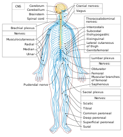
The human nervous system. Blue is PNS; yellow is CNS.
Identifiers
Acronym(s)PNS
MeSHD017933
TAA14.2.00.001
FMA9903

The peripheral nervous system (PNS) is one of two components that make up the nervous system of bilateral animals, with the other part being the central nervous system (CNS). The PNS consists of the nerves and ganglia outside the brain and spinal cord. The main function of the PNS is to connect the CNS to the limbs and organs, essentially serving as a relay between the brain and spinal cord and the rest of the body. Unlike the CNS, the PNS is not protected by the vertebral column and skull, or by the blood–brain barrier, which leaves it exposed to toxins and mechanical injuries.

The peripheral nervous system is divided into the somatic nervous system and the autonomic nervous system. In the somatic nervous system, the cranial nerves are part of the PNS with the exception of the optic nerve (cranial nerve II), along with the retina. The second cranial nerve is not a true peripheral nerve but a tract of the diencephalon. Cranial nerve ganglia originated in the CNS. However, the remaining ten cranial nerve axons extend beyond the brain and are therefore considered part of the PNS. The autonomic nervous system exerts involuntary control over smooth muscle and glands. The connection between CNS and organs allows the system to be in two different functional states: sympathetic and parasympathetic.

Structure

The peripheral nervous system is divided into the somatic nervous system, and the autonomic nervous system. The somatic nervous system is under voluntary control, and transmits signals from the brain to end organs such as muscles. The sensory nervous system is part of the somatic nervous system and transmits signals from senses such as taste and touch (including fine touch and gross touch) to the spinal cord and brain. The autonomic nervous system is a 'self-regulating' system which influences the function of organs outside voluntary control, such as the heart rate, or the functions of the digestive system.

Somatic nervous system

The somatic system includes the sensory nervous system and the somatosensory system and consists of sensory nerves and somatic nerves, and many nerves which hold both functions. 

In the head and neck, cranial nerves carry somatosensory data. There are twelve cranial nerves, ten of which originate from the brainstem, and mainly control the functions of the anatomic structures of the head with some exceptions. One unique cranial nerve is the vagus nerve, which receives sensory information from organs in the thorax and abdomen. The accessory nerve is responsible for innervating the sternocleidomastoid and trapezius muscles, neither of which being exclusively in the head.

For the rest of the body, spinal nerves are responsible for somatosensory information. These arise from the spinal cord. Usually these arise as a web ("plexus") of interconnected nerves roots that arrange to form single nerves. These nerves control the functions of the rest of the body. In humans, there are 31 pairs of spinal nerves: 8 cervical, 12 thoracic, 5 lumbar, 5 sacral, and 1 coccygeal. These nerve roots are named according to the spinal vertebrata which they are adjacent to. In the cervical region, the spinal nerve roots come out above the corresponding vertebrae (i.e., nerve root between the skull and 1st cervical vertebrae is called spinal nerve C1). From the thoracic region to the coccygeal region, the spinal nerve roots come out below the corresponding vertebrae. It is important to note that this method creates a problem when naming the spinal nerve root between C7 and T1 (so it is called spinal nerve root C8). In the lumbar and sacral region, the spinal nerve roots travel within the dural sac and they travel below the level of L2 as the cauda equina.

Cervical spinal nerves (C1–C4)

The first 4 cervical spinal nerves, C1 through C4, split and recombine to produce a variety of nerves that serve the neck and back of head.

Spinal nerve C1 is called the suboccipital nerve, which provides motor innervation to muscles at the base of the skull. C2 and C3 form many of the nerves of the neck, providing both sensory and motor control. These include the greater occipital nerve, which provides sensation to the back of the head, the lesser occipital nerve, which provides sensation to the area behind the ears, the greater auricular nerve and the lesser auricular nerve

The phrenic nerve is a nerve essential for our survival which arises from nerve roots C3, C4 and C5. It supplies the thoracic diaphragm, enabling breathing. If the spinal cord is transected above C3, then spontaneous breathing is not possible.

Brachial plexus (C5–T1)

The last four cervical spinal nerves, C5 through C8, and the first thoracic spinal nerve, T1, combine to form the brachial plexus, or plexus brachialis, a tangled array of nerves, splitting, combining and recombining, to form the nerves that subserve the upper-limb and upper back. Although the brachial plexus may appear tangled, it is highly organized and predictable, with little variation between people. See brachial plexus injuries.

Lumbosacral plexus (L1–Co1)

The anterior divisions of the lumbar nerves, sacral nerves, and coccygeal nerve form the lumbosacral plexus, the first lumbar nerve being frequently joined by a branch from the twelfth thoracic. For descriptive purposes this plexus is usually divided into three parts:

Autonomic nervous system

The autonomic nervous system (ANS) controls involuntary responses to regulate physiological functions. The brain and spinal cord of the central nervous system are connected with organs that have smooth muscle, such as the heart, bladder, and other cardiac, exocrine, and endocrine related organs, by ganglionic neurons. The most notable physiological effects from autonomic activity are pupil constriction and dilation, and salivation of saliva. The autonomic nervous system is always activated, but is either in the sympathetic or parasympathetic state. Depending on the situation, one state can overshadow the other, resulting in a release of different kinds of neurotransmitters. There is a lesser known division of the autonomic nervous system known as the enteric nervous system. Located only around the digestive tract, this system allows for local control without input from the sympathetic or the parasympathetic branches, though it can still receive and respond to signals from the rest of the body. The enteric system is responsible for various functions related to gastrointestinal system.

Sympathetic nervous system

The sympathetic system is activated during a “fight or flight” situation in which mental stress or physical danger is encountered. Neurotransmitters such as norepinephrine, and epinephrine are released, which increases heart rate and blood flow in certain areas like muscle, while simultaneously decreasing activities of non-critical functions for survival, like digestion. The systems are independent to each other, which allows activation of certain parts of the body, while others remain rested.

Parasympathetic nervous system

Primarily using the neurotransmitter acetylcholine (ACh) as a mediator, the parasympathetic system allows the body to function in a “rest and digest” state. Consequently, when the parasympathetic system dominates the body, there are increases in salivation and activities in digestion, while heart rate and other sympathetic response decrease. Unlike the sympathetic system, humans have some voluntary controls in the parasympathetic system. The most prominent examples of this control are urination and defecation.

Disease

Diseases of the peripheral nervous system can be specific to one or more nerves, or affect the system as a whole.

Any peripheral nerve or nerve root can be damaged, called a mononeuropathy. Such injuries can be because of injury or trauma, or compression. Compression of nerves can occur because of a tumour mass or injury. Alternatively, if a nerve is in an area with a fixed size it may be trapped if the other components increase in size, such as carpal tunnel syndrome and tarsal tunnel syndrome. Common symptoms of carpal tunnel syndrome include pain and numbness in the thumb, index and middle finger. In peripheral neuropathy, the function one or more nerves are damaged through a variety of means. Toxic damage may occur because of diabetes (diabetic neuropathy), alcohol, heavy metals or other toxins; some infections; autoimmune and inflammatory conditions such as amyloidosis and sarcoidosis. Peripheral neuropathy is associated with a sensory loss in a "glove and stocking" distribution that begins at the peripheral and slowly progresses upwards, and may also be associated with acute and chronic pain. Peripheral neuropathy is not just limited to the somatosensory nerves, but the autonomic nervous system too (autonomic neuropathy).
Acetyl-CoA

From Wikipedia, the free encyclopedia
 
Acetyl-CoA
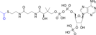
Acetyl-CoA-2D colored.svg
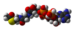
Acetyl-CoA-3D-vdW.png
Names
IUPAC name
S-[2-[3-[[(2R)-4-[[[(2R,3S,4R,5R)-5-(6-aminopurin-9-yl)-4-hydroxy-3-phosphonooxyoxolan-2-yl]methoxy-hydroxyphosphoryl]oxy-hydroxyphosphoryl]oxy-2-hydroxy-3,3-dimethylbutanoyl]amino]propanoylamino]ethyl] ethanethioate
Identifiers
3D model (JSmol)
ChEBI
ChemSpider
ECHA InfoCard 100.000.719
KEGG
MeSH Acetyl+Coenzyme+A
PubChem CID
Properties
C23H38N7O17P3S
Molar mass 809.57 g·mol−1
Except where otherwise noted, data are given for materials in their standard state (at 25 °C [77 °F], 100 kPa).

Acetyl-CoA (acetyl coenzyme A) is a molecule that participates in many biochemical reactions in protein, carbohydrate and lipid metabolism. Its main function is to deliver the acetyl group to the citric acid cycle (Krebs cycle) to be oxidized for energy production. Coenzyme A (CoASH or CoA) consists of a β-mercaptoethylamine group linked to the vitamin pantothenic acid through an amide linkage  and 3'-phosphorylated ADP. The acetyl group (indicated in blue in the structural diagram on the right) of acetyl-CoA is linked to the sulfhydryl substituent of the β-mercaptoethylamine group. This thioester linkage is a "high energy" bond, which is particularly reactive. Hydrolysis of the thioester bond is exergonic (−31.5 kJ/mol).

CoA is acetylated to acetyl-CoA by the breakdown of carbohydrates through glycolysis and by the breakdown of fatty acids through β-oxidation. Acetyl-CoA then enters the citric acid cycle, where the acetyl group is oxidized to carbon dioxide and water, and the energy released is captured in the form of 11 ATP and one GTP per acetyl group.

Konrad Bloch and Feodor Lynen were awarded the 1964 Nobel Prize in Physiology and Medicine for their discoveries linking acetyl-CoA and fatty acid metabolism. Fritz Lipmann won the Nobel Prize in 1953 for his discovery of the cofactor coenzyme A.

Direct synthesis

The acetylation of CoA is determined by the carbon sources.

Extramitochondrial

Intramitochondrial

Pyruvate dehydrogenase complex reaction
  • At high glucose levels, acetyl-CoA is produced through glycolysis. Pyruvate undergoes oxidative decarboxylation in which it loses its carboxyl group (as carbon dioxide) to form acetyl-CoA, giving off 33.5 kJ/mol of energy. The oxidative conversion of pyruvate into acetyl-CoA is referred to as the pyruvate dehydrogenase reaction. It is catalyzed by the pyruvate dehydrogenase complex. Other conversions between pyruvate and acetyl-CoA are possible. For example, pyruvate formate lyase disproportionates pyruvate into acetyl-CoA and formic acid.

Functions

Intermediates in various pathways

Pyruvic acid

From Wikipedia, the free encyclopedia
 
Pyruvic acid
Brenztraubensäure.svg
Pyruvic-acid-3D-balls.png
Names
Preferred IUPAC name
2-Oxopropanoic acid
Other names
Pyruvic acid
α-Ketopropionic acid
Acetylformic acid
Pyroracemic acid
Identifiers
3D model (JSmol)
Abbreviations Pyr
ChEBI
ChEMBL
ChemSpider
DrugBank
ECHA InfoCard 100.004.387
KEGG
PubChem CID
UNII
Properties
C3H4O3
Molar mass 88.06 g/mol
Density 1.250 g/cm3
Melting point 11.8 °C (53.2 °F; 284.9 K)
Boiling point 165 °C (329 °F; 438 K)
Acidity (pKa) 2.50
Related compounds
Other anions
pyruvate ion Pyruvate skeletal.svg
Pyruvate-3D-balls.png
acetic acid
glyoxylic acid
oxalic acid
propionic acid
acetoacetic acid
Related compounds
propionaldehyde
glyceraldehyde
methylglyoxal
sodium pyruvate
Except where otherwise noted, data are given for materials in their standard state (at 25 °C [77 °F], 100 kPa).

Pyruvic acid (CH3COCOOH) is the simplest of the alpha-keto acids, with a carboxylic acid and a ketone functional group. Pyruvate (/pˈrvt/), the conjugate base, CH3COCOO, is a key intermediate in several metabolic pathways throughout the cell.

Pyruvic acid can be made from glucose through glycolysis, converted back to carbohydrates (such as glucose) via gluconeogenesis, or to fatty acids through a reaction with acetyl-CoA. It can also be used to construct the amino acid alanine and can be converted into ethanol or lactic acid via fermentation.

Pyruvic acid supplies energy to cells through the citric acid cycle (also known as the Krebs cycle) when oxygen is present (aerobic respiration), and alternatively ferments to produce lactate when oxygen is lacking (lactic acid fermentation).

Chemistry

In 1834, Théophile-Jules Pelouze distilled tartaric acid and isolated glutaric acid and another unknown organic acid. Jöns Jacob Berzelius characterized this other acid the following year and named pyruvic acid because it was distilled using heat. Pyruvic acid is a colorless liquid with a smell similar to that of acetic acid and is miscible with water. In the laboratory, pyruvic acid may be prepared by heating a mixture of tartaric acid and potassium hydrogen sulfate, by the oxidation of propylene glycol by a strong oxidizer (e.g., potassium permanganate or bleach), or by the hydrolysis of acetyl cyanide, formed by reaction of acetyl chloride with potassium cyanide
CH3COCl + KCN → CH3COCN + KCl
CH3COCN → CH3COCOOH

Biochemistry

Pyruvate is an important chemical compound in biochemistry. It is the output of the metabolism of glucose known as glycolysis. One molecule of glucose breaks down into two molecules of pyruvate, which are then used to provide further energy, in one of two ways. Pyruvate is converted into acetyl-coenzyme A, which is the main input for a series of reactions known as the Krebs cycle (also known as the citric acid cycle or tricarboxylic acid cycle). Pyruvate is also converted to oxaloacetate by an anaplerotic reaction, which replenishes Krebs cycle intermediates; also, the oxaloacetate is used for gluconeogenesis. These reactions are named after Hans Adolf Krebs, the biochemist awarded the 1953 Nobel Prize for physiology, jointly with Fritz Lipmann, for research into metabolic processes. The cycle is also known as the citric acid cycle or tricarboxylic acid cycle, because citric acid is one of the intermediate compounds formed during the reactions. 

If insufficient oxygen is available, the acid is broken down anaerobically, creating lactate in animals and ethanol in plants and microorganisms (and carp). Pyruvate from glycolysis is converted by fermentation to lactate using the enzyme lactate dehydrogenase and the coenzyme NADH in lactate fermentation, or to acetaldehyde (with the enzyme pyruvate decarboxylase) and then to ethanol in alcoholic fermentation.

Pyruvate is a key intersection in the network of metabolic pathways. Pyruvate can be converted into carbohydrates via gluconeogenesis, to fatty acids or energy through acetyl-CoA, to the amino acid alanine, and to ethanol. Therefore, it unites several key metabolic processes.

Reference ranges for blood tests, comparing blood content of pyruvate (shown in violet near middle) with other constituents.

Pyruvic acid production by glycolysis

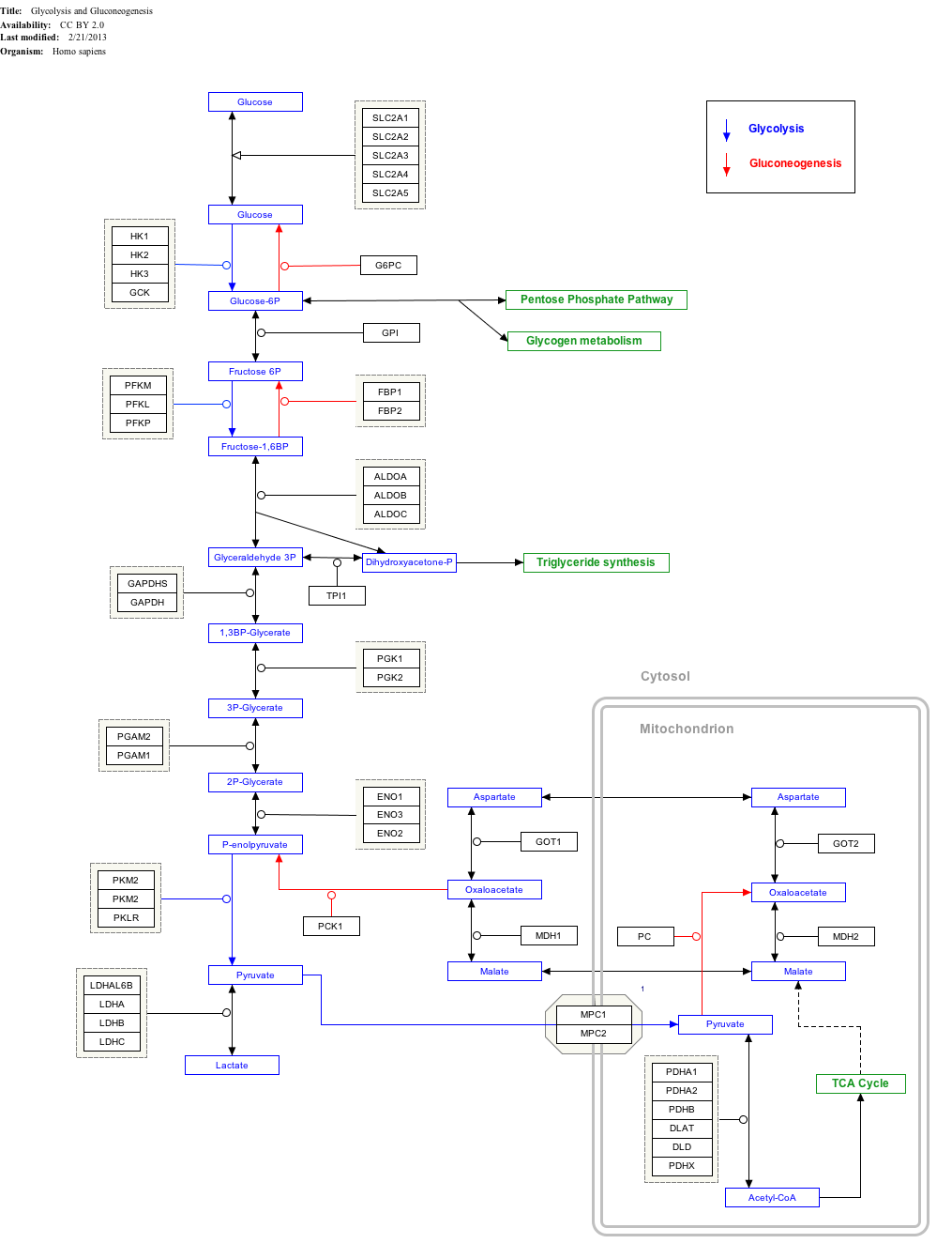
In glycolysis, phosphoenolpyruvate (PEP) is converted to pyruvate by pyruvate kinase. This reaction is strongly exergonic and irreversible; in gluconeogenesis, it takes two enzymes, pyruvate carboxylase and PEP carboxykinase, to catalyze the reverse transformation of pyruvate to PEP. 

phosphoenolpyruvate pyruvate kinase pyruvate
Phosphoenolpyruvate wpmp.svg   Pyruvic-acid-2D-skeletal.svg
ADP ATP
Biochem reaction arrow reversible YYYY horiz med.svg
ADP ATP
 
  pyruvate carboxylase and PEP carboxykinase
Compound C00074 at KEGG Pathway Database. Enzyme 2.7.1.40 at KEGG Pathway Database. Compound C00022 at KEGG Pathway Database.
[[File:
GlycolysisGluconeogenesis_WP534go to articlego to articlego to articlego to articlego to articlego to articlego to articlego to articlego to articlego to articlego to articlego to articlego to articlego to articlego to articlego to articlego to articlego to articlego to articlego to articlego to articlego to articlego to articlego to articlego to articlego to articlego to articlego to articlego to articlego to articlego to articlego to articlego to articlego to articlego to articlego to articlego to articlego to articlego to articlego to articlego to articlego to articlego to articlego to articlego to articlego to articlego to articlego to articlego to articlego to articlego to articlego to articlego to articlego to articlego to articlego to articlego to articlego to articlego to articlego to articlego to articlego to articlego to articlego to articlego to articlego to articlego to articlego to articlego to articlego to articlego to WikiPathwaysgo to articlego to Entrezgo to article
[[]]
[[]]
[[]]
[[]]
[[]]
[[]]
[[]]
[[]]
[[]]
[[]]
[[]]
[[]]
[[]]
[[]]
[[]]
[[]]
[[]]
[[]]
[[]]
[[]]
[[]]
[[]]
[[]]
[[]]
[[]]
[[]]
[[]]
[[]]
[[]]
[[]]
[[]]
[[]]
[[]]
[[]]
[[]]
[[]]
[[]]
[[]]
[[]]
[[]]
[[]]
[[]]
[[]]
[[]]
[[]]
[[]]
[[]]
[[]]
[[]]
[[]]
[[]]
[[]]
[[]]
[[]]
[[]]
[[]]
[[]]
[[]]
[[]]
[[]]
[[]]
[[]]
[[]]
[[]]
[[]]
[[]]
[[]]
[[]]
[[]]
[[]]
[[]]
[[]]
[[]]
[[]]
[[]]
GlycolysisGluconeogenesis_WP534
|{{{bSize}}}px|alt=Glycolysis and Gluconeogenesis edit]]
Glycolysis and Gluconeogenesis

Decarboxylation to acetyl CoA


pyruvate pyruvate dehydrogenase complex acetyl-CoA
Pyruvate wpmp.png   Acetyl-CoA.svg
CoA + NAD+ CO2 + NADH + H+
Biochem reaction arrow forward YYNN horiz med.svg


Carboxylation to oxaloacetate

Carboxylation by pyruvate carboxylase produces oxaloacetate

pyruvate pyruvate carboxylase oxaloacetate
Pyruvate wpmp.png   Oxaloacetate wpmp.png
ATP + CO2 ADP + Pi
Biochem reaction arrow forward YYNN horiz med.svg


Transamination to alanine

Transamination by alanine transaminase produces alanine

pyruvate alanine transaminase alanine
Pyruvate wpmp.png   L-alanine-skeletal.svg
glutamate α-ketoglutarate
Biochem reaction arrow reversible YYYY horiz med.svg
glutamate α-ketoglutarate


Reduction to lactate

Reduction by lactate dehydrogenase produces lactate

pyruvate lactate dehydrogenase lactate
Pyruvate wpmp.png   Lactic-acid-skeletal.svg
NADH NAD+
Biochem reaction arrow reversible YYYY horiz med.svg
NADH NAD+


Uses

Pyruvate is sold as a weight-loss supplement, though credible science has yet to back this claim. A systematic review of six trials found a statistically significant difference in body weight with pyruvate compared to placebo. However, all of the trials had methodological weaknesses and the magnitude of the effect was small. The review also identified adverse events associated with pyruvate such as diarrhea, bloating, gas, and increase in low-density lipoprotein (LDL) cholesterol. The authors concluded that there was insufficient evidence to support the use of pyruvate for weight loss.

There is also in vitro as well as in vivo evidence in hearts that pyruvate improves metabolism by NADH production stimulation and increases cardiac function.

Butane

From Wikipedia, the free encyclopedia ...