Search This Blog

Tuesday, November 26, 2019

Estradiol

From Wikipedia, the free encyclopedia
 
Estradiol
The chemical structure of estradiol.
A ball-and-stick model of estradiol.
Names
Pronunciation /ˌɛstrəˈdl/ ES-trə-DY-ohl
IUPAC name
(8R,9S,13S,14S,17S)-13-Methyl-6,7,8,9,11,12,14,15,16,17-decahydrocyclopenta[a]phenanthrene-3,17-diol
Other names
Oestradiol; E2; 17β-Estradiol; Estra-1,3,5(10)-triene-3,17β-diol; 17β-Oestradiol
Identifiers
3D model (JSmol)
ChEBI
ChEMBL
ChemSpider
DrugBank
ECHA InfoCard 100.000.022
KEGG
PubChem CID
UNII
Properties
C18H24O2
Molar mass 272.38 g/mol
-186.6·10−6 cm3/mol
Pharmacology
G03CA03 (WHO)
License data
Oral, sublingual, intranasal, topical/transdermal, vaginal, intramuscular or subcutaneous (as an ester), subdermal implant
Pharmacokinetics:
Oral: <5 span="">
~98%:• Albumin: 60%
SHBG: 38%
• Free: 2%
Liver (via hydroxylation, sulfation, glucuronidation)
Oral: 13–20 hours
Sublingual: 8–18 hours
Topical (gel): 36.5 hours
Urine: 54%
Feces: 6%
Except where otherwise noted, data are given for materials in their standard state (at 25 °C [77 °F], 100 kPa).

Estradiol (E2), also spelled oestradiol, is an estrogen steroid hormone and the major female sex hormone. It is involved in the regulation of the estrous and menstrual female reproductive cycles. Estradiol is responsible for the development of female secondary sexual characteristics such as the breasts, widening of the hips, and a female-associated pattern of fat distribution and is important in the development and maintenance of female reproductive tissues such as the mammary glands, uterus, and vagina during puberty, adulthood, and pregnancy. It also has important effects in many other tissues including bone, fat, skin, liver, and the brain. Though estradiol levels in males are much lower compared to those in females, estradiol has important roles in males as well. Apart from humans and other mammals, estradiol is also found in most vertebrates and crustaceans, insects, fish, and other animal species.

Estradiol is produced especially within the follicles of the ovaries, but also in other tissues including the testicles, the adrenal glands, fat, liver, the breasts, and the brain. Estradiol is produced in the body from cholesterol through a series of reactions and intermediates. The major pathway involves the formation of androstenedione, which is then converted by aromatase into estrone and is subsequently converted into estradiol. Alternatively, androstenedione can be converted into testosterone, which can then be converted into estradiol. Upon menopause in females, production of estrogens by the ovaries stops and estradiol levels decrease to very low levels.

In addition to its role as a natural hormone, estradiol is used as a medication, for instance in menopausal hormone therapy; for information on estradiol as a medication, see the estradiol (medication) article.

Biological function

Sexual development

The development of secondary sex characteristics in women is driven by estrogens, to be specific, estradiol. These changes are initiated at the time of puberty, most are enhanced during the reproductive years, and become less pronounced with declining estradiol support after menopause. Thus, estradiol produces breast development, and is responsible for changes in the body shape, affecting bones, joints, and fat deposition. In females, estradiol induces breast development, widening of the hips, a feminine fat distribution (with fat deposited particularly in the breasts, hips, thighs, and buttocks), and maturation of the vagina and vulva, whereas it mediates the pubertal growth spurt (indirectly via increased growth hormone secretion) and epiphyseal closure (thereby limiting final height) in both sexes.

Reproduction

Female reproductive system

In the female, estradiol acts as a growth hormone for tissue of the reproductive organs, supporting the lining of the vagina, the cervical glands, the endometrium, and the lining of the fallopian tubes. It enhances growth of the myometrium. Estradiol appears necessary to maintain oocytes in the ovary. During the menstrual cycle, estradiol produced by the growing follicles triggers, via a positive feedback system, the hypothalamic-pituitary events that lead to the luteinizing hormone surge, inducing ovulation. In the luteal phase, estradiol, in conjunction with progesterone, prepares the endometrium for implantation. During pregnancy, estradiol increases due to placental production. The effect of estradiol, together with estrone and estriol, in pregnancy is less clear. They may promote uterine blood flow, myometrial growth, stimulate breast growth and at term, promote cervical softening and expression of myometrial oxytocin receptors. In baboons, blocking of estrogen production leads to pregnancy loss, suggesting estradiol has a role in the maintenance of pregnancy. Research is investigating the role of estrogens in the process of initiation of labor. Actions of estradiol are required before the exposure of progesterone in the luteal phase.

Male reproductive system

The effect of estradiol (and estrogens in general) upon male reproduction is complex. Estradiol is produced by action of aromatase mainly in the Leydig cells of the mammalian testis, but also by some germ cells and the Sertoli cells of immature mammals. It functions (in vitro) to prevent apoptosis of male sperm cells. While some studies in the early 1990s claimed a connection between globally declining sperm counts and estrogen exposure in the environment, later studies found no such connection, nor evidence of a general decline in sperm counts. Suppression of estradiol production in a subpopulation of subfertile men may improve the semen analysis.

Males with certain sex chromosome genetic conditions, such as Klinefelter's syndrome, will have a higher level of estradiol.

Skeletal system

Estradiol has a profound effect on bone. Individuals without it (or other estrogens) will become tall and eunuchoid, as epiphyseal closure is delayed or may not take place. Low levels of estradiol may also predict fractures, with the highest risk occurring particularly in men with low total and high sex hormone binding globulin protein. Bone density, as well as joints, are also affected, resulting in early osteopenia and osteoporosis. Women past menopause experience an accelerated loss of bone mass due to a relative estrogen deficiency.

Skin health

The estrogen receptor, as well as the progesterone receptor, have been detected in the skin, including in keratinocytes and fibroblasts. At menopause and thereafter, decreased levels of female sex hormones result in atrophy, thinning, and increased wrinkling of the skin and a reduction in skin elasticity, firmness, and strength. These skin changes constitute an acceleration in skin aging and are the result of decreased collagen content, irregularities in the morphology of epidermal skin cells, decreased ground substance between skin fibers, and reduced capillaries and blood flow. The skin also becomes more dry during menopause, which is due to reduced skin hydration and surface lipids (sebum production). Along with chronological aging and photoaging, estrogen deficiency in menopause is one of the three main factors that predominantly influences skin aging.

Hormone replacement therapy consisting of systemic treatment with estrogen alone or in combination with a progestogen, has well-documented and considerable beneficial effects on the skin of postmenopausal women. These benefits include increased skin collagen content, skin thickness and elasticity, and skin hydration and surface lipids. Topical estrogen has been found to have similar beneficial effects on the skin. In addition, a study has found that topical 2% progesterone cream significantly increases skin elasticity and firmness and observably decreases wrinkles in peri- and postmenopausal women. Skin hydration and surface lipids, on the other hand, did not significantly change with topical progesterone. These findings suggest that progesterone, like estrogen, also has beneficial effects on the skin, and may be independently protective against skin aging.

Nervous system

Estrogens can be produced in the brain from steroid precursors. As antioxidants, they have been found to have neuroprotective function.

The positive and negative feedback loops of the menstrual cycle involve ovarian estradiol as the link to the hypothalamic-pituitary system to regulate gonadotropins.

Estrogen is considered to play a significant role in women's mental health, with links suggested between the hormone level, mood and well-being. Sudden drops or fluctuations in, or long periods of sustained low levels of estrogen may be correlated with significant mood-lowering. Clinical recovery from depression postpartum, perimenopause, and postmenopause was shown to be effective after levels of estrogen were stabilized and/or restored.

Recently, the volumes of sexually dimorphic brain structures in transgender women were found to change and approximate typical female brain structures when exposed to estrogen concomitantly with androgen deprivation over a period of months, suggesting that estrogen and/or androgens have a significant part to play in sex differentiation of the brain, both prenatally and later in life.

There is also evidence the programming of adult male sexual behavior in many vertebrates is largely dependent on estradiol produced during prenatal life and early infancy. It is not yet known whether this process plays a significant role in human sexual behavior, although evidence from other mammals tends to indicate a connection.

Estrogen has been found to increase the secretion of oxytocin and to increase the expression of its receptor, the oxytocin receptor, in the brain. In women, a single dose of estradiol has been found to be sufficient to increase circulating oxytocin concentrations.

Gynecological cancers

Estradiol has been tied to the development and progression of cancers such as breast cancer, ovarian cancer and endometrial cancer. Estradiol affects target tissues mainly by interacting with two nuclear receptors called estrogen receptor α (ERα) and estrogen receptor β (ERβ). One of the functions of these estrogen receptors is the modulation of gene expression. Once estradiol binds to the ERs, the receptor complexes then bind to specific DNA sequences, possibly causing damage to the DNA and an increase in cell division and DNA replication. Eukaryotic cells respond to damaged DNA by stimulating or impairing G1, S, or G2 phases of the cell cycle to initiate DNA repair. As a result, cellular transformation and cancer cell proliferation occurs.

Other functions

Estradiol has complex effects on the liver. It affects the production of multiple proteins, including lipoproteins, binding proteins, and proteins responsible for blood clotting. In high amounts, estradiol can lead to cholestasis, for instance cholestasis of pregnancy

Certain gynecological conditions are dependent on estrogen, such as endometriosis, leiomyomata uteri, and uterine bleeding.

Estrogen affects certain blood vessels. Improvement in arterial blood flow has been demonstrated in coronary arteries.

Biological activity

Estradiol acts primarily as an agonist of the estrogen receptor (ER), a nuclear steroid hormone receptor. There are two subtypes of the ER, ERα and ERβ, and estradiol potently binds to and activates both of these receptors. The result of ER activation is a modulation of gene transcription and expression in ER-expressing cells, which is the predominant mechanism by which estradiol mediates its biological effects in the body. Estradiol also acts as an agonist of membrane estrogen receptors (mERs), such as GPER (GPR30), a recently discovered non-nuclear receptor for estradiol, via which it can mediate a variety of rapid, non-genomic effects. Unlike the case of the ER, GPER appears to be selective for estradiol, and shows very low affinities for other endogenous estrogens, such as estrone and estriol.[40] Additional mERs besides GPER include ER-X, ERx, and Gq-mER.

ERα/ERβ are in inactive state trapped in multimolecular chaperone complexes organized around the heat shock protein 90 (HSP90), containing p23 protein, and immunophilin, and located in majority in cytoplasm and partially in nucleus. In the E2 classical pathway or estrogen classical pathway, estradiol enters the cytoplasm, where it interacts with ERs. Once bound E2, ERs dissociate from the molecular chaperone complexes and become competent to dimerize, migrate to nucleus, and to bind to specific DNA sequences (estrogen response element, ERE), allowing for gene transcription which can take place over hours and days. 

Given by subcutaneous injection in mice, estradiol is about 10-fold more potent than estrone and about 100-fold more potent than estriol. As such, estradiol is the main estrogen in the body, although the roles of estrone and estriol as estrogens are said not to be negligible.

Biochemistry

Human steroidogenesis, showing estradiol at bottom right.

Biosynthesis

Estradiol, like other steroid hormones, is derived from cholesterol. After side chain cleavage and using the Δ5 or the Δ4- pathway, androstenedione is the key intermediary. A portion of the androstenedione is converted to testosterone, which in turn undergoes conversion to estradiol by aromatase. In an alternative pathway, androstenedione is aromatized to estrone, which is subsequently converted to estradiol via 17β-hydroxysteroid dehydrogenase (17β-HSD).

During the reproductive years, most estradiol in women is produced by the granulosa cells of the ovaries by the aromatization of androstenedione (produced in the theca folliculi cells) to estrone, followed by conversion of estrone to estradiol by 17β-HSD. Smaller amounts of estradiol are also produced by the adrenal cortex, and, in men, by the testes.

Estradiol is not produced in the gonads only, in particular, fat cells produce active precursors to estradiol, and will continue to do so even after menopause. Estradiol is also produced in the brain and in arterial walls

In men, approximately 15 to 25% of circulating estradiol is produced in the testicles. The rest is synthesized via peripheral aromatization of testosterone into estradiol and of androstenedione into estrone (which is then transformed into estradiol via peripheral 17β-HSD). This peripheral aromatization occurs predominantly in adipose tissue, but also occurs in other tissues such as bone, liver, and the brain. Approximately 40 to 50 µg of estradiol is produced per day in men.

Distribution

In plasma, estradiol is largely bound to SHBG, and also to albumin. Only a fraction of 2.21% (± 0.04%) is free and biologically active, the percentage remaining constant throughout the menstrual cycle.

Metabolism

Inactivation of estradiol includes conversion to less-active estrogens, such as estrone and estriol. Estriol is the major urinary metabolite. Estradiol is conjugated in the liver to form estrogen conjugates like estradiol sulfate, estradiol glucuronide and, as such, excreted via the kidneys. Some of the water-soluble conjugates are excreted via the bile duct, and partly reabsorbed after hydrolysis from the intestinal tract. This enterohepatic circulation contributes to maintaining estradiol levels. 

Estradiol is also metabolized via hydroxylation into catechol estrogens. In the liver, it is non-specifically metabolized by CYP1A2, CYP3A4, and CYP2C9 via 2-hydroxylation into 2-hydroxyestradiol, and by CYP2C9, CYP2C19, and CYP2C8 via 17β-hydroxy dehydrogenation into estrone, with various other cytochrome P450 (CYP) enzymes and metabolic transformations also being involved.

Estradiol is additionally conjugated with an ester into lipoidal estradiol forms like estradiol palmitate and estradiol stearate to a certain extent; these esters are stored in adipose tissue and may act as a very long-lasting reservoir of estradiol.[57][58]

Excretion

Estradiol is excreted in the form of glucuronide and sulfate estrogen conjugates in urine.

Levels

Estradiol levels across the menstrual cycle in 36 normally cycling, ovulatory women, based on 956 specimens. The horizontal dashed lines are the mean integrated levels for each curve. The vertical dashed line in the center is mid-cycle.
 
Levels of estradiol in premenopausal women are highly variable throughout the menstrual cycle and reference ranges widely vary from source to source. Estradiol levels are minimal and according to most laboratories range from 20 to 80 pg/mL during the early to mid follicular phase (or the first week of the menstrual cycle, also known as menses). Levels of estradiol gradually increase during this time and through the mid to late follicular phase (or the second week of the menstrual cycle) until the pre-ovulatory phase. At the time of pre-ovulation (a period of about 24 to 48 hours), estradiol levels briefly surge and reach their highest concentrations of any other time during the menstrual cycle. Circulating levels are typically between 130 and 200 pg/mL at this time, but in some women may be as high as 300 to 400 pg/mL, and the upper limit of the reference range of some laboratories are even greater (for instance, 750 pg/mL). Following ovulation (or mid-cycle) and during the latter half of the menstrual cycle or the luteal phase, estradiol levels plateau and fluctuate between around 100 and 150 pg/mL during the early and mid luteal phase, and at the time of the late luteal phase, or a few days before menstruation, reach a low of around 40 pg/mL. The mean integrated levels of estradiol during a full menstrual cycle have variously been reported by different sources as 80, 120, and 150 pg/mL. Although contradictory reports exist, one study found mean integrated estradiol levels of 150 pg/mL in younger women whereas mean integrated levels ranged from 50 to 120 pg/mL in older women.

During the reproductive years of the human female, levels of estradiol are somewhat higher than that of estrone, except during the early follicular phase of the menstrual cycle; thus, estradiol may be considered the predominant estrogen during human female reproductive years in terms of absolute serum levels and estrogenic activity. During pregnancy, estriol becomes the predominant circulating estrogen, and this is the only time at which estetrol occurs in the body, while during menopause, estrone predominates (both based on serum levels). The estradiol produced by male humans, from testosterone, is present at serum levels roughly comparable to those of postmenopausal women (14–55 versus <35 2-="" 4-fold="" 70-year-old="" a="" also="" approximately="" are="" been="" compared="" concentrations="" estradiol="" has="" higher="" if="" in="" it="" levels="" man.="" man="" ml="" of="" p="" pg="" reported="" respectively="" that="" the="" those="" to="" woman="">

Measurement

In women, serum estradiol is measured in a clinical laboratory and reflects primarily the activity of the ovaries. The Estradiol blood test measures the amount of estradiol in the blood. It is used to check the function of the ovaries, placenta, adrenal glands. This can detect baseline estrogen in women with amenorrhea or menstrual dysfunction, and to detect the state of hypoestrogenicity and menopause. Furthermore, estrogen monitoring during fertility therapy assesses follicular growth and is useful in monitoring the treatment. Estrogen-producing tumors will demonstrate persistent high levels of estradiol and other estrogens. In precocious puberty, estradiol levels are inappropriately increased.

Ranges

Individual laboratory results should always been interpreted using the ranges provided by the laboratory that performed the test.

Reference ranges for the blood content of estradiol during the menstrual cycle

In the normal menstrual cycle, estradiol levels measure typically <50 200="" a="" again="" and="" at="" briefly="" development="" drop="" during="" end="" estradiol="" follicular="" for="" is="" levels="" luteal="" menstrual="" menstruation="" ml="" nbsp="" of="" ovulation="" p="" peak.="" peak:="" pg="" phase="" pregnancy.="" rise="" second="" the="" their="" there="" to="" unless="" with="">

During pregnancy, estrogen levels, including estradiol, rise steadily toward term. The source of these estrogens is the placenta, which aromatizes prohormones produced in the fetal adrenal gland.

Medical use

Estradiol is used as a medication, primarily in hormone therapy for menopausal symptoms as well as transgender hormone replacement therapy.

Chemistry

Structures of major endogenous estrogens
Chemical structures of major endogenous estrogens
Estrone (E1)
Estradiol (E2)
Estriol (E3)
Estetrol (E4)
Note the hydroxyl (–OH) groups: estrone (E1) has one, estradiol (E2) has two, estriol (E3) has three, and estetrol
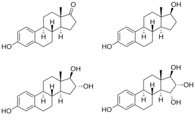
Estradiol is an estrane steroid. It is also known as 17β-estradiol (to distinguish it from 17α-estradiol) or as estra-1,3,5(10)-triene-3,17β-diol. It has two hydroxyl groups, one at the C3 position and the other at the 17β position, as well as three double bonds in the A ring. Due to its two hydroxyl groups, estradiol is often abbreviated as E2. The structurally related estrogens, estrone (E1), estriol (E3), and estetrol (E4) have one, three, and four hydroxyl groups, respectively.

History

The discovery of estrogen is usually  credited to the American scientists Edgar Allen and Edward A. Doisy. In 1923, they observed that injection of fluid from porcine ovarian follicles produced pubertal- and estrus-type changes (including vaginal, uterine, and mammary gland changes and sexual receptivity) in sexually immature, ovariectomized mice and rats. These findings demonstrated the existence of a hormone which is produced by the ovaries and is involved in sexual maturation and reproduction. At the time of its discovery, Allen and Doisy did not name the hormone, and simply referred to it as an "ovarian hormone" or "follicular hormone"; others referred to it variously as feminin, folliculin, menformon, thelykinin, and emmenin. In 1926, Parkes and Bellerby coined the term estrin to describe the hormone on the basis of it inducing estrus in animals. Estrone was isolated and purified independently by Allen and Doisy and German scientist Adolf Butenandt in 1929, and estriol was isolated and purified by Marrian in 1930; they were the first estrogens to be identified.

Estradiol, the most potent of the three major estrogens, was the last of the three to be identified. It was discovered by Schwenk and Hildebrant in 1933, who synthesized it via reduction of estrone. Estradiol was subsequently isolated and purified from sow ovaries by Doisy in 1935, with its chemical structure determined simultaneously, and was referred to variously as dihydrotheelin, dihydrofolliculin, dihydrofollicular hormone, and dihydroxyestrin. In 1935, the name estradiol and the term estrogen were formally established by the Sex Hormone Committee of the Health Organization of the League of Nations; this followed the names estrone (which was initially called theelin, progynon, folliculin, and ketohydroxyestrin) and estriol (initially called theelol and trihydroxyestrin) having been established in 1932 at the first meeting of the International Conference on the Standardization of Sex Hormones in London. Following its discovery, a partial synthesis of estradiol from cholesterol was developed by Inhoffen and Hohlweg in 1940, and a total synthesis was developed by Anner and Miescher in 1948.

Society and culture

Etymology

The name estradiol derives from estra-, Gk.οἶστρος (oistros, literally meaning "verve or inspiration"), which refers to the estranesteroidring system, and -diol, a chemical term and suffix indicating that the compound is a type of alcohol bearing two hydroxylgroups.

Adipose tissue

From Wikipedia, the free encyclopedia
 
Adipose tissue
Illu connective tissues 1.jpg
Adipose tissue is one of the main types of connective tissue.
Pronunciation/ˈædɪˌps/ (About this soundlisten)
Identifiers
MeSHD000273
FMA20110

In biology, adipose tissue, body fat, or simply fat is a loose connective tissue composed mostly of adipocytes.[1] In addition to adipocytes, adipose tissue contains the stromal vascular fraction (SVF) of cells including preadipocytes, fibroblasts, vascular endothelial cells and a variety of immune cells such as adipose tissue macrophages. Adipose tissue is derived from preadipocytes. Its main role is to store energy in the form of lipids, although it also cushions and insulates the body. Far from being hormonally inert, adipose tissue has, in recent years, been recognized as a major endocrine organ, as it produces hormones such as leptin, estrogen, resistin, and cytokine (especially TNFα). The two types of adipose tissue are white adipose tissue (WAT), which stores energy, and brown adipose tissue (BAT), which generates body heat. The formation of adipose tissue appears to be controlled in part by the adipose gene. Adipose tissue – more specifically brown adipose tissue – was first identified by the Swiss naturalist Conrad Gessner in 1551.

Anatomical features

In humans, adipose tissue is located: beneath the skin (subcutaneous fat), around internal organs (visceral fat), in bone marrow (yellow bone marrow), intermuscular (Muscular system) and in the breast (breast tissue). Adipose tissue is found in specific locations, which are referred to as adipose depots. Apart from adipocytes, which comprise the highest percentage of cells within adipose tissue, other cell types are present, collectively termed stromal vascular fraction (SVF) of cells. SVF includes preadipocytes, fibroblasts, adipose tissue macrophages, and endothelial cells. Adipose tissue contains many small blood vessels. In the integumentary system, which includes the skin, it accumulates in the deepest level, the subcutaneous layer, providing insulation from heat and cold. Around organs, it provides protective padding. However, its main function is to be a reserve of lipids, which can be oxidised to meet the energy needs of the body and to protect it from excess glucose by storing triglycerides produced by the liver from sugars, although some evidence suggests that most lipid synthesis from carbohydrates occurs in the adipose tissue itself.[4] Adipose depots in different parts of the body have different biochemical profiles. Under normal conditions, it provides feedback for hunger and diet to the brain.

Mice

The obese mouse on the left has large stores of adipose tissue. It is unable to produce the hormone leptin. This causes the mouse to be hungry and eat more, which results in obesity. For comparison, a mouse with a normal amount of adipose tissue is shown on the right.
 
Mice have eight major adipose depots, four of which are within the abdominal cavity. The paired gonadal depots are attached to the uterus and ovaries in females and the epididymis and testes in males; the paired retroperitoneal depots are found along the dorsal wall of the abdomen, surrounding the kidney, and, when massive, extend into the pelvis. The mesenteric depot forms a glue-like web that supports the intestines and the omental depot (which originates near the stomach and spleen) and - when massive - extends into the ventral abdomen. Both the mesenteric and omental depots incorporate much lymphoid tissue as lymph nodes and milky spots, respectively.

The two superficial depots are the paired inguinal depots, which are found anterior to the upper segment of the hind limbs (underneath the skin) and the subscapular depots, paired medial mixtures of brown adipose tissue adjacent to regions of white adipose tissue, which are found under the skin between the dorsal crests of the scapulae. The layer of brown adipose tissue in this depot is often covered by a "frosting" of white adipose tissue; sometimes these two types of fat (brown and white) are hard to distinguish. The inguinal depots enclose the inguinal group of lymph nodes. Minor depots include the pericardial, which surrounds the heart, and the paired popliteal depots, between the major muscles behind the knees, each containing one large lymph node. Of all the depots in the mouse, the gonadal depots are the largest and the most easily dissected, comprising about 30% of dissectible fat.

Obesity

In an obese person, excess adipose tissue hanging downward from the abdomen is referred to as a panniculus. A panniculus complicates surgery of the morbidly obese individual. It may remain as a literal "apron of skin" if a severely obese person quickly loses large amounts of fat (a common result of gastric bypass surgery). Obesity is treated through exercise, diet, and behavioral therapy. Reconstructive surgery is one method of treatment.

Visceral fat

Abdominal obesity in men - beer belly

Visceral fat or abdominal fat (also known as organ fat or intra-abdominal fat) is located inside the abdominal cavity, packed between the organs (stomach, liver, intestines, kidneys, etc.). Visceral fat is different from subcutaneous fat underneath the skin, and intramuscular fat interspersed in skeletal muscles. Fat in the lower body, as in thighs and buttocks, is subcutaneous and is not consistently spaced tissue, whereas fat in the abdomen is mostly visceral and semi-fluid. Visceral fat is composed of several adipose depots, including mesenteric, epididymal white adipose tissue (EWAT), and perirenal depots. Visceral fat is often expressed in terms of its area in cm2 (VFA, visceral fat area).

An excess of visceral fat is known as central obesity, or "belly fat", in which the abdomen protrudes excessively. New developments such as the Body Volume Index (BVI) are specifically designed to measure abdominal volume and abdominal fat. Excess visceral fat is also linked to type 2 diabetes, insulin resistance, inflammatory diseases, and other obesity-related diseases. Likewise, the accumulation of neck fat (or cervical adipose tissue) has been shown to be associated with mortality. Several studies have suggested that visceral fat can be predicted from simple anthropometric measures, and predicts mortality more accurately than body mass index or waist circumference.

Men are more likely to have fat stored in the abdomen due to sex hormone differences. Female sex hormone causes fat to be stored in the buttocks, thighs, and hips in women. When women reach menopause and the estrogen produced by the ovaries declines, fat migrates from the buttocks, hips and thighs to the waist; later fat is stored in the abdomen.

High-intensity exercise is one way to effectively reduce total abdominal fat. One study suggests at least 10 MET-hours per week of aerobic exercise is required for visceral fat reduction.

Epicardial fat

Epicardial adipose tissue (EAT) is a particular form of visceral fat deposited around the heart and found to be a metabolically active organ that generates various bioactive molecules, which might significantly affect cardiac function. Marked component differences have been observed in comparing EAT with subcutaneous fat, suggesting a depot specific impact of stored fatty acids on adipocyte function and metabolism.

Subcutaneous fat

Micro-anatomy of subcutaneous fat
 
Most of the remaining nonvisceral fat is found just below the skin in a region called the hypodermis. This subcutaneous fat is not related to many of the classic obesity-related pathologies, such as heart disease, cancer, and stroke, and some evidence even suggests it might be protective. The typically female (or gynecoid) pattern of body fat distribution around the hips, thighs, and buttocks is subcutaneous fat, and therefore poses less of a health risk compared to visceral fat.

Like all other fat organs, subcutaneous fat is an active part of the endocrine system, secreting the hormones leptin and resistin.\
 
The relationship between the subcutaneous adipose layer and total body fat in a person is often modelled by using regression equations. The most popular of these equations was formed by Durnin and Wormersley, who rigorously tested many types of skinfold, and, as a result, created two formulae to calculate the body density of both men and women. These equations present an inverse correlation between skinfolds and body density—as the sum of skinfolds increases, the body density decreases.

Factors such as sex, age, population size or other variables may make the equations invalid and unusable, and, as of 2012, Durnin and Wormersley's equations remain only estimates of a person's true level of fatness. New formulae are still being created.

Marrow fat

Marrow fat, also known as marrow adipose tissue (MAT), is a poorly understood adipose depot that resides in the bone and is interspersed with hematopoietic cells as well as bony elements. The adipocytes in this depot are derived from mesenchymal stem cells (MSC) which can give rise to fat cells, bone cells as well as other cell types. The fact that MAT increases in the setting of calorie restriction/ anorexia is a feature that distinguishes this depot from other fat depots. The exercise regulation of marrow fat suggests that it bears some physiologic similarity to other white adipose depots. Moreover, increased MAT in obesity further suggests a similarity to white fat depots.

Ectopic fat

Ectopic fat is the storage of triglycerides in tissues other than adipose tissue, that are supposed to contain only small amounts of fat, such as the liver, skeletal muscle, heart, and pancreas. This can interfere with cellular functions and hence organ function and is associated with insulin resistance in type-2 diabetes. It is stored in relatively high amounts around the organs of the abdominal cavity, but is not to be confused with visceral fat.

The specific cause for the accumulation of ectopic fat is unknown. The cause is likely a combination of genetic, environmental, and behavioral factors that are involved in excess energy intake and decreased physical activity. Substantial weight loss can reduce ectopic fat stores in all organs and this is associated with an improvement of the function of that organ.

In the latter case, non-invasive weight loss interventions like diet or exercise can decrease ectopic fat (particularly in heart and liver) in overweight or obese children and adults.

Physiology

Free fatty acids (FFAs) are liberated from lipoproteins by lipoprotein lipase (LPL) and enter the adipocyte, where they are reassembled into triglycerides by esterifying them onto glycerol. Human fat tissue contains about 87% lipids.

There is a constant flux of FFAs entering and leaving adipose tissue. The net direction of this flux is controlled by insulin and leptin—if insulin is elevated, then there is a net inward flux of FFA, and only when insulin is low can FFA leave adipose tissue. Insulin secretion is stimulated by high blood sugar, which results from consuming carbohydrates.

In humans, lipolysis (hydrolysis of triglycerides into free fatty acids) is controlled through the balanced control of lipolytic B-adrenergic receptors and a2A-adrenergic receptor-mediated antilipolysis.

Fat cells have an important physiological role in maintaining triglyceride and free fatty acid levels, as well as determining insulin resistance. Abdominal fat has a different metabolic profile—being more prone to induce insulin resistance. This explains to a large degree why central obesity is a marker of impaired glucose tolerance and is an independent risk factor for cardiovascular disease (even in the absence of diabetes mellitus and hypertension). Studies of female monkeys at Wake Forest University (2009) discovered that individuals suffering from higher stress have higher levels of visceral fat in their bodies. This suggests a possible cause-and-effect link between the two, wherein stress promotes the accumulation of visceral fat, which in turn causes hormonal and metabolic changes that contribute to heart disease and other health problems.

Recent advances in biotechnology have allowed for the harvesting of adult stem cells from adipose tissue, allowing stimulation of tissue regrowth using a patient's own cells. In addition, adipose-derived stem cells from both human and animals reportedly can be efficiently reprogrammed into induced pluripotent stem cells without the need for feeder cells. The use of a patient's own cells reduces the chance of tissue rejection and avoids ethical issues associated with the use of human embryonic stem cells. A growing body of evidence also suggests that different fat depots (i.e. abdominal, omental, pericardial) yield adipose-derived stem cells with different characteristics. These depot-dependent features include proliferation rate, immunophenotype, differentiation potential, gene expression, as well as sensitivity to hypoxic culture conditions. Oxygen levels seem to play an important role on the metabolism and in general the function of adipose-derived stem cells.

Adipose tissue is a major peripheral source of aromatase in both males and females, contributing to the production of estradiol.

Adipose tissues also secrete a type of cytokines (cell-to-cell signalling proteins) called adipokines (adipose cytokines), which play a role in obesity-associated complications. Perivascular adipose tissue releases adipokines such as adiponectin that affect the contractile function of the vessels that they surround.

Brown fat

Brown fat or brown adipose tissue (BAT) is a specialized form of adipose tissue important for adaptive thermogenesis in humans and other mammals. BAT can generate heat by "uncoupling" the respiratory chain of oxidative phosphorylation within mitochondria through tissue-specific expression of uncoupling protein 1 (UCP1). BAT is primarily located around the neck and large blood vessels of the thorax, where may effectively act in heat exchange. BAT is robustly activated upon cold exposure by the release of catecholamines from sympathetic nerves that results in UCP1 activation. BAT activation may also occur in response to overfeeding. UCP1 activity is stimulated by long chain fatty acids that are produced subsequent to β-adrenergic receptor activation. UCP1 is proposed to function as a fatty acid proton symporter, although the exact mechanism has yet to be elucidated. In contrast, UCP1 is inhibited by ATP, ADP, and GTP.

Attempts to simulate this process pharmacologically have so far been unsuccessful. Techniques to manipulate the differentiation of "brown fat" could become a mechanism for weight loss therapy in the future, encouraging the growth of tissue with this specialized metabolism without inducing it in other organs.

Until recently, brown adipose tissue was thought to be primarily limited to infants in humans, but new evidence has now overturned that belief. Metabolically active tissue with temperature responses similar to brown adipose was first reported in the neck and trunk of some human adults in 2007, and the presence of brown adipose in human adults was later verified histologically in the same anatomical regions.

Beige fat and WAT browning

Morphology of three different classes of adipocytes
 
Browning of WAT, also referred to as "beiging", occurs when adipocytes within WAT depots develop features of BAT. Beige adipocytes take on a multilocular appearance (containing several lipid droplets) and increase expression of uncoupling protein 1 (UCP1). In doing so, these normally energy-storing adipocytes become energy-releasing adipocytes. 

The calorie-burning capacity of brown and beige fat has been extensively studied as research efforts focus on therapies targeted to treat obesity and diabetes. The drug 2,4-dinitrophenol, which also acts as a chemical uncoupler similarly to UCP1, was used for weight loss in the 1930s. However, it was quickly discontinued when excessive dosing led to adverse side effects including hyperthermia and death. β3 agonists, like CL316,243, have also been developed and tested in humans. However, the use of such drugs has proven largely unsuccessful due to several challenges, including varying species receptor specificity and poor oral bioavailability.

Cold is a primary regulator of BAT processes and induces WAT browning. Browning in response to chronic cold exposure has been well documented and is a reversible process. A study in mice demonstrated that cold-induced browning can be completely reversed in 21 days, with measurable decreases in UCP1 seen within a 24-hour period. A study by Rosenwald et al. revealed that when the animals are re-exposed to a cold environment, the same adipocytes will adopt a beige phenotype, suggesting that beige adipocytes are retained.

Transcriptional regulators, as well as a growing number of other factors, regulate the induction of beige fat. Four regulators of transcription are central to WAT browning and serve as targets for many of the molecules known to influence this process. These include peroxisome proliferator-activated receptor gamma (PPARγ), PR domain containing 16 (PRDM16), peroxisome proliferator-activated receptor gamma coactivator 1 alpha (PGC-1α), and Early B-Cell Factor-2 (EBF2).

The list of molecules that influence browning has grown in direct proportion to the popularity of this topic and is constantly evolving as more knowledge is acquired. Among these molecules are irisin and fibroblast growth factor 21 (FGF21), which have been well-studied and are believed to be important regulators of browning. Irisin is secreted from muscle in response to exercise and has been shown to increase browning by acting on beige preadipocytes. FGF21, a hormone secreted mainly by the liver, has garnered a great deal of interest after being identified as a potent stimulator of glucose uptake and a browning regulator through its effects on PGC-1α. It is increased in BAT during cold exposure and is thought to aid in resistance to diet-induced obesity FGF21 may also be secreted in response to exercise and a low protein diet, although the latter has not been thoroughly investigated. Data from these studies suggest that environmental factors like diet and exercise may be important mediators of browning. In mice, it was found that beiging can occur through the production of methionine-enkephalin peptides by type 2 innate lymphoid cells in response to interleukin 33.

Genomics and bioinformatics tools to study browning

Due to the complex nature of adipose tissue and a growing list of browning regulatory molecules, great potential exists for the use of bioinformatics tools to improve study within this field. Studies of WAT browning have greatly benefited from advances in these techniques, as beige fat is rapidly gaining popularity as a therapeutic target for the treatment of obesity and diabetes.

DNA microarray is a bioinformatics tool used to quantify expression levels of various genes simultaneously, and has been used extensively in the study of adipose tissue. One such study used microarray analysis in conjunction with Ingenuity IPA software to look at changes in WAT and BAT gene expression when mice were exposed to temperatures of 28 and 6 °C. The most significantly up- and downregulated genes were then identified and used for analysis of differentially expressed pathways. It was discovered that many of the pathways upregulated in WAT after cold exposure are also highly expressed in BAT, such as oxidative phosphorylation, fatty acid metabolism, and pyruvate metabolism.[74] This suggests that some of the adipocytes switched to a beige phenotype at 6 °C. Mössenböck et al. also used microarray analysis to demonstrate that insulin deficiency inhibits the differentiation of beige adipocytes but does not disturb their capacity for browning. These two studies demonstrate the potential for the use of microarray in the study of WAT browning. 

RNA sequencing (RNA-Seq) is a powerful computational tool that allows for the quantification of RNA expression for all genes within a sample. Incorporating RNA-Seq into browning studies is of great value, as it offers better specificity, sensitivity, and a more comprehensive overview of gene expression than other methods. RNA-Seq has been used in both human and mouse studies in an attempt characterize beige adipocytes according to their gene expression profiles and to identify potential therapeutic molecules that may induce the beige phenotype. One such study used RNA-Seq to compare gene expression profiles of WAT from wild-type (WT) mice and those overexpressing Early B-Cell Factor-2 (EBF2). WAT from the transgenic animals exhibited a brown fat gene program and had decreased WAT specific gene expression compared to the WT mice. Thus, EBF2 has been identified as a potential therapeutic molecule to induce beiging. 

Chromatin immunoprecipitation with sequencing (ChIP-seq) is a method used to identify protein binding sites on DNA and assess histone modifications. This tool has enabled examination of epigenetic regulation of browning and helps elucidate the mechanisms by which protein-DNA interactions stimulate the differentiation of beige adipocytes. Studies observing the chromatin landscapes of beige adipocytes have found that adipogenesis of these cells results from the formation of cell specific chromatin landscapes, which regulate the transcriptional program and, ultimately, control differentiation. Using ChIP-seq in conjunction with other tools, recent studies have identified over 30 transcriptional and epigenetic factors that influence beige adipocyte development.

Genetics

The thrifty gene hypothesis (also called the famine hypothesis) states that in some populations the body would be more efficient at retaining fat in times of plenty, thereby endowing greater resistance to starvation in times of food scarcity. This hypothesis, originally advanced in the context of glucose metabolism and insulin resistance, has been discredited by physical anthropologists, physiologists, and the original proponent of the idea himself with respect to that context, although according to its developer it remains "as viable as when [it was] first advanced" in other contexts.

In 1995, Jeffrey Friedman, in his residency at the Rockefeller University, together with Rudolph Leibel, Douglas Coleman et al. discovered the protein leptin that the genetically obese mouse lacked. Leptin is produced in the white adipose tissue and signals to the hypothalamus. When leptin levels drop, the body interprets this as a loss of energy, and hunger increases. Mice lacking this protein eat until they are four times their normal size.

Leptin, however, plays a different role in diet-induced obesity in rodents and humans. Because adipocytes produce leptin, leptin levels are elevated in the obese. However, hunger remains, and—when leptin levels drop due to weight loss—hunger increases. The drop of leptin is better viewed as a starvation signal than the rise of leptin as a satiety signal. However, elevated leptin in obesity is known as leptin resistance. The changes that occur in the hypothalamus to result in leptin resistance in obesity are currently the focus of obesity research.

Gene defects in the leptin gene (ob) are rare in human obesity. As of July 2010, only 14 individuals from five families have been identified worldwide who carry a mutated ob gene (one of which was the first ever identified cause of genetic obesity in humans)—two families of Pakistani origin living in the UK, one family living in Turkey, one in Egypt, and one in Austria—and two other families have been found that carry a mutated ob receptor. Others have been identified as genetically partially deficient in leptin, and, in these individuals, leptin levels on the low end of the normal range can predict obesity.

Several mutations of genes involving the melanocortins (used in brain signaling associated with appetite) and their receptors have also been identified as causing obesity in a larger portion of the population than leptin mutations.

Physical properties

Adipose tissue has a density of ~0.9 g/ml. Thus, a person with more adipose tissue will float more easily than a person of the same weight with more muscular tissue, since muscular tissue has a density of 1.06 g/ml.

Body fat meter

A body fat meter is a widely available tool used to measure the percentage of fat in the human body. Different meters use various methods to determine the body fat to weight ratio. They tend to under-read body fat percentage.

In contrast with clinical tools, one relatively inexpensive type of body fat meter uses the principle of bioelectrical impedance analysis (BIA) in order to determine an individual's body fat percentage. To achieve this, the meter passes a small, harmless, electric current through the body and measures the resistance, then uses information on the person's weight, height, age, and sex to calculate an approximate value for the person's body fat percentage. The calculation measures the total volume of water in the body (lean tissue and muscle contain a higher percentage of water than fat), and estimates the percentage of fat based on this information. The result can fluctuate several percentage points depending on what has been eaten and how much water has been drunk before the analysis. Before bioelectrical impedance analysis machines were developed, there were many different ways in analyzing body composition such as skin fold methods using calipers, underwater weighing, whole body air displacement plethysmography (ADP) and DXA.

Animal studies

Within the fat (adipose) tissue of CCR2 deficient mice, there is an increased number of eosinophils, greater alternative Macrophage activation, and a propensity towards type 2 cytokine expression. Furthermore, this effect was exaggerated when the mice became obese from a high fat diet.

Nylon

From Wikipedia, the free encyclopedia ...