Search This Blog

Friday, January 31, 2020

Synaptogenesis

From Wikipedia, the free encyclopedia
https://en.wikipedia.org/wiki/Synaptogenesis

Synaptogenesis is the formation of synapses between neurons in the nervous system. Although it occurs throughout a healthy person's lifespan, an explosion of synapse formation occurs during early brain development, known as exuberant synaptogenesis. Synaptogenesis is particularly important during an individual's critical period, during which there is a certain degree of synaptic pruning due to competition for neural growth factors by neurons and synapses. Processes that are not used, or inhibited during their critical period will fail to develop normally later on in life.

Formation of the neuromuscular junction


Function

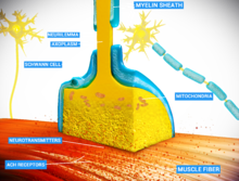
The neuromuscular junction (NMJ) is the most well-characterized synapse in that it provides a simple and accessible structure that allows for easy manipulation and observation. The synapse itself is composed of three cells: the motor neuron, the myofiber, and the Schwann cell. In a normally functioning synapse, a signal will cause the motor neuron to depolarize, by releasing the neurotransmitter acetylcholine (ACh). Acetylcholine travels across the synaptic cleft where it reaches acetylcholine receptors (AChR) on the plasma membrane of the myofiber, the sarcolemma. As the AChRs open ion channels, the membrane depolarizes, causing muscle contraction. The entire synapse is covered in a myelin sheath provided by the Schwann cell to insulate and encapsulate the junction. Another important part of the neuromuscular system and central nervous system are the astrocytes. While originally they were thought to have only functioned as support for the neurons, they play an important role in functional plasticity of synapses.

Origin and movement of cells

During development, each of the three germ layer cell types arises from different regions of the growing embryo. The individual myoblasts originate in the mesoderm and fuse to form a multi-nucleated myotube. During or shortly after myotube formation, motoneurons from the neural tube form preliminary contacts with the myotube. The Schwann cells arise from the neural crest and are led by the axons to their destination. Upon reaching it, they form a loose, unmyelinated covering over the innervating axons. The movement of the axons (and subsequently the Schwann cells) is guided by the growth cone, a filamentous projection of the axon that actively searches for neurotrophins released by the myotube.

The specific patterning of synapse development at the neuromuscular junction shows that the majority of muscles are innervated at their midpoints. Although it may seem that the axons specifically target the midpoint of the myotube, several factors reveal that this is not a valid claim. It appears that after the initial axonal contact, the newly formed myotube proceeds to grow symmetrically from that point of innervation. Coupled with the fact that AChR density is the result of axonal contact instead of the cause, the structural patterns of muscle fibers can be attributed to both myotatic growth as well as axonal innervation.

The preliminary contact formed between the motoneuron and the myotube generates synaptic transmission almost immediately, but the signal produced is very weak. There is evidence that Schwann cells may facilitate these preliminary signals by increasing the amount of spontaneous neurotransmitter release through small molecule signals. After about a week, a fully functional synapse is formed following several types of differentiation in both the post-synaptic muscle cell and the pre-synaptic motoneuron. This pioneer axon is of crucial importance because the new axons that follow have a high propensity for forming contacts with well-established synapses.

Post-synaptic differentiation

The most noticeable difference in the myotube following contact with the motoneuron is the increased concentration of AChR in the plasma membrane of the myotube in the synapse. This increased amount of AChR allows for more effective transmission of synaptic signals, which in turn leads to a more-developed synapse. The density of AChR is > 10,000/μm2 and approximately 10/μm2 around the edge. This high concentration of AChR in the synapse is achieved through clustering of AChR, up-regulation of the AChR gene transcription in the post-synaptic nuclei, and down-regulation of the AChR gene in the non-synaptic nuclei. The signals that initiate post-synaptic differentiation may be neurotransmitters released directly from the axon to the myotube, or they may arise from changes activated in the extracellular matrix of the synaptic cleft.

Clustering

AChR experiences multimerization within the post-synaptic membrane largely due to the signaling molecule Agrin. The axon of the motoneuron releases agrin, a proteoglycan that initiates a cascade that eventually leads to AChR association. Agrin binds to a muscle-specific kinase (MuSK) receptor in the post-synaptic membrane, and this in turn leads to downstream activation of the cytoplasmic protein Rapsyn. Rapsyn contains domains that allow for AChR association and multimerization, and it is directly responsible for AChR clustering in the post-synaptic membrane: rapsyn-deficient mutant mice fail to form AChR clusters.

Synapse-specific transcription

The increased concentration of AChR is not simply due to a rearrangement of pre-existing synaptic components. The axon also provides signals that regulate gene expression within the myonuclei directly beneath the synapse. This signaling provides for localized up-regulation of transcription of AChR genes and consequent increase in local AChR concentration. The two signaling molecules released by the axon are calcitonin gene-related peptide (CGRP) and neuregulin, which trigger a series of kinases that eventually lead to transcriptional activation of the AChR genes.

Extrasynaptic repression

Repression of the AChR gene in the non-synaptic nuclei is an activity-dependent process involving the electrical signal generated by the newly formed synapse. Reduced concentration of AChR in the extrasynaptic membrane in addition to increased concentration in the post-synaptic membrane helps ensure the fidelity of signals sent by the axon by localizing AChR to the synapse. Because the synapse begins receiving inputs almost immediately after the motoneuron comes into contact with the myotube, the axon quickly generates an action potential and releases ACh. The depolarization caused by AChR induces muscle contraction and simultaneously initiates repression of AChR gene transcription across the entire muscle membrane. Note that this affects gene transcription at a distance: the receptors that are embedded within the post-synaptic membrane are not susceptible to repression.

Pre-synaptic differentiation

Although the mechanisms regulating pre-synaptic differentiation are unknown, the changes exhibited at the developing axon terminal are well characterized. The pre-synaptic axon shows an increase in synaptic volume and area, an increase of synaptic vesicles, clustering of vesicles at the active zone, and polarization of the pre-synaptic membrane. These changes are thought to be mediated by neurotrophin and cell adhesion molecule release from muscle cells, thereby emphasizing the importance of communication between the motoneuron and the myotube during synaptogenesis. Like post-synaptic differentiation, pre-synaptic differentiation is thought to be due to a combination of changes in gene expression and a redistribution of pre-existing synaptic components. Evidence for this can be seen in the up-regulation of genes expressing vesicle proteins shortly after synapse formation as well as their localization at the synaptic terminal.

Synaptic maturation

Immature synapses are multiply innervated at birth, due the high propensity for new axons to innervate at a pre-existing synapse. As the synapse matures, the synapses segregate and eventually all axonal inputs except for one retract in a process called synapse elimination. Furthermore, the post-synaptic end plate grows deeper and creates folds through invagination to increase the surface area available for neurotransmitter reception. At birth, Schwann cells form loose, unmyelinated covers over groups of synapses, but as the synapse matures, Schwann cells become dedicated to a single synapse and form a myelinated cap over the entire neuromuscular junction.

Synapse elimination

The process of synaptic pruning known as synapse elimination is a presumably activity-dependent process that involves competition between axons. Hypothetically, a synapse strong enough to produce an action potential will trigger the myonuclei directly across from the axon to release synaptotrophins that will strengthen and maintain well-established synapses. This synaptic strengthening is not conferred upon the weaker synapses, thereby starving them out. It has also been suggested that in addition to the synaptotrophins released to the synapse exhibiting strong activity, the depolarization of the post-synaptic membrane causes release of synaptotoxins that ward off weaker axons.

Synapse formation specificity

A remarkable aspect of synaptogenesis is the fact that motoneurons are able to distinguish between fast and slow-twitch muscle fibers; fast-twitch muscle fibers are innervated by "fast" motoneurons, and slow-twitch muscle fibers are innervated by "slow" motoneurons. There are two hypothesized paths by which the axons of motoneurons achieve this specificity, one in which the axons actively recognize the muscles that they innervate and make selective decisions based on inputs, and another that calls for more indeterminate innervation of muscle fibers. In the selective paths, the axons recognize the fiber type, either by factors or signals released specifically by the fast or slow-twitch muscle fibers. In addition, selectivity can be traced to the lateral position that the axons are predeterminately arranged in order to link them to the muscle fiber that they will eventually innervate. The hypothesized non-selective pathways indicate that the axons are guided to their destinations by the matrix through which they travel. Essentially, a path is laid out for the axon and the axon itself is not involved in the decision-making process. Finally, the axons may non-specifically innervate muscle fibers and cause the muscles to acquire the characteristics of the axon that innervates them. In this path, a "fast" motoneuron can convert any muscle fiber into a fast-twitch muscle fiber. There is evidence for both selective and non-selective paths in synapse formation specificity, leading to the conclusion that the process is a combination of several factors.

Central nervous system synapse formation

Although the study of synaptogenesis within the central nervous system (CNS) is much more recent than that of the NMJ, there is promise of relating the information learned at the NMJ to synapses within the CNS. Many similar structures and basic functions exist between the two types of neuronal connections. At the most basic level, the CNS synapse and the NMJ both have a nerve terminal that is separated from the postsynaptic membrane by a cleft containing specialized extracellular material. Both structures exhibit localized vesicles at the active sites, clustered receptors at the post-synaptic membrane, and glial cells that encapsulate the entire synaptic cleft. In terms of synaptogenesis, both synapses exhibit differentiation of the pre- and post-synaptic membranes following initial contact between the two cells. This includes the clustering of receptors, localized up-regulation of protein synthesis at the active sites, and neuronal pruning through synapse elimination.

Despite these similarities in structure, there is a fundamental difference between the two connections. The CNS synapse is strictly neuronal and does not involve muscle fibers: for this reason the CNS uses different neurotransmitter molecules and receptors. More importantly, neurons within the CNS often receive multiple inputs that must be processed and integrated for successful transfer of information. Muscle fibers are innervated by a single input and operate in an all or none fashion. Coupled with the plasticity that is characteristic of the CNS neuronal connections, it is easy to see how increasingly complex CNS circuits can become.

Factors regulating synaptogenesis in the CNS


Signaling

The main method of synaptic signaling in the NMJ is through use of the neurotransmitter acetylcholine and its receptor. The CNS homolog is glutamate and its receptors, and one of special significance is the N-methyl-D-aspartate (NMDA) receptor. It has been shown that activation of NMDA receptors initiates synaptogenesis through activation of downstream products. The heightened level of NMDA receptor activity during development allows for increased influx of calcium, which acts as a secondary signal. Eventually, immediate early genes (IEG) are activated by transcription factors and the proteins required for neuronal differentiation are translated. The NMDA receptor function is associated with the estrogen receptor in hippocampal neurons. Experiments conducted with estradiol show that exposure to the estrogen significantly increases synaptic density and protein concentration.

Synaptic signaling during synaptogenesis is not only activity-dependent, but is also dependent on the environment in which the neurons are located. For instance, brain-derived neurotrophic factor (BDNF) is produced by the brain and regulates several functions within the developing synapse, including enhancement of transmitter release, increased concentration of vesicles, and cholesterol biosynthesis. Cholesterol is essential to synaptogenesis because the lipid rafts that it forms provide a scaffold upon which numerous signaling interactions can occur. BDNF-null mutants show significant defects in neuronal growth and synapse formation. Aside from neurotrophins, cell-adhesion molecules are also essential to synaptogenesis. Often the binding of pre-synaptic cell-adhesion molecules with their post-synaptic partners triggers specializations that facilitate synaptogenesis. Indeed, a defect in genes encoding neuroligin, a cell-adhesion molecule found in the post-synaptic membrane, has been linked to cases of autism and mental retardation. Finally, many of these signaling processes can be regulated by matrix metalloproteinases (MMPs) as the targets of many MMPs are these specific cell-adhesion molecules.

Morphology

The special structure found in the CNS that allows for multiple inputs is the dendritic spine, the highly dynamic site of excitatory synapses. This morphological dynamism is due to the specific regulation of the actin cytoskeleton, which in turn allows for regulation of synapse formation. Dendritic spines exhibit three main morphologies: filopodia, thin spines, and mushroom spines. The filopodia play a role in synaptogenesis through initiation of contact with axons of other neurons. Filopodia of new neurons tend to associate with multiply synapsed axons, while the filopodia of mature neurons tend to sites devoid of other partners. The dynamism of spines allows for the conversion of filopodia into the mushroom spines that are the primary sites of glutamate receptors and synaptic transmission.

Environmental enrichment

Rats raised with environmental enrichment have 25% more synapses than controls. This effect occurs whether a more stimulating environment is experienced immediately following birth, after weaning, or during maturity. Stimulation effects not only synaptogenesis upon pyramidal neurons but also stellate ones.

Contributions of the Wnt protein family

The (Wnt) family, includes several embryonic morphogens that contribute to early pattern formation in the developing embryo. Recently data have emerged showing that the Wnt protein family has roles in the later development of synapse formation and plasticity. Wnt contribution to synaptogenesis has been verified in both the central nervous system and the neuromuscular junction.

Central nervous system

Wnt family members contribute to synapse formation in the cerebellum by inducing presynaptic and postsynaptic terminal formation. This brain region contains three main neuronal cell types- Purkinje cells, granule cells and mossy fiber cells. Wnt-3 expression contributes to Purkinje cell neurite outgrowth and synapse formation. Granule cells express Wnt-7a to promote axon spreading and branching in their synaptic partner, mossy fiber cells. Retrograde secretion of Wnt-7a to mossy fiber cells causes growth cone enlargement by spreading microtubules. Furthermore, Wnt-7a retrograde signaling recruits synaptic vesicles and presynaptic proteins to the synaptic active zone. Wnt-5a performs a similar function on postsynaptic granule cells; this Wnt stimulates receptor assembly and clustering of the scaffolding protein PSD-95.

In the hippocampus Wnts in conjunction with cell electrical activity promote synapse formation. Wnt7b is expressed in maturing dendrites, and the expression of the Wnt receptor Frizzled (Fz), increases highly with synapse formation in the hippocampus. NMDA glutamate receptor activation increases Wnt2 expression. Long term potentiation (LTP) due to NMDA activation and subsequent Wnt expression leads to Fz-5 localization at the postsynaptic active zone. Furthermore, Wnt7a and Wnt2 signaling after NMDA receptor mediated LTP leads to increased dendritic arborization and regulates activity induced synaptic plasticity. Blocking Wnt expression in the hippocampus mitigates these activity dependent effects by reducing dendritic arborization and subsequently, synaptic complexity.

Neuromuscular junction

Similar mechanisms of action by Wnts in the central nervous system are observed in the neuromuscular junction (NMJ) as well. In the Drosophila NMJ mutations in the Wnt5 receptor Derailed (drl) reduce the number of and density of synaptic active zones. The major neurotransmitter in this system is glutamate. Wnt is needed to localize glutamatergic receptors on postsynaptic muscle cells. As a result, Wnt mutations diminish evoked currents on the postsynaptic muscle.

In the vertebrate NMJ, motor neuron expression of Wnt-11r contributes to acetylcholine receptor (AChR) clustering in the postsynaptic density of muscle cells. Wnt-3 is expressed by muscle fibers and is secreted retrogradely onto motor neurons. In motor neurons, Wnt-3 works with Agrin to promote growth cone enlargement, axon branching and synaptic vesicle clustering.

Dendritic spine

From Wikipedia, the free encyclopedia
https://en.wikipedia.org/wiki/Dendritic_spine
 
Dendritic spine
Dendritic spines.jpg
Spiny dendrite of a striatal medium spiny neuron.
Spline types 3D.png
Common types of dendritic spines.
Details
Identifiers
Latingemmula dendritica
MeSHD049229
THH2.00.06.1.00036

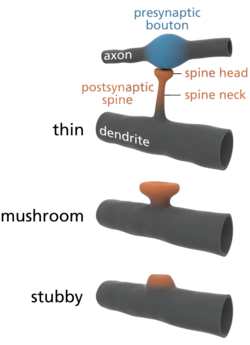
A dendritic spine (or spine) is a small membranous protrusion from a neuron's dendrite that typically receives input from a single axon at the synapse. Dendritic spines serve as a storage site for synaptic strength and help transmit electrical signals to the neuron's cell body. Most spines have a bulbous head (the spine head), and a thin neck that connects the head of the spine to the shaft of the dendrite. The dendrites of a single neuron can contain hundreds to thousands of spines. In addition to spines providing an anatomical substrate for memory storage and synaptic transmission, they may also serve to increase the number of possible contacts between neurons.

Structure

Dendritic spines are small with spine head volumes ranging 0.01 μm3 to 0.8 μm3. Spines with strong synaptic contacts typically have a large spine head, which connects to the dendrite via a membranous neck. The most notable classes of spine shape are "thin", "stubby", "mushroom", and "branched". Electron microscopy studies have shown that there is a continuum of shapes between these categories. The variable spine shape and volume is thought to be correlated with the strength and maturity of each spine-synapse. 

Distribution

Dendritic spines usually receive excitatory input from axons, although sometimes both inhibitory and excitatory connections are made onto the same spine head. Excitatory axon proximity to dendritic spines is not sufficient to predict the presence of a synapse, as demonstrated by the Lichtman lab in 2015.

Spines are found on the dendrites of most principal neurons in the brain, including the pyramidal neurons of the neocortex, the medium spiny neurons of the striatum, and the Purkinje cells of the cerebellum. Dendritic spines occur at a density of up to 5 spines/1 μm stretch of dendrite. Hippocampal and cortical pyramidal neurons may receive tens of thousands of mostly excitatory inputs from other neurons onto their equally numerous spines, whereas the number of spines on Purkinje neuron dendrites is an order of magnitude larger. 

Cytoskeleton and organelles

The cytoskeleton of dendritic spines is particularly important in their synaptic plasticity; without a dynamic cytoskeleton, spines would be unable to rapidly change their volumes or shapes in responses to stimuli. These changes in shape might affect the electrical properties of the spine. The cytoskeleton of dendritic spines is primarily made of filamentous actin (F-actin). tubulin Monomers and microtubule-associated proteins (MAPs) are present, and organized microtubules are present. Because spines have a cytoskeleton of primarily actin, this allows them to be highly dynamic in shape and size. The actin cytoskeleton directly determines the morphology of the spine, and actin regulators, small GTPases such as Rac, RhoA, and CDC42, rapidly modify this cytoskeleton. Overactive Rac1 results in consistently smaller dendritic spines.

In addition to their electrophysiological activity and their receptor-mediated activity, spines appear to be vesicularly active and may even translate proteins. Stacked discs of the smooth endoplasmic reticulum (SERs) have been identified in dendritic spines. Formation of this "spine apparatus" depends on the protein synaptopodin and is believed to play an important role in calcium handling. "Smooth" vesicles have also been identified in spines, supporting the vesicular activity in dendritic spines. The presence of polyribosomes in spines also suggests protein translational activity in the spine itself, not just in the dendrite. 

Physiology


Receptor activity

Dendritic spines express glutamate receptors (e.g. AMPA receptor and NMDA receptor) on their surface. The TrkB receptor for BDNF is also expressed on the spine surface, and is believed to play a role in spine survival. The tip of the spine contains an electron-dense region referred to as the "postsynaptic density" (PSD). The PSD directly apposes the active zone of its synapsing axon and comprises ~10% of the spine's membrane surface area; neurotransmitters released from the active zone bind receptors in the postsynaptic density of the spine. Half of the synapsing axons and dendritic spines are physically tethered by calcium-dependent cadherin, which forms cell-to-cell adherent junctions between two neurons. 

Glutamate receptors (GluRs) are localized to the postsynaptic density, and are anchored by cytoskeletal elements to the membrane. They are positioned directly above their signalling machinery, which is typically tethered to the underside of the plasma membrane, allowing signals transmitted by the GluRs into the cytosol to be further propagated by their nearby signalling elements to activate signal transduction cascades. The localization of signalling elements to their GluRs is particularly important in ensuring signal cascade activation, as GluRs would be unable to affect particular downstream effects without nearby signallers.

Signalling from GluRs is mediated by the presence of an abundance of proteins, especially kinases, that are localized to the postsynaptic density. These include calcium-dependent calmodulin, CaMKII (calmodulin-dependent protein kinase II), PKC (Protein Kinase C), PKA (Protein Kinase A), Protein Phosphatase-1 (PP-1), and Fyn tyrosine kinase. Certain signallers, such as CaMKII, are upregulated in response to activity.

Spines are particularly advantageous to neurons by compartmentalizing biochemical signals. This can help to encode changes in the state of an individual synapse without necessarily affecting the state of other synapses of the same neuron. The length and width of the spine neck has a large effect on the degree of compartmentalization, with thin spines being the most biochemically isolated spines. 

Plasticity

Dendritic spines are very "plastic", that is, spines change significantly in shape, volume, and number in small time courses. Because spines have a primarily actin cytoskeleton, they are dynamic, and the majority of spines change their shape within seconds to minutes because of the dynamicity of actin remodeling. Furthermore, spine number is very variable and spines come and go; in a matter of hours, 10-20% of spines can spontaneously appear or disappear on the pyramidal cells of the cerebral cortex, although the larger "mushroom"-shaped spines are the most stable.

Spine maintenance and plasticity is activity-dependent and activity-independent. BDNF partially determines spine levels, and low levels of AMPA receptor activity is necessary to maintain spine survival, and synaptic activity involving NMDA receptors encourages spine growth. Furthermore, two-photon laser scanning microscopy and confocal microscopy have shown that spine volume changes depending on the types of stimuli that are presented to a synapse. 

Importance to learning and memory


Evidence of importance

A depiction of spine formation and elimination.
Experience-dependent spine formation and elimination

Spine plasticity is implicated in motivation, learning, and memory. In particular, long-term memory is mediated in part by the growth of new dendritic spines (or the enlargement of pre-existing spines) to reinforce a particular neural pathway. Because dendritic spines are plastic structures whose lifespan is influenced by input activity, spine dynamics may play an important role in the maintenance of memory over a lifetime. 

Age-dependent changes in the rate of spine turnover suggest that spine stability impacts developmental learning. In youth, dendritic spine turnover is relatively high and produces a net loss of spines. This high rate of spine turnover may characterize critical periods of development and reflect learning capacity in adolescence—different cortical areas exhibit differing levels of synaptic turnover during development, possibly reflecting varying critical periods for specific brain regions. In adulthood, however, most spines remain persistent, and the half-life of spines increases. This stabilization occurs due to a developmentally regulated slow-down of spine elimination, a process which may underlie the stabilization of memories in maturity.

Experience-induced changes in dendritic spine stability also point to spine turnover as a mechanism involved in the maintenance of long-term memories, though it is unclear how sensory experience affects neural circuitry. Two general models might describe the impact of experience on structural plasticity. On the one hand, experience and activity may drive the discrete formation of relevant synaptic connections that store meaningful information in order to allow for learning. On the other hand, synaptic connections may be formed in excess, and experience and activity may lead to the pruning of extraneous synaptic connections.

In lab animals of all ages, environmental enrichment has been related to dendritic branching, spine density, and overall number of synapses. In addition, skill training has been shown to lead to the formation and stabilization of new spines while destabilizing old spines, suggesting that the learning of a new skill involves a rewiring process of neural circuits. Since the extent of spine remodeling correlates with success of learning, this suggests a crucial role of synaptic structural plasticity in memory formation. In addition, changes in spine stability and strengthening occur rapidly and have been observed within hours after training.

Conversely, while enrichment and training are related to increases in spine formation and stability, long-term sensory deprivation leads to an increase in the rate of spine elimination and therefore impacts long-term neural circuitry. Upon restoring sensory experience after deprivation in adolescence, spine elimination is accelerated, suggesting that experience plays an important role in the net loss of spines during development. In addition, other sensory deprivation paradigms—such as whisker trimming—have been shown to increase the stability of new spines.

Research in neurological diseases and injuries shed further light on the nature and importance of spine turnover. After stroke, a marked increase in structural plasticity occurs near the trauma site, and a five- to eightfold increase from control rates in spine turnover has been observed. Dendrites disintegrate and reassemble rapidly during ischemia—as with stroke, survivors showed an increase in dendritic spine turnover. While a net loss of spines is observed in Alzheimer's disease and cases of intellectual disability, cocaine and amphetamine use have been linked to increases in dendritic branching and spine density in the prefrontal cortex and the nucleus accumbens. Because significant changes in spine density occur in various brain diseases, this suggests a balanced state of spine dynamics in normal circumstances, which may be susceptible to disequilibrium under varying pathological conditions.

There is also some evidence for loss of dendritic spines as a consequence of aging. One study using mice has noted a correlation between age-related reductions in spine densities in the hippocampus and age-dependent declines in hippocampal learning and memory.

Importance contested

Despite experimental findings that suggest a role for dendritic spine dynamics in mediating learning and memory, the degree of structural plasticity's importance remains debatable. For instance, studies estimate that only a small portion of spines formed during training actually contribute to lifelong learning. In addition, the formation of new spines may not significantly contribute to the connectivity of the brain, and spine formation may not bear as much of an influence on memory retention as other properties of structural plasticity, such as the increase in size of spine heads.

Modeling

Theoreticians have for decades hypothesized about the potential electrical function of spines, yet our inability to examine their electrical properties has until recently stopped theoretical work from progressing too far. Recent advances in imaging techniques along with increased use of two-photon glutamate uncaging have led to a wealth of new discoveries; we now suspect that there are voltage-dependent sodium, potassium, and calcium channels in the spine heads.

Cable theory provides the theoretical framework behind the most "simple" method for modelling the flow of electrical currents along passive neural fibres. Each spine can be treated as two compartments, one representing the neck, the other representing the spine head. The compartment representing the spine head alone should carry the active properties. 

Baer and Rinzel's continuum model

To facilitate the analysis of interactions between many spines, Baer & Rinzel formulated a new cable theory for which the distribution of spines is treated as a continuum. In this representation, spine head voltage is the local spatial average of membrane potential in adjacent spines. The formulation maintains the feature that there is no direct electrical coupling between neighboring spines; voltage spread along dendrites is the only way for spines to interact.

Spike-diffuse-spike model

The SDS model was intended as a computationally simple version of the full Baer and Rinzel model. It was designed to be analytically tractable and have as few free parameters as possible while retaining those of greatest significance, such as spine neck resistance. The model drops the continuum approximation and instead uses a passive dendrite coupled to excitable spines at discrete points. Membrane dynamics in the spines are modelled using integrate and fire processes. The spike events are modelled in a discrete fashion with the wave form conventionally represented as a rectangular function. 

Modeling spine calcium transients

Calcium transients in spines are a key trigger for synaptic plasticity. NMDA receptors, which have a high permeability for calcium, only conduct ions if the membrane potential is suffiently depolarized. The amount of calcium entering a spine during synaptic activity therefore depends on the depolarization of the spine head. Evidence from calcium imaging experiments (two-photon microscopy) and from compartmental modelling indicates that spines with high resistance necks experience larger calcium transients during synaptic activity.

Development

Dendritic spines can develop directly from dendritic shafts or from dendritic filopodia. During synaptogenesis, dendrites rapidly sprout and retract filopodia, small membrane organelle-lacking membranous protrusions. Recently, I-BAR protein MIM was found to contribute to the initiation process. During the first week of birth, the brain is predominated by filopodia, which eventually develop synapses. However, after this first week, filopodia are replaced by spiny dendrites but also small, stubby spines that protrude from spiny dendrites. In the development of certain filopodia into spines, filopodia recruit presynaptic contact to the dendrite, which encourages the production of spines to handle specialized postsynaptic contact with the presynaptic protrusions. 

Spines, however, require maturation after formation. Immature spines have impaired signaling capabilities, and typically lack "heads" (or have very small heads), only necks, while matured spines maintain both heads and necks.

Clinical significance

Cognitive disorders such as ADHD, autism, intellectual disability, and fragile X syndrome, may be resultant from abnormalities in dendritic spines, especially the number of spines and their maturity. The ratio of matured to immature spines is important in their signaling, as immature spines have impaired synaptic signaling. Fragile X syndrome is characterized by an overabundance of immature spines that have multiple filopodia in cortical dendrites. 

History

Dendritic spines were first described at the end of the 19th century by Santiago Ramón y Cajal on cerebellar neurons. Ramón y Cajal then proposed that dendritic spines could serve as contacting sites between neurons. This was demonstrated more than 50 years later thanks to the emergence of electron microscopy. Until the development of confocal microscopy on living tissues, it was commonly admitted that spines were formed during embryonic development and then would remain stable after birth. In this paradigm, variations of synaptic weight were considered as sufficient to explain memory processes at the cellular level. But since about a decade ago, new techniques of confocal microscopy demonstrated that dendritic spines are indeed motile and dynamic structures that undergo a constant turnover, even after birth.

Chemical synapse

From Wikipedia, the free encyclopedia
https://en.wikipedia.org/wiki/Chemical_synapse
 
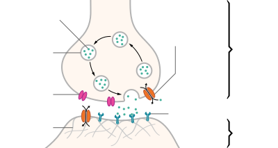
Artistic interpretation of the major elements in chemical synaptic transmission. An electrochemical wave called an action potential travels along the axon of a neuron. When the action potential reaches the presynaptic terminal, it provokes the release of a synaptic vesicle, secreting its quanta of neurotransmitter molecules. The neurotransmitter binds to chemical receptor molecules located in the membrane of another neuron, the postsynaptic neuron, on the opposite side of the synaptic cleft.

Chemical synapses are biological junctions through which neurons' signals can be sent to each other and to non-neuronal cells such as those in muscles or glands. Chemical synapses allow neurons to form circuits within the central nervous system. They are crucial to the biological computations that underlie perception and thought. They allow the nervous system to connect to and control other systems of the body. 

At a chemical synapse, one neuron releases neurotransmitter molecules into a small space (the synaptic cleft) that is adjacent to another neuron. The neurotransmitters are contained within small sacs called synaptic vesicles, and are released into the synaptic cleft by exocytosis. These molecules then bind to neurotransmitter receptors on the postsynaptic cell. Finally, the neurotransmitters are cleared from the synapse through one of several potential mechanisms including enzymatic degradation or re-uptake by specific transporters either on the presynaptic cell or on some other neuroglia to terminate the action of the neurotransmitter. 

The adult human brain is estimated to contain from 1014 to 5 × 10¹⁴ (100–500 trillion) synapses. Every cubic millimeter of cerebral cortex contains roughly a billion (short scale, i.e. 109) of them. The number of synapses in the human cerebral cortex has separately been estimated at 0.15 quadrillion (150 trillion).
 
The word "synapse" comes from "synaptein", which Sir Charles Scott Sherrington and colleagues coined from the Greek "syn-" ("together") and "haptein" ("to clasp"). Chemical synapses are not the only type of biological synapse: electrical and immunological synapses also exist. Without a qualifier, however, "synapse" commonly refers to chemical synapse.

Structure

Structure of a typical chemical synapse
Distinguish between pre- and post- synapse
"The connection linking neuron to neuron is the synapse. Signal flows
in one direction, from the presynaptic neuron to the postsynaptic neuron
via the synapse which acts as a variable attenuator."  In brief,
the direction of the signal flow determines the prefix for the involved
synapses.

Synapses are functional connections between neurons, or between neurons and other types of cells. A typical neuron gives rise to several thousand synapses, although there are some types that make far fewer. Most synapses connect axons to dendrites, but there are also other types of connections, including axon-to-cell-body, axon-to-axon, and dendrite-to-dendrite. Synapses are generally too small to be recognizable using a light microscope except as points where the membranes of two cells appear to touch, but their cellular elements can be visualized clearly using an electron microscope.

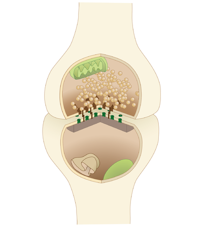
Chemical synapses pass information directionally from a presynaptic cell to a postsynaptic cell and are therefore asymmetric in structure and function. The presynaptic axon terminal, or synaptic bouton, is a specialized area within the axon of the presynaptic cell that contains neurotransmitters enclosed in small membrane-bound spheres called synaptic vesicles (as well as a number of other supporting structures and organelles, such as mitochondria and endoplasmic reticulum). Synaptic vesicles are docked at the presynaptic plasma membrane at regions called active zones

Immediately opposite is a region of the postsynaptic cell containing neurotransmitter receptors; for synapses between two neurons the postsynaptic region may be found on the dendrites or cell body. Immediately behind the postsynaptic membrane is an elaborate complex of interlinked proteins called the postsynaptic density (PSD). 

Proteins in the PSD are involved in anchoring and trafficking neurotransmitter receptors and modulating the activity of these receptors. The receptors and PSDs are often found in specialized protrusions from the main dendritic shaft called dendritic spines.

Synapses may be described as symmetric or asymmetric. When examined under an electron microscope, asymmetric synapses are characterized by rounded vesicles in the presynaptic cell, and a prominent postsynaptic density. Asymmetric synapses are typically excitatory. Symmetric synapses in contrast have flattened or elongated vesicles, and do not contain a prominent postsynaptic density. Symmetric synapses are typically inhibitory. 

The synaptic cleft —also called synaptic gap— is a gap between the pre- and postsynaptic cells that is about 20 nm (0.02 μ) wide. The small volume of the cleft allows neurotransmitter concentration to be raised and lowered rapidly.

An autapse is a chemical (or electrical) synapse formed when the axon of one neuron synapses with its own dendrites. 

Signaling in chemical synapses


Overview

Here is a summary of the sequence of events that take place in synaptic transmission from a presynaptic neuron to a postsynaptic cell. Each step is explained in more detail below. Note that with the exception of the final step, the entire process may run only a few hundred microseconds, in the fastest synapses.

  1. The process begins with a wave of electrochemical excitation called an action potential traveling along the membrane of the presynaptic cell, until it reaches the synapse.
  2. The electrical depolarization of the membrane at the synapse causes channels to open that are permeable to calcium ions.
  3. Calcium ions flow through the presynaptic membrane, rapidly increasing the calcium concentration in the interior.
  4. The high calcium concentration activates a set of calcium-sensitive proteins attached to vesicles that contain a neurotransmitter chemical.
  5. These proteins change shape, causing the membranes of some "docked" vesicles to fuse with the membrane of the presynaptic cell, thereby opening the vesicles and dumping their neurotransmitter contents into the synaptic cleft, the narrow space between the membranes of the pre- and postsynaptic cells.
  6. The neurotransmitter diffuses within the cleft. Some of it escapes, but some of it binds to chemical receptor molecules located on the membrane of the postsynaptic cell.
  7. The binding of neurotransmitter causes the receptor molecule to be activated in some way. Several types of activation are possible, as described in more detail below. In any case, this is the key step by which the synaptic process affects the behavior of the postsynaptic cell.
  8. Due to thermal vibration, the motion of atoms, vibrating about their equilibrium positions in a crystalline solid, neurotransmitter molecules eventually break loose from the receptors and drift away.
  9. The neurotransmitter is either reabsorbed by the presynaptic cell, and then repackaged for future release, or else it is broken down metabolically.

Neurotransmitter release

Release of neurotransmitter occurs at the end of axonal branches.
 
The release of a neurotransmitter is triggered by the arrival of a nerve impulse (or action potential) and occurs through an unusually rapid process of cellular secretion (exocytosis). Within the presynaptic nerve terminal, vesicles containing neurotransmitter are localized near the synaptic membrane. The arriving action potential produces an influx of calcium ions through voltage-dependent, calcium-selective ion channels at the down stroke of the action potential (tail current). Calcium ions then bind to synaptotagmin proteins found within the membranes of the synaptic vesicles, allowing the vesicles to fuse with the presynaptic membrane. The fusion of a vesicle is a stochastic process, leading to frequent failure of synaptic transmission at the very small synapses that are typical for the central nervous system. Large chemical synapses (e.g. the neuromuscular junction), on the other hand, have a synaptic release probability of 1. Vesicle fusion is driven by the action of a set of proteins in the presynaptic terminal known as SNAREs. As a whole, the protein complex or structure that mediates the docking and fusion of presynaptic vesicles is called the active zone. The membrane added by the fusion process is later retrieved by endocytosis and recycled for the formation of fresh neurotransmitter-filled vesicles. 

An exception to the general trend of neurotransmitter release by vesicular fusion is found in the type II receptor cells of mammalian taste buds. Here the neurotransmitter ATP is released directly from the cytoplasm into the synaptic cleft via voltage gated channels.

Receptor binding

Receptors on the opposite side of the synaptic gap bind neurotransmitter molecules. Receptors can respond in either of two general ways. First, the receptors may directly open ligand-gated ion channels in the postsynaptic cell membrane, causing ions to enter or exit the cell and changing the local transmembrane potential. The resulting change in voltage is called a postsynaptic potential. In general, the result is excitatory in the case of depolarizing currents, and inhibitory in the case of hyperpolarizing currents. Whether a synapse is excitatory or inhibitory depends on what type(s) of ion channel conduct the postsynaptic current(s), which in turn is a function of the type of receptors and neurotransmitter employed at the synapse. The second way a receptor can affect membrane potential is by modulating the production of chemical messengers inside the postsynaptic neuron. These second messengers can then amplify the inhibitory or excitatory response to neurotransmitters.

Termination

After a neurotransmitter molecule binds to a receptor molecule, it must be removed to allow for the postsynaptic membrane to continue to relay subsequent EPSPs and/or IPSPs. This removal can happen through one or more processes:
  • The neurotransmitter may diffuse away due to thermally-induced oscillations of both it and the receptor, making it available to be broken down metabolically outside the neuron or to be reabsorbed.
  • Enzymes within the subsynaptic membrane may inactivate/metabolize the neurotransmitter.
  • Reuptake pumps may actively pump the neurotransmitter back into the presynaptic axon terminal for reprocessing and re-release following a later action potential.

Synaptic strength

The strength of a synapse has been defined by Sir Bernard Katz as the product of (presynaptic) release probability pr, quantal size q (the postsynaptic response to the release of a single neurotransmitter vesicle, a 'quantum'), and n, the number of release sites. "Unitary connection" usually refers to an unknown number of individual synapses connecting a presynaptic neuron to a postsynaptic neuron. The amplitude of postsynaptic potentials (PSPs) can be as low as 0.4 mV to as high as 20 mV. The amplitude of a PSP can be modulated by neuromodulators or can change as a result of previous activity. Changes in the synaptic strength can be short-term, lasting seconds to minutes, or long-term (long-term potentiation, or LTP), lasting hours. Learning and memory are believed to result from long-term changes in synaptic strength, via a mechanism known as synaptic plasticity

Receptor desensitization

Desensitization of the postsynaptic receptors is a decrease in response to the same neurotransmitter stimulus. It means that the strength of a synapse may in effect diminish as a train of action potentials arrive in rapid succession – a phenomenon that gives rise to the so-called frequency dependence of synapses. The nervous system exploits this property for computational purposes, and can tune its synapses through such means as phosphorylation of the proteins involved.

Synaptic plasticity

Synaptic transmission can be changed by previous activity. These changes are called synaptic plasticity and may result in either a decrease in the efficacy of the synapse, called depression, or an increase in efficacy, called potentiation. These changes can either be long-term or short-term. Forms of short-term plasticity include synaptic fatigue or depression and synaptic augmentation. Forms of long-term plasticity include long-term depression and long-term potentiation. Synaptic plasticity can be either homosynaptic (occurring at a single synapse) or heterosynaptic (occurring at multiple synapses).

Homosynaptic plasticity

Homosynaptic Plasticity (or also homotropic modulation) is a change in the synaptic strength that results from the history of activity at a particular synapse. This can result from changes in presynaptic calcium as well as feedback onto presynaptic receptors, i.e. a form of autocrine signaling. Homosynaptic plasticity can affect the number and replenishment rate of vesicles or it can affect the relationship between calcium and vesicle release. Homosynaptic plasticity can also be postsynaptic in nature. It can result in either an increase or decrease in synaptic strength.

One example is neurons of the sympathetic nervous system (SNS), which release noradrenaline, which, besides affecting postsynaptic receptors, also affects presynaptic α2-adrenergic receptors, inhibiting further release of noradrenaline. This effect is utilized with clonidine to perform inhibitory effects on the SNS.

Heterosynaptic plasticity

Heterosynaptic Plasticity (or also heterotropic modulation) is a change in synaptic strength that results from the activity of other neurons. Again, the plasticity can alter the number of vesicles or their replenishment rate or the relationship between calcium and vesicle release. Additionally, it could directly affect calcium influx. Heterosynaptic plasticity can also be postsynaptic in nature, affecting receptor sensitivity. 

One example is again neurons of the sympathetic nervous system, which release noradrenaline, which, in addition, generates an inhibitory effect on presynaptic terminals of neurons of the parasympathetic nervous system.

Integration of synaptic inputs

In general, if an excitatory synapse is strong enough, an action potential in the presynaptic neuron will trigger an action potential in the postsynaptic cell. In many cases the excitatory postsynaptic potential (EPSP) will not reach the threshold for eliciting an action potential. When action potentials from multiple presynaptic neurons fire simultaneously, or if a single presynaptic neuron fires at a high enough frequency, the EPSPs can overlap and summate. If enough EPSPs overlap, the summated EPSP can reach the threshold for initiating an action potential. This process is known as summation, and can serve as a high pass filter for neurons.

On the other hand, a presynaptic neuron releasing an inhibitory neurotransmitter, such as GABA, can cause an inhibitory postsynaptic potential (IPSP) in the postsynaptic neuron, bringing the membrane potential farther away from the threshold, decreasing its excitability and making it more difficult for the neuron to initiate an action potential. If an IPSP overlaps with an EPSP, the IPSP can in many cases prevent the neuron from firing an action potential. In this way, the output of a neuron may depend on the input of many different neurons, each of which may have a different degree of influence, depending on the strength and type of synapse with that neuron. John Carew Eccles performed some of the important early experiments on synaptic integration, for which he received the Nobel Prize for Physiology or Medicine in 1963. 

Volume transmission

When a neurotransmitter is released at a synapse, it reaches its highest concentration inside the narrow space of the synaptic cleft, but some of it is certain to diffuse away before being reabsorbed or broken down. If it diffuses away, it has the potential to activate receptors that are located either at other synapses or on the membrane away from any synapse. The extrasynaptic activity of a neurotransmitter is known as volume transmission. It is well established that such effects occur to some degree, but their functional importance has long been a matter of controversy.

Recent work indicates that volume transmission may be the predominant mode of interaction for some special types of neurons. In the mammalian cerebral cortex, a class of neurons called neurogliaform cells can inhibit other nearby cortical neurons by releasing the neurotransmitter GABA into the extracellular space. Along the same vein, GABA released from neurogliaform cells into the extracellular space also acts on surrounding astrocytes, assigning a role for volume transmission in the control of ionic and neurotransmitter homeostasis. Approximately 78% of neurogliaform cell boutons do not form classical synapses. This may be the first definitive example of neurons communicating chemically where classical synapses are not present.

Relationship to electrical synapses

An electrical synapse is an electrically conductive link between two abutting neurons that is formed at a narrow gap between the pre- and postsynaptic cells, known as a gap junction. At gap junctions, cells approach within about 3.5 nm of each other, rather than the 20 to 40 nm distance that separates cells at chemical synapses. As opposed to chemical synapses, the postsynaptic potential in electrical synapses is not caused by the opening of ion channels by chemical transmitters, but rather by direct electrical coupling between both neurons. Electrical synapses are faster than chemical synapses. Electrical synapses are found throughout the nervous system, including in the retina, the reticular nucleus of the thalamus, the neocortex, and in the hippocampus. While chemical synapses are found between both excitatory and inhibitory neurons, electrical synapses are most commonly found between smaller local inhibitory neurons. Electrical synapses can exist between two axons, two dendrites, or between an axon and a dendrite. In some fish and amphibians, electrical synapses can be found within the same terminal of a chemical synapse, as in Mauthner cells.

Effects of drugs

One of the most important features of chemical synapses is that they are the site of action for the majority of psychoactive drugs. Synapses are affected by drugs such as curare, strychnine, cocaine, morphine, alcohol, LSD, and countless others. These drugs have different effects on synaptic function, and often are restricted to synapses that use a specific neurotransmitter. For example, curare is a poison that stops acetylcholine from depolarizing the postsynaptic membrane, causing paralysis. Strychnine blocks the inhibitory effects of the neurotransmitter glycine, which causes the body to pick up and react to weaker and previously ignored stimuli, resulting in uncontrollable muscle spasms. Morphine acts on synapses that use endorphin neurotransmitters, and alcohol increases the inhibitory effects of the neurotransmitter GABA. LSD interferes with synapses that use the neurotransmitter serotonin. Cocaine blocks reuptake of dopamine and therefore increases its effects.

History

During the 1950s, Bernard Katz and Paul Fatt observed spontaneous miniature synaptic currents at the frog neuromuscular junction. Based on these observations, they developed the 'quantal hypothesis' that is the basis for our current understanding of neurotransmitter release as exocytosis and for which Katz received the Nobel Prize in Physiology or Medicine in 1970. In the late 1960s, Ricardo Miledi and Katz advanced the hypothesis that depolarization-induced influx of calcium ions triggers exocytosis.

United States labor law

From Wikipedia, the free encyclopedia https://en.wikipedia.org/wiki/Uni...