Search This Blog

Monday, February 24, 2020

Wilson's disease

From Wikipedia, the free encyclopedia

Wilson's disease
Other namesWilson disease, hepatolenticular degeneration
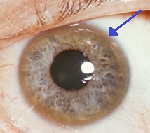
Kayser-Fleischer ringArrow.jpg
A brown ring on the edge of the cornea (Kayser–Fleischer ring) is common in Wilson's disease, especially when neurological symptoms are present.
SpecialtyGastroenterology
SymptomsSwelling of the legs, yellowish skin, personality changes
Usual onsetAge 5 to 35
CausesGenetic
Differential diagnosisChronic liver disease, Parkinson's disease, multiple sclerosis, others
TreatmentDietary changes, chelating agents, zinc supplements, liver transplant
Frequency~1 per 30,000

Wilson's disease is a genetic disorder in which excess copper builds up in the body. Symptoms are typically related to the brain and liver. Liver-related symptoms include vomiting, weakness, fluid build up in the abdomen, swelling of the legs, yellowish skin and itchiness. Brain-related symptoms include tremors, muscle stiffness, trouble speaking, personality changes, anxiety and seeing or hearing things that others do not.

Wilson's disease is caused by a mutation in the Wilson disease protein (ATP7B) gene. This protein transports excess copper into bile, where it is excreted in waste products.[1] The condition is autosomal recessive; for a person to be affected, they must inherit a mutated copy of the gene from both parents. Diagnosis may be difficult and often involves a combination of blood tests, urine tests and a liver biopsy. Genetic testing may be used to screen family members of those affected.

Wilson's disease is typically treated with dietary changes and medication. Dietary changes involve eating a low-copper diet and not using copper cookware. Medications used include chelating agents such as trientine and d-penicillamine and zinc supplements.[1] Complications of Wilson's disease can include liver failure, liver cancer and kidney problems. A liver transplant may be helpful in those in whom other treatments are not effective or if liver failure occurs.

Wilson's disease occurs in about 1 in 30,000 people. Symptoms usually begin between the ages of 5 and 35 years. It was first described in 1854 by German pathologist Friedrich Theodor von Frerichs and is named after British neurologist Samuel Wilson.

Signs and symptoms

The main sites of copper accumulation are the liver and the brain, and consequently liver disease and neuropsychiatric symptoms are the main features that lead to diagnosis. People with liver problems tend to come to medical attention earlier, generally as children or teenagers, than those with neurological and psychiatric symptoms, who tend to be in their twenties or older. Some are identified only because relatives have been diagnosed with Wilson's disease; many of these, when tested, turn out to have been experiencing symptoms of the condition but have not received a diagnosis.

Liver disease

Liver disease may present itself as tiredness, increased bleeding tendency or confusion (due to hepatic encephalopathy) and portal hypertension. The latter, a condition in which the pressure in the portal vein is markedly increased, leads to esophageal varices, blood vessels in the esophagus that may bleed in a life-threatening fashion, as well as enlargement of the spleen (splenomegaly) and accumulation of fluid in the abdominal cavity (ascites). On examination, signs of chronic liver disease such as spider angiomata (small distended blood vessels, usually on the chest) may be observed. Chronic active hepatitis has caused cirrhosis of the liver in most by the time they develop symptoms. While most people with cirrhosis have an increased risk of hepatocellular carcinoma (liver cancer), this risk is relatively very low in Wilson's disease.

About 5% of all people are diagnosed only when they develop fulminant acute liver failure, often in the context of a hemolytic anemia (anemia due to the destruction of red blood cells). This leads to abnormalities in protein production (identified by deranged coagulation) and metabolism by the liver. The deranged protein metabolism leads to the accumulation of waste products such as ammonia in the bloodstream. When these irritate the brain, the person develops hepatic encephalopathy (confusion, coma, seizures and finally life-threatening swelling of the brain).

Neuropsychiatric symptoms

About half the people with Wilson's disease have neurological or psychiatric symptoms. Most initially have mild cognitive deterioration and clumsiness, as well as changes in behavior. Specific neurological symptoms usually then follow, often in the form of parkinsonism (cogwheel rigidity, bradykinesia or slowed movements and a lack of balance are the most common parkinsonian features) with or without a typical hand tremor, masked facial expressions, slurred speech, ataxia (lack of coordination) or dystonia (twisting and repetitive movements of part of the body). Seizures and migraine appear to be more common in Wilson's disease. A characteristic tremor described as "wing-beating tremor" is encountered in many people with Wilson's; this is absent at rest but can be provoked by abducting the arms and flexing the elbows toward the midline.

Cognition can also be affected in Wilson's disease. This comes in two, not mutually exclusive, categories: frontal lobe disorder (may present as impulsivity, impaired judgement, promiscuity, apathy and executive dysfunction with poor planning and decision making) and subcortical dementia (may present as slow thinking, memory loss and executive dysfunction, without signs of aphasia, apraxia or agnosia). It is suggested that these cognitive involvements are related and closely linked to psychiatric manifestations of the disease.

Psychiatric problems due to Wilson's disease may include behavioral changes, depression, anxiety disorders, and psychosis. Psychiatric symptoms are commonly seen in conjunction with neurological symptoms and are rarely manifested on their own. These symptoms are often poorly defined and can sometimes be attributed to other causes. Because of this, diagnosis of Wilson's disease is rarely made when only psychiatric symptoms are present.

Other organ systems

Medical conditions have been linked with copper accumulation in Wilson's disease:

Genetics

Wilson's disease has an autosomal recessive pattern of inheritance.

The Wilson's disease gene (ATP7B) is on chromosome 13 (13q14.3) and is expressed primarily in the liver, kidney, and placenta. The gene codes for a P-type (cation transport enzyme) ATPase that transports copper into bile and incorporates it into ceruloplasmin. Mutations can be detected in 90% of cases. Most (60%) are homozygous for ATP7B mutations (two abnormal copies), and 30% have only one abnormal copy. Ten percent have no detectable mutation.

Although 300 mutations of ATP7B have been described, in most populations the cases of Wilson's disease are due to a small number of mutations specific for that population. For instance, in Western populations the H1069Q mutation (replacement of a histidine by a glutamine at position 1069 in the protein) is present in 37–63% of cases, while in China this mutation is very uncommon and R778L (arginine to leucine at 778) is found more often. Relatively little is known about the relative impact of various mutations, although the H1069Q mutation seems to predict later onset and predominantly neurological problems, according to some studies.

A normal variation in the PRNP gene can modify the course of the disease by delaying the age of onset and affecting the type of symptoms that develop. This gene produces prion protein, which is active in the brain and other tissues and also appears to be involved in transporting copper. A role for the ApoE gene was initially suspected but could not be confirmed.

The condition is inherited in an autosomal recessive pattern. In order to inherit it, both of the parents of an individual must carry an affected gene. Most have no family history of the condition. People with only one abnormal gene are called carriers (heterozygotes) and may have mild, but medically insignificant, abnormalities of copper metabolism.

Wilson's disease is the most common from a group of hereditary diseases that cause copper overload in the liver. All can cause cirrhosis at a young age. The other members of the group are Indian childhood cirrhosis (ICC), endemic Tyrolean infantile cirrhosis and idiopathic copper toxicosis. These are not related to ATP7B mutations: for example, ICC has been linked to mutations in the KRT8 and the KRT18 gene.

Pathophysiology

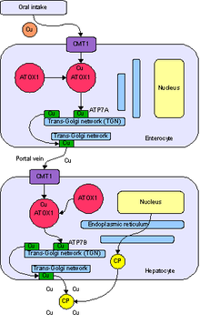
Normal absorption and distribution of copper. Cu = copper, CP = ceruloplasmin, green = ATP7B carrying copper.

Copper is needed by the body for a number of functions, predominantly as a cofactor for a number of enzymes such as ceruloplasmin, cytochrome c oxidase, dopamine β-hydroxylase, superoxide dismutase and tyrosinase.

Copper enters the body through the digestive tract. A transporter protein on the cells of the small bowel, copper membrane transporter 1 (Ctr1; SLC31A1), carries copper inside the cells, where some is bound to metallothionein and part is carried by ATOX1 to an organelle known as the trans-Golgi network. Here, in response to rising concentrations of copper, an enzyme called ATP7A (Menkes' protein) releases copper into the portal vein to the liver. Liver cells also carry the CMT1 protein, and metallothionein and ATOX1 bind it inside the cell, but here it is ATP7B that links copper to ceruloplasmin and releases it into the bloodstream, as well as removing excess copper by secreting it into bile. Both functions of ATP7B are impaired in Wilson's disease. Copper accumulates in the liver tissue; ceruloplasmin is still secreted, but in a form that lacks copper (termed apoceruloplasmin) and is rapidly degraded in the bloodstream.

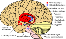
When the amount of copper in the liver overwhelms the proteins that normally bind it, it causes oxidative damage through a process known as Fenton chemistry; this damage eventually leads to chronic active hepatitis, fibrosis (deposition of connective tissue) and cirrhosis. The liver also releases copper into the bloodstream that is not bound to ceruloplasmin. This free copper precipitates throughout the body but particularly in the kidneys, eyes and brain. In the brain, most copper is deposited in the basal ganglia, particularly in the putamen and globus pallidus (together called the lenticular nucleus); these areas normally participate in the coordination of movement as well as playing a significant role in neurocognitive processes such as the processing of stimuli and mood regulation. Damage to these areas, again by Fenton chemistry, produces the neuropsychiatric symptoms seen in Wilson's disease.

It is not clear why Wilson's disease causes hemolysis, but various lines of evidence suggest that a high level of free (non-ceruloplasmin bound) copper has a direct effect on either oxidation of hemoglobin, inhibition of energy-supplying enzymes in the red blood cell, or direct damage to the cell membrane.

Diagnosis

Location of the basal ganglia, the part of the brain affected by Wilson's disease

Wilson's disease may be suspected on the basis of any of the symptoms mentioned above, or when a close relative has been found to have Wilson's. Most have slightly abnormal liver function tests such as a raised aspartate transaminase, alanine transaminase and bilirubin level. If the liver damage is significant, albumin may be decreased due to an inability of damaged liver cells to produce this protein; likewise, the prothrombin time (a test of coagulation) may be prolonged as the liver is unable to produce proteins known as clotting factors. Alkaline phosphatase levels are relatively low in those with Wilson's-related acute liver failure. If there are neurological symptoms, magnetic resonance imaging (MRI) of the brain is usually performed; this shows hyperintensities in the part of the brain called the basal ganglia in the T2 setting. MRI may also demonstrate the characteristic "face of the giant panda" pattern.

There is no totally reliable test for Wilson's disease, but levels of ceruloplasmin and copper in the blood, as well of the amount of copper excreted in urine during a 24-hour period, are together used to form an impression of the amount of copper in the body. The gold standard—or most ideal test—is a liver biopsy.

Ceruloplasmin

Ceruloplasmin

Levels of ceruloplasmin are abnormally low (<0 .2="" 80="" a="" at="" be="" can="" cases.="" g="" however="" href="https://en.wikipedia.org/wiki/Inflammation" in="" it="" levels="" nbsp="" normal="" of="" ongoing="" people="" present="" title="Inflammation" with="">inflammation
as it is an acute phase protein. Low ceruloplasmin is also found in Menkes disease and aceruloplasminemia, which are related to, but much rarer than Wilson's disease.

The combination of neurological symptoms, Kayser–Fleischer rings and a low ceruloplasmin level is considered sufficient for the diagnosis of Wilson's disease. In many cases, however, further tests are needed.

Serum and urine copper

Serum copper is low, which may seem paradoxical given that Wilson's disease is a disease of copper excess. However, 95% of plasma copper is carried by ceruloplasmin which is often low in Wilson's disease. Urine copper is elevated in Wilson's disease and is collected for 24 hours in a bottle with a copper-free liner. Levels above 100 μg/24h (1.6 μmol/24h) confirm Wilson's disease, and levels above 40 μg/24h (0.6 μmol/24h) are strongly indicative. High urine copper levels are not unique to Wilson's disease; they are sometimes observed in autoimmune hepatitis and in cholestasis (any disease obstructing the flow of bile from the liver to the small bowel).

In children, the penicillamine test may be used. A 500 mg oral dose of penicillamine is administered, and urine collected for 24 hours. If this contains more than 1600 μg (25 μmol), it is a reliable indicator of Wilson's disease. This test has not been validated in adults.

Liver biopsy

Once other investigations have indicated Wilson's disease, the ideal test is the removal of a small amount of liver tissue through a liver biopsy. This is assessed microscopically for the degree of steatosis and cirrhosis, and histochemistry and quantification of copper are used to measure the severity of the copper accumulation. A level of 250 μg of copper per gram of dried liver tissue confirms Wilson's disease. Occasionally, lower levels of copper are found; in that case, the combination of the biopsy findings with all other tests could still lead to a formal diagnosis of Wilson's.

In the earlier stages of the disease, the biopsy typically shows steatosis (deposition of fatty material), increased glycogen in the nucleus, and areas of necrosis (cell death). In more advanced disease, the changes observed are quite similar to those seen in autoimmune hepatitis, such as infiltration by inflammatory cells, piecemeal necrosis and fibrosis (scar tissue). In advanced disease, finally, cirrhosis is the main finding. In acute liver failure, degeneration of the liver cells and collapse of the liver tissue architecture is seen, typically on a background of cirrhotic changes. Histochemical methods for detecting copper are inconsistent and unreliable, and taken alone are regarded as insufficient to establish a diagnosis.

Genetic testing

Mutation analysis of the ATP7B gene, as well as other genes linked to copper accumulation in the liver, may be performed. Once a mutation is confirmed, it is possible to screen family members for the disease as part of clinical genetics family counseling. Regional distributions of genes associated with Wilson's disease are important to follow, as this can help clinicians design appropriate screening strategies. Since mutations of the WD gene vary between populations, research and genetic testing done in countries like the USA or United Kingdom can pose problems as they tend to have more mixed populations.

Treatment

Diet

In general, a diet low in copper-containing foods is recommended with the avoidance of mushrooms, nuts, chocolate, dried fruit, liver, sesame seeds and sesame oil, and shellfish.

Medication

Medical treatments are available for Wilson's disease. Some increase the removal of copper from the body, while others prevent the absorption of copper from the diet.

Generally, penicillamine is the first treatment used. This binds copper (chelation) and leads to excretion of copper in the urine. Hence, monitoring of the amount of copper in the urine can be done to ensure a sufficiently high dose is taken. Penicillamine is not without problems: about 20% experience a side effect or complication of penicillamine treatment, such as drug-induced lupus (causing joint pains and a skin rash) or myasthenia (a nerve condition leading to muscle weakness). In those who presented with neurological symptoms, almost half experience a paradoxical worsening in their symptoms. While this phenomenon is observed in other treatments for Wilson's, it is usually taken as an indication for discontinuing penicillamine and commencing second-line treatment. Those intolerant to penicillamine may instead be commenced on trientine hydrochloride, which also has chelating properties. Some recommend trientine as first-line treatment, but experience with penicillamine is more extensive. A further agent, under clinical investigation by Wilson Therapeutics, with known activity in Wilson's disease is tetrathiomolybdate. This is regarded as experimental, though some studies have shown a beneficial effect.

Once all results have returned to normal, zinc (usually in the form of a zinc acetate prescription called Galzin) may be used instead of chelators to maintain stable copper levels in the body. Zinc stimulates metallothionein, a protein in gut cells that binds copper and prevents their absorption and transport to the liver. Zinc therapy is continued unless symptoms recur or if the urinary excretion of copper increases.

In rare cases where none of the oral treatments are effective, especially in severe neurological disease, dimercaprol (British anti-Lewisite) is occasionally necessary. This treatment is injected intramuscularly (into a muscle) every few weeks and has unpleasant side effects such as pain.

People who are asymptomatic (for instance, those diagnosed through family screening or only as a result of abnormal test results) are generally treated, as the copper accumulation may cause long-term damage in the future. It is unclear whether these people are best treated with penicillamine or zinc acetate.

Physical and occupational therapies

Physiotherapy and occupational therapy are beneficial for patients with the neurologic form of the disease. The copper chelating treatment may take up to six months to start working, and these therapies can assist in coping with ataxia, dystonia, and tremors, as well as preventing the development of contractures that can result from dystonia.

Transplantation

Liver transplantation is an effective cure for Wilson's disease but is used only in particular scenarios because of the risks and complications associated with the procedure. It is used mainly in people with fulminant liver failure who fail to respond to medical treatment or in those with advanced chronic liver disease. Liver transplantation is avoided in severe neuropsychiatric illness, in which its benefit has not been demonstrated.

Prognosis

Left untreated, Wilson's disease tends to become progressively worse and is eventually fatal. With early detection and treatment, most of those affected can live relatively normal lives. Liver and neurologic damage that occurs prior to treatment may improve, but it is often permanent.

History

The disease bears the name of the British physician Samuel Alexander Kinnier Wilson (1878–1937), a neurologist who described the condition, including the pathological changes in the brain and liver, in 1912. Wilson's work had been predated by, and drew on, reports from German neurologist Carl Westphal (in 1883), who termed it "pseudosclerosis"; by the British neurologist William Gowers (in 1888); by the Finnish neuropathologist Ernst Alexander Homén (in 1889-1892), who noted the hereditary nature of the disease; and by Adolph Strümpell (in 1898), who noted hepatic cirrhosis. Neuropathologist John Nathaniel Cumings made the link with copper accumulation in both the liver and the brain in 1948. The occurrence of hemolysis was noted in 1967.

Cumings, and simultaneously the New Zealand neurologist Derek Denny-Brown, working in the United States, first reported effective treatment with metal chelator British anti-Lewisite in 1951. This treatment had to be injected but was one of the first therapies available in the field of neurology, a field that classically was able to observe and diagnose but had few treatments to offer. The first effective oral chelation agent, penicillamine, was discovered in 1956 by British neurologist John Walshe. In 1982, Walshe also introduced trientine, and was the first to develop tetrathiomolybdate for clinical use. Zinc acetate therapy initially made its appearance in the Netherlands, where physicians Schouwink and Hoogenraad used it in 1961 and in the 1970s, respectively, but it was further developed later by Brewer and colleagues at the University of Michigan.

The genetic basis of Wilson's disease and linkage to ATP7B mutations was elucidated in the 1980s and 1990s by several research groups.

Other animals

Hereditary copper accumulation has been described in Bedlington Terriers, where it generally only affects the liver. It is due to mutations in the COMMD1 (or MURR1) gene. Despite this findings, COMMD1 mutations could not be detected in humans with non-Wilsonian copper accumulation states (such as Indian childhood cirrhosis) to explain their genetic origin.

Tardive dyskinesia

From Wikipedia, the free encyclopedia

Tardive dyskinesia
Other namesLinguofacial dyskinesia, tardive dystonia, tardive oral dyskinesia
Dopamine-3d-CPK.png
Tardive dyskinesia is believed to involve the neurotransmitter dopamine.
Pronunciation
SpecialtyNeurology, psychiatry
SymptomsInvoluntary, repetitive body movements
CausesNeuroleptic medications (antipsychotics, metoclopramide)
Diagnostic methodBased on symptoms after ruling out other potential causes
Differential diagnosisHuntington's disease, cerebral palsy, Tourette syndrome, dystonia
PreventionUsing lowest possible dose of neuroleptic medication
TreatmentStopping neuroleptic medication if possible, switching to clozapine
MedicationValbenazine, tetrabenazine, botulinum toxin
PrognosisVariable
Frequency20% (atypical antipsychotics) 30% (typical antipsychotics)

Tardive dyskinesia (TD) is a disorder that results in involuntary, repetitive body movements, which may include grimacing, sticking out the tongue, or smacking the lips. Additionally, there may be rapid jerking movements or slow writhing movements. In about 20% of people, the disorder interferes with daily functioning.

Tardive dyskinesia occurs in some people as a result of long-term use of dopamine-receptor-blocking medications such as antipsychotics and metoclopramide. These medications are usually used for mental illness but may also be given for gastrointestinal or neurological problems. The condition typically develops only after months to years of use. The diagnosis is based on the symptoms after ruling out other potential causes.

Efforts to prevent the condition include either using the lowest possible dose or discontinuing use of neuroleptics. Treatment includes stopping the neuroleptic medication if possible or switching to clozapine. Other medications such as valbenazine, tetrabenazine, or botulinum toxin may be used to lessen the symptoms. With treatment, some see a resolution of symptoms, while others do not.

Rates in those on atypical antipsychotics are about 20%, while those on typical antipsychotics have rates of about 30%. The risk of acquiring the condition is greater in older people. The term "tardive dyskinesia" first came into use in 1964.

Signs and symptoms

Tardive dyskinesia is characterized by repetitive, involuntary movements. Some examples of these types of involuntary movements include:

Rapid, involuntary movements of the limbs, torso, and fingers may also occur. In some cases, an individual's legs can be so affected that walking becomes difficult or impossible. These symptoms are the opposite of people who are diagnosed with Parkinson's disease. People with Parkinson's have difficulty moving, whereas people with tardive dyskinesia have difficulty not moving.

Respiratory irregularity, such as grunting and difficulty breathing, is another symptom associated with tardive dyskinesia, although studies have shown that the rate of people affected is relatively low.

Tardive dyskinesia is often misdiagnosed as a mental illness rather than a neurological disorder, and as a result, people are prescribed neuroleptic drugs, which increase the probability that the person will develop a severe and disabling case, and shortening the typical survival period.

Other closely related neurological disorders have been recognized as variants of tardive dyskinesia. Tardive dystonia is similar to standard dystonia but permanent. Tardive akathisia involves painful feelings of inner tension and anxiety and a compulsive drive to move the body. In some extreme cases, afflicted individuals experience so much internal torture that they lose their ability to sit still. Tardive tourettism is a tic disorder featuring the same symptoms as Tourette syndrome. The two disorders are extremely close in nature and often can only be differentiated by the details of their respective onsets. Tardive myoclonus, a rare disorder, presents as brief jerks of muscles in the face, neck, trunk, and extremities.

"AIMS Examination": This test is used when psychotropic medications have been prescribed because people sometimes develop tardive dyskinesia due to prolonged use of antipsychotic medications. The Abnormal Involuntary Movement Scale (AIMS) examination is a test used to identify the symptoms of tardive dyskinesia (TD). The test is not meant to tell whether there is an absence or presence of tardive dyskinesia. It just scales to level of symptoms indicated by the actions observed. The levels range from none to severe. The AIMS examination was constructed in the 1970s to measure involuntary facial, trunk, and limb movements. It is best to do this test before and after the administration of the psychotropic drugs. Taking the AIMS consistently can help to track severity of TD over time.

Causes

Tardive dyskinesia was first described in the 1950s shortly after the introduction of chlorpromazine and other antipsychotic drugs. However, the exact mechanism of the disorder remains largely uncertain. The most compelling line of evidence suggests that tardive dyskinesia may result primarily from neuroleptic-induced dopamine supersensitivity in the nigrostriatal pathway, with the D2 dopamine receptor being most affected. Neuroleptics act primarily on this dopamine system, and older neuroleptics, which have greater affinity for the D2 binding site, are associated with high risk for tardive dyskinesia. The D2 hypersensitivity hypothesis is also supported by evidence of a dose–response relationship, withdrawal effects, studies on D2 agonists and antagonists, animal studies, and genetic polymorphism research.

Given similar doses of the same neuroleptic, differences among individuals still exist in the likelihood of developing tardive dyskinesia. Such individual differences may be due to genetic polymorphisms, which code for D2 receptor binding site affinity, or prior exposure to environmental toxins. Decreased functional reserve or cognitive dysfunction, associated with aging, mental retardation, alcohol and drug abuse, or traumatic head injuries, has also been shown to increase risk of developing the disorder among those treated with neuroleptics. Antipsychotic drugs can sometimes camouflage the signs of tardive dyskinesia from occurring in the early stages; this can happen from the individual having an increased dose of an antipsychotic drug. Often the symptoms of tardive dyskinesia are not apparent until the individual comes off of the antipsychotic drugs; however, when tardive dyskinesia worsens, the signs become visible.

Other dopamine antagonists and antiemetics can cause tardive dyskinesia, such as metoclopramide and promethazine, used to treat gastrointestinal disorders. Atypical antipsychotics are considered lower-risk for causing TD than their typical counterparts with their relative rates of TD of 13.1% and 32.4% respectively in short-term trials with haloperidol being the main typical antipsychotic utilised in said trials.[18] Quetiapine and clozapine are considered the lowest risk agents for precipitating TD.[18] From 2008, there have been reported cases of the anti-psychotic medication aripiprazole, a partial agonist at D2 receptors, leading to tardive dyskinesia.[19] As of 2013, reports of tardive dyskinesia in aripiprazole have grown in number.[20] The available research seems to suggest that the concurrent prophylactic use of a neuroleptic and an antiparkinsonian drug is useless to avoid early extrapyramidal side-effects and may render the person more sensitive to tardive dyskinesia. Since 1973 the use of these drugs has been found to be associated with the development of tardive dyskinesia.[21][22]

Risk factors

An increased risk of tardive dyskinesia has been associated with smoking in some studies, although a negative study does exist. There seems to be a cigarette smoke-exposure-dependent risk for TD in people who are antipsychotic-treated . Elderly peoples are also at a heightened risk for developing TD, as are females and those with organic brain injuries or diabetes mellitus and those with the negative symptoms of schizophrenia. TD is also more common in those that experience acute neurological side effects from antipsychotic drug treatment. Racial discrepancies in TD rate also exist, with Africans and African Americans having higher rates of TD after exposure to antipsychotics. Certain genetic risk factors for TD have been identified including polymorphisms in the genes encoding the D3, 5-HT2A and 5-HT2C receptors.

Prevention

Prevention of tardive dyskinesia is achieved by using the lowest effective dose of a neuroleptic for the shortest time. However, with diseases of chronic psychosis such as schizophrenia, this strategy must be balanced with the fact that increased dosages of neuroleptics are more beneficial in preventing recurrence of psychosis. If tardive dyskinesia is diagnosed, the causative drug should be discontinued. Tardive dyskinesia may persist after withdrawal of the drug for months, years or even permanently. Some studies suggest that physicians should consider using atypical antipsychotics as a substitute to typical antipsychotics for people requiring medication. These agents are associated with fewer neuromotor side effects and a lower risk of developing tardive dyskinesia.

Studies have tested the use of melatonin, high dosage vitamins, and different antioxidants in concurrence with antipsychotic drugs (often used to treat schizophrenia) as a way of preventing and treating tardive dyskinesia. Although further research is needed, studies reported a much lower percentage of individuals developing tardive dyskinesia than the current rate of people for those taking antipsychotic drugs. Tentative evidence supports the use of vitamin E for prevention.

Treatment

Valbenazine was approved by the FDA for tardive dyskinesia in April 2017. Tetrabenazine, which is a dopamine depleting drug, is sometimes used to treat tardive dyskinesia and other movement disorders (e.g. Huntington's chorea). Deutetrabenazine, an isotopic isomer of tetrabenazine, was approved by the FDA for tardive dyskinesia in August 2017. Vitamin B6 has been reported to be an effective treatment for TD in two randomised double-blind placebo-controlled trials, but the overall evidence for its effectiveness is considered "weak." Clonidine may also be useful in the treatment of TD, although dose-limiting hypotension and sedation may hinder its usage. Botox injections are used for minor focal dystonia, but not in more advanced tardive dyskinesia. As of 2018 evidence is insufficient to support the use of benzodiazepines, baclofen, progabide, sodium valproate, gaboxadol, or calcium channel blockers (e.g. diltiazem).

Epidemiology

Tardive dyskinesia most commonly occurs in people with psychiatric conditions who are treated with antipsychotic medications for many years. The average rate of people affected has been estimated to be around 30% for individuals taking antipsychotic medication, such as that used to treat schizophrenia. A study being conducted at the Yale University School of Medicine has estimated that "32% of people develop persistent tics after 5 years on major tranquilizers, 57% by 15 years, and 68% by 25 years." More drastic data was found during a longitudinal study conducted on individuals 45 years of age and older who were taking antipsychotic drugs. According to this research study, 26% of people developed tardive dyskinesia after just one year on the medication. Another 60% of this at-risk group developed the disorder after 3 years, and 23% developed severe cases of tardive dyskinesia within 3 years. According to these estimates, the majority of people will eventually develop the disorder if they remain on the drugs long enough.

Elderly people are more prone to develop tardive dyskinesia, and elderly women are more at-risk than elderly men. The risk is much lower for younger men and women, and also more equal across the sexes. People who have undergone electroconvulsive therapy or have a history of diabetes or alcohol abuse also have a higher risk of developing tardive dyskinesia.

Several studies have recently been conducted comparing the number of people affected of tardive dyskinesia with second generation, or more modern, antipsychotic drugs to that of first generation drugs. The newer antipsychotics appear to have a substantially reduced potential for causing tardive dyskinesia. However, some studies express concern that the number of people affected has decreased far less than expected, cautioning against the overestimation of the safety of modern antipsychotics.

A physician can evaluate and diagnose a person with tardive dyskinesia by conducting a systematic examination. The physician should ask the person to relax, and look for symptoms like facial grimacing, eye or lip movements, tics, respiratory irregularities, and tongue movements. In some cases, people experience nutritional problems, so a physician can also look for a gain or loss in weight.

Apart from the underlying psychiatric disorder, tardive dyskinesia may cause afflicted people to become socially isolated. It also increases the risk of body dysmorphic disorder (BDD) and can even lead to suicide. Emotional or physical stress can increase the severity of dyskinetic movements, whereas relaxation and sedation have the opposite effect.

Adverse effect

From Wikipedia, the free encyclopedia
 
An adverse effect is an undesired harmful effect resulting from a medication or other intervention such as surgery. An adverse effect may be termed a "side effect", when judged to be secondary to a main or therapeutic effect. If it results from an unsuitable or incorrect dosage or procedure, this is called a medical error and not a complication. Adverse effects are sometimes referred to as "iatrogenic" because they are generated by a physician/treatment. Some adverse effects occur only when starting, increasing or discontinuing a treatment.

Using a drug or other medical intervention which is contraindicated may increase the risk of adverse effects. Adverse effects may cause complications of a disease or procedure and negatively affect its prognosis. They may also lead to non-compliance with a treatment regimen. Adverse effects of medical treatment resulted in 142,000 deaths in 2013 up from 94,000 deaths in 1990 globally.

The harmful outcome is usually indicated by some result such as morbidity, mortality, alteration in body weight, levels of enzymes, loss of function, or as a pathological change detected at the microscopic, macroscopic or physiological level. It may also be indicated by symptoms reported by a patient. Adverse effects may cause a reversible or irreversible change, including an increase or decrease in the susceptibility of the individual to other chemicals, foods, or procedures, such as drug interactions.

Classification

In terms of drugs, adverse events may be defined as: “Any untoward medical occurrence in a patient or clinical investigation subject administered a pharmaceutical product and which does not necessarily have to have a causal relationship with this treatment.”

In clinical trials, a distinction is made between an adverse event and a serious adverse event. Generally, any event which causes death, permanent damage, birth defects, or requires hospitalization is considered a serious adverse event. The results of trials are often included in the labelling of the medication to provide information both for patients and the prescribing physicians.

The term "life-threatening" in the context of a serious adverse event refers to an event in which the patient was at risk of death at the time of the event; it does not refer to an event which hypothetically might have caused death if it were more severe.

Reporting systems

In many countries, adverse effects are required by law to be reported, researched in clinical trials and included into the patient information accompanying medical devices and drugs for sale to the public. Investigators in human clinical trials are obligated to report these events in clinical study reports. Research suggests that these events are often inadequately reported in publicly available reports. Because of the lack of these data and uncertainty about methods for synthesising them, individuals conducting systematic reviews and meta-analyses of therapeutic interventions often unknowingly overemphasise health benefit. To balance the overemphasis on benefit, scholars have called for more complete reporting of harm from clinical trials.

United Kingdom

The Yellow Card Scheme is a United Kingdom initiative run by the Medicines and Healthcare products Regulatory Agency (MHRA) and the Commission on Human Medicines (CHM) to gather information on adverse effects to medicines. This includes all licensed medicines, from medicines issued on prescription to medicines bought over the counter from a supermarket. The scheme also includes all herbal supplements and unlicensed medicines found in cosmetic treatments. Adverse drug reactions (ADRs) can be reported by a number of health care professionals including physicians, pharmacists and nurses, as well as patients.

United States

In the United States several reporting systems have been built, such as the Vaccine Adverse Event Reporting System (VAERS), the Manufacturer and User Facility Device Experience Database (MAUDE) and the Special Nutritionals Adverse Event Monitoring System. MedWatch is the main reporting center, operated by the Food and Drug Administration.

Australia

In Australia, adverse effect reporting is administered by the Adverse Drug Reactions Advisory Committee (ADRAC), a subcommittee of the Australian Drug Evaluation Committee (ADEC). Reporting is voluntary, and ADRAC requests healthcare professionals to report all adverse reactions to its current drugs of interest, and serious adverse reactions to any drug. ADRAC publishes the Australian Adverse Drug Reactions Bulletin every two months. The Government's Quality Use of Medicines program is tasked with acting on this reporting to reduce and minimize the number of preventable adverse effects each year.

New Zealand

Adverse reaction reporting is an important component of New Zealand's pharmacovigilance activities. The Centre for Adverse Reactions Monitoring (CARM) in Dunedin is New Zealand's national monitoring centre for adverse reactions. It collects and evaluates spontaneous reports of adverse reactions to medicines, vaccines, herbal products and dietary supplements from health professionals in New Zealand. Currently the CARM database holds over 80,000 reports and provides New Zealand-specific information on adverse reactions to these products, and serves to support clinical decision making when unusual symptoms are thought to be therapy related.

Canada

In Canada, adverse reaction reporting is an important component of the surveillance of marketed health products conducted by the Health Products and Food Branch (HPFB) of Health Canada. Within HPFB, the Marketed Health Products Directorate leads the coordination and implementation of consistent monitoring practices with regards to assessment of signals and safety trends, and risk communications concerning regulated marketed health products. 

MHPD also works closely with international organizations to facilitate the sharing of information. Adverse reaction reporting is mandatory for the industry and voluntary for consumers and health professionals.

Limitations

In principle, medical professionals are required to report all adverse effects related to a specific form of therapy. In practice, it is at the discretion of the professional to determine whether a medical event is at all related to the therapy. For example, a leg fracture in a skiing accident in a patient who years before took antibiotics for pneumonia is not likely to get reported.

As a result, routine adverse effects reporting often may not include long-term and subtle effects that may ultimately be attributed to a therapy.

Part of the difficulty is identifying the source of a complaint. A headache in a patient taking medication for influenza may be caused by the underlying disease or may be an adverse effect of the treatment. In patients with end-stage cancer, death is a very likely outcome and whether the drug is the cause or a bystander is often difficult to discern.

By situation

Medical procedures

Surgery may have a number of undesirable or harmful effects, such as infection, hemorrhage, inflammation, scarring, loss of function, or changes in local blood flow. They can be reversible or irreversible, and a compromise must be found by the physician and the patient between the beneficial or life-saving consequences of surgery versus its adverse effects. For example, a limb may be lost to amputation in case of untreatable gangrene, but the patient's life is saved. Presently, one of the greatest advantages of minimally invasive surgery, such as laparoscopic surgery, is the reduction of adverse effects. 

Other nonsurgical physical procedures, such as high-intensity radiation therapy, may cause burns and alterations in the skin. In general, these therapies try to avoid damage to healthy tissues while maximizing the therapeutic effect. 

Vaccination may have adverse effects due to the nature of its biological preparation, sometimes using attenuated pathogens and toxins. Common adverse effects may be fever, malaise and local reactions in the vaccination site. Very rarely, there is a serious adverse effect, such as eczema vaccinatum, a severe, sometimes fatal complication which may result in persons who have eczema or atopic dermatitis

Diagnostic procedures may also have adverse effects, depending much on whether they are invasive, minimally invasive or noninvasive. For example, allergic reactions to radiocontrast materials often occur, and a colonoscopy may cause the perforation of the intestinal wall.

Medications

Adverse effects can occur as a collateral or side effect of many interventions, but they are particularly important in pharmacology, due to its wider, and sometimes uncontrollable, use by way of self-medication. Thus, responsible drug use becomes an important issue here. Adverse effects, like therapeutic effects of drugs, are a function of dosage or drug levels at the target organs, so they may be avoided or decreased by means of careful and precise pharmacokinetics, the change of drug levels in the organism in function of time after administration.

Adverse effects may also be caused by drug interaction. This often occurs when patients fail to inform their physician and pharmacist of all the medications they are taking, including herbal and dietary supplements. The new medication may interact agonistically or antagonistically (potentiate or decrease the intended therapeutic effect), causing significant morbidity and mortality around the world. Drug-drug and food-drug interactions may occur, and so-called "natural drugs" used in alternative medicine can have dangerous adverse effects. For example, extracts of St John's wort (Hypericum perforatum), a phytotherapic used for treating mild depression are known to cause an increase in the cytochrome P450 enzymes responsible for the metabolism and elimination of many drugs, so patients taking it are likely to experience a reduction in blood levels of drugs they are taking for other purposes, such as cancer chemotherapeutic drugs, protease inhibitors for HIV and hormonal contraceptives

The scientific field of activity associated with drug safety is increasingly government-regulated, and is of major concern for the public, as well as to drug manufacturers. The distinction between adverse and nonadverse effects is a major undertaking when a new drug is developed and tested before marketing it. This is done in toxicity studies to determine the nonadverse effect level (NOAEL). These studies are used to define the dosage to be used in human testing (phase I), as well as to calculate the maximum admissible daily intake. Imperfections in clinical trials, such as insufficient number of patients or short duration, sometimes lead to public health disasters, such as those of fenfluramine (the so-called fen-phen episode), thalidomide and, more recently, of cerivastatin (Baycol, Lipobay) and rofecoxib (Vioxx), where drastic adverse effects were observed, such as teratogenesis, pulmonary hypertension, stroke, heart disease, neuropathy, and a significant number of deaths, causing the forced or voluntary withdrawal of the drug from the market. 

Most drugs have a large list of nonsevere or mild adverse effects which do not rule out continued usage. These effects, which have a widely variable incidence according to individual sensitivity, include nausea, dizziness, diarrhea, malaise, vomiting, headache, dermatitis, dry mouth, etc. These can be considered a form of pseudo-allergic reaction, as not all users experience these effects; many users experience none at all.

The Medication Appropriateness Tool for Comorbid Health Conditions in Dementia (MATCH-D) warns that people with dementia are more likely to experience adverse effects, and that they are less likely to be able to reliably report symptoms.

Drugs contain side effects which is the reason why commercials or advertisements put many disclaimers about the unwanted symptoms after taking the drug(s).

Examples with specific medications

Controversies

Sometimes, putative medical adverse effects are regarded as controversial and generate heated discussions in society and lawsuits against drug manufacturers. One example is the recent controversy as to whether autism was linked to the MMR vaccine (or by thiomersal, a mercury-based preservative used in some vaccines). No link has been found in several large studies, and despite removal of thimerosal from vaccines a decade ago the rate of autism has not decreased as would be expected if it had been the causative agent.

Another instance is the potential adverse effects of silicone breast implants, which led to hundreds of thousands of litigations against manufacturers of gel-based implants, due to allegations of damage to the immune system which have not yet been conclusively proven.

Due to the exceedingly high impact on public health of widely used medications, such as hormonal contraception and hormone replacement therapy, which may affect millions of users, even marginal probabilities of adverse effects of a severe nature, such as breast cancer, have led to public outcry and changes in medical therapy, although its benefits largely surpassed the statistical risks.

Cooperative

From Wikipedia, the free encyclopedia ...