Search This Blog

Friday, March 13, 2020

Pediatric gynaecology

From Wikipedia, the free encyclopedia
  
Pediatric gynaecology
SystemFemale reproductive system
Significant diseasesGynaecological cancers, menstrual bleeding
SpecialistPediatric gynaecologist

Pediatric gynaecology or pediatric gynecology is the medical practice dealing with the health of the vagina, vulva, uterus, and ovaries of infants, children, and adolescents. Its counterpart is pediatric andrology, which deals with medical issues specific to the penis and testes.

Etymology

The word "gynaecology" comes from the Greek γυνή gyne. "woman" and -logia, "study."

Examination

Assessment of the external genitalia and breast development are often part of routine physical examinations. Physicians also can advise pediatric gynecology patients on anatomy and sexuality. Assessment can include an examination of the vulva, and rarely involve the introduction of instruments into the vagina. Many young patients prefer to have a parent, usually a mother, in the examination room. Two main positions for examination can be used, depending on the patient's preference and the specific examination being performed, including the frog-leg position (with the head of the examination table raised or lowered), the lithotomy position with stirrups, or either of these with a parent holding the child. A hand mirror can be provided to allow the child to participate and to educate the child about their anatomy. Anesthesia or sedation should only be used when the examination is being performed in an emergency situation; otherwise it is recommended that the clinician see a reluctant child with a gynecologic complaint over several visits to foster trust.

Examination of the external genitalia should be done by gently moving the labia minora to either side, or gently moving them towards the anterior (front) side of the body to expose the vaginal introitus. Routine physical examinations by a pediatrician typically include a visual examination of breasts and vulva; more extensive examinations may be performed by a pediatrician in response to a specific complaint. Rarely, an internal examination may be necessary, and may need to be conducted under anesthesia. Cases where an internal examination may be necessary include vaginal bleeding, retained foreign bodies, and potential tumors.

Diseases and conditions

There are a number of common pediatric gynecologic conditions and complaints, both pathological and benign.

Intersex conditions

A pediatric gynecologist can care for children with a number of intersex conditions, including Swyer syndrome (46,XY karyotype).

Amenorrhea

Amenorrhea, the lack of a menstrual period, may indicate a congenital anomaly of the reproductive tract. Typically obvious on an external visual examination of a child's vulva, imperforate hymen is the presence of a hymen that completely covers the introitus. Other anomalies that can cause amenorrhea include Müllerian agenesis affecting the uterus, cervix, and/or vagina; obstructed uterine horn; OHVIRA syndrome; and the presence of a transverse vaginal septum. OHVIRA and uterine horn obstruction can also cause increasingly painful menstruation (dysmenorrhea) in the months following menarche.

Abnormal vaginal bleeding

Vaginal bleeding not associated with menarche may be cause for concern in a child. In the first few days of life, some amount of vaginal bleeding is normal, prompted by the drop in transplacental hormones. Causes of vaginal bleeding in children include trauma, condyloma acuminata, lichen sclerosus, vulvovaginitis, tumors, urethral prolapse, precocious puberty, exogenous hormone exposure, and retained foreign body. Most causes can be diagnosed with a visual examination of the vulva and a careful medical history, but some may require vaginoscopy or a speculum exam.

Vulvovaginitis

Vulvovaginitis in children may be "nonspecific", or caused by irritation with no known infectious cause, or infectious, caused by a pathogenic organism. Nonspecific vulvovaginitis may be triggered by fecal contamination, sexual abuse, chronic diseases, foreign bodies, nonestrogenized epithelium, chemical irritants, eczema, seborrhea, or immunodeficiency. It is treated with topical steroids; antibiotics may be given in cases where itching has resulted in a secondary infection.

Infectious vulvovaginitis can be caused by group A beta-hemolytic Streptococcus (7–20% of cases), Haemophilus influenzae, Streptococcus pneumoniae, Staphylococcus aureus, Shigella, Yersinia, or common STI organisms (Neisseria gonorrhoeae, Chlamydia trachomatis, Trichomonas vaginalis, herpes simplex virus, and human papillomavirus). Symptoms and treatment of infectious vulvovaginitis vary depending on the organism causing it. Shigella infections of the reproductive tract usually coexist with infectious of the gastrointestinal tract and cause mucous, purulent discharge. They are treated with trimethoprim-sulfamethoxazole. Streptococcus infections cause similar symptoms to nonspecific vulvovaginitis and are treated with amoxicillin. STI-associated vulvovaginitis may be caused by sexual abuse or vertical transmission, and are treated and diagnosed like adult infections.

Vulvitis

Vulvitis, inflammation of the vulva, can have a variety of etiologies in children and adolescents, including allergic dermatitis, contact dermatitis, lichen sclerosus, and infections with bacteria, fungi, and parasites. Dermatitis in infants is commonly caused by a soiled diaper being left on for an extended period of time. Increasing the frequency of diaper changes and topical application of emollients are sufficient to resolve most cases. Dermatitis of the vulva in older children is usually caused by exposure to an irritant (e.g. scented products that come into contact with the vulva, laundry detergent, soaps, etc.) and is treated with preventing exposure and encouraging sitz baths with baking soda as the vulvar skin heals. Other treatment options for vulvar dermatitis include oral hydroxyzine hydrochloride or topical hydrocortisone.

Lichen sclerosus is another common cause of vulvitis in children, and it often affects an hourglass or figure eight-shaped area of skin around the anus and vulva. Symptoms of a mild case include skin fissures, loss of skin pigment (hypopigmentation), skin atrophy, a parchment-like texture to the skin, dysuria, itching, discomfort, and excoriation. In more severe cases, the vulva may become discolored, developing dark purple bruising (ecchymosis), bleeding, scarring, attenuation of the labia minora, and fissures and bleeding affecting the posterior fourchette. Its cause is unknown, but likely genetic or autoimmune, and it is unconnected to malignancy in children. If the skin changes are not obvious on visual inspection, a biopsy of the skin may be performed to acquire an exact diagnosis. Treatment for vulvar lichen sclerosis may consist of topical hydrocortisone in mild cases, or stronger topical steroids (e.g. clobetasol propionate). Preliminary studies show that 75% of cases do not resolve with puberty.

Organisms responsible for vulvitis in children include pinworms (Enterobius vermicularis), Candida yeast, and group A hemolytic Streptococcus. Though pinworms mainly affect the perianal area, they can cause itching and irritation to the vulva as well. Pinworms are treated with albendazole. Vulvar Candida infections are uncommon in children, and generally occur in infants after antibiotic therapy, and in children with diabetes or immunodeficiency. Candida infections cause a red raised vulvar rash with satellite lesions and clear borders, and are diagnosed by microscopically examining a sample treated with potassium hydroxide for hyphae. They are treated with topical butoconazole, clotrimazole, or miconazole. Streptococcus infections are characterized by a dark red discoloration of the vulva and introitus, and cause pain, itching, bleeding, and dysuria. They are treated with antibiotics.

Breast abnormalities

An abnormal mass in a child's developing breast or early development of breast tissue may prompt concern. Neonates can have small breast buds at birth or white discharge (witches' milk), caused by exposure to transplacental hormones in utero. These phenomena are not pathological and typically disappear over the first weeks to months of life. Accessory nipples (polythelia) occur in 1% of children along the embryonic milk line and are benign in most cases. They may be removed surgically if they develop glandular tissue and cause pain, have discharge, or develop fibroadenomas.

Some asymmetric breast growth is normal in early adolescence, but asymmetry may be caused by trauma, fibroadenoma, or cysts. Most non-pathological asymmetry resolves spontaneously by the end of puberty; if it does not, surgical intervention is possible. Some adolescents may develop tuberous breasts, wherein the normal fat and glandular tissue grows directly away from the chest due to the adherence of breast fascia to the underlying muscle. Hormone replacement therapy or oral contraceptives are used to encourage outward growth of the breast base. Hypertrophy of breast tissue may or may not be a problem for an individual adolescent; back pain, kyphosis, shoulder pain, and psychologic distress may be cause for breast reduction surgery after development is complete. On the opposite end of the spectrum, breast tissue may not develop for a variety of reasons. The most common cause is low levels of estrogen (hypoestrogenism), which may result from chronic disease, radiation or chemotherapy, Poland syndrome, extreme physical activity, or gonadal dysgenesis. Amastia, which occurs when a child is born without glandular breast tissue, is rare.

More than 99% of breast masses in children and adolescents are benign, and include fibrocystic breast changes, cysts, fibroadenomas, lymph nodes, and abscesses. Fibroadenomas make up 68–94% of all pediatric breast masses, and can be simply observed to ensure their stability, or excised if they are symptomatic, large, and/or enlarging.

Mastitis

Mastitis, infection of the breast tissue, occurs most commonly in neonates and children over 10, though it is rare overall in children. Most often caused by S. aureus, mastitis in children is caused by a variety of factors, including trauma, nipple piercing, lactation and/or pregnancy, or shaving periareolar hair. The development of abscesses from mastitis is more common in children than in adults.

Precocious puberty

Precocious puberty occurs when children younger than 8 experience changes indicative of puberty, including development of breast buds (thelarche), pubic hair, and a growth spurt. Thelarche before 8 is considered abnormal. Though not all precocious puberty has a specific pathological cause, it may indicate a serious medical problem and is thoroughly evaluated. In most cases, the cause of precocious puberty cannot be identified. "Central precocious puberty" or "true precocious puberty" stems from early activation of the hypothalamic-pituitary-ovarian axis. It occurs in 1 in 5,000 to 1 in 10,000 people and can be caused by a lesion in the central nervous system or have no apparent cause. "Peripheral precocious puberty" or "GnRH independent precocious puberty" does not involve the hypothalamic-pituitary-ovarian axis, instead, it involves other sources of hormones. The causes of peripheral precocious puberty include adrenal or ovarian tumors, congenital adrenal hyperplasia, and exogenous hormone exposure.

Premature thelarche

Premature development of breast tissue is not necessarily indicative of precocious puberty; if it occurs without a corresponding growth spurt and with normal bone age, it does not represent pubertal development. It is associated with low birthweight and slightly elevated estradiol. Most premature breast development regresses spontaneously, and monitoring for other signs of precocious puberty is usually the only necessary management.

Labial adhesion

Labial adhesion is a fusion between the labia minora that may be small and posterior – and generally asymptomatic – or may involve the entire labia and seal off the vaginal introitus entirely. It is generally only treated when it causes urinary symptoms; otherwise it normally resolves when the vaginal mucosa becomes estrogenized at the onset of puberty. Treatments include topical application of estrogens or betamethasone with gentle traction on the labia, followed with vitamin A, vitamin D, and/or petroleum jelly to prevent re-adhesion. The labia may be separated manually with local anesthesia or surgically under general anesthesia (in a procedure called introitoplasty) if topical treatment is unsuccessful. This is followed with estrogen treatment to prevent recurrence.

Ovarian mass

Ovarian masses in children are typically cystic, but 1% are malignant ovarian cancers. 30–70% of neonates with ovaries have cysts; they are caused by transplacental hormones in utero or by the postnatal spike in gonadotropins. Neonatal ovarian cysts usually affect one ovary, do not cause symptoms, are classed as simple, and disappear by the age of 4 months. In rare cases, neonatal ovarian cysts may result in ovarian torsion, autoamputation of the ovary, intracystic hemorrhage, rupture, and compression of surrounding organs. Cysts smaller than 5 centimeters in diameter may be monitored with ultrasonography; larger cysts are more likely to cause complications are either drained by percutaneous aspiration or surgically removed.

In older children, cystic ovarian masses may cause a visible change in body shape, chronic pain, and precocious puberty; complications with these cysts cause acute, severe abdominal pain. Transabdominal ultrasonography can be used to diagnose and image pediatric ovarian cysts, because transvaginal probes are not recommended for use in children. Complex cysts are likely to be benign mature cystic teratoma, whereas the most common malignancies in this age group are malignant germ cell tumors and epithelial ovarian cancer.

Complaints

Common pediatric gynecologic complaints include vaginal discharge, pre-menarche bleeding, itching, and accounts of sexual abuse.

A mass in the inguinal area may be a hernia or may be a testis in an intersex child.

Prepubertal anatomy

The vaginal mucosa in prepubertal children is markedly different from that of postpubertal adolescents; it is thin and red colored.

In neonates, the uterus is spade-shaped, contains fluid 25% of the time, and often has a visible endometrial stripe. This is normal and due to the hormones that have passed to the neonate across the placenta. The shape of the uterus is influenced by the anteroposterior diameter of the cervix, which is larger than the fundus at this age. By premenarchal age, the uterus is tubular, because the fundus and the cervix are the same diameter. The ovaries are small in neonates and grow throughout childhood to a volume of 2–4 cubic centimeters. On vaginoscopy, the prepubertal cervix is usually level with the proximal vagina.

Puberty

During puberty, the vaginal mucosa becomes estrogenized and becomes a dull pink color and gains moisture. Secondary sex characteristics develop under the influence of estrogen on the hypothalamic-pituitary-gonadal axis, typically between the ages of 8 and 13. These characteristics include breast buds, pubic hair, and accelerated growth. Higher body mass index is correlated with earlier puberty.
Gynaecology

From Wikipedia, the free encyclopedia
  
Gynaecology
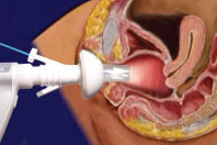
Dilating vaginal speculum inflating vagina and light illuminating.jpg
A dilating vaginal speculum, a tool for examining the vagina, in a model of the female reproductive system
SystemFemale reproductive system
SubdivisionsOncology, Maternal medicine, Maternal-fetal medicine
Significant diseasesGynaecological cancers, infertility, dysmenorrhea
Significant testsLaparoscopy
SpecialistGynaecologist

Gynaecology or gynecology (see spelling differences) is the medical practice dealing with the health of the female reproductive system (vagina, uterus, and ovaries). Outside medicine, the term means "the science of women". Its counterpart is andrology, which deals with medical issues specific to the male reproductive system.

Almost all modern gynaecologists are also obstetricians. In many areas, the specialities of gynaecology and obstetrics overlap.

Etymology

The word "gynaecology" comes from the oblique stem (γυναικ-) of Greek γυνή (gyne), "woman", and -logia, "study".

History

The Kahun Gynaecological Papyrus, dated to about 1800 BC, deals with women's health —gynaecological diseases, fertility, pregnancy, contraception, etc. The text is divided into thirty-four sections, each section dealing with a specific problem and containing diagnosis and treatment; no prognosis is suggested. Treatments are non surgical, comprising applying medicines to the affected body part or swallowing them. The womb is at times seen as the source of complaints manifesting themselves in other body parts.

The Hippocratic Corpus contains several gynaecological treatises dating to the 5th/4th centuries BC. Aristotle is another strong source for medical texts from the 4th century BC with his descriptions of biology primarily found in History of Animals, Parts of Animals, Generation of Animals. The gynaecological treatise Gynaikeia by Soranus of Ephesus (1st/2nd century AD) is extant (together with a 6th-century Latin paraphrase by Muscio, a physician of the same school). He was the chief representative of the school of physicians known as the "Methodists".

J. Marion Sims is widely considered the father of modern gynaecology. Now criticized for his practices, Sims developed some of his techniques by operating on slaves, many of whom were not given anaesthesia. Sims performed surgeries on 12 enslaved women in his homemade backyard hospital for four years. While performing these surgeries he invited men physicians and students to watch invasive and painful procedures while the women were exposed. On one of the women, named Anarcha, he performed 30 surgeries without anesthesia. Due to having so many enslaved women, he would rotate from one to another, continuously trying to perfect the repair of their fistulas. Physicians and students lost interest in assisting Sims over the course of his backyard practice, and he recruited other enslaved women, who were healing from their own surgeries, to assist him. In 1855 Sims went on to found the Woman's Hospital in New York, the first hospital specifically for female disorders.

Examination

The historic taboo associated with the examination of female genitalia has long inhibited the science of gynaecology. This 1822 drawing by Jacques-Pierre Maygrier shows a "compromise" procedure, in which the physician is kneeling before the woman but cannot see her genitalia. Modern gynaecology no longer uses such a position.

In some countries, women must first see a general practitioner (GP; also known as a family practitioner (FP)) prior to seeing a gynaecologist. If their condition requires training, knowledge, surgical procedure, or equipment unavailable to the GP, the patient is then referred to a gynaecologist. In the United States, however, law and many health insurance plans allow gynaecologists to provide primary care in addition to aspects of their own specialty. With this option available, some women opt to see a gynaecological surgeon for non-gynaecological problems without another physician's referral. 

As in all of medicine, the main tools of diagnosis are clinical history and examination. Gynaecological examination is quite intimate, more so than a routine physical exam. It also requires unique instrumentation such as the speculum. The speculum consists of two hinged blades of concave metal or plastic which are used to retract the tissues of the vagina and permit examination of the cervix, the lower part of the uterus located within the upper portion of the vagina. Gynaecologists typically do a bimanual examination (one hand on the abdomen and one or two fingers in the vagina) to palpate the cervix, uterus, ovaries and bony pelvis. It is not uncommon to do a rectovaginal examination for complete evaluation of the pelvis, particularly if any suspicious masses are appreciated. Male gynaecologists may have a female chaperone for their examination. An abdominal or vaginal ultrasound can be used to confirm any abnormalities appreciated with the bimanual examination or when indicated by the patient's history.

Diseases

Examples of conditions dealt with by a gynaecologist are:
There is some crossover in these areas. For example, a woman with urinary incontinence may be referred to a urologist.

Therapies

As with all surgical specialties, gynaecologists may employ medical or surgical therapies (or many times, both), depending on the exact nature of the problem that they are treating. Pre- and post-operative medical management will often employ many standard drug therapies, such as antibiotics, diuretics, antihypertensives, and antiemetics. Additionally, gynaecologists make frequent use of specialized hormone-modulating therapies (such as Clomifene citrate and hormonal contraception) to treat disorders of the female genital tract that are responsive to pituitary or gonadal signals.

Surgery, however, is the mainstay of gynaecological therapy. For historical and political reasons, gynaecologists were previously not considered "surgeons", although this point has always been the source of some controversy. Modern advancements in both general surgery and gynaecology, however, have blurred many of the once rigid lines of distinction. The rise of sub-specialties within gynaecology which are primarily surgical in nature (for example urogynaecology and gynaecological oncology) have strengthened the reputations of gynaecologists as surgical practitioners, and many surgeons and surgical societies have come to view gynaecologists as comrades of sorts. As proof of this changing attitude, gynaecologists are now eligible for fellowship in both the American College of Surgeons and Royal Colleges of Surgeons, and many newer surgical textbooks include chapters on (at least basic) gynaecological surgery.

Some of the more common operations that gynaecologists perform include:
  1. Dilation and curettage (removal of the uterine contents for various reasons, including completing a partial miscarriage and diagnostic sampling for dysfunctional uterine bleeding refractive to medical therapy)
  2. Hysterectomy (removal of the uterus)
  3. Oophorectomy (removal of the ovaries)
  4. Tubal ligation (a type of permanent sterilization)
  5. Hysteroscopy (inspection of the uterine cavity)
  6. Diagnostic laparoscopy – used to diagnose and treat sources of pelvic and abdominal pain; perhaps most famously used to provide a definitive diagnosis of endometriosis.
  7. Exploratory laparotomy – may be used to investigate the level of progression of benign or malignant disease, or to assess and repair damage to the pelvic organs.
  8. Various surgical treatments for urinary incontinence, including cystoscopy and sub-urethral slings.
  9. Surgical treatment of pelvic organ prolapse, including correction of cystocele and rectocele.
  10. Appendectomy – often performed to remove site of painful endometriosis implantation or prophylactically (against future acute appendicitis) at the time of hysterectomy or Caesarean section. May also be performed as part of a staging operation for ovarian cancer.
  11. Cervical Excision Procedures (including cryosurgery) – removal of the surface of the cervix containing pre-cancerous cells which have been previously identified on Pap smear.

Specialist training

Gynaecologist
Occupation
NamesDoctor, Medical Specialist
Occupation type
Specialty
Activity sectors
Medicine
Description
Education required
Fields of
employment
Hospitals, Clinics

In the UK the Royal College of Obstetricians and Gynaecologists, based in London, encourages the study and advancement of both the science and practice of obstetrics and gynaecology. This is done through postgraduate medical education and training development, and the publication of clinical guidelines and reports on aspects of the specialty and service provision. The RCOG International Office works with other international organisations to help lower maternal morbidity and mortality in under-resourced countries.

Gynaecologic oncology is a subspecialty of gynaecology, dealing with gynaecology-related cancer.

Gender of physicians

Gynaecology has historically been dominated by male doctors. However, in recent times, as many of the barriers to access the education and training required to successfully practice gynaecology were removed, women have started to outnumber men in the field. Despite this, male gynaecologists typically make more on average than their female counterparts, although both groups work the same number of hours on average.

Possible reasons reported for the decrease in male gynaecologists range from there being a perception of a lack of respect from other doctors towards them, distrust about their motivations for wanting to work exclusively with female sexual organs and questions about their overall character, as well as a concern about being associated with other male gynaecologists who have been arrested for sex offences and limited future employment opportunities.

Surveys have also shown a large and consistent majority of women are uncomfortable having intimate exams done by a male doctor. Women are also more likely to be embarrassed and vague with personal information if the professional is a man, so as a result talk more openly and in greater details when discussing their sexual history with another woman rather than a man, leading to questions about the ability of male gynaecologists to offer quality care to patients. This, when coupled with more women choosing female physicians has decreased the employment opportunities for men choosing to become gynaecologists.

In the United States, it has been reported that 4 in 5 students choosing a residency in gynaecology are now female. In Sweden, to comply with discrimination laws, patients may not choose a doctor—regardless of specialty—based on factors such as ethnicity or gender and declining to see a doctor solely because of preference regarding e.g. the practitioner's skin color or gender may legally be viewed as refusing care. In Turkey, due to patient preference to be seen by another female, there are now few male gynaecologists working in the field.

There have been a number of legal challenges in the US against healthcare providers who have started hiring based on the gender of physicians. Dr Mircea Veleanu argued, in part, that his former employers discriminated against him by accommodating the wishes of female patients who had requested female doctors for intimate exams. A male nurse complained about an advert for an all-female obstetrics and gynaecology practice in Columbia, Maryland claiming this was a form of sexual discrimination. Dr David Garfinkel, a New Jersey-based ob-gyn sued his former employer after being fired due to, as he claimed, "because I was male, I wasn't drawing as many patients as they'd expected".

So far, all legal challenges by male gynaecologists to remove patient choice have failed due to there being protection in law for 'bona fide occupational qualification' which in previous cases involving wash-room attendants and male nurses have recognized justification for gender-based requirements for certain jobs.

Obstetrics

From Wikipedia, the free encyclopedia

Obstetrics is the field of study concentrated on pregnancy, childbirth, and the postpartum period. As a medical specialty, obstetrics is combined with gynaecology under the discipline known as obstetrics and gynecology (OB/GYN), which is a surgical field.

Main areas

Prenatal care

Prenatal care is important in screening for various complications of pregnancy. This includes routine office visits with physical exams and routine lab tests:

First trimester

Genetic screening for Down syndrome (trisomy 21) and trisomy 18, the national standard in the United States, is rapidly evolving away from the AFP-Quad screen for Down syndrome, done typically in the second trimester at 16–18 weeks. The newer integrated screen (formerly called F.A.S.T.E.R for First And Second Trimester Early Results) can be done at 10 plus weeks to 13 plus weeks with an ultrasound of the fetal neck (thicker nuchal skin correlates with higher risk of Down syndrome being present) and two chemicals (analytes) PAPP-A and βHCG (pregnancy hormone level itself). It gives an accurate risk profile very early. A second blood screen at 15 to 20 weeks refines the risk more accurately. The cost is higher than an "AFP-quad" screen due to the ultrasound and second blood test, but it is quoted to have a 93% pick up rate as opposed to 88% for the standard AFP/QS. This is an evolving standard of care in the United States.

Second trimester

  • MSAFP/quad. screen (four simultaneous blood tests) (maternal serum AFP, inhibin A, estriol, & βHCG) – elevations, low numbers or odd patterns correlate with neural tube defect risk and increased risks of trisomy 18 or trisomy 21
  • Ultrasound either abdominal or transvaginal to assess cervix, placenta, fluid and baby
  • Amniocentesis is the national standard (in what country) for women over 35 or who reach 35 by mid pregnancy or who are at increased risk by family history or prior birth history.

Third trimester

  • Hematocrit (if low, the mother receives iron supplements)
  • Group B Streptococcus screen. If positive, the woman receives IV penicillin or ampicillin while in labor—or, if she is allergic to penicillin, an alternative therapy, such as IV clindamycin or IV vancomycin.
  • Glucose loading test (GLT) – screens for gestational diabetes; if > 140 mg/dL, a glucose tolerance test (GTT) is administered; a fasting glucose > 105 mg/dL suggests gestational diabetes.
Most doctors do a sugar load in a drink form of 50 grams of glucose in cola, lime or orange and draw blood an hour later (plus or minus 5 minutes). The standard modified criteria have been lowered to 135 since the late 1980s. 

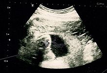
The result of an Ultrasonography: a black and white image that shows a clear view of the interior abdomen.

Fetal assessments

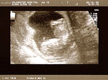
A dating scan at 12 weeks.
 
Obstetric ultrasonography is routinely used for dating the gestational age of a pregnancy from the size of the fetus, determine the number of fetuses and placentae, evaluate for an ectopic pregnancy and first trimester bleeding, the most accurate dating being in first trimester before the growth of the foetus has been significantly influenced by other factors. Ultrasound is also used for detecting congenital anomalies (or other foetal anomalies) and determining the biophysical profiles (BPP), which are generally easier to detect in the second trimester when the foetal structures are larger and more developed. Specialised ultrasound equipment can also evaluate the blood flow velocity in the umbilical cord, looking to detect a decrease/absence/reversal or diastolic blood flow in the umbilical artery.

X-rays and computerized tomography (CT) are not used, especially in the first trimester, due to the ionizing radiation, which has teratogenic effects on the foetus. No effects of magnetic resonance imaging (MRI) on the foetus have been demonstrated, but this technique is too expensive for routine observation. Instead, obstetric ultrasonography is the imaging method of choice in the first trimester and throughout the pregnancy, because it emits no radiation, is portable, and allows for realtime imaging.

The safety of frequent ultrasound scanning has not be confirmed. Despite this, increasing numbers of women are choosing to have additional scans for no medical purpose, such as gender scans, 3D and 4D scans. A normal gestation would reveal a gestational sac, yolk sac, and fetal pole. The gestational age can be assessed by evaluating the mean gestational sac diameter (MGD) before week 6, and the crown-rump length after week 6. Multiple gestation is evaluated by the number of placentae and amniotic sacs present.

Other tools used for assessment include:

Intercurrent diseases

A pregnant woman may have intercurrent diseases, that is, other diseases or conditions (not directly caused by the pregnancy) that may become worse or be a potential risk to the pregnancy.

Induction and labour

Induction is a method of artificially or prematurely stimulating labour in a woman. Reasons to induce can include pre-eclampsia, foetal distress, placental malfunction, intrauterine growth retardation and failure to progress through labour increasing the risk of infection and foetal distresses.

Induction may be achieved via several methods:
  • Disturbance of cervical memebranes
  • Pessary of Prostin cream, prostaglandin E2
  • Intravaginal or oral administration of misoprostol
  • Cervical insertion of a 30-mL Foley catheter
  • Rupturing the amniotic membranes
  • Intravenous infusion of synthetic oxytocin (Pitocin or Syntocinon)
During labour, the obstetrician carries out the following tasks:

Complications and emergencies

The main emergencies include:
  • Ectopic pregnancy is when an embryo implants in the uterine (Fallopian) tube or (rarely) on the ovary or inside the peritoneal cavity. This may cause massive internal bleeding.
  • Pre-eclampsia is a disease defined by a combination of signs and symptoms that are related to maternal hypertension. The cause is unknown, and markers are being sought to predict its development from the earliest stages of pregnancy. Some unknown factors cause vascular damage in the endothelium, causing hypertension. If severe, it progresses to eclampsia, where seizures occur, which can be fatal. Preeclamptic patients with the HELLP syndrome show liver failure and Disseminated intravascular coagulation (DIC). The only treatment is to deliver the foetus. Women may still develop pre-eclampsia following delivery.
  • Placental abruption is where the placenta detaches from the uterus and the woman and foetus can bleed to death if not managed appropriately.
  • Foetal distress where the foetus is getting compromised in the uterine environment.
  • Shoulder dystocia where one of the foetus' shoulders becomes stuck during vaginal birth. There are many risk factors, including macrosmic (large) foetus, but many are also unexplained.
  • Uterine rupture can occur during obstructed labour and endanger foetal and maternal life.
  • Prolapsed cord can only happen after the membranes have ruptured. The umbilical cord delivers before the presenting part of the foetus. If the foetus is not delivered within minutes, or the pressure taken off the cord, the foetus dies.
  • Obstetrical hemorrhage may be due to a number of factors such as placenta previa, uterine rupture or tears, uterine atony, retained placenta or placental fragments, or bleeding disorders.
  • Puerperal sepsis is an ascending infection of the genital tract. It may happen during or after labour. Signs to look out for include signs of infection (pyrexia or hypothermia, raised heart rate and respiratory rate, reduced blood pressure), and abdominal pain, offensive lochia (blood loss) increased lochia, clots, diarrhea and vomiting.

Postnatal care

Postnatal care is care provided to the mother following parturition.

A woman in the Western world who is delivering in a hospital may leave the hospital as soon as she is medically stable and chooses to leave, which can be as early as a few hours postpartum, though the average for spontaneous vaginal delivery (SVD) is 1–2 days, and the average caesarean section postnatal stay is 3–4 days.

During this time the mother is monitored for bleeding, bowel and bladder function, and baby care. The infant's health is also monitored.

Certain things must be kept in mind as the physician proceeds with the post-natal care.
  1. General condition of the patient.
  2. Check for vital signs (pulse, blood pressure, temperature, respiratory rate, (pain) at times)
  3. Palour?
  4. Oedema?
  5. Dehydration?
  6. Fundus (height following parturition, and the feel of the fundus) (Per abdominal examination)
  7. If an episiotomy or a C-section was performed, check for the dressing. Intact, pus, oozing, haematomas?
  8. Lochia (colour, amount, odour)?
  9. Bladder (keep the patient catheterized for 12 hours following local anaesthesia and 24–48 hours after general anaesthesia) ? (check for bladder function)
  10. Bowel movements?
  11. More bowel movements?
  12. Follow up with the neonate to check if they are healthy.

Veterinary obstetrics

History

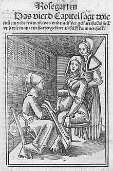
Two midwives assisting a woman in labour on a birthing chair in the 16th century, from a work by Eucharius Rößlin.

Prior to the 18th century, caring for pregnant women in Europe was confined exclusively to women, and rigorously excluded men. The expectant mother would invite close female friends and family members to her home to keep her company during childbirth. Skilled midwives managed all aspects of the labour and delivery. The presence of physicians and surgeons was very rare and only occurred if a serious complication had taken place and the midwife had exhausted all measures at her disposal. Calling a surgeon was very much a last resort and having men deliver women in this era was seen as offending female modesty.

Before the 18th century

Prior to the 18th and 19th centuries, midwifery was well established but obstetrics was not recognized as a specific medical specialty. However, the subject matter and interest in the female reproductive system and sexual practice can be traced back to Ancient Egypt and Ancient Greece. Soranus of Ephesus sometimes is called the most important figure in ancient gynecology. Living in the late first century A.D. and early second century, he studied anatomy and had opinions and techniques on abortion, contraception –most notably coitus interruptus– and birth complications. After his death, techniques and works of gynecology declined; very little of his works were recorded and survived to the late 18th century when gynecology and obstetrics reemerged as a medical specialism.

18th century

The 18th century marked the beginning of many advances in European midwifery, based on better knowledge of the physiology of pregnancy and labour. By the end of the century, medical professionals began to understand the anatomy of the uterus and the physiological changes that take place during labour. The introduction of forceps in childbirth also took place at this time. All these medical advances in obstetrics were a lever for the introduction of men into an arena previously managed and run by women—midwifery.

The addition of the male-midwife (or man-midwife) is historically a significant change to the profession of obstetrics. In the 18th century medical men began to train in area of childbirth and believed with their advanced knowledge in anatomy that childbirth could be improved. In France these male-midwives were referred to as accoucheurs, a title later used all over Europe. The founding of lying-in hospitals also contributed to the medicalization and male-dominance of obstetrics. These early maternity hospitals were establishments where women would come to have their babies delivered, as opposed to the practice since time immemorial of the midwife attending the home of the woman in labour. This institution provided male-midwives with endless patients to practice their techniques on and was a way for these men to demonstrate their knowledge.

Many midwives of the time bitterly opposed the involvement of men in childbirth. Some male practitioners also opposed the involvement of medical men like themselves in midwifery and even went as far as to say that male-midwives only undertook midwifery solely for perverse erotic satisfaction. The accoucheurs argued that their involvement in midwifery was to improve the process of childbirth. These men also believed that obstetrics would forge ahead and continue to strengthen.

19th century

18th century physicians expected that obstetrics would continue to grow, but the opposite happened. Obstetrics entered a stage of stagnation in the 19th century, which lasted until about the 1880s. The central explanation for the lack of advancement during this time was the rejection of obstetrics by the medical community. The 19th century marked an era of medical reform in Europe and increased regulation over the profession. Major European institutions such as The College of Physicians and Surgeons considered delivering babies ungentlemanly work and refused to have anything to do with childbirth as a whole. Even when Medical Act 1858 was introduced, which stated that medical students could qualify as doctors, midwifery was entirely ignored. This made it nearly impossible to pursue an education in midwifery and also have the recognition of being a doctor or surgeon. Obstetrics was pushed to the side.

By the late 19th century, the foundation of modern-day obstetrics and midwifery began developing. Delivery of babies by doctors became popular and readily accepted, but midwives continued to play a role in childbirth. Midwifery also changed during this era due to increased regulation and the eventual need for midwives to become certified. Many European countries by the late 19th century were monitoring the training of midwives and issued certification based on competency. Midwives were no longer uneducated in the formal sense.

As midwifery began to develop, so did the profession of obstetrics near the end of the century. Childbirth was no longer unjustifiably despised by the medical community as it once had been at the beginning of the century. But obstetrics was underdeveloped compared to other medical specialites. Many male physicians would deliver children but very few would have referred to themselves as obstetricians. The end of the 19th century did mark a significant accomplishment in the profession with the advancements in asepsis and anaesthesia, which paved the way for the mainstream introduction and later success of the Caesarean Section.

Before the 1880s mortality rates in lying-hospitals would reach unacceptably high levels and became an area of public concern. Much of these maternal deaths were due to puerperal fever, then known as childbed fever. In the 1800s Dr. Ignaz Semmelweis noticed that women giving birth at home had a much lower incidence of childbed fever than those giving birth by physicians in lying-hospitals. His investigation discovered that washing hands with an antiseptic solution before a delivery reduced childbed fever fatalities by 90%. So it was concluded that it was physicians who had been spreading disease from one labouring mother to the next. Despite the publication of this information, doctors still would not wash. It was not until the 20th century when advancements in aseptic technique and the understanding of disease would play a significant role in the decrease of maternal mortality rates among many populations.

History of obstetrics in America

The development of obstetrics as a practice for accredited doctors happened at the turn of the 18th century and thus was very differently developed in Europe and in the Americas due to the independence of many countries in the Americas from European powers. “Unlike in Europe and the British Isles, where midwifery laws were national, in America, midwifery laws were local and varied widely”.

Gynaecology and Obstetrics gained attention in the American medical field at the end of the nineteenth century through the development of such procedures as the ovariotomy. These procedures then were shared with European surgeons who replicated the surgeries. This was a period when antiseptic, aseptic or anaesthetic measures were just being introduced to surgical and observational procedures and without these procedures surgeries were dangerous and often fatal. Following are two surgeons noted for their contributions to these fields include Ephraim McDowell and James Marion Sims

Ephraim McDowell developed a surgical practice in 1795 and performed the first ovariotomy in 1809 on a 47-year-old widow who then lived on for thirty-one more years. He had attempted to share this with John Bell whom he had practiced under who had retired to Italy. Bell was said to have died without seeing the document but it was published by an associate in Extractions of Diseased Ovaria in 1825. By the mid-century the surgery was both successfully and unsuccessfully being performed. Pennsylvanian surgeons the Attlee brothers made this procedure very routine for a total of 465 surgeries–John Attlee performed 64 successfully of 78 while his brother William reported 387– between the years of 1843 and 1883. By the middle of the nineteenth century this procedure was successfully performed in Europe by English surgeons Sir Spencer Wells and Charles Clay as well as French surgeons Eugène Koeberlé, Auguste Nélaton and Jules Péan.

J. Marion Sims was the surgeon responsible for being the first treating a vesicovaginal fistula – a condition linked to many caused mainly by prolonged pressing of the feotus against the pelvis or other causes such as rape, hysterectomy, or other operations– and also having been doctor to many European royals and the 20th President of the United States James A. Garfield after he had been shot. Sims does have a controversial medical past. Under the beliefs at the time about pain and the prejudice towards African people, he had practiced his surgical skills and developed skills on slaves. These women were the first patients of modern gynecology. One of the women he operated on was named Anarcha Westcott, the woman he first treated for a fistula.

Historical role of gender

Women and men inhabited very different roles in natal care up to the 18th century. The role of a physician was exclusively held by men who went to university, an overly male institution, who would theorize anatomy and the process of reproduction based on theological teaching and philosophy. Many beliefs about the female body and menstruation in the 17th and 18th centuries were inaccurate; clearly resulting from the lack of literature about the practice. Many of the theories of what caused menstruation prevailed from Hippocratic philosophy. Midwives of this time were those assisted in the birth and care of both born and unborn children, and as the name suggests, this position was held mainly by women.

During the birth of a child, men were rarely present. Women from the neighbourhood or family would join in on the process of birth and assist in many different ways. The one position where men would help with the birth of a child would be in the sitting position, usually when performed on the side of a bed to support the mother.

Men were introduced into the field of obstetrics in the nineteenth century and resulted in a change of the focus of this profession. Gynaecology directly resulted as a new and separate field of study from obstetrics and focused on the curing of illness and indispositions of female sexual organs. This had some relevance to some conditions as menopause, uterine and cervical problems, and childbirth could leave the mother in need of extensive surgery to repair tissue. But, there was also a large blame of the uterus for completely unrelated conditions. This led to many social consequences of the nineteenth century.

Green development

From Wikipedia, the free encyclopedia https://en.wikipedia.org/w...