Search This Blog

Sunday, October 11, 2020

Urea cycle

From Wikipedia, the free encyclopedia

The urea cycle (also known as the ornithine cycle) is a cycle of biochemical reactions that produces urea (NH2)2CO from ammonia (NH3). This cycle occurs in ureotelic organisms. The urea cycle converts highly toxic ammonia to urea for excretion. This cycle was the first metabolic cycle to be discovered (Hans Krebs and Kurt Henseleit, 1932), five years before the discovery of the TCA cycle. This cycle was described in more detail later on by Ratner and Cohen. The urea cycle takes place primarily in the liver and, to a lesser extent, in the kidneys.

Function

Amino acid catabolism results in waste ammonia. All animals need a way to excrete this product. Most aquatic organisms, or ammonotelic organisms, excrete ammonia without converting it. Organisms that cannot easily and safely remove nitrogen as ammonia convert it to a less toxic substance such as urea via the urea cycle, which occurs mainly in the liver. Urea produced by the liver is then released into the bloodstream where it travels to the kidneys and is ultimately excreted in urine. The urea cycle is essential to these organisms, because if the nitrogen or ammonia are not eliminated from the organism it can be very detrimental. In species including birds and most insects, the ammonia is converted into uric acid or its urate salt, which is excreted in solid form.

Reactions

The entire process converts two amino groups, one from NH+
4
and one from Aspartate, and a carbon atom from HCO
3
, to the relatively nontoxic excretion product urea at the cost of four "high-energy" phosphate bonds (3 ATP hydrolyzed to 2 ADP and one AMP). The conversion from ammonia to urea happens in five main steps. The first is needed for ammonia to enter the cycle and the following four are all a part of the cycle itself. To enter the cycle, ammonia is converted to carbamoyl phosphate. The urea cycle consists of four enzymatic reactions: one mitochondrial and three cytosolic.

Reactions of the urea cycle

Step Reactants Products Catalyzed by Location
1 NH3 + HCO
3
+ 2ATP
carbamoyl phosphate + 2ADP + Pi CPS1 mitochondria
2 carbamoyl phosphate + ornithine citrulline + Pi OTC, zinc, biotin mitochondria
3 citrulline + aspartate + ATP argininosuccinate + AMP + PPi ASS cytosol
4 argininosuccinate arginine + fumarate ASL cytosol
5 arginine + H2O ornithine + urea ARG1, manganese cytosol
The reactions of the urea cycle
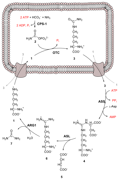
Urea cycle.svg

1 L-ornithine
2 carbamoyl phosphate
3 L-citrulline
4 argininosuccinate
5 fumarate
6 L-arginine
7 urea
L-Asp L-aspartate
CPS-1 carbamoyl phosphate synthetase I
OTC Ornithine transcarbamoylase
ASS argininosuccinate synthetase
ASL argininosuccinate lyase
ARG1 arginase 1

First reaction: entering the urea cycle

Before the urea cycle begins ammonia is converted to carbamoyl phosphate. The reaction is catalyzed by carbamoyl phosphate synthetase I and requires the use of two ATP molecules. The carbamoyl phosphate then enters the urea cycle.

Steps of the urea cycle

  1. Carbamoyl phosphate is converted to citrulline. With catalysis by ornithine transcarbamoylase, the carbamoyl phosphate group is donated to ornithine and releases a phosphate group.
  2. A condensation reaction occurs between the amino group of aspartate and the carbonyl group of citrulline to form argininosuccinate. This reaction is ATP dependent and is catalyzed by argininosuccinate synthetase.
  3. Argininosuccinate undergoes cleavage by argininosuccinase to form arginine and fumarate.
  4. Arginine is cleaved by arginase to form urea and ornithine. The ornithine is then transported back to the mitochondria to begin the urea cycle again.

Overall reaction equation

In the first reaction, NH+
4
+ HCO
3
is equivalent to NH3 + CO2 + H2O.

Thus, the overall equation of the urea cycle is:

Since fumarate is obtained by removing NH3 from aspartate (by means of reactions 3 and 4), and PPi + H2O → 2 Pi, the equation can be simplified as follows:

Note that reactions related to the urea cycle also cause the production of 2 NADH, so the overall reaction releases slightly more energy than it consumes. The NADH is produced in two ways:

We can summarize this by combining the reactions:

The two NADH produced can provide energy for the formation of 5 ATP (cytosolic NADH provides 2.5 ATP with the malate-aspartate shuttle in human liver cell), a net production of two high-energy phosphate bond for the urea cycle. However, if gluconeogenesis is underway in the cytosol, the latter reducing equivalent is used to drive the reversal of the GAPDH step instead of generating ATP.

The fate of oxaloacetate is either to produce aspartate via transamination or to be converted to phosphoenolpyruvate, which is a substrate for gluconeogenesis.

Products of the urea cycle

As stated above many vertebrates use the urea cycle to create urea out of ammonium so that the ammonium does not damage the body. Though this is helpful, there are other effects of the urea cycle. For example: consumption of two ATP, production of urea, generation of H+, the combining of HCO3- and NH4+ to forms where it can be regenerated, and finally the consumption of NH4+.

Regulation

N-Acetylglutamic acid

The synthesis of carbamoyl phosphate and the urea cycle are dependent on the presence of N-acetylglutamic acid (NAcGlu), which allosterically activates CPS1. NAcGlu is an obligate activator of carbamoyl phosphate synthetase. Synthesis of NAcGlu by N-acetylglutamate synthase (NAGS) is stimulated by both Arg, allosteric stimulator of NAGS, and Glu, a product in the transamination reactions and one of NAGS's substrates, both of which are elevated when free amino acids are elevated. So Glu not only is a substrate for NAGS but also serves as an activator for the urea cycle.

Substrate concentrations

The remaining enzymes of the cycle are controlled by the concentrations of their substrates. Thus, inherited deficiencies in cycle enzymes other than ARG1 do not result in significant decreases in urea production (if any cycle enzyme is entirely missing, death occurs shortly after birth). Rather, the deficient enzyme's substrate builds up, increasing the rate of the deficient reaction to normal.

The anomalous substrate buildup is not without cost, however. The substrate concentrations become elevated all the way back up the cycle to NH+
4
, resulting in hyperammonemia (elevated [NH+
4
]P).

Although the root cause of NH+
4
toxicity is not completely understood, a high [NH+
4
] puts an enormous strain on the NH+
4
-clearing system, especially in the brain (symptoms of urea cycle enzyme deficiencies include intellectual disability and lethargy). This clearing system involves GLUD1 and GLUL, which decrease the 2-oxoglutarate (2OG) and Glu pools. The brain is most sensitive to the depletion of these pools. Depletion of 2OG decreases the rate of TCAC, whereas Glu is both a neurotransmitter and a precursor to GABA, another neurotransmitter. 

Link with the citric acid cycle

The urea cycle and the citric acid cycle are independent cycles but are linked. One of the nitrogens in the urea cycle is obtained from the transamination of oxaloacetate to aspartate. The fumarate that is produced in step three is also an intermediate in the citric acid cycle and is returned to that cycle.

Urea cycle disorders

Urea cycle disorders are rare and affect about one in 35,000 people in the United States. Genetic defects in the enzymes involved in the cycle can occur, which usually manifest within a few days after birth. The recently born child will typically experience varying bouts of emesis and periods of lethargy.

Ultimately the infant may go into a coma and develop brain damage. Newborns with UCD are at a much higher risk of complications or death due to untimely screening tests and misdiagnosed cases. The most common misdiagnosis is neonatal sepsis. Signs of UCD can be present within the first 2-3 days of life, however, the present method to get confirmation by test results can take too long and can potentially cause complications such as coma or death.

Urea cycle disorders may also be diagnosed in adults, and symptoms may include delirium episodes, lethargy, and symptoms similar to that of a stroke. On top of these symptoms if the urea cycle begins to malfunction in the liver the patient may obtain cirrhosis which can also lead to sarcopenia (the loss of muscle mass). Mutations lead to deficiencies of the various enzymes and transporters involved in the urea cycle and cause urea cycle disorders. If individuals with a defect in any of the six enzymes used in the cycle ingest amino acids beyond what is necessary for the minimum daily requirements, then the ammonia that is produced will not be able to be converted to urea. These individuals can experience hyperammonemia or the buildup of a cycle intermediate.

Individual disorders


All urea cycle defects, except OTC deficiency, are inherited in an autosomal recessive manner. OTC deficiency is inherited as an X-linked recessive disorder, although some females can show symptoms. Most urea cycle disorders are associated with hyperammonemia, however argininemia and some forms of argininosuccinic aciduria do not present with elevated ammonia.


Le Chatelier's principle


From Wikipedia, the free encyclopedia
Jump to navigation Jump to search

Le Chatelier's principle (pronounced UK: /lə ʃæˈtɛlj/ or US: /ˈʃɑːtəlj/), also called Chatelier's principle or "The Equilibrium Law", is a principle of chemistry used to predict the effect of a change in conditions on chemical equilibria. The principle is named after French chemist Henry Louis Le Chatelier, and sometimes also credited to Karl Ferdinand Braun, who discovered it independently. It can be stated as:

When any system at equilibrium for a long period of time is subjected to a change in concentration, temperature, volume, or pressure, (1) the system changes to a new equilibrium, and (2) this change partly counteracts the applied change.

It is common to treat the principle as a more general observation of systems, such as:

When a settled system is disturbed, it will adjust to diminish the change that has been made to it:

or, "roughly stated",

Any change in status quo prompts an opposing reaction in the responding system.

The concept of systemic maintenance of an equilibrium state despite perturbations has a variety of names, depending upon the discipline using it (e.g. homeostasis, an idea which encompasses the concept, is commonly used in biology), and has been studied in a variety of contexts, chiefly in the natural sciences. In chemistry, the principle is used to manipulate the outcomes of reversible reactions, often to increase their yield. In pharmacology, the binding of ligands to receptors may shift the equilibrium according to Le Chatelier's principle, thereby explaining the diverse phenomena of receptor activation and desensitization. In economics, the principle has been generalized to help explain the price equilibrium of efficient economic systems.

Phenomena in apparent contradiction to Le Chatelier's principle can also arise in systems of simultaneous equilibrium (see response reactions).

As a physical law

Le Chatelier's principle describes the qualitative behavior of systems where there is an externally induced, instantaneous change in one parameter of a system; it states that a behavioral shift occurs in the system so as to oppose (partly cancel) the parameter change. The duration of adjustment depends on the strength of the negative feedback to the initial shock. Where a shock initially induces positive feedback (such as thermal runaway), the new equilibrium can be far from the old one, and can take a long time to reach. In some dynamic systems, the end-state cannot be determined from the shock. The principle is typically used to describe closed negative-feedback systems, but applies, in general, to thermodynamically closed and isolated systems in nature, since the second law of thermodynamics ensures that the disequilibrium caused by an instantaneous shock must have a finite half-life. The principle has analogs throughout the entire physical world.

While well rooted in chemical equilibrium and extended into economic theory, Le Chatelier's principle can also be used in describing mechanical systems in that a system put under stress will respond in such a way as to reduce or minimize that stress. Moreover, the response will generally be via the mechanism that most easily relieves that stress. Shear pins and other such sacrificial devices are design elements that protect systems against stress applied in undesired manners to relieve it so as to prevent more extensive damage to the entire system, a practical engineering application of Le Chatelier's principle.

Chemistry

Effect of change in concentration

Changing the concentration of a chemical will shift the equilibrium to the side that would counter that change in concentration. The chemical system will attempt to partly oppose the change affected to the original state of equilibrium. In turn, the rate of reaction, extent, and yield of products will be altered corresponding to the impact on the system.

This can be illustrated by the equilibrium of carbon monoxide and hydrogen gas, reacting to form methanol.

CO + 2 H2 ⇌ CH3OH

Suppose we were to increase the concentration of CO in the system. Using Le Chatelier's principle, we can predict that the amount of methanol will increase, decreasing the total change in CO. If we are to add a species to the overall reaction, the reaction will favor the side opposing the addition of the species. Likewise, the subtraction of a species would cause the reaction to "fill the gap" and favor the side where the species was reduced. This observation is supported by the collision theory. As the concentration of CO is increased, the frequency of successful collisions of that reactant would increase also, allowing for an increase in forward reaction, and generation of the product. Even if the desired product is not thermodynamically favored, the end-product can be obtained if it is continuously removed from the solution.

The effect of a change in concentration is often exploited synthetically for condensation reactions (i.e., reactions that extrude water) that are equilibrium processes (e.g., formation of an ester from carboxylic acid and alcohol or an imine from an amine and aldehyde). This can be achieved by physically sequestering water, by adding desiccants like anhydrous magnesium sulfate or molecular sieves, or by continuous removal of water by distillation, often facilitated by a Dean-Stark apparatus.

Effect of change in temperature

The reversible reaction N2O4(g) ⇌ 2NO2(g) is endothermic, so the equilibrium position can be shifted by changing the temperature.
When heat is added and the temperature increases, the reaction shifts to the right and the flask turns reddish brown due to an increase in NO2. This demonstrates Le Chatelier's principle: the equilibrium shifts in the direction that consumes energy.
When heat is removed and the temperature decreases, the reaction shifts to the left and the flask turns colorless due to an increase in N2O4: again, according to Le Chatelier's principle.

The effect of changing the temperature in the equilibrium can be made clear by 1) incorporating heat as either a reactant or a product, and 2) assuming that an increase in temperature increases the heat content of a system. When the reaction is exothermicH is negative and energy is released), heat is included as a product, and when the reaction is endothermicH is positive and energy is consumed), heat is included as a reactant. Hence, whether increasing or decreasing the temperature would favor the forward or the reverse reaction can be determined by applying the same principle as with concentration changes.

Take, for example, the reversible reaction of nitrogen gas with hydrogen gas to form ammonia:

N2(g) + 3 H2(g) ⇌ 2 NH3(g)    ΔH = -92 kJ mol−1

Because this reaction is exothermic, it produces heat:

N2(g) + 3 H2(g) ⇌ 2 NH3(g) + heat

If the temperature were increased, the heat content of the system would increase, so the system would consume some of that heat by shifting the equilibrium to the left, thereby producing less ammonia. More ammonia would be produced if the reaction were run at a lower temperature, but a lower temperature also lowers the rate of the process, so, in practice (the Haber process) the temperature is set at a compromise value that allows ammonia to be made at a reasonable rate with an equilibrium concentration that is not too unfavorable.

In exothermic reactions, an increase in temperature decreases the equilibrium constant, K, whereas in endothermic reactions, an increase in temperature increases K.

Le Chatelier's principle applied to changes in concentration or pressure can be understood by giving K a constant value. The effect of temperature on equilibria, however, involves a change in the equilibrium constant. The dependence of K on temperature is determined by the sign of ΔH. The theoretical basis of this dependence is given by the Van 't Hoff equation.

Effect of change in pressure

The equilibrium concentrations of the products and reactants do not directly depend on the total pressure of the system. They may depend on the partial pressures of the products and reactants, but if the number of moles of gaseous reactants is equal to the number of moles of gaseous products, pressure has no effect on equilibrium.

Changing total pressure by adding an inert gas at constant volume does not affect the equilibrium concentrations (see Effect of adding an inert gas below).

Changing total pressure by changing the volume of the system changes the partial pressures of the products and reactants and can affect the equilibrium concentrations (see §Effect of change in volume below).

Effect of change in volume

Changing the volume of the system changes the partial pressures of the products and reactants and can affect the equilibrium concentrations. With a pressure increase due to a decrease in volume, the side of the equilibrium with fewer moles is more favorable and with a pressure decrease due to an increase in volume, the side with more moles is more favorable. There is no effect on a reaction where the number of moles of gas is the same on each side of the chemical equation.

Considering the reaction of nitrogen gas with hydrogen gas to form ammonia:

N2 + 3 H24 moles2 NH32 moles    ΔH = -92kJ mol−1

Note the number of moles of gas on the left-hand side and the number of moles of gas on the right-hand side. When the volume of the system is changed, the partial pressures of the gases change. If we were to decrease pressure by increasing volume, the equilibrium of the above reaction will shift to the left, because the reactant side has a greater number of moles than does the product side. The system tries to counteract the decrease in partial pressure of gas molecules by shifting to the side that exerts greater pressure. Similarly, if we were to increase pressure by decreasing volume, the equilibrium shifts to the right, counteracting the pressure increase by shifting to the side with fewer moles of gas that exert less pressure. If the volume is increased because there are more moles of gas on the reactant side, this change is more significant in the denominator of the equilibrium constant expression, causing a shift in equilibrium.

Effect of adding an inert gas

An inert gas (or noble gas), such as helium, is one that does not react with other elements or compounds. Adding an inert gas into a gas-phase equilibrium at constant volume does not result in a shift. This is because the addition of a non-reactive gas does not change the equilibrium equation, as the inert gas appears on both sides of the chemical reaction equation. For example, if A and B react to form C and D, but X does not participate in the reaction: . While it is true that the total pressure of the system increases, the total pressure does not have any effect on the equilibrium constant; rather, it is a change in partial pressures that will cause a shift in the equilibrium. If, however, the volume is allowed to increase in the process, the partial pressures of all gases would be decreased resulting in a shift towards the side with the greater number of moles of gas. The shift will never occur on the side with fewer moles of gas. It is also known as Le Chatelier's postulate.

Effect of a catalyst

A catalyst increases the rate of a reaction without being consumed in the reaction. The use of a catalyst does not affect the position and composition of the equilibrium of a reaction, because both the forward and backward reactions are sped up by the same factor.

For example, consider the Haber process for the synthesis of ammonia (NH3):

N2 + 3 H2 ⇌ 2 NH3

In the above reaction, iron (Fe) and molybdenum (Mo) will function as catalysts if present. They will accelerate any reactions, but they do not affect the state of the equilibrium.

General statement of Le Chatelier's principle

Le Chatelier's principle refers to states of thermodynamic equilibrium. The latter are stable against perturbations that satisfy certain criteria; this is essential to the definition of thermodynamic equilibrium.

For this, a state of thermodynamic equilibrium is most conveniently described through a fundamental relation that specifies a cardinal function of state, of the energy kind, or of the entropy kind, as a function of state variables chosen to fit the thermodynamic operations through which a perturbation is to be applied.

In theory and, nearly, in some practical scenarios, a body can be in a stationary state with zero macroscopic flows and rates of chemical reaction (for example, when no suitable catalyst is present), yet not in thermodynamic equilibrium, because it is metastable or unstable; then Le Chatelier's principle does not necessarily apply.

General statements related to Le Chatelier's principle

A body can also be in a stationary state with non-zero rates of flow and chemical reaction; sometimes the word "equilibrium" is used in reference to such states, though by definition they are not thermodynamic equilibria. Sometimes, it is proposed to consider Le Chatelier's principle for such states. For this exercise, rates of flow and of chemical reaction must be considered. Such rates are not supplied by equilibrium thermodynamics. For such states, it has turned out to be difficult or unfeasible to make valid and very general statements that echo Le Chatelier's principle. Prigogine and Defay demonstrate that such a scenario may or may not exhibit moderation, depending upon exactly what conditions are imposed after the perturbation.

Economics

In economics, a similar concept also named after Le Chatelier was introduced by American economist Paul Samuelson in 1947. There the generalized Le Chatelier principle is for a maximum condition of economic equilibrium: Where all unknowns of a function are independently variable, auxiliary constraints—"just-binding" in leaving initial equilibrium unchanged—reduce the response to a parameter change. Thus, factor-demand and commodity-supply elasticities are hypothesized to be lower in the short run than in the long run because of the fixed-cost constraint in the short run.

Since the change of the value of an objective function in a neighbourhood of the maximum position is described by the envelope theorem, Le Chatelier's principle can be shown to be a corollary thereof.

Frostbite

From Wikipedia, the free encyclopedia
 
Frostbite
Other namesFrostnip
Frost bite.jpg
Frostbitten toes two to three days after mountain climbing
SpecialtyDermatology Emergency medicine, orthopedics
SymptomsNumbness, feeling cold, clumsy, pale color
ComplicationsHypothermia, compartment syndrome
TypesSuperficial, deep
CausesTemperatures below freezing
Risk factorsAlcohol, smoking, mental health problems, certain medications, prior cold injury
Diagnostic methodBased on symptoms
Differential diagnosisFrostnip, pernio, trench foot
PreventionAvoid cold, wear proper clothing, maintain hydration and nutrition, stay active without becoming exhausted
TreatmentRewarming, medication, surgery
MedicationIbuprofen, tetanus vaccine, iloprost, thrombolytics
FrequencyUnknown

Frostbite occurs when exposure to low temperatures causes freezing of the skin or other tissues. The initial symptom is typically numbness. This may be followed by clumsiness with a white or bluish color to the skin. Swelling or blistering may occur following treatment. The hands, feet, and face are most commonly affected. Complications may include hypothermia or compartment syndrome.

People who are exposed to low temperatures for prolonged periods, such as winter sports enthusiasts, military personnel, and homeless individuals, are at greatest risk. Other risk factors include drinking alcohol, smoking, mental health problems, certain medications, and prior injuries due to cold. The underlying mechanism involves injury from ice crystals and blood clots in small blood vessels following thawing. Diagnosis is based on symptoms. Severity may be divided into superficial (1st and 2nd degree) or deep (3rd and 4th degree). A bone scan or MRI may help in determining the extent of injury.

Prevention is through wearing proper clothing, maintaining hydration and nutrition, avoiding low temperatures, and staying active without becoming exhausted. Treatment is by rewarming. This should be done only when refreezing is not a concern. Rubbing or applying snow to the affected part is not recommended. The use of ibuprofen and tetanus toxoid is typically recommended. For severe injuries iloprost or thrombolytics may be used. Surgery is sometimes necessary. Amputation, however, should generally be delayed for a few months to allow determination of the extent of injury.

The number of cases of frostbite is unknown. Rates may be as high as 40% a year among those who mountaineer. The most common age group affected is those 30 to 50 years old. Evidence of frostbite occurring in people dates back 5,000 years. Frostbite has also played an important role in a number of military conflicts. The first formal description of the condition was in 1813 by Dominique Jean Larrey, a physician in Napoleon's army, during its invasion of Russia.

Signs and symptoms

Frostbite

Areas that are usually affected include cheeks, ears, nose and fingers and toes. Frostbite is often preceded by frostnip. The symptoms of frostbite progress with prolonged exposure to cold. Historically, frostbite has been classified by degrees according to skin and sensation changes, similar to burn classifications. However, the degrees do not correspond to the amount of long term damage. A simplification of this system of classification is superficial (first or second degree) or deep injury (third or fourth degree).

First degree

  • First degree frostbite is superficial, surface skin damage that is usually not permanent.
  • Early on, the primary symptom is loss of feeling in the skin. In the affected areas, the skin is numb, and possibly swollen, with a reddened border.
  • In the weeks after injury, the skin's surface may slough off.

Second degree

  • In second degree frostbite, the skin develops clear blisters early on, and the skin's surface hardens.
  • In the weeks after injury, this hardened, blistered skin dries, blackens, and peels.
  • At this stage, lasting cold sensitivity and numbness can develop.

Third degree

  • In third degree frostbite, the layers of tissue below the skin freeze.
  • Symptoms include blood blisters and "blue-grey discoloration of the skin".
  • In the weeks after injury, pain persists and a blackened crust (eschar) develops.
  • There can be longterm ulceration and damage to growth plates.

Fourth degree

Frostbite 12 days later
  • In fourth degree frostbite, structures below the skin are involved like muscles, tendon, and bone.
  • Early symptoms include a colorless appearance of the skin, a hard texture, and painless rewarming.
  • Later, the skin becomes black and mummified. The amount of permanent damage can take one month or more to determine. Autoamputation can occur after two months.

Causes

Risk factors

The major risk factor for frostbite is exposure to cold through geography, occupation and/or recreation. Inadequate clothing and shelter are major risk factors. Frostbite is more likely when the body's ability to produce or retain heat is impaired. Physical, behavioral, and environmental factors can all contribute to the development of frostbite. Immobility and physical stress (such as malnutrition or dehydration) are also risk factors. Disorders and substances that impair circulation contribute, including diabetes, Raynaud's phenomenon, tobacco and alcohol use. Homeless individuals and individuals with some mental illnesses may be at higher risk.

Mechanism

Freezing

In frostbite, cooling of the body causes narrowing of the blood vessels (vasoconstriction). Temperatures below −4 °C (25 °F) are required to form ice crystals in the tissues. The process of freezing causes ice crystals to form in the tissue, which in turn causes damage at the cellular level. Ice crystals can damage cell membranes directly. In addition, ice crystals can damage small blood vessels at the site of injury. Scar tissue forms when fibroblasts replace the dead cells.

Rewarming

Rewarming causes tissue damage through reperfusion injury, which involves vasodilation, swelling (edema), and poor blood flow (stasis). Platelet aggregation is another possible mechanism of injury. Blisters and spasm of blood vessels (vasospasm) can develop after rewarming.

Non-freezing cold injury

The process of frostbite differs from the process of non-freezing cold injury (NFCI). In NFCI, temperature in the tissue decreases gradually. This slower temperature decrease allows the body to try to compensate through alternating cycles of closing and opening blood vessels (vasoconstriction and vasodilation). If this process continues, inflammatory mast cells act in the area. Small clots (microthrombi) form and can cut off blood to the affected area (known as ischemia) and damage nerve fibers. Rewarming causes a series of inflammatory chemicals such as prostaglandins to increase localized clotting.

Pathophysiology

The pathological mechanism by which frostbite causes body tissue injury can be characterized by four stages: Prefreeze, freeze-thaw, vascular stasis, and the late ischemic stage.

  1. Prefreeze phase: involves the cooling of tissues without ice crystal formation.
  2. Freeze-thaw phase: ice-crystals form, resulting in cellular damage and death.
  3. Vascular stasis phase: marked by blood coagulation or the leaking of blood out of the vessels.
  4. Late ischemic phase: characterized by inflammatory events, ischemia and tissue death.

Diagnosis

Frostbite is diagnosed based on signs and symptoms as described above, and by patient history. Other conditions that can have a similar appearance or occur at the same time include:

  • Frostnip is similar to frostbite, but without ice crystal formation in the skin. Whitening of the skin and numbness reverse quickly after rewarming.
  • Trench foot is damage to nerves and blood vessels that results exposure to wet, cold (non-freezing) conditions. This is reversible if treated early.
  • Pernio or chilblains are inflammation of the skin from exposure to wet, cold (non-freezing) conditions. They can appear as various types of ulcers and blisters.
  • Bullous pemphigoid is a condition that causes itchy blisters over the body that can mimic frostbite. It does not require exposure to cold to develop.
  • Levamisole toxicity is a vasculitis that can appear similar to frostbite. It is caused by contamination of cocaine by levamisole. Skin lesions can look similar those of frostbite, but do not require cold exposure to occur.

People who have hypothermia often have frostbite as well. Since hypothermia is life-threatening this should be treated first. Technetium-99 or MR scans are not required for diagnosis, but might be useful for prognostic purposes.

Prevention

The Wilderness Medical Society recommends covering the skin and scalp, taking in adequate nutrition, avoiding constrictive footwear and clothing, and remaining active without causing exhaustion. Supplemental oxygen might also be of use at high elevations. Repeated exposure to cold water makes people more susceptible to frostbite. Additional measures to prevent frostbite include:

  • Avoiding temperatures below −15 °C (5 °F)
  • Avoiding moisture, including in the form of sweat and/or skin emollients
  • Avoiding alcohol and drugs that impair circulation or natural protective responses
  • Layering clothing
  • Using chemical or electric warming devices
  • Recognizing early signs of frostnip and frostbite

Treatment

Individuals with frostbite or potential frostbite should go to a protected environment and get warm fluids. If there is no risk of re-freezing, the extremity can be exposed and warmed in the groin or underarm of a companion. If the area is allowed to refreeze, there can be worse tissue damage. If the area cannot be reliably kept warm, the person should be brought to a medical facility without rewarming the area. Rubbing the affected area can also increase tissue damage. Aspirin and ibuprofen can be given in the field to prevent clotting and inflammation. Ibuprofen is often preferred to aspirin because aspirin may block a subset of prostaglandins that are important in injury repair.

The first priority in people with frostbite should be to assess for hypothermia and other life-threatening complications of cold exposure. Before treating frostbite, the core temperature should be raised above 35 °C. Oral or intravenous (IV) fluids should be given.

Other considerations for standard hospital management include:

  • wound care: blisters can be drained by needle aspiration, unless they are bloody (hemorrhagic). Aloe vera gel can be applied before breathable, protective dressings or bandages are put on.
  • antibiotics: if there is trauma, skin infection (cellulitis) or severe injury
  • tetanus toxoid: should be administered according to local guidelines. Uncomplicated frostbite wounds are not known to encourage tetanus.
  • pain control: NSAIDs or opioids are recommended during the painful rewarming process.

Rewarming

If the area is still partially or fully frozen, it should be rewarmed in the hospital with a warm bath with povidone iodine or chlorhexidine antiseptic. Active rewarming seeks to warm the injured tissue as quickly as possible without burning. The faster tissue is thawed, the less tissue damage occurs.

According to Handford and colleagues, "The Wilderness Medical Society and State of Alaska Cold Injury Guidelines recommend a temperature of 37–39 °C, which decreases the pain experienced by the patient whilst only slightly slowing rewarming time." Warming takes 15 minutes to 1 hour. Rewarming can be very painful, so pain management is important.

Medications

People with potential for large amputations and who present within 24 hours of injury can be given TPA with heparin. These medications should be withheld if there are any contraindications. Bone scans or CT angiography can be done to assess damage.

Blood vessel dilating medications such as iloprost may prevent blood vessel blockage. This treatment might be appropriate in grades 2–4 frostbite, when people get treatment within 48 hours. In addition to vasodilators, sympatholytic drugs can be used to counteract the detrimental peripheral vasoconstriction that occurs during frostbite.

Surgery

Various types of surgery might be indicated in frostbite injury, depending on the type and extent of damage. Debridement or amputation of necrotic tissue is usually delayed unless there is gangrene or systemic infection (sepsis). This has led to the adage "Frozen in January, amputate in July". If symptoms of compartment syndrome develop, fasciotomy can be done to attempt to preserve blood flow.

Prognosis

3 weeks after initial frostbite

Tissue loss and autoamputation are potential consequences of frostbite. Permanent nerve damage including loss of feeling can occur. It can take several weeks to know what parts of the tissue will survive. Time of exposure to cold is more predictive of lasting injury than temperature the individual was exposed to. The classification system of grades, based on the tissue response to initial rewarming and other factors is designed to predict degree of longterm recovery.

Grades

Grade 1: if there is no initial lesion on the area, no amputation or lasting effects are expected

Grade 2: if there is a lesion on the distal body part, tissue and fingernails can be destroyed

Grade 3: if there is a lesion on the intermediate or near body part, autoamputation and loss of function can occur

Grade 4: if there is a lesion very near the body (such as the carpals of the hand), the limb can be lost. Sepsis and/or other systemic problems are expected.

A number of long term sequelae can occur after frostbite. These include transient or permanent changes in sensation, paresthesia, increased sweating, cancers, and bone destruction/arthritis in the area affected.

Epidemiology

There is a lack of comprehensive statistics about the epidemiology of frostbite. In the United States, frostbite is more common in northern states. In Finland, annual incidence was 2.5 per 100,000 among civilians, compared with 3.2 per 100,000 in Montreal. Research suggests that men aged 30–49 are at highest risk, possibly due to occupational or recreational exposures to cold.

History

Frostbite has been described in military history for millennia. The Greeks encountered and discussed the problem of frostbite as early as 400 BCE. Researchers have found evidence of frostbite in humans dating back 5,000 years, in an Andean mummy. Napoleon's Army was the first documented instance of mass cold injury in the early 1800s. According to Zafren, nearly 1 million combatants fell victim to frostbite in the First and Second World Wars, and the Korean War.

Society and culture

Mountaineer Nigel Vardy in hospital after suffering frostbite when benighted on Denali in 1999. His nose, fingers and toes were subsequently amputated.

Several notable cases of frostbite include: Captain Lawrence Oates, an English army captain and Antarctic explorer who in 1912 died of complications of frostbite; noted American rock climber Hugh Herr, who in 1982 lost both legs below the knee to frostbite after being stranded on Mount Washington (New Hampshire) in a blizzard; Beck Weathers, a survivor of the 1996 Mount Everest disaster who lost his nose and hands to frostbite; Scottish mountaineer Jamie Andrew, who in 1999 had all four limbs amputated due to sepsis from frostbite sustained after becoming trapped for four nights whilst climbing Les Droites in the Mont Blanc massif.

Research directions

Evidence is insufficient to determine whether or not hyperbaric oxygen therapy as an adjunctive treatment can assist in tissue salvage. Cases have been reported, but no randomized control trial has been performed on humans.

Medical sympathectomy using intravenous reserpine has also been attempted with limited success.

 Studies have suggested that administration of tissue plasminogen activator (tPa) either intravenously or intra-arterially may decrease the likelihood of eventual need for amputation.

Necrosis

From Wikipedia, the free encyclopedia
 
Structural changes of cells undergoing necrosis and apoptosis

Necrosis (from Ancient Greek νέκρωσις, nékrōsis, "death") is a form of cell injury which results in the premature death of cells in living tissue by autolysis. Necrosis is caused by factors external to the cell or tissue, such as infection, or trauma which result in the unregulated digestion of cell components. In contrast, apoptosis is a naturally occurring programmed and targeted cause of cellular death. While apoptosis often provides beneficial effects to the organism, necrosis is almost always detrimental and can be fatal.

Cellular death due to necrosis does not follow the apoptotic signal transduction pathway, but rather various receptors are activated and result in the loss of cell membrane integrity and an uncontrolled release of products of cell death into the extracellular space. This initiates in the surrounding tissue an inflammatory response, which attracts leukocytes and nearby phagocytes which eliminate the dead cells by phagocytosis. However, microbial damaging substances released by leukocytes would create collateral damage to surrounding tissues. This excess collateral damage inhibits the healing process. Thus, untreated necrosis results in a build-up of decomposing dead tissue and cell debris at or near the site of the cell death. A classic example is gangrene. For this reason, it is often necessary to remove necrotic tissue surgically, a procedure known as debridement.

Classification

Structural signs that indicate irreversible cell injury and the progression of necrosis include dense clumping and progressive disruption of genetic material, and disruption to membranes of cells and organelles.

Morphological patterns

There are six distinctive morphological patterns of necrosis:

  1. Coagulative necrosis is characterized by the formation of a gelatinous (gel-like) substance in dead tissues in which the architecture of the tissue is maintained, and can be observed by light microscopy. Coagulation occurs as a result of protein denaturation, causing albumin to transform into a firm and opaque state. This pattern of necrosis is typically seen in hypoxic (low-oxygen) environments, such as infarction. Coagulative necrosis occurs primarily in tissues such as the kidney, heart and adrenal glands. Severe ischemia most commonly causes necrosis of this form.
  2. Liquefactive necrosis (or colliquative necrosis), in contrast to coagulative necrosis, is characterized by the digestion of dead cells to form a viscous liquid mass. This is typical of bacterial, or sometimes fungal, infections because of their ability to stimulate an inflammatory response. The necrotic liquid mass is frequently creamy yellow due to the presence of dead leukocytes and is commonly known as pus. Hypoxic infarcts in the brain presents as this type of necrosis, because the brain contains little connective tissue but high amounts of digestive enzymes and lipids, and cells therefore can be readily digested by their own enzymes.
  3. Gangrenous necrosis can be considered a type of coagulative necrosis that resembles mummified tissue. It is characteristic of ischemia of lower limb and the gastrointestinal tracts. If superimposed infection of dead tissues occurs, then liquefactive necrosis ensues (wet gangrene).
  4. Caseous necrosis can be considered a combination of coagulative and liquefactive necrosis, typically caused by mycobacteria (e.g. tuberculosis), fungi and some foreign substances. The necrotic tissue appears as white and friable, like clumped cheese. Dead cells disintegrate but are not completely digested, leaving granular particles. Microscopic examination shows amorphous granular debris enclosed within a distinctive inflammatory border. Some granulomas contain this pattern of necrosis.
  5. Fat necrosis is specialized necrosis of fat tissue, resulting from the action of activated lipases on fatty tissues such as the pancreas. In the pancreas it leads to acute pancreatitis, a condition where the pancreatic enzymes leak out into the peritoneal cavity, and liquefy the membrane by splitting the triglyceride esters into fatty acids through fat saponification. Calcium, magnesium or sodium may bind to these lesions to produce a chalky-white substance. The calcium deposits are microscopically distinctive and may be large enough to be visible on radiographic examinations. To the naked eye, calcium deposits appear as gritty white flecks.
  6. Fibrinoid necrosis is a special form of necrosis usually caused by immune-mediated vascular damage. It is marked by complexes of antigen and antibodies, referred to as immune complexes deposited within arterial walls together with fibrin.

Other clinical classifications of necrosis

  1. There are also very specific forms of necrosis such as gangrene (term used in clinical practices for limbs which have suffered severe hypoxia), gummatous necrosis (due to spirochaetal infections) and hemorrhagic necrosis (due to the blockage of venous drainage of an organ or tissue).
  2. Some spider bites may lead to necrosis. In the United States, only spider bites from the brown recluse spider (genus Loxosceles) reliably progress to necrosis. In other countries, spiders of the same genus, such as the Chilean recluse in South America, are also known to cause necrosis. Claims that yellow sac spiders and hobo spiders possess necrotic venom have not been substantiated.
  3. In blind mole rats (genus Spalax), the process of necrosis replaces the role of the systematic apoptosis normally used in many organisms. Low oxygen conditions, such as those common in blind mole rats' burrows, usually cause cells to undergo apoptosis. In adaptation to higher tendency of cell death, blind mole rats evolved a mutation in the tumor suppressor protein p53 (which is also used in humans) to prevent cells from undergoing apoptosis. Human cancer patients have similar mutations, and blind mole rats were thought to be more susceptible to cancer because their cells cannot undergo apoptosis. However, after a specific amount of time (within 3 days according to a study conducted at the University of Rochester), the cells in blind mole rats release interferon-beta (which the immune system normally uses to counter viruses) in response to over-proliferation of cells caused by the suppression of apoptosis. In this case, the interferon-beta triggers cells to undergo necrosis, and this mechanism also kills cancer cells in blind mole rats. Because of tumor suppression mechanisms such as this, blind mole rats and other spalacids are resistant to cancer.

Causes

Necrotic leg wound caused by a brown recluse spider bite

Necrosis may occur due to external or internal factors.

External factors

External factors may involve mechanical trauma (physical damage to the body which causes cellular breakdown), damage to blood vessels (which may disrupt blood supply to associated tissue), and ischemia. Thermal effects (extremely high or low temperature) can result in necrosis due to the disruption of cells.

In frostbite, crystals form, increasing the pressure of remaining tissue and fluid causing the cells to burst. Under extreme conditions tissues and cells die through an unregulated process of destruction of membranes and cytosol.

Internal factors

Internal factors causing necrosis include: trophoneurotic disorders (diseases that occur due to defective nerve action in a part of an organ which results in failure of nutrition); injury and paralysis of nerve cells. Pancreatic enzymes (lipases) are the major cause of fat necrosis.

Necrosis can be activated by components of the immune system, such as the complement system; bacterial toxins; activated natural killer cells; and peritoneal macrophages. Pathogen-induced necrosis programs in cells with immunological barriers (intestinal mucosa) may alleviate invasion of pathogens through surfaces affected by inflammation. Toxins and pathogens may cause necrosis; toxins such as snake venoms may inhibit enzymes and cause cell death. Necrotic wounds have also resulted from the stings of Vespa mandarinia. 

Pathological conditions are characterized by inadequate secretion of cytokines. Nitric oxide (NO) and reactive oxygen species (ROS) are also accompanied by intense necrotic death of cells. A classic example of a necrotic condition is ischemia which leads to a drastic depletion of oxygen, glucose, and other trophic factors and induces massive necrotic death of endothelial cells and non-proliferating cells of surrounding tissues (neurons, cardiomyocytes, renal cells, etc.). Recent cytological data indicates that necrotic death occurs not only during pathological events but it is also a component of some physiological process.

Activation-induced death of primary T lymphocytes and other important constituents of the immune response are caspase-independent and necrotic by morphology; hence, current researchers have demonstrated that necrotic cell death can occur not only during pathological processes, but also during normal processes such as tissue renewal, embryogenesis, and immune response.

Pathogenesis

Pathways

Until recently, necrosis was thought to be an unregulated process. However, there are two broad pathways in which necrosis may occur in an organism.

The first of these two pathways initially involves oncosis, where swelling of the cells occurs. Affected cells then proceed to blebbing, and this is followed by pyknosis, in which nuclear shrinkage transpires. In the final step of this pathway cell nuclei are dissolved into the cytoplasm, which is referred to as karyolysis.

The second pathway is a secondary form of necrosis that is shown to occur after apoptosis and budding. In these cellular changes of necrosis, the nucleus breaks into fragments (known as karyorrhexis).

Histopathological changes

The nucleus changes in necrosis and characteristics of this change are determined by the manner in which its DNA breaks down:

  • Karyolysis: the chromatin of the nucleus fades due to the loss of the DNA by degradation.
  • Karyorrhexis: the shrunken nucleus fragments to complete dispersal.
  • Pyknosis: the nucleus shrinks, and the chromatin condenses.

Other typical cellular changes in necrosis include:

On a larger histologic scale, pseudopalisades (false palisades) are hypercellular zones that typically surrounds necrotic tissue. Pseudopalisading necrosis indicates an aggressive tumor.

Treatment

There are many causes of necrosis, and as such treatment is based upon how the necrosis came about. Treatment of necrosis typically involves two distinct processes: Usually, the underlying cause of the necrosis must be treated before the dead tissue itself can be dealt with.

  • Debridement, referring to the removal of dead tissue by surgical or non-surgical means, is the standard therapy for necrosis. Depending on the severity of the necrosis, this may range from removal of small patches of skin to complete amputation of affected limbs or organs. Chemical removal of necrotic tissue is another option in which enzymatic debriding agents, categorised as proteolytic, fibrinolytic or collagenases, are used to target the various components of dead tissue. In select cases, special maggot therapy using Lucilia sericata larvae has been employed to remove necrotic tissue and infection.
  • In the case of ischemia, which includes myocardial infarction, the restriction of blood supply to tissues causes hypoxia and the creation of reactive oxygen species (ROS) that react with, and damage proteins and membranes. Antioxidant treatments can be applied to scavenge the ROS.
  • Wounds caused by physical agents, including physical trauma and chemical burns, can be treated with antibiotics and anti-inflammatory drugs to prevent bacterial infection and inflammation. Keeping the wound clean from infection also prevents necrosis.
  • Chemical and toxic agents (e.g. pharmaceutical drugs, acids, bases) react with the skin leading to skin loss and eventually necrosis. Treatment involves identification and discontinuation of the harmful agent, followed by treatment of the wound, including prevention of infection and possibly the use of immunosuppressive therapies such as anti-inflammatory drugs or immunosuppressants. In the example of a snake bite, the use of anti-venom halts the spread of toxins whilst receiving antibiotics to impede infection.

Even after the initial cause of the necrosis has been halted, the necrotic tissue will remain in the body. The body's immune response to apoptosis, which involves the automatic breaking down and recycling of cellular material, is not triggered by necrotic cell death due to the apoptotic pathway being disabled.

In plants

If calcium is deficient, pectin cannot be synthesized, and therefore the cell walls cannot be bonded and thus an impediment of the meristems. This will lead to necrosis of stem and root tips and leaf edges. For example, necrosis of tissue can occur in Arabidopsis thaliana due to plant pathogens.

Cacti such as the Saguaro and Cardon in the Sonoran Desert experience necrotic patch formation regularly; a species of Dipterans called Drosophila mettleri has developed a p450 detoxification system to enable it to use the exudates released in these patches to both nest and feed larvae.

Inhalant

From Wikipedia, the free encyclopedia https://en.wikipedia.org/w...