Search This Blog

Thursday, October 12, 2023

Artificial heart valve

From Wikipedia, the free encyclopedia
 
Artificial heart valve
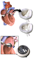
Different types of artificial heart valves
Specialtycardiology
In artificial heart valve is a one-way valve implanted into a person's heart to replace a heart valve that is not functioning properly (valvular heart disease). Artificial heart valves can be separated into three broad classes: mechanical heart valves, bioprosthetic tissue valves and engineered tissue valves.

The human heart contains four valves: tricuspid valve, pulmonary valve, mitral valve and aortic valve. Their main purpose is to keep blood flowing in the proper direction through the heart, and from the heart into the major blood vessels connected to it (the pulmonary artery and the aorta). Heart valves can malfunction for a variety of reasons, which can impede the flow of blood through the valve (stenosis) and/or let blood flow backwards through the valve (regurgitation). Both processes put strain on the heart and may lead to serious problems, including heart failure. While some dysfunctional valves can be treated with drugs or repaired, others need to be replaced with an artificial valve.

Background

3D Medical Animation still shot of Artificial Heart Valve

A heart contains four valves (tricuspid, pulmonary, mitral and aortic valves) which open and close as blood passes through the heart. Blood enters the heart in the right atrium and passes through the tricuspid valve to the right ventricle. From there, blood is pumped through the pulmonary valve to enter the lungs. After being oxygenated, blood passes to the left atrium, where is it pumped through the mitral valve to the left ventricle. The left ventricle pumps blood to the aorta through the aortic valve.

There are many potential causes of heart valve damage, such as birth defects, age related changes, and effects from other disorders, such as rheumatic fever and infections causing endocarditis. High blood pressure and heart failure which can enlarge the heart and arteries, and scar tissue can form after a heart attack or injury.

The three main types of artificial heart valves are mechanical, biological (bioprosthetic/tissue), and tissue-engineered valves. In the US, UK and the European Union, the most common type of artificial heart valve is the bioprosthetic valve. Mechanical valves are more commonly used in Asia and Latin America. Companies that manufacture heart valves include Edwards Lifesciences, Medtronic, Abbott (St. Jude Medical), CryoLife, and LifeNet Health.

Mechanical valves

Mechanical valves come in three main types – caged ball, tilting-disc and bileaflet – with various modifications on these designs. Caged ball valves are no longer implanted. Bileaflet valves are the most common type of mechanical valve implanted in patients today.

Caged ball valves

Caged ball valve

The first artificial heart valve was the caged ball valve, a type of ball check valve, in which a ball is housed inside a cage. When the heart contracts and the blood pressure in the chamber of the heart exceeds the pressure on the outside of the chamber, the ball is pushed against the cage and allows blood to flow. When the heart finishes contracting, the pressure inside the chamber drops and the ball moves back against the base of the valve forming a seal.

In 1952, Charles A. Hufnagel implanted caged ball heart valves into ten patients (six of whom survived the operation), marking the first success in prosthetic heart valves. A similar valve was invented by Miles 'Lowell' Edwards and Albert Starr in 1960, commonly referred to as the Starr-Edwards silastic ball valve. This consisted of a silicone ball enclosed in a methyl metacrylate cage welded to a ring. The Starr-Edwards valve was first implanted in a human on August 25, 1960, and was discontinued by Edwards Lifesciences in 2007.

Caged ball valves are strongly associated with blood clot formation, so people who have one required a high degree of anticoagulation, usually with a target INR of 3.0–4.5.

Tilting-disc valves

tilting-disc valve

Introduced in 1969, the first clinically available tilting-disc valve was the Bjork-Shiley valve. Tilting‑disc valves, a type of swing check valve, are made of a metal ring covered by an ePTFE fabric. The metal ring holds, by means of two metal supports, a disc that opens when the heart beats to let blood flow through, then closes again to prevent blood flowing backwards. The disc is usually made of an extremely hard carbon material (pyrolytic carbon), enabling the valve to function for years without wearing out.

Bileaflet valves

Bileaflet valve

Introduced in 1979, bileaflet valves are made of two semicircular leaflets that revolve around struts attached to the valve housing. With a larger opening than caged ball or tilting-disc valves, they carry a lower risk of blood clots. They are, however, vulnerable to blood backflow.

Advantages of mechanical valves

The major advantage of mechanical valves over bioprosthetic valves is their greater durability. Made from metal and/or pyrolytic carbon, they can last 20–30 years.

Disadvantages of mechanical valves

One of the major drawbacks of mechanical heart valves is that they are associated with an increased risk of blood clots. Clots formed by red blood cell and platelet damage can block blood vessels leading to stroke. People with mechanical valves need to take anticoagulants (blood thinners), such as warfarin, for the rest of their life. Mechanical heart valves can also cause mechanical hemolytic anemia, a condition where the red blood cells are damaged as they pass through the valve. Cavitation, the rapid formation of microbubbles in a fluid such as blood due to a localized drop of pressure, can lead to mechanical heart valve failure, so cavitation testing is an essential part of the valve design verification process.

Many of the complications associated with mechanical heart valves can be explained through fluid mechanics. For example, blood clot formation is a side effect of high shear stresses created by the design of the valves. From an engineering perspective, an ideal heart valve would produce minimal pressure drops, have small regurgitation volumes, minimize turbulence, reduce prevalence of high stresses, and not create flow separations in the vicinity of the valve.

Implanted mechanical valves can cause foreign body rejection. The blood may coagulate and eventually result in a hemostasis. The usage of anticoagulation drugs will be interminable to prevent thrombosis.

Bioprosthetic tissue valves

Bioprosthetic valves are usually made from animal tissue (heterograft/xenograft) attached to a metal or polymer support. Bovine (cow) tissue is most commonly used, but some are made from porcine (pig) tissue. The tissue is treated to prevent rejection and calcification.

Alternatives to animal tissue valves are sometimes used, where valves are used from human donors, as in aortic homografts and pulmonary autografts. An aortic homograft is an aortic valve from a human donor, retrieved either after their death or from a heart that is removed to be replaced during a heart transplant. A pulmonary autograft, also known as the Ross procedure, is where the aortic valve is removed and replaced with the patient's own pulmonary valve (the valve between the right ventricle and the pulmonary artery). A pulmonary homograft (a pulmonary valve taken from a cadaver) is then used to replace the patient's own pulmonary valve. This procedure was first performed in 1967 and is used primarily in children, as it allows the patient's own pulmonary valve (now in the aortic position) to grow with the child.

Advantages of bioprosthetic heart valves

Bioprosthetic valves are less likely than mechanical valves to cause blood clots, so do not require lifelong anticoagulation. As a result, people with bioprosthetic valves have a lower risk of bleeding than those with mechanical valves.

Disadvantages of bioprosthetic heart valves

Tissue valves are less durable than mechanical valves, typically lasting 10–20 years. This means that people with bioprosthetic valves have a higher incidence of requiring another aortic valve replacement in their lifetime. Bioprosthetic valves tend to deteriorate more quickly in younger patients.

In recent years, scientists have developed a new tissue preservation technology, with the aim of improving the durability of bioprosthetic valves. In sheep and rabbit studies, tissue preserved using this new technology had less calcification than control tissue. A valve containing this tissue is now marketed, but long-term durability data in patients are not yet available.

Current bioprosthetic valves lack longevity, and will calcify over time. When a valve calcifies, the valve cusps become stiff and thick and cannot close completely. Moreover, bioprosthetic valves can't grow with or adapt to the patient: if a child has bioprosthetic valves they will need to get the valves replaced several times to fit their physical growth.

Tissue-engineered valves

For over 30 years researchers have been trying to grow heart valves in vitro. These tissue‑engineered valves involve seeding human cells on to a scaffold. The two main types of scaffold are natural scaffolds, such as decellularized tissue, or scaffolds made from degradable polymers. The scaffold acts as an extracellular matrix, guiding tissue growth into the correct 3D structure of the heart valve. Some tissue-engineered heart valves have been tested in clinical trials, but none are commercially available.

Tissue engineered heart valves can be person-specific and 3D modeled to fit an individual recipient 3D printing is used because of its high accuracy and precision of dealing with different biomaterials. Cells that are used for tissue engineered heart valves are expected to secrete the extracellular matrix (ECM). Extracellular matrix provides support to maintain the shape of the valves and determines the cell activities.

Scientists can follow the structure of heart valves to produce something that looks similar to them, but since tissue engineered valves lack the natural cellular basis, they either fail to perform their functions like natural heart valves, or function when they are implanted but gradually degrade over time. An ideal tissue engineered heart valve would be non‐thrombogenic, biocompatible, durable, resistant to calcification, grow with the surrounding heart, and exhibit a physiological hemodynamic profile. To achieve these goals, the scaffold should be carefully chosen—there are three main candidates: decellularized ECM (xenografts or homografts), natural polymers, and synthetic polymers.

Differences between mechanical and tissue valves

Mechanical and tissue valves are made of different materials. Mechanical valves are generally made of titanium and carbon. Tissue valves are made up of human or animal tissue. The valves composed of human tissue, known as allografts or homografts, are from donors' human hearts.

Mechanical valves can be a better choice for younger people and people at risk of valve deterioration due to its durability. It is also preferable for people who are already taking blood thinners and people who would be unlikely to tolerate another valve replacement operation.

Tissue valves are better for older age groups as another valve replacement operation may not be needed in their lifetime. Due to the risk of forming blood clots for mechanical valves and severe bleeding as a major side effect of taking blood-thinning medications, people who have a risk of blood bleeding and are not willing to take warfarin may also consider tissue valves. Other patients who may be more suitable for tissue valves are people who have other planned surgeries and unable to take blood-thinning medications. People who plan to become pregnant may also consider tissue valves as warfarin causes risks in pregnancy.

Functional requirements of artificial heart valves

An artificial heart valve should ideally function like a natural heart valve. The functioning of natural heart valves is characterized by many advantages:

  • Minimal regurgitation – This means that the amount of blood leaking backwards through the valve as it closes is small. Some degree of valvular regurgitation is inevitable and natural, up to around 5 ml per beat. However, several heart valve pathologies (e.g. rheumatic endocarditis) may lead to clinically significant valvular regurgitation. A desirable characteristic of heart valve prostheses is that regurgitation is minimal over the full range of physiological heart function.
  • Minimal transvalvular pressure gradient – Whenever a fluid flows through a restriction, such as a valve, a pressure gradient arises over the restriction. This pressure gradient is a result of the increased resistance to flow through the restriction. Natural heart valves have a low transvalvular pressure gradient as they present little obstruction to the flow through themselves, normally less than 16 mmHg. A desirable characteristic of heart valve prostheses is that their transvalvular pressure gradient is as small as possible.
  • Non-thrombogenic – Natural heart valves are lined with an endothelium comparable with the endothelium lining the heart chambers, so they are not normally thrombogenic (i.e. they don't cause blood clots). Blood clots can be hazardous because they can lodge in, and block, downstream arteries (e.g. coronary arteries, leading to heart attack [myocardial infarction]; or cerebral arteries, leading to stroke). A desirable characteristic of artificial heart valves is that they are non- or minimally thrombogenic.
  • Self-repairing – Valve leaflets retain some capacity for repair thanks to regenerative cells (e.g. fibroblasts) in the connective tissue from which the leaflets are composed. As the human heart beats approximately 3.4×10⁹ times during a typical human lifespan, this limited but nevertheless present repair capacity is critically important. No heart valve prostheses can currently self-repair, but tissue-engineered valves may eventually offer such capabilities.

Artificial heart valve repair

Artificial heart valves are expected to last from 10 to 30 years.

The most common problems with artificial heart valves are various forms of degeneration, including gross billowing of leaflets, ischemic mitral valve pathology, and minor chordal lengthening. The repairing process of the artificial heart valve regurgitation and stenosis usually requires an open-heart surgery, and a repair or partial replacement of regurgitant valves is usually preferred.

Researchers are investigating catheter-based surgery that allows repair of an artificial heart valve without large incisions.

Researchers are investigating Interchangeable Prosthetic Heart Valve that allows redo and fast-track repair of an artificial heart valve. 

Additional images


Cardiac surgery

From Wikipedia, the free encyclopedia
 
Cardiac surgery
Two cardiac surgeons performing coronary artery bypass surgery. Note the use of a steel retractor to forcefully maintain the exposure of the heart.
ICD-9-CM35-37
MeSHD006348
OPS-301 code5-35...5-37
Cardiac surgery
SpecialtyCardiothoracic surgery

Cardiac surgery, or cardiovascular surgery, is surgery on the heart or great vessels performed by cardiac surgeons. It is often used to treat complications of ischemic heart disease (for example, with coronary artery bypass grafting); to correct congenital heart disease; or to treat valvular heart disease from various causes, including endocarditis, rheumatic heart disease, and atherosclerosis. It also includes heart transplantation.

History

19th century

The earliest operations on the pericardium (the sac that surrounds the heart) took place in the 19th century and were performed by Francisco Romero (1801) in the city of Almería (Spain), Dominique Jean Larrey (1810), Henry Dalton (1891), and Daniel Hale Williams (1893). The first surgery on the heart itself was performed by Axel Cappelen on 4 September 1895 at Rikshospitalet in Kristiania, now Oslo. Cappelen ligated a bleeding coronary artery in a 24-year-old man who had been stabbed in the left axilla and was in deep shock upon arrival. Access was through a left thoracotomy. The patient awoke and seemed fine for 24 hours but became ill with a fever and died three days after the surgery from mediastinitis.

20th century

Surgery on the great vessels (e.g., aortic coarctation repair, Blalock–Thomas–Taussig shunt creation, closure of patent ductus arteriosus) became common after the turn of the century. However, operations on the heart valves were unknown until, in 1925, Henry Souttar operated successfully on a young woman with mitral valve stenosis. He made an opening in the appendage of the left atrium and inserted a finger in order to palpate and explore the damaged mitral valve. The patient survived for several years, but Souttar's colleagues considered the procedure unjustified, and he could not continue.

Alfred Blalock, Helen Taussig, and Vivien Thomas performed the first successful palliative pediatric cardiac operation at Johns Hopkins Hospital on 29 November 1944, in a one-year-old girl with Tetralogy of Fallot.

Cardiac surgery changed significantly after World War II. In 1947, Thomas Sellors of Middlesex Hospital in London operated on a Tetralogy of Fallot patient with pulmonary stenosis and successfully divided the stenosed pulmonary valve. In 1948, Russell Brock, probably unaware of Sellors's work, used a specially designed dilator in three cases of pulmonary stenosis. Later that year, he designed a punch to resect a stenosed infundibulum, which is often associated with Tetralogy of Fallot. Many thousands of these "blind" operations were performed until the introduction of cardiopulmonary bypass made direct surgery on valves possible.

Also in 1948, four surgeons carried out successful operations for mitral valve stenosis resulting from rheumatic fever. Horace Smithy of Charlotte used a valvulotome to remove a portion of a patient's mitral valve, while three other doctors—Charles Bailey of Hahnemann University Hospital in Philadelphia; Dwight Harken in Boston; and Russell Brock of Guy's Hospital in London—adopted Souttar's method. All four men began their work independently of one another within a period of a few months. This time, Souttar's technique was widely adopted, with some modifications.

The first successful intracardiac correction of a congenital heart defect using hypothermia was performed by lead surgeon Dr. F. John Lewis (Dr. C. Walton Lillehei assisted) at the University of Minnesota on 2 September 1952. In 1953, Alexander Alexandrovich Vishnevsky conducted the first cardiac surgery under local anesthesia. In 1956, Dr. John Carter Callaghan performed the first documented open-heart surgery in Canada.

Types of cardiac surgery

Open-heart surgery

Open-heart surgery is any kind of surgery in which a surgeon makes a large incision (cut) in the chest to open the rib cage and operate on the heart. "Open" refers to the chest, not the heart. Depending on the type of surgery, the surgeon also may open the heart.

Dr. Wilfred G. Bigelow of the University of Toronto found that procedures involving opening the patient's heart could be performed better in a bloodless and motionless environment. Therefore, during such surgery, the heart is temporarily stopped, and the patient is placed on cardiopulmonary bypass, meaning a machine pumps their blood and oxygen. Because the machine cannot function the same way as the heart, surgeons try to minimize the time a patient spends on it.

Cardiac surgery at Gemelli Hospital in Rome

Cardiopulmonary bypass was developed after surgeons realized the limitations of hypothermia in cardiac surgery: Complex intracardiac repairs take time, and the patient needs blood flow to the body (particularly to the brain), as well as heart and lung function. In July 1952, Forest Dodrill was the first to use a mechanical pump in a human to bypass the left side of the heart whilst allowing the patient's lungs to oxygenate the blood, in order to operate on the mitral valve. In 1953, Dr. John Heysham Gibbon of Jefferson Medical School in Philadelphia reported the first successful use of extracorporeal circulation by means of an oxygenator, but he abandoned the method after subsequent failures. In 1954, Dr. Lillehei performed a series of successful operations with the controlled cross-circulation technique, in which the patient's mother or father was used as a "heart-lung machine". Dr. John W. Kirklin at the Mayo Clinic was the first to use a Gibbon-type pump-oxygenator.

Nazih Zuhdi performed the first total intentional hemodilution open-heart surgery on Terry Gene Nix, age 7, on 25 February 1960 at Mercy Hospital in Oklahoma City. The operation was a success; however, Nix died three years later. In March 1961, Zuhdi, Carey, and Greer performed open-heart surgery on a child, aged 3+12, using the total intentional hemodilution machine.

Modern beating-heart surgery

In the early 1990s, surgeons began to perform off-pump coronary artery bypass, done without cardiopulmonary bypass. In these operations, the heart continues beating during surgery, but is stabilized to provide an almost still work area in which to connect a conduit vessel that bypasses a blockage. The conduit vessel that is often used is the Saphenous vein. This vein is harvested using a technique known as endoscopic vessel harvesting (EVH).

Heart transplant

In 1945, the Soviet pathologist Nikolai Sinitsyn successfully transplanted a heart from one frog to another frog and from one dog to another dog.

Norman Shumway is widely regarded as the father of human heart transplantation, although the world's first adult heart transplant was performed by a South African cardiac surgeon, Christiaan Barnard, using techniques developed by Shumway and Richard Lower. Barnard performed the first transplant on Louis Washkansky on 3 December 1967 at Groote Schuur Hospital in Cape Town. Adrian Kantrowitz performed the first pediatric heart transplant on 6 December 1967 at Maimonides Hospital (now Maimonides Medical Center) in Brooklyn, New York, barely three days later. Shumway performed the first adult heart transplant in the United States on 6 January 1968 at Stanford University Hospital.

Coronary Artery Bypass Grafting (CABG)

Coronary artery bypass grafting, also called revascularization, is a common surgical procedure to create an alternative path to deliver blood supply to the heart and body, with the goal of preventing clot formation. This can be done in many ways, and the arteries used can be taken from several areas of the body. Arteries are typically harvested from the chest, arm, or wrist and then attached to a portion of the coronary artery, relieving pressure and limiting clotting factors in that area of the heart.

The procedure is typically performed because of coronary artery disease (CAD), in which a plaque-like substance builds up in the coronary artery, the main pathway carrying oxygen-rich blood to the heart. This can cause a blockage and/or a rupture, which can lead to a heart attack.

Minimally invasive surgery

As an alternative to open-heart surgery, which involves a five- to eight-inch incision in the chest wall, a surgeon may perform an endoscopic procedure by making very small incisions through which a camera and specialized tools are inserted.

In robot-assisted heart surgery, a machine controlled by a cardiac surgeon is used to perform a procedure. The main advantage to this is the size of the incision required: three small port holes instead of an incision big enough for the surgeon's hands. The use of robotics in heart surgery continues to be evaluated, but early research has shown it to be a safe alternative to traditional techniques.

Post-surgical procedures

As with any surgical procedure, cardiac surgery requires postoperative precautions to avoid complications. Incision care is needed to avoid infection and minimize scarring. Swelling and loss of appetite are common.

Recovery from open-heart surgery begins with about 48 hours in an intensive care unit, where heart rate, blood pressure, and oxygen levels are closely monitored. Chest tubes are inserted to drain blood around the heart and lungs. After discharge from the hospital, compression socks may be recommended in order to regulate blood flow.

Risks

The advancement of cardiac surgery and cardiopulmonary bypass techniques has greatly reduced the mortality rates of these procedures. For instance, repairs of congenital heart defects are currently estimated to have 4–6% mortality rates.

A major concern with cardiac surgery is neurological damage. Stroke occurs in 2–3% of all people undergoing cardiac surgery, and the rate is higher in patients with other risk factors for stroke. A more subtle complication attributed to cardiopulmonary bypass is postperfusion syndrome, sometimes called "pumphead". The neurocognitive symptoms of postperfusion syndrome were initially thought to be permanent, but turned out to be transient, with no permanent neurological impairment.

In order to assess the performance of surgical units and individual surgeons, a popular risk model has been created called the EuroSCORE. It takes a number of health factors from a patient and, using precalculated logistic regression coefficients, attempts to quantify the probability that they will survive to discharge. Within the United Kingdom, the EuroSCORE was used to give a breakdown of all cardiothoracic surgery centres and to indicate whether the units and their individuals surgeons performed within an acceptable range. The results are available on the Care Quality Commission website.

Another important source of complications are the neuropsychological and psychopathologic changes following open-heart surgery. One example is Skumin syndrome, described by Victor Skumin in 1978, which is a "cardioprosthetic psychopathological syndrome" associated with mechanical heart valve implants and characterized by irrational fear, anxiety, depression, sleep disorder, and weakness.

Risk reduction

Pharmacological and non-pharmacological prevention approaches may reduce the risk of atrial fibrillation after an operation and reduce the length of hospital stays, however there is no evidence that this improves mortality.

Non-pharmacologic approaches

Preoperative physical therapy may reduce postoperative pulmonary complications, such as pneumonia and atelectasis, in patients undergoing elective cardiac surgery and may decrease the length of hospital stay by more than three days on average. There is evidence that quitting smoking at least four weeks before surgery may reduce the risk of postoperative complications.

Pharmacological approaches

Beta-blocking medication is sometimes prescribed during cardiac surgery. There is some low certainty evidence that this perioperative blockade of beta-adrenergic receptors may reduce the incidence of atrial fibrillation and ventricular arrhythmias in patients undergoing cardiac surgery.

Vascular surgery

From Wikipedia, the free encyclopedia
 
Vascular surgery
Open infrarenal aortic repair model
ICD-9-CM38-39
MeSHD014656
OPS-301 code5-38...5-39

Vascular surgery is a surgical subspecialty in which vascular diseases involving the arteries, veins, or lymphatic vessels, are managed by medical therapy, minimally-invasive catheter procedures and surgical reconstruction. The specialty evolved from general and cardiovascular surgery where it refined the management of just the vessels, no longer treating the heart or other organs. Modern vascular surgery includes open surgery techniques, endovascular (minimally invasive) techniques and medical management of vascular diseases - unlike the parent specialities. The vascular surgeon is trained in the diagnosis and management of diseases affecting all parts of the vascular system excluding the coronaries and intracranial vasculature. Vascular surgeons also are called to assist other physicians to carry out surgery near vessels, or to salvage vascular injuries that include hemorrhage control, dissection, occlusion or simply for safe exposure of vascular structures.

History

Early leaders of the field included Russian surgeon Nikolai Korotkov, noted for developing early surgical techniques, American interventional radiologist Charles Theodore Dotter who is credited with inventing minimally invasive angioplasty (1964), and Australian Robert Paton, who helped the field achieve recognition as a specialty. Edwin Wylie of San Francisco was one of the early American pioneers who developed and fostered advanced training in vascular surgery and pushed for its recognition as a specialty in the United States in the 1970s. The most notable historic figure in vascular surgery is the 1912 Nobel Prize winning surgeon, Alexis Carrel for his techniques used to suture vessels.

Evolution

Medical science has advanced significantly since 1507, when Leonardo da Vinci drew this diagram of the internal organs and vascular systems of a woman.

The specialty continues to be based on operative arterial and venous surgery but since the early 1990s has evolved greatly. There is now considerable emphasis on minimally invasive alternatives to surgery. The field was originally pioneered by interventional radiologists like Dr. Charles Dotter, who invented angioplasty using serial dilatation of vessels.

The surgeon Dr. Thomas J. Fogarty invented a balloon catheter, designed to remove clots from occluded vessels, which was used as the eventual model to do endovascular angioplasty. Further development of the field has occurred via joint efforts between interventional radiology, vascular surgery, and interventional cardiology. This area of vascular surgery is called Endovascular Surgery or Interventional Vascular Radiology, a term that some in the specialty append to their primary qualification as Vascular Surgeon. Endovascular and endovenous procedures (e.g., EVAR) can now form the bulk of a vascular surgeon's practice.

The treatment of the aorta, the body's largest artery, dates back to Greek surgeon Antyllus, who first performed surgeries for various aneurysms in the second century AD. Modern treatment of aortic diseases stems from development and advancements from Michael DeBakey and Denton Cooley. In 1955, DeBakey and Cooley performed the first replacement of a thoracic aneurysm with a homograft. In 1958, they began using the Dacron graft, resulting in a revolution for surgeons in the repair of aortic aneurysms. He also was first to perform cardiopulmonary bypass to repair the ascending aorta, using antegrade perfusion of the brachiocephalic artery.

Dr. Ted Diethrich, one of Dr. DeBakey's associates, went on to pioneer many of the minimally invasive techniques that later became hallmarks of endovascular surgery. Dietrich later founded the Arizona Heart Hospital in 1998 and served as its medical director from 1998 to 2010. In 2000, Diethrich performed the first endovascular aneurysm repair (EVAR) for ruptured abdominal aortic aneurysm. Dietrich trained several future leaders in the field of endovascular surgery at the Arizona Heart Hospital including Venkatesh Ramaiah, MD who served as medical director of the institution following Dietrich's death in 2017.

The development of endovascular surgery has been accompanied by a gradual separation of vascular surgery from its origin in general surgery. Most vascular surgeons would now confine their practice to vascular surgery and, similarly, general surgeons would not be trained or practise the larger vascular surgery operations or most endovascular procedures. More recently, professional vascular surgery societies and their training program have formally separated vascular surgery into a separate specialty with its own training program, meetings and accreditation. Notable societies are Society for Vascular Surgery (SVS), USA; Australia and New Zealand Society of Vascular Surgeons (ANZSVS). Local societies also exist (e.g., New South Wales Vascular and Melbourne Vascular Surgical Association (MVSA)). Larger societies of surgery actively separate and encourage specialty surgical societies under their umbrella (e.g., Royal Australasian College of Surgeons (RACS)).

Currently

Arterial and venous disease treatment by angiography, stenting, and non-operative varicose vein treatment sclerotherapy, endovenous laser treatment have largely replaced major surgery in many first world countries. These procedures provide reasonable outcomes that are comparable to surgery with the advantage of short hospital stay (day or overnight for most cases) with lower morbidity and mortality rates. Historically performed by interventional radiologists, vascular surgeons have become increasingly proficient with endovascular methods. The durability of endovascular arterial procedures is generally good, especially when viewed in the context of their common clinical usage i.e. arterial disease occurring in elderly patients and usually associated with concurrent significant patient comorbidities especially ischemic heart disease. The cost savings from shorter hospital stays and less morbidity are considerable but are somewhat balanced by the high cost of imaging equipment, construction and staffing of dedicated procedural suites, and of the implant devices themselves. The benefits for younger patients and in venous disease are less persuasive but there are strong trends towards nonoperative treatment options driven by patient preference, health insurance company costs, trial demonstrating comparable efficacy at least in the medium term.

A recent trend in the United States is the stand-alone day angiography facility associated with a private vascular surgery clinic, thus allowing treatment of most arterial endovascular cases conveniently and possibly with lesser overall community cost. Similar non-hospital treatment facilities for non-operative vein treatment have existed for some years and are now widespread in many countries.

NHS England conducted a review of all 70 vascular surgery sites across England in 2018 as part of its Getting It Right First Time programme. The review specified that vascular hubs should perform at least 60 abdominal aortic aneurysm procedures and 40 carotid endarterectomies a year. 12 trusts missed both targets and many more missed one of them. A programme of concentrating vascular surgery in fewer centres is proceeding.

Vascular surgery encompasses surgery of the aorta, carotid arteries, and lower extremities, including the iliac, femoral, vascular trauma and tibial arteries. Vascular surgery also involves surgery of veins, for conditions such as May–Thurner syndrome and for varicose veins. In some regions, vascular surgery also includes dialysis access surgery and transplant surgery.

Management of arterial diseases

Infrarenal aortic occlusion imaged with magnetic resonance angiography (MRA).

The management of arterial pathology excluding coronary and intracranial disease is within the scope of vascular surgeons. Disease states generally arise from narrowing of the arterial system known as stenosis or abnormal dilation referred to as an aneurysm. There are multiple mechanisms by which the arterial lumen can narrow, the most common of which is atherosclerosis. Symptomatic stenosis may also result from a complication of arterial dissection. Other less common causes of stenosis include fibromuscular dysplasia, radiation induced fibrosis or cystic adventitial disease. Dilation of an artery which retains histologic layers is called an aneurysm. An aneurysms can be fusiform (concentric dilation), saccular (outpouching) or a combination of the two. Arterial dilation which does not contain three histologic layers is considered a pseudoaneurysm. Additionally, there are a number of congenital vascular anomalies which lead to symptomatic disease that are managed by the vascular surgeon, a few of which include aberrant subclavian artery, popliteal artery entrapment syndrome or persistent sciatic artery. Vascular surgeons treat arterial diseases with a range of therapies including lifestyle modification, medications, endovascular therapy and surgery.

Aneurysms

Aortic aneurysms

The aorta is the largest artery in the body and the descending aorta has both a thoracic and an abdominal component. A thoracic aortic aneurysm is located in the chest, and an abdominal aortic aneurysm is located in the abdomen. Not pictured here are aneurysms which span both cavities and are referred to as thoracoabdominal aortic aneurysms.
Abdominal

An abdominal aortic aneurysm (AAA) refers to aneurysmal dilation of the aorta confined to the abdominal cavity. Most commonly, aneurysms are asymptomatic and located in the infrarenal position. Often, they are discovered incidentally or on screening exams in patients with risk factors such as a history of smoking. Patients with aneurysms which have a diameter less than 5 cm are at <1% rupture risk per year. When the aneurysm meets size criteria it can be treated with aortic replacement or EVAR.

Thoracic

Thoracic aortic aneurysms are contained in the chest. Aneurysms of the descending aorta can often be treated with thoracic endovascular aortic repair or TEVAR. Treating aneurysms which involve the ascending aorta are generally within the scope of cardiac surgeons, but upcoming endovascular technology may allow for a more minimally invasive approach in some patients.

Thoracoabdominal

Thoroacoabdominal aneurysms are those which span the chest and abdominal cavities. The Crawford classification was developed and describes five types of thoracoabdominal aneurysms.

Other arterial aneurysms

In addition to treating aneurysms which arise from the aorta, vascular surgeons also treat aneurysms elsewhere in the body.

Visceral artery

Visceral artery aneurysms include those isolated to the renal artery, splenic artery, celiac artery, and hepatic artery. Of these, data shows that splenic artery aneurysms are the most common.

Indications for repair differ slightly between arteries. For instance, current guidelines recommend repair of renal and splenic artery aneurysms greater than 3 cm, and those of any size in women of childbearing age; whereas celiac and hepatic artery aneurysms are indicated for repair when their size is greater than 2 cm. This is in contrast to superior mesenteric artery aneurysms which should be repaired regardless of size when they are discovered.

Popliteal artery

A popliteal artery aneurysm is an arterial aneurysm localized in the popliteal artery which courses behind the knee. Unlike aneurysms located in the abdomen, popliteal artery aneurysm rarely present with rupture but rather with symptoms of acute limb ischemia due to embolization of thrombus. Thus, when a patient presents with an asymptomatic popliteal aneurysm that is greater than 2 cm in diameter a vascular surgeon are able to offer vascular bypass or endovascular exclusion depending on several factors.

Arterial dissections

Early classification schemes of aortic dissection. Stanford type A are those which originate in the ascending aorta whereas Stanford type B originate distal to the left subclavian artery (descending aorta). The Debakey classification describes where the original tear is and the extent of the dissection.

The artery wall is composed of three concentric layers: the intima, media and adventitia. In general, an arterial dissection is a tear in the innermost layer of the arterial wall that makes a separation which allows blood to flow, and collect, between the layers. Arterial dissections include: an aortic dissection (aorta), a coronary artery dissection (coronary artery), two types of cervical artery dissection involving one of the arteries in the neck – a carotid artery dissection (carotid artery), and a vertebral artery dissection (vertebral artery), a pulmonary artery dissection is an extremely rare condition as a complication of chronic pulmonary hypertension.

Whereas cardiac surgeons are usually in charge of managing type A dissections, type B dissections are typically managed by vascular surgeons. The most common risk factor for type B aortic dissection is hypertension. The first line treatment for type B aortic dissection is aimed at reducing both heart rate and blood pressure and is referred to as anti-impulse therapy.

A thoracic aortic stent graft, seen on chest X-ray which was placed during a TEVAR procedure.

Should initial medical management fail or there is the involvement of a major branch of the aorta, vascular surgery may be needed for these type B dissections. Treatment may include thoracic endovascular aortic repair (TEVAR) with or without extra-anatomic bypass such as carotid-carotid bypass, carotid-subclavian bypass, or subclavian-carotid transposition.

Visceral artery dissection

Visceral artery dissections are arterial dissections involving the superior mesenteric artery, celiac artery, renal arteries, hepatic artery and others. When they are an extension of an aortic dissection, this condition is managed simultaneously with aortic treatment. In isolation, visceral artery dissections are discovered incidentally in up to a third of patients and in these cases may be managed medically by a vascular surgeon. In cases where the dissection results in organ damage it is generally accepted by vascular surgeons that surgery is necessary. Surgical management strategies depend on the associated complications, surgical ability and patient preference.

Mesenteric ischemia

Mesenteric ischemia results from the acute or chronic obstruction of the superior mesenteric artery (SMA). The SMA arises from the abdominal aorta and usually supplies blood from the distal duodenum through two-thirds of the transverse colon and the pancreas.

Chronic mesenteric ischemia

The symptoms of chronic mesenteric ischemia can be classified as abdominal angina which is abdominal pain which occurs a fixed period of time after eating. Due to this, patient's may avoid eating, resulting in unintended weight loss. The first surgical treatment is thought to be performed by R.S. Shaw and described in the New England Journal of Medicine in 1958. The procedure Shaw described is referred to as mesenteric endarterectomy. Since then, many advances in treatment have been made in minimally invasive, endovascular techniques including angioplasty and stenting.

Acute mesenteric ischemia

Acute mesenteric ischemia (AMI) results from the sudden occlusion of the superior mesenteric artery.

Renovascular hypertension

The renal arteries supply oxygenated blood to the kidneys. The kidneys serve to filter the flood and control blood pressure through the renin-angiotensin system. One cause of resistant hypertension is atherosclerotic disease in the renal arteries and is generally referred to as renovascular hypertension. If renovascular hypertension is diagnosed and maximal medical fails to control high blood pressure, the vascular surgeon may offer surgical treatment, either endovascular or open surgical reconstruction.

Cerebrovascular disease

Carotid ultrasound.
Carotid endarterectomy.

Vascular surgeons are responsible for treating extracranial cerebrovascular disease as well as the interpretation of non-invasive vascular imaging relating to extracranial and intracranial circulation such as carotid ultrasonography and transcranial doppler. The most common of cerebrovascular conditions treated by vascular surgeons is carotid artery stenosis which is a narrowing of the carotid arteries and may be either clinically symptomatic or asymptomatic (silent). Carotid artery stenosis is caused by atherosclerosis whereby the buildup of atheromatous plaque inside the artery causes narrowing.

Symptoms of carotid artery stenosis can include transient ischemic attack or stroke. Both symptomatic and asymptomatic carotid stenosis can be diagnosed with the aid of carotid duplex ultrasound which allows for the estimation of severity of narrowing as well as characterize the plaque. Treatment can include medical therapy, carotid endarterectomy or carotid stenting.

The Society for Vascular Surgery publishes clinical practice guidelines for the management of extracranial cerebrovascular disease. Less common diseases involving cerebral circulation treated by vascular surgeons include vertebrobasilar insufficiency, subclavian steal syndrome, carotid artery dissection, vertebral artery dissection, carotid body tumor and carotid artery aneurysm among others.

Peripheral artery disease

Peripheral artery disease PAD is the abnormal narrowing of the arteries which supply the limbs. Patients with this condition can present with intermittent claudication which is pain mainly in the calves and thighs while walking. If there is progression, a patient may also present with chronic limb threatening ischemia which encompasses pain at rest and non-healing wounds. Vascular surgeons are experts in the diagnosis, medical management, endovascular and open surgical treatment of PAD.

A vascular surgeon may diagnose PAD using a combination of history, physical exam and medical imaging. Medical imaging may include ankle-brachial index, doppler ultrasonography and computed tomography angiography, among others. Treatments are individualized and may include medical therapy, endovascular intervention or open surgical options including angioplasty, stenting, atherectomy, endarterectomy and vascular bypass, among others.

Management of venous diseases

Chronic venous disease

Chronic venous insufficiency is the abnormal pooling of blood in the lower extremity venous system which can lead to reticular veins, varicose veins, chronic edema and inflammation among other things. Population data suggests that chronic venous insufficiency affects up to 40% of females and 17% of males. When chronic insufficiency leads to pain, swelling and skin changes it is referred to as chronic venous disease. Chronic venous insufficiency (CVI) is distinguished from post-thrombotic syndrome (PTS) in that CVI is primarily an issue of valvular incompetence of the superficial or deep veins whereas PTS may occur as a long-term complication of deep venous thrombosis.

The vascular surgeon has several modalities to treat lower extremity venous disease which including medical, interventional and surgical procedures. For instance, venous ulceration may be treated with Unna's boots, superficial venous reflux with radiofrequency, laser ablation or vein stripping if indicated. When indicated, insufficiency in the deep veins may be treated with reconstruction of the venous valves with internal or external valvuloplasty.

Varicose veins

A medical illustration of lower extremity varicose veins.

Lower extremity varicose veins is the condition in which the superficial veins become tortuous(snakelike) and dilated (enlarged) to greater than 3mm in the upright position. Incompetent or faulty valves are often present in these veins when investigated with duplex ultrasonography. Vascular treatments can include compression stockings, venous ablation or vein stripping, depending on specific patient presentation, severity of disease, among other things.

Nonthrombotic iliac vein lesions

Nonthrombotic iliac vein lesions (NIVL) include May-Thurner Syndrome (MTS) whereby there is compression of the left iliac venous outflow usually by the right iliac artery leading to left leg discomfort, pain, swelling and varicose veins. NIVL encompasses compression of the iliac veins on either the right or left side. Vascular surgeons may offer different treatment modalities depending on the patient presentation. Minimally invasive diagnostic and therapeutic options might include intravascular ultrasound, venography and iliac vein stenting whereas surgical management may be offered in refractory cases. Surgical management strategies involve reconstruction or bypass of the affected segment such as cross-pubic venous bypass, also known as the Palma procedure.

Deep vein thrombosis

Deep vein thrombosis (DVT) is the formation of thrombus in a deep vein. DVT is more likely to occur in the lower extremity than the upper extremity or jugular vein. When a DVT involves the pelvic and lower extremity veins it can sometimes be classified as an iliofemoral DVT. Some evidence to suggests that performing an intervention in these cases may be beneficial whereas other evidence does not. Overall, the data shows that there may be a reduction in the incidence in post-thrombotic syndrome in patients who undergo certain procedures for iliofemoral DVT but it is not without risks. A vascular surgeon may offer venogram, endovascular suction or mechanical thrombectomy and in some cases pharmacomechanical thrombectomy. Some lower extremity DVT can be severe enough to cause a condition called phlegmasia cerulea dolens or phlegmasia alba dolens and can be limb-threatening events. When phlegmasia is present, intervention is often warranted and may include venous thrombectomy.

Post-thrombotic syndrome

Post-thrombotic syndrome (PTS) is a medical condition that sometimes occurs as a long-term complication of DVT and is characterized by long term edema and skin changes following DVT. Presenting symptoms may include itchiness, pain, cramps and paresthesia. It is estimated that between 20% and 50% of patients will experience some degree of PTS. A treatment strategy for PTS may involve the use of compression stockings.

Pulmonary embolism

Surgical management of an acute pulmonary embolism (pulmonary thrombectomy) is uncommon and has largely been abandoned because of poor long-term outcomes. However, recently, it has gone through a resurgence with the revision of the surgical technique and is thought to benefit certain people. Chronic pulmonary embolism leading to pulmonary hypertension (known as chronic thromboembolic hypertension) is treated with a surgical procedure known as a pulmonary thromboendarterectomy.

Compressive venopathies

Compression of large veins by adjacent structures or masses may lead to distinct clinical syndromes including May–Thurner syndrome (MTS), nutcracker syndrome and superior vena cava syndrome to name a few. Treatment modalities include venography, intravascular ultrasound and venous stenting as well as more invasive open venous reconstruction and bypass.

Management of hemodialysis access

Patients with chronic kidney disease may have progression of disease which requires renal replacement therapy to filter their blood. One strategy for this therapy is hemodialysis, which is a procedure that involves filtering a patient's blood to remove waste products and returning their blood back to them. One method which avoids repeated arterial trauma is to create an arteriovenous fistula (AVF). The first procedure described for this purpose is named the Cimino fistula, after one of the surgeons who first had success with it. Vascular surgeons may create an AVF for a patient as well as undertake minimally invasive procedures to ensure the fistula remains patent.

Management of vascular trauma

One way that vascular trauma may be understood is by categorizing vascular injury by three criteria: mechanism of injury, anatomical site of injury and contextual circumstances. Mechanism of injury refers to etiology, e.g. iatrogenic, blunt, penetrating, blast injury, etc. Anatomical site functionally refers to whether there is compressible versus non-compressible hemorrhage, while contextual circumstances refers to injuries sustained in the civilian or military realm. Each context can be further broken down: military into combatant vs. noncombatant and civil into urban vs rural trauma. This categorization scheme is of both epidemiologic and clinical significance. For instance, arterial injury in military combatants currently occurs predominantly in males in their twenties who are exposed to improvised explosive devices or gunshot wounds; whereas in the civilian realm, one study conducted in the United States showed the most common mechanisms to include motor vehicle collisions, firearm injuries, stab wounds and falls from heights.

Blunt thoracic aortic injury

Advances in vascular surgery, specifically endovascular technologies, have led to a dramatic change in the operative approach to blunt thoracic aortic injury (BTAI). BTAI results from a high speed insult to the thorax such as a motor vehicle collision or a fall from a height. One widely-used classification scheme is based on the extent of injury to the anatomic layers of the aorta as seen with computed tomography angiography or intravascular ultrasound. Grade 1 BTAI are those which tear the aortic intima; grade 2 injuries refer to intramural hematoma; grade 3 injuries are pseudoaneurysm and are only contained by adventitial tissue; and grade 4 refer to free rupture of blood into the chest and surrounding tissue. When indicated, first line intervention involves TEVAR.

Politics of Europe

From Wikipedia, the free encyclopedia ...