| Human brain | |
|---|---|

Human brain and skull
| |

Cerebral lobes: the frontal lobe (pink), parietal lobe (green) and occipital lobe (blue)
| |
| Details | |
| Precursor | Neural tube |
| System | Central nervous system Neuroimmune system |
| Artery | Internal carotid arteries, vertebral arteries |
| Vein | Internal jugular vein, internal cerebral veins; external veins: (superior, middle, and inferior cerebral veins), basal vein, and cerebellar veins |
| Identifiers | |
| Latin | Cerebrum |
| Greek | ἐγκέφαλος (enképhalos) |
| MeSH | D001921 |
| TA | A14.1.03.001 |
| FMA | 50801 |
The human brain is the central organ of the human nervous system, and with the spinal cord makes up the central nervous system. The brain consists of the cerebrum, the brainstem and the cerebellum. It controls most of the activities of the body, processing, integrating, and coordinating the information it receives from the sense organs, and making decisions as to the instructions sent to the rest of the body. The brain is contained in, and protected by, the skull bones of the head.
The cerebrum is the largest part of the human brain. It is divided into two cerebral hemispheres. The cerebral cortex is an outer layer of grey matter, covering the core of white matter. The cortex is split into the neocortex and the much smaller allocortex. The neocortex is made up of six neuronal layers, while the allocortex has three or four. Each hemisphere is conventionally divided into four lobes – the frontal, temporal, parietal, and occipital lobes. The frontal lobe is associated with executive functions including self-control, planning, reasoning, and abstract thought, while the occipital lobe is dedicated to vision. Within each lobe, cortical areas are associated with specific functions, such as the sensory, motor and association regions. Although the left and right hemispheres are broadly similar in shape and function, some functions are associated with one side, such as language in the left and visual-spatial ability in the right. The hemispheres are connected by commissural nerve tracts, the largest being the corpus callosum.
The cerebrum is connected by the brainstem to the spinal cord. The brainstem consists of the midbrain, the pons, and the medulla oblongata. The cerebellum is connected to the brainstem by pairs of tracts. Within the cerebrum is the ventricular system, consisting of four interconnected ventricles in which cerebrospinal fluid is produced and circulated. Underneath the cerebral cortex are several important structures, including the thalamus, the epithalamus, the pineal gland, the hypothalamus, the pituitary gland, and the subthalamus; the limbic structures, including the amygdala and the hippocampus; the claustrum, the various nuclei of the basal ganglia; the basal forebrain structures, and the three circumventricular organs. The cells of the brain include neurons and supportive glial cells. There are more than 86 billion neurons in the brain, and a more or less equal number of other cells. Brain activity is made possible by the interconnections of neurons and their release of neurotransmitters in response to nerve impulses. Neurons connect to form neural pathways, neural circuits, and elaborate network systems. The whole circuitry is driven by the process of neurotransmission.
The brain is protected by the skull, suspended in cerebrospinal fluid, and isolated from the bloodstream by the blood–brain barrier. However, the brain is still susceptible to damage, disease, and infection. Damage can be caused by trauma, or a loss of blood supply known as a stroke. The brain is susceptible to degenerative disorders, such as Parkinson's disease, dementias including Alzheimer's disease, and multiple sclerosis. Psychiatric conditions, including schizophrenia and clinical depression, are thought to be associated with brain dysfunctions. The brain can also be the site of tumours, both benign and malignant; these mostly originate from other sites in the body.
The study of the anatomy of the brain is neuroanatomy, while the study of its function is neuroscience. A number of techniques are used to study the brain. Specimens from other animals, which may be examined microscopically, have traditionally provided much information. Medical imaging technologies such as functional neuroimaging, and electroencephalography (EEG) recordings are important in studying the brain. The medical history of people with brain injury has provided insight into the function of each part of the brain. Brain research has evolved over time, with philosophical, experimental, and theoretical phases. The next phase has been predicted to be one of simulating brain activity.
In culture, the philosophy of mind has for centuries attempted to address the question of the nature of consciousness and the mind-body problem. The pseudoscience of phrenology attempted to localise personality attributes to regions of the cortex in the 19th century. In science fiction, brain transplants are imagined in tales such as the 1942 Donovan's Brain.
Structure
Gross anatomy
The adult human brain weighs on average about 1.2–1.4 kg (2.6–3.1 lb) which is about 2% of the total body weight, with a volume of around 1260 cm3 in men and 1130 cm3 in women, although there is substantial individual variation. Neurological differences between the sexes have not been shown to correlate in any simple way with IQ or other measures of cognitive performance.
The cerebrum, consisting of the cerebral hemispheres, forms the largest part of the brain and overlies the other brain structures. The outer region of the hemispheres, the cerebral cortex, is grey matter, consisting of cortical layers of neurons. Each hemisphere is divided into four main lobes – the frontal lobe, parietal lobe, temporal lobe, and occipital lobe. Three other lobes are included by some sources which are a central lobe, a limbic lobe, and an insular lobe. The central lobe comprises the precentral gyrus and the postcentral gyrus and is included since it forms a distinct functional role.
The brainstem, resembling a stalk, attaches to and leaves the cerebrum at the start of the midbrain area. The brainstem includes the midbrain, the pons, and the medulla oblongata. Behind the brainstem is the cerebellum (Latin: little brain).
The cerebrum, brainstem, cerebellum, and spinal cord are covered by three membranes called meninges. The membranes are the tough dura mater; the middle arachnoid mater and the more delicate inner pia mater. Between the arachnoid mater and the pia mater is the subarachnoid space and subarachnoid cisterns, which contain the cerebrospinal fluid. The outermost membrane of the cerebral cortex is the basement membrane of the pia mater called the glia limitans and is an important part of the blood–brain barrier.
The living brain is very soft, having a gel-like consistency similar to soft tofu. The cortical layers of neurons constitute much of the cerebral grey matter, while the deeper subcortical regions of myelinated axons, make up the white matter. The white matter of the brain makes up about half of the total brain volume.
Structural and functional areas of the human brain.

.png)
Functional areas of the human brain. Dashed areas shown are commonly left hemisphere dominant
Cerebrum

Major gyri and sulci on the lateral surface of the cortex

Lobes of the brain
The cerebrum is the largest part of the brain, and is divided into nearly symmetrical left and right hemispheres by a deep groove, the longitudinal fissure. Asymmetry between the lobes is noted as a petalia. The hemispheres are connected by five commissures that span the longitudinal fissure, the largest of these is the corpus callosum.
Each hemisphere is conventionally divided into four main lobes; the frontal lobe, parietal lobe, temporal lobe, and occipital lobe, named according to the skull bones that overlie them. Each lobe is associated with one or two specialised functions though there is some functional overlap between them. The surface of the brain is folded into ridges (gyri) and grooves (sulci), many of which are named, usually according to their position, such as the frontal gyrus of the frontal lobe or the central sulcus separating the central regions of the hemispheres. There are many small variations in the secondary and tertiary folds.
The outer part of the cerebrum is the cerebral cortex, made up of grey matter arranged in layers. It is 2 to 4 millimetres (0.079 to 0.157 in) thick, and deeply folded to give a convoluted appearance.[21] Beneath the cortex is the cerebral white matter. The largest part of the cerebral cortex is the neocortex, which has six neuronal layers. The rest of the cortex is of allocortex, which has three or four layers.
The cortex is mapped by divisions into about fifty different functional areas known as Brodmann's areas. These areas are distinctly different when seen under a microscope. The cortex is divided into two main functional areas – a motor cortex and a sensory cortex. The primary motor cortex, which sends axons down to motor neurons
in the brainstem and spinal cord, occupies the rear portion of the
frontal lobe, directly in front of the somatosensory area. The primary sensory areas receive signals from the sensory nerves and tracts by way of relay nuclei in the thalamus. Primary sensory areas include the visual cortex of the occipital lobe, the auditory cortex in parts of the temporal lobe and insular cortex, and the somatosensory cortex in the parietal lobe. The remaining parts of the cortex, are called the association areas. These areas receive input from the sensory areas and lower parts of the brain and are involved in the complex cognitive processes of perception, thought, and decision-making. The main functions of the frontal lobe are to control attention, abstract thinking, behaviour, problem solving tasks, and physical reactions and personality. The occipital lobe is the smallest lobe; its main functions are visual reception, visual-spatial processing, movement, and colour recognition. There is a smaller occipital lobule in the lobe known as the cuneus. The temporal lobe controls auditory and visual memories, language, and some hearing and speech.

Cortical folds and white matter in horizontal bisection of head
The cerebrum contains the ventricles where the cerebrospinal fluid is produced and circulated. Below the corpus callosum is the septum pellucidum, a membrane that separates the lateral ventricles. Beneath the lateral ventricles is the thalamus and to the front and below this is the hypothalamus. The hypothalamus leads on to the pituitary gland. At the back of the thalamus is the brainstem.
The basal ganglia, also called basal nuclei, are a set of structures deep within the hemispheres involved in behaviour and movement regulation. The largest component is the striatum, others are the globus pallidus, the substantia nigra and the subthalamic nucleus. Part of the dorsal striatum, the putamen, and the globus pallidus, lie separated from the lateral ventricles and thalamus by the internal capsule, whereas the caudate nucleus stretches around and abuts the lateral ventricles on their outer sides. At the deepest part of the lateral sulcus between the insular cortex and the striatum is a thin neuronal sheet called the claustrum.
Below and in front of the striatum are a number of basal forebrain structures. These include the nucleus accumbens, nucleus basalis, diagonal band of Broca, substantia innominata, and the medial septal nucleus. These structures are important in producing the neurotransmitter, acetylcholine,
which is then distributed widely throughout the brain. The basal
forebrain, in particular the nucleus basalis, is considered to be the
major cholinergic output of the central nervous system to the striatum and neocortex.
Cerebellum

Human brain viewed from below, showing cerebellum and brainstem
The cerebellum is divided into an anterior lobe, a posterior lobe, and the flocculonodular lobe. The anterior and posterior lobes are connected in the middle by the vermis. The cerebellum has a much thinner outer cortex that is narrowly furrowed horizontally.
Viewed from underneath between the two lobes is the third lobe the flocculonodular lobe. The cerebellum rests at the back of the cranial cavity, lying beneath the occipital lobes, and is separated from these by the cerebellar tentorium, a sheet of fibre.
It is connected to the midbrain of the brainstem by the superior cerebellar peduncles, to the pons by the middle cerebellar peduncles, and to the medulla by the inferior cerebellar peduncles. The cerebellum consists of an inner medulla of white matter and an outer cortex of richly folded grey matter.
The cerebellum's anterior and posterior lobes appear to play a role in
the coordination and smoothing of complex motor movements, and the
flocculonodular lobe in the maintenance of balance although debate exists as to its cognitive, behavioural and motor functions.
Brainstem
The brainstem lies beneath the cerebrum and consists of the midbrain, pons and medulla. It lies in the back part of the skull, resting on the part of the base known as the clivus, and ends at the foramen magnum, a large opening in the occipital bone. The brainstem continues below this as the spinal cord, protected by the vertebral column.
Ten of the twelve pairs of cranial nerves emerge directly from the brainstem. The brainstem also contains many cranial nerve nuclei and nuclei of peripheral nerves, as well as nuclei involved in the regulation of many essential processes including breathing, control of eye movements and balance. The reticular formation, a network of nuclei of ill-defined formation, is present within and along the length of the brainstem. Many nerve tracts, which transmit information to and from the cerebral cortex to the rest of the body, pass through the brainstem.
Microanatomy
The human brain is primarily composed of neurons, glial cells, neural stem cells, and blood vessels. Types of neuron include interneurons, pyramidal cells including Betz cells, motor neurons (upper and lower motor neurons), and cerebellar Purkinje cells. Betz cells are the largest cells (by size of cell body) in the nervous system.
The adult human brain is estimated to contain 86±8 billion neurons,
with a roughly equal number (85±10 billion) of non-neuronal cells. Out of these neurons, 16 billion (19%) are located in the cerebral cortex, and 69 billion (80%) are in the cerebellum.
Types of glial cell are astrocytes (including Bergmann glia), oligodendrocytes, ependymal cells (including tanycytes), radial glial cells, microglia, and a subtype of oligodendrocyte progenitor cells. Astrocytes are the largest of the glial cells. They are stellate cells with many processes radiating from their cell bodies. Some of these processes end as perivascular end-feet on capillary walls. The glia limitans of the cortex is made up of astrocyte foot processes that serve in part to contain the cells of the brain.
Mast cells are white blood cells that interact in the neuroimmune system in the brain. Mast cells in the central nervous system are present in the meninges;
they mediate neuroimmune responses in inflammatory conditions and help
to maintain the blood–brain barrier, particularly in brain regions where
the barrier is absent. Across systems, mast cells serve as the main effector cell through which pathogens can affect the gut–brain axis.
Some 400 genes are shown to be brain-specific. In all neurons, ELAVL3 is expressed, and in pyramidal neurons, NRGN and REEP2 are also expressed. GAD1 – essential for the biosynthesis of the neurotransmitter GABA – is expressed in interneurons. Proteins expressed in glial cells are astrocyte markers GFAP, and S100B. Myelin basic protein, and the transcription factor, OLIG2 are expressed in oligodendrocytes.
Cerebrospinal fluid

Cerebrospinal fluid circulates in spaces around and within the brain
Cerebrospinal fluid is a clear, colourless transcellular fluid that circulates around the brain in the subarachnoid space, in the ventricular system, and in the central canal of the spinal cord. It also fills some gaps in the subarachnoid space, known as subarachnoid cisterns. The four ventricles, two lateral, a third, and a fourth ventricle, all contain choroid plexus that produces cerebrospinal fluid. The third ventricle lies in the midline and is connected to the lateral ventricles. A single duct, the cerebral aqueduct between the pons and the cerebellum, connects the third ventricle to the fourth ventricle. Three separate openings, the middle and two lateral apertures, drain the cerebrospinal fluid from the fourth ventricle to the cisterna magna
one of the major cisterns. From here, cerebrospinal fluid circulates
around the brain and spinal cord in the subarachnoid space, between the
arachnoid mater and pia mater.
At any one time, there is about 150mL of cerebrospinal fluid – most
within the subarachnoid space. It is constantly being regenerated and
absorbed, and replaces about once every 5–6 hours.
A glymphatic system has been described
as the lymphatic drainage system of the brain. The brain-wide
glymphatic pathway includes drainage routes from the cerebrospinal
fluid, and from the meningeal lymphatic vessels that are associated with the dural sinuses, and run alongside the cerebral blood vessels. The pathway drains interstitial fluid from the tissue of the brain.
Blood supply

Two circulations joining at the circle of Willis

Diagram showing features of cerebral outer membranes and supply of blood vessels
The internal carotid arteries supply oxygenated blood to the front of the brain and the vertebral arteries supply blood to the back of the brain. These two circulations join together in the circle of Willis, a ring of connected arteries that lies in the interpeduncular cistern between the midbrain and pons.
The internal carotid arteries are branches of the common carotid arteries. They enter the cranium through the carotid canal, travel through the cavernous sinus and enter the subarachnoid space. They then enter the circle of Willis, with two branches, the anterior cerebral arteries emerging. These branches travel forward and then upward along the longitudinal fissure, and supply the front and midline parts of the brain. One or more small anterior communicating arteries join the two anterior cerebral arteries shortly after they emerge as branches. The internal carotid arteries continue forward as the middle cerebral arteries. They travel sideways along the sphenoid bone of the eye socket, then upwards through the insula cortex, where final branches arise. The middle cerebral arteries send branches along their length.
The vertebral arteries emerge as branches of the left and right subclavian arteries. They travel upward through transverse foramina – spaces in the cervical vertebrae and then emerge as two vessels, one on the left and one on the right of the medulla. They give off one of the three cerebellar branches. The vertebral arteries join in front of the middle part of the medulla to form the larger basilar artery, which sends multiple branches to supply the medulla and pons, and the two other anterior and superior cerebellar branches. Finally, the basilar artery divides into two posterior cerebral arteries.
These travel outwards, around the superior cerebellar peduncles, and
along the top of the cerebellar tentorium, where it sends branches to
supply the temporal and occipital lobes. Each posterior cerebral artery sends a small posterior communicating artery to join with the internal carotid arteries.
Blood drainage
Cerebral veins drain deoxygenated blood from the brain. The brain has two main networks of veins: an exterior or superficial network, on the surface of the cerebrum that has three branches, and an interior network. These two networks communicate via anastomosing (joining) veins. The veins of the brain drain into larger cavities the dural venous sinuses usually situated between the dura mater and the covering of the skull. Blood from the cerebellum and midbrain drains into the great cerebral vein. Blood from the medulla and pons of the brainstem have a variable pattern of drainage, either into the spinal veins or into adjacent cerebral veins.
The blood in the deep part of the brain drains, through a venous plexus into the cavernous sinus at the front, and the superior and inferior petrosal sinuses at the sides, and the inferior sagittal sinus at the back. Blood drains from the outer brain into the large superior sagittal sinus, which rests in the midline on top of the brain. Blood from here joins with blood from the straight sinus at the confluence of sinuses.
Blood from here drains into the left and right transverse sinuses. These then drain into the sigmoid sinuses, which receive blood from the cavernous sinus and superior and inferior petrosal sinuses. The sigmoid drains into the large internal jugular veins.
The blood–brain barrier
The larger arteries throughout the brain supply blood to smaller capillaries. These smallest of blood vessels in the brain, are lined with cells joined by tight junctions and so fluids do not seep in or leak out to the same degree as they do in other capillaries, thereby creating the blood–brain barrier. Pericytes play a major role in the formation of the tight junctions.
The barrier is less permeable to larger molecules, but is still
permeable to water, carbon dioxide, oxygen, and most fat-soluble
substances (including anaesthetics and alcohol). The blood–brain barrier is not present in the circumventricular organs, structures in the brain that may need to respond to changes in body fluids, such as the pineal gland, area postrema, and some areas of the hypothalamus. There is a similar blood–cerebrospinal fluid barrier,
which serves the same purpose as the blood–brain barrier, but
facilitates the transport of different substances into the brain due to
the distinct structural characteristics between the two barrier systems.
Development

Neurulation and neural crest cells

Primary and secondary vesicle stages of development in the early embryo to the fifth week

Brain of a human embryo in the sixth week of development
At the beginning of the third week of development, the embryonic ectoderm forms a thickened strip called the neural plate. By the fourth week of development the neural plate has widened to give a broad cephalic end, a less broad middle part and a narrow caudal end. These swellings are known as the primary brain vesicles and represent the beginnings of the forebrain, midbrain and hindbrain.
Neural crest cells (derived from the ectoderm) populate the lateral edges of the plate at the neural folds. In the fourth week in the neurulation stage the neural folds close to form the neural tube, bringing together the neural crest cells at the neural crest.
The neural crest runs the length of the tube with cranial neural crest
cells at the cephalic end and caudal neural crest cells at the tail.
Cells detach from the crest and migrate in a craniocaudal (head to tail) wave inside the tube. Cells at the cephalic end give rise to the brain, and cells at the caudal end give rise to the spinal cord.
The tube flexes as it grows, forming the crescent-shaped cerebral
hemispheres at the head. The cerebral hemispheres first appear on day
32.
Early in the fourth week the cephalic part bends sharply forward in a cephalic flexure.
This flexed part becomes the forebrain (prosencephalon); the adjoining
curving part becomes the midbrain (mesencephalon) and the part caudal to
the flexure becomes the hindbrain (rhombencephalon). These areas are
formed as swellings known as the three primitive vesicles. In the fifth week of development five brain vesicles have formed. The forebrain separates into two vesicles – an anterior telencephalon and a posterior diencephalon.
The telencephalon gives rise to the cerebral cortex, basal ganglia, and
related structures. The diencephalon gives rise to the thalamus and
hypothalamus. The hindbrain also splits into two areas – the
metencephalon and the myelencephalon. The metencephalon gives rise to
the cerebellum and pons. The myelencephalon gives rise to the medulla
oblongata. Also during the fifth week, the brain divides into repeating segments called neuromeres. In the hindbrain these are known as rhombomeres.
A characteristic of the brain is the cortical folding known as gyrification. During fetal development,
the cortex starts off smooth. By the gestational age of 24 weeks, the
wrinkled morphology showing the fissures that begin to mark out the
lobes of the brain is evident.
Scientists do not have a clear answer as to why the cortex wrinkles and
folds, but they have linked gyrification with intelligence and neurological disorders, and have proposed a number of gyrification theories. These theories include those based on mechanical buckling, axonal tension, and differential tangential expansion.
The first cleft to appear in the fourth month is the lateral cerebral fossa.
The expanding caudal end of the hemisphere has to curve over in a
forward direction to fit into the restricted space. This covers the
fossa and turns it into a much deeper ridge known as the lateral sulcus and this marks out the temporal lobe. By the sixth month other sulci have formed that demarcate the frontal, parietal, and occipital lobes. A gene present in the human genome (ArhGAP11B) may play a major role in gyrification and encephalisation.
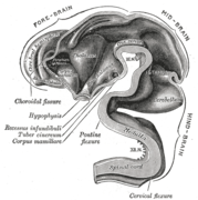
 Brain of human embryo at 4.5 weeks, showing interior of forebrain
 Brain interior at 5 weeks
 Brain viewed at midline at 3 months


Function

Motor and sensory regions of the brain
Motor control
The motor system of the brain is responsible for the generation and control of movement. Generated movements pass from the brain through nerves to motor neurons in the body, which control the action of muscles. The corticospinal tract carries movements from the brain, through the spinal cord, to the torso and limbs. The cranial nerves carry movements related to the eyes, mouth and face.
Gross movement – such as locomotion and the movement of arms and legs – is generated in the motor cortex, divided into three parts: the primary motor cortex, found in the prefrontal gyrus
and has sections dedicated to the movement of different body parts.
These movements are supported and regulated by two other areas, lying anterior to the primary motor cortex: the premotor area and the supplementary motor area.
The hands and mouth have a much larger area dedicated to them than
other body parts, allowing finer movement; this has been visualised in a
motor homunculus. Impulses generated from the motor cortex travel along the corticospinal tract along the front of the medulla and cross over (decussate) at the medullary pyramids. These then travel down the spinal cord, with most connecting to interneurons, in turn connecting to lower motor neurons within the grey matter that then transmit the impulse to move to muscles themselves. The cerebellum and basal ganglia, play a role in fine, complex and coordinated muscle movements.
Connections between the cortex and the basal ganglia control muscle
tone, posture and movement initiation, and are referred to as the extrapyramidal system.
Sensory

Cortical areas

Routing of neural signals from the two eyes to the brain
The sensory nervous system is involved with the reception and processing of sensory information.
This information is received through the cranial nerves, through tracts
in the spinal cord, and directly at centres of the brain exposed to the
blood. The brain also receives and interprets information from the special senses of vision, smell, hearing, and taste. Mixed motor and sensory signals are also integrated.
From the skin, the brain receives information about fine touch, pressure, pain, vibration and temperature. From the joints, the brain receives information about joint position. The sensory cortex
is found just near the motor cortex, and, like the motor cortex, has
areas related to sensation from different body parts. Sensation
collected by a sensory receptor on the skin is changed to a nerve signal, that is passed up a series of neurons through tracts in the spinal cord. The dorsal column–medial lemniscus pathway
contains information about fine touch, vibration and position of
joints. Neurons travel up the back part of the spinal cord to the back
part of the medulla, where they connect with second-order neurons that immediately swap sides. These neurons then travel upwards into the ventrobasal complex in the thalamus where they connect with third-order neurons and travel up to the sensory cortex. The spinothalamic tract
carries information about pain, temperature, and gross touch. Neurons
travel up the spinal cord and connect with second-order neurons in the reticular formation of the brainstem for pain and temperature, and also at the ventrobasal complex of the medulla for gross touch.
Vision is generated by light that hits the retina of the eye. Photoreceptors in the retina transduce the sensory stimulus of light into an electrical nerve signal that is sent to the visual cortex
in the occipital lobe. Vision from the left visual field is received on
the right side of each retina (and vice versa) and passes through the optic nerve until some information changes sides,
so that all information about one side of the visual field passes
through tracts in the opposite side of the brain. The nerves reach the
brain at the lateral geniculate nucleus, and travel through the optic radiation to reach the visual cortex.
Hearing and balance are both generated in the inner ear. The movement of liquids within the inner ear is generated by motion (for balance) and transmitted vibrations generated by the ossicles (for sound). This creates a nerve signal that passes through the vestibulocochlear nerve. From here, it passes through to the cochlear nuclei, the superior olivary nucleus, the medial geniculate nucleus, and finally the auditory radiation to the auditory cortex.
The sense of smell is generated by receptor cells in the epithelium of the olfactory mucosa in the nasal cavity. This information passes through a relatively permeable part of the skull to the olfactory nerve. This nerve transmits to the neural circuitry of the olfactory bulb from where information is passed to the olfactory cortex.
Taste is generated from receptors on the tongue and passed along the facial and glossopharyngeal nerves into the solitary tract in the brainstem. Some taste information is also passed from the pharynx into this area via the vagus nerve. Information is then passed from here through the thalamus into the gustatory cortex.
Regulation
Autonomic functions of the brain include the regulation, or rhythmic control of the heart rate and rate of breathing, and maintaining homeostasis.
Blood pressure and heart rate are influenced by the vasomotor centre of the medulla, which causes arteries and veins to be somewhat constricted at rest. It does this by influencing the sympathetic and parasympathetic nervous systems via the vagus nerve. Information about blood pressure is generated by baroreceptors in aortic bodies in the aortic arch, and passed to the brain along the afferent fibres of the vagus nerve. Information about the pressure changes in the carotid sinus comes from carotid bodies located near the carotid artery and this is passed via a nerve joining with the glossopharyngeal nerve. This information travels up to the solitary nucleus in the medulla. Signals from here influence the vasomotor centre to adjust vein and artery constriction accordingly.
The brain controls the rate of breathing, mainly by respiratory centres in the medulla and pons. The respiratory centres control respiration, by generating motor signals that are passed down the spinal cord, along the phrenic nerve to the diaphragm and other muscles of respiration. This is a mixed nerve
that carries sensory information back to the centres. There are four
respiratory centres, three with a more clearly defined function, and an
apneustic centre with a less clear function. In the medulla a dorsal
respiratory group causes the desire to breathe in and receives sensory information directly from the body. Also in the medulla, the ventral respiratory group influences breathing out during exertion. In the pons the pneumotaxic centre influences the duration of each breath, and the apneustic centre seems to have an influence on inhalation. The respiratory centres directly senses blood carbon dioxide and pH. Information about blood oxygen, carbon dioxide and pH levels are also sensed on the walls of arteries in the peripheral chemoreceptors
of the aortic and carotid bodies. This information is passed via the
vagus and glossopharyngeal nerves to the respiratory centres. High
carbon dioxide, an acidic pH, or low oxygen stimulate the respiratory
centres. The desire to breathe in is also affected by pulmonary stretch receptors
in the lungs which, when activated, prevent the lungs from
overinflating by transmitting information to the respiratory centres via
the vagus nerve.
The hypothalamus in the diencephalon, is involved in regulating many functions of the body. Functions include neuroendocrine regulation, regulation of the circadian rhythm, control of the autonomic nervous system,
and the regulation of fluid, and food intake. The circadian rhythm is
controlled by two main cell groups in the hypothalamus. The anterior
hypothalamus includes the suprachiasmatic nucleus and the ventrolateral preoptic nucleus which through gene expression cycles, generates a roughly 24 hour circadian clock. In the circadian day an ultradian rhythm takes control of the sleeping pattern. Sleep
is an essential requirement for the body and brain and allows the
closing down and resting of the body's systems. There are also findings
that suggest that the daily build-up of toxins in the brain are removed
during sleep. Whilst awake the brain consumes a fifth of the body's total energy needs. Sleep necessarily reduces this use and gives time for the restoration of energy-giving ATP. The effects of sleep deprivation show the absolute need for sleep.
The lateral hypothalamus contains orexinergic neurons that control appetite and arousal through their projections to the ascending reticular activating system. The hypothalamus controls the pituitary gland through the release of peptides such as oxytocin, and vasopressin, as well as dopamine into the median eminence.
Through the autonomic projections, the hypothalamus is involved in
regulating functions such as blood pressure, heart rate, breathing,
sweating, and other homeostatic mechanisms. The hypothalamus also plays a role in thermal regulation, and when stimulated by the immune system, is capable of generating a fever. The hypothalamus is influenced by the kidneys – when blood pressure falls, the renin
released by the kidneys stimulates a need to drink. The hypothalamus
also regulates food intake through autonomic signals, and hormone
release by the digestive system.
Language

Broca's area and Wernicke's area are linked by the arcuate fasciculus.
While language functions were traditionally thought to be localized to Wernicke's area and Broca's area, it is now mostly accepted that a wider network of cortical regions contributes to language functions.
The study on how language is represented, processed, and acquired by the brain is called neurolinguistics, which is a large multidisciplinary field drawing from cognitive neuroscience, cognitive linguistics, and psycholinguistics.
Lateralisation
The cerebrum has a contralateral organisation
with each hemisphere of the brain interacting primarily with one half
of the body: the left side of the brain interacts with the right side of
the body, and vice versa. The developmental cause for this is
uncertain. Motor connections from the brain to the spinal cord, and sensory connections from the spinal cord to the brain, both cross sides
in the brainstem. Visual input follows a more complex rule: the optic
nerves from the two eyes come together at a point called the optic chiasm, and half of the fibres from each nerve split off to join the other.
The result is that connections from the left half of the retina, in
both eyes, go to the left side of the brain, whereas connections from
the right half of the retina go to the right side of the brain.
Because each half of the retina receives light coming from the opposite
half of the visual field, the functional consequence is that visual
input from the left side of the world goes to the right side of the
brain, and vice versa.
Thus, the right side of the brain receives somatosensory input from the
left side of the body, and visual input from the left side of the
visual field.
The left and right sides of the brain appear symmetrical, but they function asymmetrically.
For example, the counterpart of the left-hemisphere motor area
controlling the right hand is the right-hemisphere area controlling the
left hand. There are, however, several important exceptions, involving
language and spatial cognition. The left frontal lobe is dominant for
language. If a key language area in the left hemisphere is damaged, it
can leave the victim unable to speak or understand, whereas equivalent damage to the right hemisphere would cause only minor impairment to language skills.
A substantial part of current understanding of the interactions between the two hemispheres has come from the study of "split-brain
patients"—people who underwent surgical transection of the corpus
callosum in an attempt to reduce the severity of epileptic seizures.
These patients do not show unusual behaviour that is immediately
obvious, but in some cases can behave almost like two different people
in the same body, with the right hand taking an action and then the left
hand undoing it.
These patients, when briefly shown a picture on the right side of the
point of visual fixation, are able to describe it verbally, but when the
picture is shown on the left, are unable to describe it, but may be
able to give an indication with the left hand of the nature of the
object shown.
Emotion
Emotions are generally defined as two-step multicomponent processes involving elicitation, followed by psychological feelings, appraisal, expression, autonomic responses, and action tendencies.
Attempts to localize basic emotions to certain brain regions have been
controversial, with some research finding no evidence for specific
locations corresponding to emotions, and instead circuitry involved in
general emotional processes. The amygdala, orbitofrontal cortex, mid and anterior insula cortex and lateral prefrontal cortex, appeared to be involved in generating the emotions, while weaker evidence was found for the ventral tegmental area, ventral pallidum and nucleus accumbens in incentive salience. Others, however, have found evidence of activation of specific regions, such as the basal ganglia in happiness, the subcallosal cingulate cortex in sadness, and amygdala in fear.
Cognition
The brain is responsible for cognition, which functions through numerous processes and executive functions. Executive functions include the ability to filter information and tune out irrelevant stimuli with attentional control and cognitive inhibition, the ability to process and manipulate information held in working memory, the ability to think about multiple concepts simultaneously and switch tasks with cognitive flexibility, the ability to inhibit impulses and prepotent responses with inhibitory control, and the ability to determine the relevance of information or appropriateness of an action. Higher order executive functions require the simultaneous use of multiple basic executive functions, and include planning and fluid intelligence (i.e., reasoning and problem solving).
The prefrontal cortex plays a significant role in mediating executive functions. Planning involves activation of the dorsolateral prefrontal cortex (DLPFC), anterior cingulate cortex, angular prefrontal cortex, right prefrontal cortex, and supramarginal gyrus. Working memory manipulation involves the DLPFC, inferior frontal gyrus, and areas of the parietal cortex. Inhibitory control involves multiple areas of the prefrontal cortex, as well as the caudate nucleus and subthalamic nucleus.
Physiology
Neurotransmission
Brain activity is made possible by the interconnections of neurons that are linked together to reach their targets. A neuron consists of a cell body, axon, and dendrites.
Dendrites are often extensive branches that receive information in the
form of signals from the axon terminals of other neurons. The signals
received may cause the neuron to initiate an action potential
(an electrochemical signal or nerve impulse) which is sent along its
axon to the axon terminal, to connect with the dendrites or with the
cell body of another neuron. An action potential is initiated at the initial segment of an axon, which contains a complex of proteins. When an action potential, reaches the axon terminal it triggers the release of a neurotransmitter at a synapse that propagates a signal that acts on the target cell. These chemical neurotransmitters include dopamine, serotonin, GABA, glutamate, and acetylcholine. GABA is the major inhibitory neurotransmitter in the brain, and glutamate is the major excitatory neurotransmitter. Neurons link at synapses to form neural pathways, neural circuits, and large elaborate network systems such as the salience network and the default mode network, and the activity between them is driven by the process of neurotransmission.
Metabolism

PET image of the human brain showing energy consumption
The brain consumes up to 20% of the energy used by the human body, more than any other organ. In humans, blood glucose is the primary source of energy for most cells and is critical for normal function in a number of tissues, including the brain. The human brain consumes approximately 60% of blood glucose in fasted, sedentary individuals. Brain metabolism normally relies upon blood glucose as an energy source, but during times of low glucose (such as fasting, endurance exercise, or limited carbohydrate intake), the brain uses ketone bodies for fuel with a smaller need for glucose. The brain can also utilize lactate during exercise. The brain stores glucose in the form of glycogen, albeit in significantly smaller amounts than that found in the liver or skeletal muscle. Long-chain fatty acids cannot cross the blood–brain barrier, but the liver can break these down to produce ketone bodies. However, short-chain fatty acids (e.g., butyric acid, propionic acid, and acetic acid) and the medium-chain fatty acids, octanoic acid and heptanoic acid, can cross the blood–brain barrier and be metabolized by brain cells.
Although the human brain represents only 2% of the body weight,
it receives 15% of the cardiac output, 20% of total body oxygen
consumption, and 25% of total body glucose utilization. The brain mostly uses glucose for energy, and deprivation of glucose, as can happen in hypoglycemia, can result in loss of consciousness.
The energy consumption of the brain does not vary greatly over time,
but active regions of the cortex consume somewhat more energy than
inactive regions: this fact forms the basis for the functional brain
imaging methods PET and fMRI. These functional imaging techniques provide a three-dimensional image of metabolic activity.
The function of sleep
is not fully understood; however, there is evidence that sleep enhances
the clearance of metabolic waste products, some of which are
potentially neurotoxic, from the brain and may also permit repair. Evidence suggests that the increased clearance of metabolic waste during sleep occurs via increased functioning of the glymphatic system. Sleep may also have an effect on cognitive function by weakening unnecessary connections.
Research
The brain is not fully understood, and research is ongoing. Neuroscientists, along with researchers from allied disciplines, study how the human brain works. The boundaries between the specialties of neuroscience, neurology and other disciplines such as psychiatry have faded as they are all influenced by basic research in neuroscience.
Neuroscience research has expanded considerably in recent decades. The "Decade of the Brain", an initiative of the United States Government in the 1990s, is considered to have marked much of this increase in research, and was followed in 2013 by the BRAIN Initiative. The Human Connectome Project
was a five-year study launched in 2009 to analyse the anatomical and
functional connections of parts of the brain, and has provided much
data.
Methods
Information
about the structure and function of the human brain comes from a
variety of experimental methods, including animals and humans.
Information about brain trauma and stroke has provided information about
the function of parts of the brain and the effects of brain damage. Neuroimaging is used to visualise the brain and record brain activity. Electrophysiology is used to measure, record and monitor the electrical activity of the cortex. Measurements may be of local field potentials of cortical areas, or of the activity of a single neuron. An electroencephalogram can record the electrical activity of the cortex using electrodes placed non-invasively on the scalp.
Invasive measures include electrocorticography, which uses electrodes placed directly on the exposed surface of the brain. This method is used in cortical stimulation mapping, used in the study of the relationship between cortical areas and their systemic function. By using much smaller microelectrodes, single-unit recordings can be made from a single neuron that give a high spatial resolution and high temporal resolution. This has enabled the linking of brain activity to behaviour, and the creation of neuronal maps.
The development of cerebral organoids
has opened ways for studying the growth of the brain, and of the
cortex, and for understanding disease development, offering further
implications for therapeutic applications.
Imaging
Functional neuroimaging techniques show changes in brain activity that relate to the function of specific brain areas. One technique is functional magnetic resonance imaging (fMRI) which has the advantages over earlier methods of SPECT and PET of not needing the use of radioactive materials and of offering a higher resolution. Another technique is functional near-infrared spectroscopy. These methods rely on the haemodynamic response that shows changes in brain activity in relation to changes in blood flow, useful in mapping functions to brain areas. Resting state fMRI
looks at the interaction of brain regions whilst the brain is not performing a specific task. This is also used to show the default mode network.
Any electrical current generates a magnetic field; neural oscillations induce weak magnetic fields, and in functional magnetoencephalography the current produced can show localised brain function in high resolution. Tractography uses MRI and image analysis to create 3D images of the nerve tracts of the brain. Connectograms give a graphical representation of the neural connections of the brain.
Differences in brain structure can be measured in some disorders, notably schizophrenia and dementia. Different biological approaches using imaging have given more insight for example into the disorders of depression and obsessive-compulsive disorder. A key source of information about the function of brain regions is the effects of damage to them.
Advances in neuroimaging
have enabled objective insights into mental disorders, leading to
faster diagnosis, more accurate prognosis, and better monitoring.
Gene and protein expression
Bioinformatics
is a field of study that includes the creation and advancement of
databases, and computational and statistical techniques, that can be
used in studies of the human brain, particularly in the areas of gene and protein expression. Bioinformatics and studies in genomics, and functional genomics, generated the need for DNA annotation, a transcriptome technology, identifying genes, and their and location and function. GeneCards is a major database.
As of 2017, just under 20,000 protein-coding genes are seen to be expressed in the human, and some 400 of these genes are brain-specific. The data that has been provided on gene expression
in the brain has fuelled further research into a number of disorders.
The long term use of alcohol for example, has shown altered gene
expression in the brain, and cell-type specific changes that may relate
to alcohol use disorder. These changes have been noted in the synaptic transcriptome in the prefrontal cortex, and are seen as a factor causing the drive to alcohol dependence, and also to other substance abuses.
Other related studies have also shown evidence of synaptic alterations and their loss, in the ageing brain.
Changes in gene expression alter the levels of proteins in various
pathways and this has been shown to be evident in synaptic contact
dysfunction or loss. This dysfunction has been seen to affect many
structures of the brain and has a marked effect on inhibitory neurons
resulting in a decreased level of neurotransmission, and subsequent
cognitive decline and disease.
Clinical significance
Injury
Injury to the brain can manifest in many ways. Traumatic brain injury, for example received in contact sport, after a fall, or a traffic or work accident, can be associated with both immediate and longer-term problems. Immediate problems may include bleeding within the brain, this may compress the brain tissue or damage its blood supply. Bruising to the brain may occur. Bruising may cause widespread damage to the nerve tracts that can lead to a condition of diffuse axonal injury. A fractured skull, injury to a particular area, deafness, and concussion
are also possible immediate developments. In addition to the site of
injury, the opposite side of the brain may be affected, termed a contrecoup injury. Longer-term issues that may develop include posttraumatic stress disorder, and hydrocephalus. Chronic traumatic encephalopathy can develop following multiple head injuries.
Disease
Neurodegenerative diseases result in progressive damage to different parts of the brain's function, and worsen with age. Common examples include dementia such as Alzheimer's disease, alcoholic dementia or vascular dementia; Parkinson's disease; and other rarer infectious, genetic, or metabolic causes such as Huntington's disease, motor neuron diseases, HIV dementia, syphilis-related dementia and Wilson's disease. Neurodegenerative diseases can affect different parts of the brain, and can affect movement, memory, and cognition.
The brain, although protected by the blood–brain barrier, can be affected by infections including viruses, bacteria and fungi. Infection may be of the meninges (meningitis), the brain matter (encephalitis), or within the brain matter (such as a cerebral abscess). Rare prion diseases including Creutzfeldt–Jakob disease and its variant, and kuru may also affect the brain.
Tumours
Brain tumours can be either benign or cancerous. Most malignant tumours arise from another part of the body, most commonly from the lung, breast and skin. Cancers of brain tissue can also occur, and originate from any tissue in and around the brain. Meningioma, cancer of the meninges around the brain, is more common than cancers of brain tissue.
Cancers within the brain may cause symptoms related to their size or
position, with symptoms including headache and nausea, or the gradual
development of focal symptoms such as gradual difficulty seeing,
swallowing, talking, or as a change of mood.
Cancers are in general investigated through the use of CT scans and MRI
scans. A variety of other tests including blood tests and lumbar
puncture may be used to investigate for the cause of the cancer and
evaluate the type and stage of the cancer. The corticosteroid dexamethasone is often given to decrease the swelling
of brain tissue around a tumour. Surgery may be considered, however
given the complex nature of many tumours or based on tumour stage or
type, radiotherapy or chemotherapy may be considered more suitable.
Mental disorders
Mental disorders, such as depression, schizophrenia, bipolar disorder, posttraumatic stress disorder, attention deficit hyperactivity disorder, obsessive-compulsive disorder, Tourette syndrome, and addiction, are known to relate to the functioning of the brain. Treatment for mental disorders may include psychotherapy, psychiatry, social intervention and personal recovery work or cognitive behavioural therapy; the underlying issues and associated prognoses vary significantly between individuals.
Epilepsy
Epileptic seizures are thought to relate to abnormal electrical activity. Seizure activity can manifest as absence of consciousness, focal effects such as limb movement or impediments of speech, or be generalized in nature. Status epilepticus refers to a seizure or series of seizures that have not terminated within 5 minutes. Seizures have a large number of causes, however many seizures occur without a definitive cause being found. In a person with epilepsy,
risk factors for further seizures may include sleeplessness, drug and
alcohol intake, and stress. Seizures may be assessed using blood tests, EEG and various medical imaging techniques based on the medical history and exam findings. In addition to treating an underlying cause and reducing exposure to risk factors, anticonvulsant medications can play a role in preventing further seizures.
Congenital
Some brain disorders such as Tay–Sachs disease are congenital, and linked to genetic and chromosomal mutations. A rare group of congenital cephalic disorders known as lissencephaly is characterised by the lack of, or inadequacy of, cortical folding. Normal development of the brain can be affected during pregnancy by nutritional deficiencies, teratogens, infectious diseases, and by the use of recreational drugs, including alcohol (which may result in fetal alcohol spectrum disorders).
Stroke

CT scan of a cerebral hemorrhage, showing an intraparenchymal bleed (bottom arrow) with surrounding edema (top arrow)
A stroke is a decrease in blood supply to an area of the brain causing cell death and brain injury. This can lead to a wide range of symptoms, including the "FAST" symptoms of facial droop, arm weakness, and speech difficulties (including with speaking and finding words or forming sentences).
Symptoms relate to the function of the affected area of the brain and
can point to the likely site and cause of the stroke. Difficulties with
movement, speech, or sight usually relate to the cerebrum, whereas imbalance, double vision, vertigo and symptoms affecting more than one side of the body usually relate to the brainstem or cerebellum.
Most strokes result from loss of blood supply, typically because of an embolus, rupture of a fatty plaque or narrowing of small arteries. Strokes can also result from bleeding within the brain. Transient ischaemic attacks (TIAs) are strokes in which symptoms resolve within 24 hours. Investigation into the stroke will involve a medical examination (including a neurological examination) and the taking of a medical history, focusing on the duration of the symptoms and risk factors (including high blood pressure, atrial fibrillation, and smoking). Further investigation is needed in younger patients. An ECG and biotelemetry may be conducted to identify atrial fibrillation; an ultrasound can investigate narrowing of the carotid arteries; an echocardiogram can be used to look for clots within the heart, diseases of the heart valves or the presence of a patent foramen ovale. Blood tests are routinely done as part of the workup including diabetes tests and a lipid profile.
Some treatments for stroke are time-critical. These include clot dissolution or surgical removal of a clot for ischaemic strokes, and decompression for haemorrhagic strokes. As stroke is time critical, hospitals and even pre-hospital care of stroke involves expedited investigations – usually a CT scan to investigate for a haemorrhagic stroke and a CT or MR angiogram to evaluate arteries that supply the brain. MRI scans,
not as widely available, may be able to demonstrate the affected area
of the brain more accurately, particularly with ischaemic stroke.
Having experienced a stroke, a person may be admitted to a stroke unit, and treatments may be directed as preventing future strokes, including ongoing anticoagulation (such as aspirin or clopidogrel), antihypertensives, and lipid-lowering drugs. A multidisciplinary team including speech pathologists, physiotherapists, occupational therapists, and psychologists plays a large role in supporting a person affected by a stroke and their rehabilitation.
A history of stroke increases the risk of developing dementia by around
70%, and recent stroke increases the risk by around 120%.
Brain death
Brain death refers to an irreversible total loss of brain function. This is characterised by coma, loss of reflexes, and apnoea, however, the declaration of brain death varies geographically and is not always accepted. In some countries there is also a defined syndrome of brainstem death. Declaration of brain death can have profound implications as the declaration, under the principle of medical futility, will be associated with the withdrawal of life support, and as those with brain death often have organs suitable for organ donation. The process is often made more difficult by poor communication with patients' families.
When brain death is suspected, reversible differential diagnoses such as, electrolyte, neurological and drug-related cognitive suppression need to be excluded. Testing for reflexes can be of help in the decision, as can the absence of response and breathing. Clinical observations, including a total lack of responsiveness, a known diagnosis, and neural imaging evidence, may all play a role in the decision to pronounce brain death.
Society and culture
Neuroanthropology
is the study of the relationship between culture and the brain. It
explores how the brain gives rise to culture, and how culture influences
brain development. Cultural differences and their relation to brain development and structure are researched in different fields.
The mind

The skull of Phineas Gage,
with the path of the iron rod that passed through it without killing
him, but altering his cognition. The case helped to convince people that
mental functions were localized in the brain.
The philosophy of the mind studies such issues as the problem of understanding consciousness and the mind–body problem. The relationship between the brain and the mind
is a significant challenge both philosophically and scientifically.
This is because of the difficulty in explaining how mental activities,
such as thoughts and emotions, can be implemented by physical structures
such as neurons and synapses, or by any other type of physical mechanism. This difficulty was expressed by Gottfried Leibniz in the analogy known as Leibniz's Mill:
One is obliged to admit that perception and what depends upon it is inexplicable on mechanical principles, that is, by figures and motions. In imagining that there is a machine whose construction would enable it to think, to sense, and to have perception, one could conceive it enlarged while retaining the same proportions, so that one could enter into it, just like into a windmill. Supposing this, one should, when visiting within it, find only parts pushing one another, and never anything by which to explain a perception.
- — Leibniz, Monadology
Doubt about the possibility of a mechanistic explanation of thought drove René Descartes, and most other philosophers along with him, to dualism: the belief that the mind is to some degree independent of the brain.
There has always, however, been a strong argument in the opposite
direction. There is clear empirical evidence that physical manipulations
of, or injuries to, the brain (for example by drugs or by lesions,
respectively) can affect the mind in potent and intimate ways. In the 19th century, the case of Phineas Gage,
a railway worker who was injured by a stout iron rod passing through
his brain, convinced both researchers and the public that cognitive
functions were localised in the brain.
Following this line of thinking, a large body of empirical evidence for
a close relationship between brain activity and mental activity has led
most neuroscientists and contemporary philosophers to be materialists, believing that mental phenomena are ultimately the result of, or reducible to, physical phenomena.
Brain size
The size of the brain and a person's intelligence are not strongly related. Studies tend to indicate small to moderate correlations (averaging around 0.3 to 0.4) between brain volume and IQ.
The most consistent associations are observed within the frontal,
temporal, and parietal lobes, the hippocampi, and the cerebellum, but
these only account for a relatively small amount of variance in IQ,
which itself has only a partial relationship to general intelligence and
real-world performance.
Other animals, including whales and elephants have larger brains than humans. However, when the brain-to-body mass ratio is taken into account, the human brain is almost twice as large as that of a bottlenose dolphin, and three times as large as that of a chimpanzee. However, a high ratio does not of itself demonstrate intelligence: very small animals have high ratios and the treeshrew has the largest quotient of any mammal.
In popular culture

Phrenology summarized in an 1883 chart
Research has disproved some common misconceptions about the brain. These include both ancient and modern myths. It is not true that neurons are not replaced after the age of two; nor that only ten per cent of the brain is used. Popular culture has also oversimplified the lateralisation of the brain, suggesting that functions are completely specific to one side of the brain or the other. Akio Mori coined the term game brain for the unreliably supported theory that spending long periods playing video games harmed the brain's pre-frontal region and the expression of emotion and creativity.
Historically, the brain featured in popular culture through phrenology, a pseudoscience
that assigned personality attributes to different regions of the
cortex. The cortex remains important in popular culture as covered in
books and satire. The brain features in science fiction, with themes such as brain transplants and cyborgs (beings with features like partly artificial brains). The 1942 science fiction book (adapted three times for cinema) Donovan's Brain tells the tale of an isolated brain kept alive in vitro, gradually taken over by a malign intelligence.
History
Early history

Hieroglyph for the word "brain" (c.1700 BC)
The Edwin Smith Papyrus, an ancient Egyptian medical treatise written in the 17th century BC, contains the earliest recorded reference to the brain. The hieroglyph
for brain, occurring eight times in this papyrus, describes the
symptoms, diagnosis, and prognosis of two traumatic injuries to the
head. The papyrus mentions the external surface of the brain, the
effects of injury (including seizures and aphasia), the meninges, and cerebrospinal fluid.
In the fifth century BC, Alcmaeon of Croton in Magna Grecia, first considered the brain to be the seat of the mind. Also in the fifth century BC in Athens, the unknown author of On the Sacred Disease, a medical treatise which is part of the Hippocratic Corpus and traditionally attributed to Hippocrates, believed the brain to be the seat of intelligence. Aristotle, in his biology initially believed the heart to be the seat of intelligence,
and saw the brain as a cooling mechanism for the blood. He reasoned
that humans are more rational than the beasts because, among other
reasons, they have a larger brain to cool their hot-bloodedness. Aristotle did describe the meninges and distinguished between the cerebrum and cerebellum. Herophilus of Chalcedon
in the fourth and third centuries BC distinguished the cerebrum and the
cerebellum, and provided the first clear description of the ventricles; and with Erasistratus of Ceos
experimented on living brains. Their works are now mostly lost, and we
know about their achievements due mostly to secondary sources. Some of
their discoveries had to be re-discovered a millennium after their
deaths. Anatomist physician Galen in the second century AD, during the time of the Roman Empire,
dissected the brains of sheep, monkeys, dogs, and pigs. He concluded
that, as the cerebellum was denser than the brain, it must control the muscles,
while as the cerebrum was soft, it must be where the senses were
processed. Galen further theorized that the brain functioned by movement
of animal spirits through the ventricles.
Renaissance
In 1316, Mondino de Luzzi's Anathomia began the modern study of brain anatomy.
Niccolò Massa discovered in 1536 that the ventricles were filled with fluid. Archangelo Piccolomini of Rome was the first to distinguish between the cerebrum and cerebral cortex. In 1543 Andreas Vesalius published his seven-volume De humani corporis fabrica. The seventh book covered the brain and eye, with detailed images of the ventricles, cranial nerves, pituitary gland, meninges, structures of the eye, the vascular supply to the brain and spinal cord, and an image of the peripheral nerves.
Vesalius rejected the common belief that the ventricles were
responsible for brain function, arguing that many animals have a similar
ventricular system to humans, but no true intelligence.
René Descartes proposed the theory of dualism to tackle the issue of the brain's relation to the mind. He suggested that the pineal gland
was where the mind interacted with the body after recording the brain
mechanisms responsible for circulating cerebrospinal fluid.
This dualism likely provided impetus for later anatomists to further
explore the relationship between the anatomical and functional aspects
of brain anatomy.
Thomas Willis is considered a second pioneer in the study of neurology and brain science. In 1664 in Cerebri Anatome (Latin: Anatomy of the brain), followed by Cerebral Pathology
in 1667. In these he described the structure of the cerebellum, the
ventricles, the cerebral hemispheres, the brainstem, and the cranial
nerves, studied its blood supply; and proposed functions associated with
different areas of the brain.
The circle of Willis was named after his investigations into the blood
supply of the brain, and he was the first to use the word "neurology."
Willis removed the brain from the body when examining it, and rejected
the commonly held view that the cortex only consisted of blood vessels
and the view of the last two millennia that the cortex was only
incidentally important.
In the late 19th century, Emil du Bois-Reymond and Hermann von Helmholtz, following the work of their teacher Johannes Peter Müller showed the electrical inpulses which pass along nerves; but unlike Müller's views, that such impulses were able to be observed. Richard Caton in 1875 demonstrated electrical impulses in the cerebral hemispheres of rabbits and monkeys. In the 1820s, Jean Pierre Flourens pioneered the experimental method of damaging specific parts of animal brains describing the effects on movement and behavior.


One of Leonardo da Vinci's sketches of the human skull
Modern period

Drawing by Camillo Golgi of vertical section of rabbit hippocampus, from his "Sulla fina anatomia degli organi centrali del sistema nervoso", 1885

Drawing of cells in chick cerebellum by Santiago Ramón y Cajal, from "Estructura de los centros nerviosos de las aves", Madrid, 1905
Studies of the brain became more sophisticated with the use of the microscope and the development of a silver staining method by Camillo Golgi during the 1880s. This was able to show the intricate structures of single neurons. This was used by Santiago Ramón y Cajal and led to the formation of the neuron doctrine,
the then revolutionary hypothesis that the neuron is the functional
unit of the brain. He used microscopy to uncover many cell types, and
proposed functions for the cells he saw. For this, Golgi and Cajal are considered the founders of twentieth century neuroscience, both sharing the Nobel prize in 1906 for their studies and discoveries in this field.
Charles Sherrington published his influential 1906 work The Integrative Action of the Nervous System
examining the function of reflexes, evolutionary development of the
nervous system, functional specialisation of the brain, and layout and
cellular function of the central nervous system. John Farquhar Fulton, founded the Journal of Neurophysiology and published the first comprehensive textbook on the physiology of the nervous system during 1938. Neuroscience during the twentieth century began to be recognised as a distinct unified academic discipline, with David Rioch, Francis O. Schmitt, and Stephen Kuffler playing critical roles in establishing the field. Rioch originated the integration of basic anatomical and physiological research with clinical psychiatry at the Walter Reed Army Institute of Research, starting in the 1950s. During the same period, Schmitt established the Neuroscience Research Program,
an inter-university and international organisation, bringing together
biology, medicine, psychological and behavioural sciences. The word
neuroscience itself arises from this program.
Paul Broca associated regions of the brain with specific functions, in particular language in Broca's area, following work on brain-damaged patients. John Hughlings Jackson described the function of the motor cortex by watching the progression of epileptic seizures through the body. Carl Wernicke described a region associated with language comprehension and production. Korbinian Brodmann divided regions of the brain based on the appearance of cells. By 1950, Sherrington, Papez, and MacLean had identified many of the brainstem and limbic system functions. The capacity of the brain to re-organise and change with age, and a recognised critical development period, were attributed to neuroplasticity, pioneered by Margaret Kennard, who experimented on monkeys during the 1930-40s.
Harvey Cushing (1869–1939) is recognised as the first proficient brain surgeon in the world. In 1937, Walter Dandy began the practice of vascular neurosurgery by performing the first surgical clipping of an intracranial aneurysm.
Comparative anatomy
The human brain has many properties that are common to all vertebrate brains. Many of its features are common to all mammalian brains, most notably a six-layered cerebral cortex and a set of associated structures, including the hippocampus and amygdala. The cortex is proportionally larger in humans than in many other mammals. Humans have more association cortex, sensory and motor parts than smaller mammals such as the rat and the cat.
As a primate brain, the human brain has a much larger cerebral cortex, in proportion to body size, than most mammals, and a highly developed visual system.
As a hominid brain, the human brain is substantially enlarged even in comparison to the brain of a typical monkey. The sequence of human evolution from Australopithecus (four million years ago) to Homo sapiens (modern humans) was marked by a steady increase in brain size. As brain size increased, this altered the size and shape of the skull, from about 600 cm3 in Homo habilis to an average of about 1520 cm3 in Homo neanderthalensis. Differences in DNA, gene expression, and gene–environment interactions help explain the differences between the function of the human brain and other primates.


