Search This Blog

Wednesday, March 4, 2020

Robert Boyle

From Wikipedia, the free encyclopedia


Robert Boyle

The Shannon Portrait of the Hon Robert Boyle.jpg
Born25 January 1627
Died31 December 1691 (aged 64)
NationalityIrish
EducationEton College
Known for
Scientific career
FieldsPhysics, chemistry
Notable studentsRobert Hooke
Influences Katherine Boyle Jones
InfluencedIsaac Newton

Robert Boyle FRS (/bɔɪl/; 25 January 1627 – 31 December 1691) was an Anglo-Irish natural philosopher, chemist, physicist, and inventor. Boyle is largely regarded today as the first modern chemist (a title some give to 8th century Islamic scholar Jabir ibn Hayyan), and therefore one of the founders of modern chemistry, and one of the pioneers of modern experimental scientific method. He is best known for Boyle's law, which describes the inversely proportional relationship between the absolute pressure and volume of a gas, if the temperature is kept constant within a closed system. Among his works, The Sceptical Chymist is seen as a cornerstone book in the field of chemistry. He was a devout and pious Anglican and is noted for his writings in theology.

Biography

Early years

Boyle was born at Lismore Castle, in County Waterford, Ireland, the seventh son and fourteenth child of The 1st Earl of Cork ('the Great Earl of Cork') and Catherine Fenton. Lord Cork, then known simply as Richard Boyle, had arrived in Dublin from England in 1588 during the Tudor plantations of Ireland and obtained an appointment as a deputy escheator. He had amassed enormous wealth and landholdings by the time Robert was born, and had been created Earl of Cork in October 1620. Catherine Fenton, Countess of Cork, was the daughter of Sir Geoffrey Fenton, the former Secretary of State for Ireland, who was born in Dublin in 1539, and Alice Weston, the daughter of Robert Weston, who was born in Lismore in 1541.

As a child, Boyle was fostered to a local family, as were his elder brothers. Boyle received private tutoring in Latin, Greek, and French and when he was eight years old, following the death of his mother, he was sent to Eton College in England. His father's friend, Sir Henry Wotton, was then the provost of the college.

During this time, his father hired a private tutor, Robert Carew, who had knowledge of Irish, to act as private tutor to his sons in Eton. However, "only Mr. Robert sometimes desires it [Irish] and is a little entered in it", but despite the "many reasons" given by Carew to turn their attentions to it, "they practice the French and Latin but they affect not the Irish". After spending over three years at Eton, Robert travelled abroad with a French tutor. They visited Italy in 1641 and remained in Florence during the winter of that year studying the "paradoxes of the great star-gazer" Galileo Galilei, who was elderly but still living in 1641.

Middle years

Robert returned to England from continental Europe in mid-1644 with a keen interest in scientific research. His father, Lord Cork, had died the previous year and had left him the manor of Stalbridge in Dorset as well as substantial estates in County Limerick in Ireland that he had acquired. Robert then made his residence at Stalbridge House, between 1644 and 1652, and conducted many experiments there. From that time, Robert devoted his life to scientific research and soon took a prominent place in the band of enquirers, known as the "Invisible College", who devoted themselves to the cultivation of the "new philosophy". They met frequently in London, often at Gresham College, and some of the members also had meetings at Oxford.

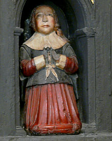
Sculpture of a young boy, thought to be Boyle, on his parents' monument in St Patrick's Cathedral, Dublin.

Having made several visits to his Irish estates beginning in 1647, Robert moved to Ireland in 1652 but became frustrated at his inability to make progress in his chemical work. In one letter, he described Ireland as "a barbarous country where chemical spirits were so misunderstood and chemical instruments so unprocurable that it was hard to have any Hermetic thoughts in it."

In 1654, Boyle left Ireland for Oxford to pursue his work more successfully. An inscription can be found on the wall of University College, Oxford, the High Street at Oxford (now the location of the Shelley Memorial), marking the spot where Cross Hall stood until the early 19th century. It was here that Boyle rented rooms from the wealthy apothecary who owned the Hall.

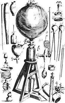
Reading in 1657 of Otto von Guericke's air pump, he set himself, with the assistance of Robert Hooke, to devise improvements in its construction, and with the result, the "machina Boyleana" or "Pneumatical Engine", finished in 1659, he began a series of experiments on the properties of air. An account of Boyle's work with the air pump was published in 1660 under the title New Experiments Physico-Mechanical, Touching the Spring of the Air, and its Effects.

Among the critics of the views put forward in this book was a Jesuit, Francis Line (1595–1675), and it was while answering his objections that Boyle made his first mention of the law that the volume of a gas varies inversely to the pressure of the gas, which among English-speaking people is usually called Boyle's Law after his name. The person who originally formulated the hypothesis was Henry Power in 1661. Boyle in 1662 included a reference to a paper written by Power, but mistakenly attributed it to Richard Towneley. In continental Europe the hypothesis is sometimes attributed to Edme Mariotte, although he did not publish it until 1676 and was likely aware of Boyle's work at the time.

One of Robert Boyle's notebooks (1690-1691) held by the Royal Society of London. The Royal Society archives holds 46 volumes of philosophical, scientific and theological papers by Boyle and seven volumes of his correspondence.

In 1663 the Invisible College became The Royal Society of London for Improving Natural Knowledge, and the charter of incorporation granted by Charles II of England named Boyle a member of the council. In 1680 he was elected president of the society, but declined the honour from a scruple about oaths.

He made a "wish list" of 24 possible inventions which included "the prolongation of life", the "art of flying", "perpetual light", "making armour light and extremely hard", "a ship to sail with all winds, and a ship not to be sunk", "practicable and certain way of finding longitudes", "potent drugs to alter or exalt imagination, waking, memory and other functions and appease pain, procure innocent sleep, harmless dreams, etc." They are extraordinary because all but a few of the 24 have come true.

It was during his time at Oxford that Boyle was a Chevalier. The Chevaliers are thought to have been established by royal order a few years before Boyle's time at Oxford. The early part of Boyle's residence was marked by the actions of the victorious parliamentarian forces, consequently this period marked the most secretive period of Chevalier movements and thus little is known about Boyle's involvement beyond his membership.

In 1668 he left Oxford for London where he resided at the house of his elder sister Katherine Jones, Lady Ranelagh, in Pall Mall. He experimented in the laboratory she had in her home and attended her salon of intellectuals interested in the sciences. The siblings maintained "a lifelong intellectual partnership, where brother and sister shared medical remedies, promoted each other’s scientific ideas, and edited each other’s manuscripts." His contemporaries widely acknowledged Katherine's influence on his work, but later historiographers dropped discussion of her accomplishments and relationship to her brother from their histories.

Later years

Plaque at the site of Boyle and Hooke's experiments in Oxford

In 1669 his health, never very strong, began to fail seriously and he gradually withdrew from his public engagements, ceasing his communications to the Royal Society, and advertising his desire to be excused from receiving guests, "unless upon occasions very extraordinary", on Tuesday and Friday forenoon, and Wednesday and Saturday afternoon. In the leisure thus gained he wished to "recruit his spirits, range his papers", and prepare some important chemical investigations which he proposed to leave "as a kind of Hermetic legacy to the studious disciples of that art", but of which he did not make known the nature. His health became still worse in 1691, and he died on 31 December that year, just a week after the death of his sister, Katherine, in whose home he had lived and with whom he had shared scientific pursuits for more than twenty years. Boyle died from paralysis. He was buried in the churchyard of St Martin-in-the-Fields, his funeral sermon being preached by his friend, Bishop Gilbert Burnet. In his will, Boyle endowed a series of lectures that came to be known as the Boyle Lectures.

Scientific investigator

Boyle's air pump

Boyle's great merit as a scientific investigator is that he carried out the principles which Francis Bacon espoused in the Novum Organum. Yet he would not avow himself a follower of Bacon, or indeed of any other teacher.

On several occasions he mentions that to keep his judgment as unprepossessed as might be with any of the modern theories of philosophy, until he was "provided of experiments" to help him judge of them. He refrained from any study of the atomical and the Cartesian systems, and even of the Novum Organum itself, though he admits to "transiently consulting" them about a few particulars. Nothing was more alien to his mental temperament than the spinning of hypotheses. He regarded the acquisition of knowledge as an end in itself, and in consequence he gained a wider outlook on the aims of scientific inquiry than had been enjoyed by his predecessors for many centuries. This, however, did not mean that he paid no attention to the practical application of science nor that he despised knowledge which tended to use.

Fig. 3: Illustration of Excerptum ex collectionibus philosophicis anglicis... novum genus lampadis à Rob. Boyle ... published in Acta Eruditorum, 1682

Robert Boyle was an alchemist; and believing the transmutation of metals to be a possibility, he carried out experiments in the hope of achieving it; and he was instrumental in obtaining the repeal, in 1689, of the statute of Henry IV against multiplying gold and silver. With all the important work he accomplished in physics – the enunciation of Boyle's law, the discovery of the part taken by air in the propagation of sound, and investigations on the expansive force of freezing water, on specific gravities and refractive powers, on crystals, on electricity, on colour, on hydrostatics, etc. – chemistry was his peculiar and favourite study. His first book on the subject was The Sceptical Chymist, published in 1661, in which he criticised the "experiments whereby vulgar Spagyrists are wont to endeavour to evince their Salt, Sulphur and Mercury to be the true Principles of Things." For him chemistry was the science of the composition of substances, not merely an adjunct to the arts of the alchemist or the physician.

He endorsed the view of elements as the undecomposable constituents of material bodies; and made the distinction between mixtures and compounds. He made considerable progress in the technique of detecting their ingredients, a process which he designated by the term "analysis". He further supposed that the elements were ultimately composed of particles of various sorts and sizes, into which, however, they were not to be resolved in any known way. He studied the chemistry of combustion and of respiration, and conducted experiments in physiology, where, however, he was hampered by the "tenderness of his nature" which kept him from anatomical dissections, especially vivisections, though he knew them to be "most instructing".

Theological interests

In addition to philosophy, Boyle devoted much time to theology, showing a very decided leaning to the practical side and an indifference to controversial polemics. At the Restoration of the king in 1660, he was favourably received at court and in 1665 would have received the provostship of Eton College had he agreed to take holy orders, but this he refused to do on the ground that his writings on religious subjects would have greater weight coming from a layman than a paid minister of the Church. 

Moreover, Boyle incorporated his scientific interests into his theology, believing that natural philosophy could provide powerful evidence for the existence of God. In works such as Disquisition about the Final Causes of Natural Things (1688), for instance, he criticised contemporary philosophers – such as René Descartes – who denied that the study of nature could reveal much about God. Instead, Boyle argued that natural philosophers could use the design apparently on display in some parts of nature to demonstrate God's involvement with the world. He also attempted to tackle complex theological questions using methods derived from his scientific practices. In Some Physico-Theological Considerations about the Possibility of the Resurrection (1675), he used a chemical experiment known as the reduction to the pristine state as part of an attempt to demonstrate the physical possibility of the resurrection of the body. Throughout his career, Boyle tried to show that science could lend support to Christianity.

As a director of the East India Company he spent large sums in promoting the spread of Christianity in the East, contributing liberally to missionary societies and to the expenses of translating the Bible or portions of it into various languages. Boyle supported the policy that the Bible should be available in the vernacular language of the people. An Irish language version of the New Testament was published in 1602 but was rare in Boyle's adult life. In 1680–85 Boyle personally financed the printing of the Bible, both Old and New Testaments, in Irish. In this respect, Boyle's attitude to the Irish language differed from the English Ascendancy class in Ireland at the time, which was generally hostile to the language and largely opposed the use of Irish (not only as a language of religious worship).

Boyle also had a monogenist perspective about race origin. He was a pioneer studying races, and he believed that all human beings, no matter how diverse their physical differences, came from the same source: Adam and Eve. He studied reported stories of parents' giving birth to different coloured albinos, so he concluded that Adam and Eve were originally white and that Caucasians could give birth to different coloured races. Boyle also extended the theories of Robert Hooke and Isaac Newton about colour and light via optical projection (in physics) into discourses of polygenesis, speculating that maybe these differences were due to "seminal impressions". Taking this into account, it might be considered that he envisioned a good explanation for complexion at his time, due to the fact that now we know that skin colour is disposed by genes, which are actually contained in the semen. Boyle's writings mention that at his time, for "European Eyes", beauty was not measured so much in colour of skin, but in "stature, comely symmetry of the parts of the body, and good features in the face". Various members of the scientific community rejected his views and described them as "disturbing" or "amusing".

In his will, Boyle provided money for a series of lectures to defend the Christian religion against those he considered "notorious infidels, namely atheists, deists, pagans, Jews and Muslims", with the provision that controversies between Christians were not to be mentioned.

Awards and honours


As a founder of the Royal Society, he was elected a Fellow of the Royal Society (FRS) in 1663. Boyle's law is named in his honour. The Royal Society of Chemistry issues a Robert Boyle Prize for Analytical Science, named in his honour. The Boyle Medal for Scientific Excellence in Ireland, inaugurated in 1899, is awarded jointly by the Royal Dublin Society and The Irish Times. Launched in 2012, The Robert Boyle Summer School organized by the Waterford Institute of Technology with support from Lismore Castle, is held annually to honor the heritage of Robert Boyle.

Important works

Title page of The Sceptical Chymist (1661)
 
Boyle's self-flowing flask, a perpetual motion machine, appears to fill itself through siphon action ("hydrostatic perpetual motion") and involves the "hydrostatic paradox" This is not possible in reality; a siphon requires its "output" to be lower than the "input".
 
The following are some of the more important of his works:
  • 1660 – New Experiments Physico-Mechanical: Touching the Spring of the Air and their Effects
  • 1661 – The Sceptical Chymist
  • 1662 – Whereunto is Added a Defence of the Authors Explication of the Experiments, Against the Obiections of Franciscus Linus and Thomas Hobbes (a book-length addendum to the second edition of New Experiments Physico-Mechanical)
  • 1663 – Considerations touching the Usefulness of Experimental Natural Philosophy (followed by a second part in 1671)
  • 1664 – Experiments and Considerations Touching Colours, with Observations on a Diamond that Shines in the Dark
  • 1665 – New Experiments and Observations upon Cold
  • 1666 – Hydrostatical Paradoxes
  • 1666 – Origin of Forms and Qualities according to the Corpuscular Philosophy. (A continuation of his work on the spring of air demonstrated that a reduction in ambient pressure could lead to bubble formation in living tissue. This description of a viper in a vacuum was the first recorded description of decompression sickness.)
  • 1669 – A Continuation of New Experiments Physico-mechanical, Touching the Spring and Weight of the Air, and Their Effects
  • 1670 – Tracts about the Cosmical Qualities of Things, the Temperature of the Subterraneal and Submarine Regions, the Bottom of the Sea, &tc. with an Introduction to the History of Particular Qualities
  • 1672 – Origin and Virtues of Gems
  • 1673 – Essays of the Strange Subtilty, Great Efficacy, Determinate Nature of Effluviums
  • 1674 – Two volumes of tracts on the Saltiness of the Sea, Suspicions about the Hidden Realities of the Air, Cold, Celestial Magnets
  • 1674 – Animadversions upon Mr. Hobbes's Problemata de Vacuo
  • 1676 – Experiments and Notes about the Mechanical Origin or Production of Particular Qualities, including some notes on electricity and magnetism
  • 1678 – Observations upon an artificial Substance that Shines without any Preceding Illustration
  • 1680 – The Aerial Noctiluca
  • 1682 – New Experiments and Observations upon the Icy Noctiluca (a further continuation of his work on the air)
  • 1684 – Memoirs for the Natural History of the Human Blood
  • 1685 – Short Memoirs for the Natural Experimental History of Mineral Waters
  • 1686 – A Free Enquiry into the Vulgarly Received Notion of Nature
  • 1690 – Medicina Hydrostatica
  • 1691 – Experimenta et Observationes Physicae
Among his religious and philosophical writings were:
  • 1648/1660 – Seraphic Love, written in 1648, but not published until 1660
  • 1663 – An Essay upon the Style of the Holy Scriptures
  • 1664 – Excellence of Theology compared with Natural Philosophy
  • 1665 – Occasional Reflections upon Several Subjects, which was ridiculed by Swift in Meditation Upon a Broomstick, and by Butler in An Occasional Reflection on Dr Charlton's Feeling a Dog's Pulse at Gresham College
  • 1675 – Some Considerations about the Reconcileableness of Reason and Religion, with a Discourse about the Possibility of the Resurrection
  • 1687 – The Martyrdom of Theodora, and of Didymus
  • 1690 – The Christian Virtuoso

X-ray microscope

From Wikipedia, the free encyclopedia
 
An X-ray microscopy image of a living 10-days-old canola plant.

An X-ray microscope uses electromagnetic radiation in the soft X-ray band to produce magnified images of objects. Since X-rays penetrate most objects, there is no need to specially prepare them for X-ray microscopy observations.

Unlike visible light, X-rays do not reflect or refract easily, and they are invisible to the human eye. Therefore, an X-ray microscope exposes film or uses a charge-coupled device (CCD) detector to detect X-rays that pass through the specimen. It is a contrast imaging technology using the difference in absorption of soft X-rays in the water window region (wavelengths: 2.34-4.4 nm, energies: 280-530 eV) by the carbon atom (main element composing the living cell) and the oxygen atom (main element for water).

Microfocus X-ray also achieves high magnification by projection. A microfocus X-ray tube produces X-rays from an extremely small focal spot (5 µm down to 0.1 µm). The X-rays are in the more conventional X-ray range (20 to 300 kV), and they are not re-focused.

Invention and Development

The history of X-ray microscopy can be traced back to the early 20th century. After the German physicist Rontgen discovered X-rays in 1895, scientists soon illuminated an object using an X-ray point source and captured the shadow images of the object with a resolution of several microns. In 1918, Einstein pointed out that the refractive index for X rays in most mediums should be just slightly less than 1, which means refractive optical parts would be difficult to use for X-ray applications.

Early X-ray microscopes by Paul Kirkpatrick and Albert Baez used grazing incidence reflective X-ray optics to focus the X-rays, which grazed X-rays off parabolic curved mirrors at a very high angle of incidence. An alternative method of focusing X-rays is to use a tiny Fresnel zone plate of concentric gold or nickel rings on a silicon dioxide substrate. Sir Lawrence Bragg produced some of the first usable X-ray images with his apparatus in the late 1940s.

Indirect drive laser inertial confinement fusion uses a "hohlraum" which is irradiated with laser beam cones from either side on its inner surface to bathe a fusion microcapsule inside with smooth high intensity X-rays. The highest energy X-rays which penetrate the hohlraum can be visualized using an X-ray microscope such as here, where X-radiation is represented in orange/red.

In the 1950s Sterling Newberry produced a shadow X-ray microscope which placed the specimen between the source and a target plate, this became the basis for the first commercial X-ray microscopes from the General Electric Company.

After a silent period in the 1960s, X-ray microscopy regained people's attention in the 1970s. In 1972, Horowitz and Howell built the first synchrotron-based X-ray microscope at the Cambridge Electron Accelerator. This microscope scanned samples using synchrotron radiation from a tiny pinhole and showed the abilities of both transmission and fluorescence microscopy. Other developments in this period include the first holographic demonstration by Sadao Aoki and Seishi Kikuta in Japan, the first TXMs using zone plates by Schmahl et al., and Stony Brook’s experiments in STXM.

The uses of synchrotron light sources brought new possibilities for X-ray microscopy in the 1980s. However, as new synchrotron source-based microscopes were built in many groups, people realized that it was difficult to perform such experiments due to insufficient technological capabilities at that time, such as poor coherent illuminations, poor quality x-ray optical elements, and user-unfriendly light sources.

Entering the 1990s, new instruments and new light-sources greatly fueled the improvement of X-ray microscopy. Microscopy methods including tomography, cryo, and cryo-tomography were successfully demonstrated. With rapid development, X-ray microscopy found itsnew applications in soil science, geochemistry, polymer sciences, and magnetism. The hardware was also miniaturized so that researchers could perform experiments in their own laboratories.

Extremely high intensity sources of 9.25 keV X-rays for X-ray phase-contrast microscopy, from a focal spot about 10 um x 10 um, may be obtained with a non-syncrotron X-ray source which uses a focused electron beam and a liquid metal anode. This was demonstratred in 2003, and in 2017 was used to image mouse brain at a voxel size of about one cubic micrometer (see below).

With the applications continuing to grow, X-ray microscopy has become a routine, proven technique used in environmental and soil sciences, geo- and cosmo-chemistry, polymer sciences, biology, magnetism, material sciences. With this increasing demand for X-ray microscopy in these fields, microscopes based on synchrotron, liquid metal anode, and other laboratory light sources are being built around the world. X-ray optics and components are also being commercialized rapidly.

Instrumentation

X-ray optics

Synchrotron Light Sources

Advanced Light Source

The Advanced Light Source (ALS) in Berkeley, California, is home to XM-1, a full-field soft X-ray microscope operated by the Center for X-ray Optics and dedicated to various applications in modern nanoscience, such as nanomagnetic materials, environmental and materials sciences and biology. XM-1 uses an X-ray lens to focus X-rays on a CCD, in a manner similar to an optical microscope. XM-1 held the world record in spatial resolution with Fresnel zone plates down to 15 nm and is able to combine high spatial resolution with a sub-100ps time resolution to study e.g. ultrafast spin dynamics. In July 2012, a group at DESY claimed a record spatial resolution of 10 nm, by using the hard X-ray scanning microscope at PETRA III.

The ALS is also home to the world's first soft x-ray microscope designed for biological and biomedical research. This new instrument, XM-2 was designed and built by scientists from the National Center for X-ray Tomography. XM-2 is capable of producing 3-dimensional tomograms of cells.

Liquid metal anode X-ray source

Extremely high intensity sources of 9.25 keV X-rays (gallium K-alpha line) for X-ray phase-contrast microscopy, from a focal spot about 10 um x 10 um, may be obtained with an X-ray source which uses a liquid metal galinstan anode. This was demonstratred in 2003. The metal flows from a nozzle downward at a high rate of speed and the high intensity electron source is focused upon it. The rapid flow of metal carries current, but the physical flow prevents a great deal of anode heating (due to forced-convective heat removal), and the high boiling point of galinstan inhibits vaporization of the anode. The technique has been used to image mouse brain in three dimensions at a voxel size of about one cubic micrometer.

Detection devices

Scanning Transmission

Sources of soft X-rays suitable for microscopy, such as synchrotron radiation sources, have fairly low brightness of the required wavelengths, so an alternative method of image formation is scanning transmission soft X-ray microscopy. Here the X-rays are focused to a point and the sample is mechanically scanned through the produced focal spot. At each point the transmitted X-rays are recorded using a detector such as a proportional counter or an avalanche photodiode. This type of Scanning Transmission X-ray Microscope (STXM) was first developed by researchers at Stony Brook University and was employed at the National Synchrotron Light Source at Brookhaven National Laboratory.

Resolution

The resolution of X-ray microscopy lies between that of the optical microscope and the electron microscope. It has an advantage over conventional electron microscopy in that it can view biological samples in their natural state. Electron microscopy is widely used to obtain images with nanometer to sub-Angstrom level resolution but the relatively thick living cell cannot be observed as the sample has to be chemically fixed, dehydrated, embedded in resin, then sliced ultra thin. However, it should be mentioned that cryo-electron microscopy allows the observation of biological specimens in their hydrated natural state, albeit embedded in water ice. Until now, resolutions of 30 nanometer are possible using the Fresnel zone plate lens which forms the image using the soft x-rays emitted from a synchrotron. Recently, the use of soft x-rays emitted from laser-produced plasmas rather than synchrotron radiation is becoming more popular.

Analysis

Additionally, X-rays cause fluorescence in most materials, and these emissions can be analyzed to determine the chemical elements of an imaged object. Another use is to generate diffraction patterns, a process used in X-ray crystallography. By analyzing the internal reflections of a diffraction pattern (usually with a computer program), the three-dimensional structure of a crystal can be determined down to the placement of individual atoms within its molecules. X-ray microscopes are sometimes used for these analyses because the samples are too small to be analyzed in any other way.

Biological Applications

One early applications of X-ray microscopy in biology was contact imaging, pioneered by Goby in 1913. In this technique, soft x-rays irradiate a specimen and expose the x-ray sensitive emulsions beneath it. Then, magnified tomographic images of the emulsions, which correspond to the x-ray opacity maps of the specimen, are recorded using a light microscope or an electron microscope. A unique advantage that X-ray contact imaging offered over electron microscopy was the ability to image wet biological materials. Thus, it was used to study the micro and nanoscale structures of plants, insects, and human cells. However, several factors, including emulsion distortions, poor illumination conditions, and low resolutions of ways to examine the emulsions, limit the resolution of contacting imaging. Electron damage of the emulsions and diffraction effects can also result in artifacts in the final images.

X-ray microscopy has its unique advantages in terms of nanoscale resolution and high penetration ability, both of which are needed in biological studies. With the recent significant progress in instruments and focusing, the three classic forms of optics—diffractive, reflective, refractive optics—have all successfully expanded into the X-ray range and have been used to investigate the structures and dynamics at cellular and sub-cellular scales. In 2005, Shapiro et al. reported cellular imaging of yeasts at a 30 nm resolution using coherent soft X-ray diffraction microscopy. In 2008, X-ray imaging of an unstained virus was demonstrated. A year later, X-ray diffraction was further applied to visualize the three-dimensional structure of an unstained human chromosome. X-ray microscopy has thus shown its great ability to circumvent the diffractive limit of classic light microscopes; however, further enhancement of the resolution is limited by detector pixels, optical instruments, and source sizes. 

A longstanding major concern of X-ray microscopy is radiation damage, as high energy X-rays produce strong radicals and trigger harmful reactions in wet specimens. As a result, biological samples are usually fixated or freeze-dried before being irradiated with high-power X-rays. Rapid cryo-treatments are also commonly used in order to preserve intact hydrated structures.

Microscope

From Wikipedia, the free encyclopedia
 

Microscope
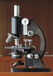
Compound Microscope (cropped).JPG
Microscope
UsesSmall sample observation
Notable experimentsDiscovery of cells
Related itemsOptical microscope Electron microscope

A microscope (from the Ancient Greek: μικρός, mikrós, "small" and σκοπεῖν, skopeîn, "to look" or "see") is an instrument used to see objects that are too small to be seen by the naked eye. Microscopy is the science of investigating small objects and structures using such an instrument. Microscopic means invisible to the eye unless aided by a microscope.

There are many types of microscopes, and they may be grouped in different ways. One way is to describe the way the instruments interact with a sample to create images, either by sending a beam of light or electrons to a sample in its optical path, or by scanning across, and a short distance from the surface of a sample using a probe. The most common microscope (and the first to be invented) is the optical microscope, which uses light to pass through a sample to produce an image. Other major types of microscopes are the fluorescence microscope, the electron microscope (both the transmission electron microscope and the scanning electron microscope) and the various types of scanning probe microscopes.

History

18th-century microscopes from the Musée des Arts et Métiers, Paris

Although objects resembling lenses date back 4000 years and there are Greek accounts of the optical properties of water-filled spheres (5th century BC) followed by many centuries of writings on optics, the earliest known use of simple microscopes (magnifying glasses) dates back to the widespread use of lenses in eyeglasses in the 13th century. The earliest known examples of compound microscopes, which combine an objective lens near the specimen with an eyepiece to view a real image, appeared in Europe around 1620. The inventor is unknown although many claims have been made over the years. Several revolve around the spectacle-making centers in the Netherlands including claims it was invented in 1590 by Zacharias Janssen (claim made by his son) and/or Zacharias' father, Hans Martens, claims it was invented by their neighbor and rival spectacle maker, Hans Lippershey (who applied for the first telescope patent in 1608), and claims it was invented by expatriate Cornelis Drebbel who was noted to have a version in London in 1619. Galileo Galilei (also sometimes cited as compound microscope inventor) seems to have found after 1610 that he could close focus his telescope to view small objects and, after seeing a compound microscope built by Drebbel exhibited in Rome in 1624, built his own improved version. Giovanni Faber coined the name microscope for the compound microscope Galileo submitted to the Accademia dei Lincei in 1625 (Galileo had called it the "occhiolino" or "little eye").

Rise of modern light microscopes

Carl Zeiss binocular compound microscope, 1914

The first detailed account of the microscopic anatomy of organic tissue based on the use of a microscope did not appear until 1644, in Giambattista Odierna's L'occhio della mosca, or The Fly's Eye.

The microscope was still largely a novelty until the 1660s and 1670s when naturalists in Italy, the Netherlands and England began using them to study biology. Italian scientist Marcello Malpighi, called the father of histology by some historians of biology, began his analysis of biological structures with the lungs. Robert Hooke's Micrographia had a huge impact, largely because of its impressive illustrations. A significant contribution came from Antonie van Leeuwenhoek who achieved up to 300 times magnification using a simple single lens microscope. He sandwiched a very small glass ball lens between the holes in two metal plates riveted together, and with an adjustable-by-screws needle attached to mount the specimen. Then, Van Leeuwenhoek re-discovered red blood cells (after Jan Swammerdam) and spermatozoa, and helped popularise the use of microscopes to view biological ultrastructure. On 9 October 1676, van Leeuwenhoek reported the discovery of micro-organisms.

The performance of a light microscope depends on the quality and correct use of the condensor lens system to focus light on the specimen and the objective lens to capture the light from the specimen and form an image. Early instruments were limited until this principle was fully appreciated and developed from the late 19th to very early 20th century, and until electric lamps were available as light sources. In 1893 August Köhler developed a key principle of sample illumination, Köhler illumination, which is central to achieving the theoretical limits of resolution for the light microscope. This method of sample illumination produces even lighting and overcomes the limited contrast and resolution imposed by early techniques of sample illumination. Further developments in sample illumination came from the discovery of phase contrast by Frits Zernike in 1953, and differential interference contrast illumination by Georges Nomarski in 1955; both of which allow imaging of unstained, transparent samples.

Electron microscopes

Electron microscope constructed by Ernst Ruska in 1933

In the early 20th century a significant alternative to the light microscope was developed, an instrument that uses a beam of electrons rather than light to generate an image. The German physicist, Ernst Ruska, working with electrical engineer Max Knoll, developed the first prototype electron microscope in 1931, a transmission electron microscope (TEM). The transmission electron microscope works on similar principles to an optical microscope but uses electrons in the place of light and electromagnets in the place of glass lenses. Use of electrons, instead of light, allows for much higher resolution. 

Development of the transmission electron microscope was quickly followed in 1935 by the development of the scanning electron microscope by Max Knoll. Although TEMs were being used for research before WWII, and became popular afterwards, the SEM was not commercially available until 1965. 

Transmission electron microscopes became popular following the Second World War. Ernst Ruska, working at Siemens, developed the first commercial transmission electron microscope and, in the 1950s, major scientific conferences on electron microscopy started being held. In 1965, the first commercial scanning electron microscope was developed by Professor Sir Charles Oatley and his postgraduate student Gary Stewart, and marketed by the Cambridge Instrument Company as the "Stereoscan".

One of the latest discoveries made about using an electron microscope is the ability to identify a virus. Since this microscope produces a visible, clear image of small organelles, in an electron microscope there is no need for reagents to see the virus or harmful cells, resulting in a more efficient way to detect pathogens.

Scanning probe microscopes

From 1981 to 1983 Gerd Binnig and Heinrich Rohrer worked at IBM in Zurich, Switzerland to study the quantum tunnelling phenomenon. They created a practical instrument, a scanning probe microscope from quantum tunnelling theory, that read very small forces exchanged between a probe and the surface of a sample. The probe approaches the surface so closely that electrons can flow continuously between probe and sample, making a current from surface to probe. The microscope was not initially well received due to the complex nature of the underlying theoretical explanations. In 1984 Jerry Tersoff and D.R. Hamann, while at AT&T's Bell Laboratories in Murray Hill, New Jersey began publishing articles that tied theory to the experimental results obtained by the instrument. This was closely followed in 1985 with functioning commercial instruments, and in 1986 with Gerd Binnig, Quate, and Gerber's invention of the atomic force microscope, then Binnig's and Rohrer's Nobel Prize in Physics for the SPM.

New types of scanning probe microscope have continued to be developed as the ability to machine ultra-fine probes and tips has advanced.

Fluorescence microscopes

Fluorescence microscope with the filter cube turret above the objective lenses, coupled with a camera.

The most recent developments in light microscope largely centre on the rise of fluorescence microscopy in biology. During the last decades of the 20th century, particularly in the post-genomic era, many techniques for fluorescent staining of cellular structures were developed. The main groups of techniques involve targeted chemical staining of particular cell structures, for example, the chemical compound DAPI to label DNA, use of antibodies conjugated to fluorescent reporters, see immunofluorescence, and fluorescent proteins, such as green fluorescent protein. These techniques use these different fluorophores for analysis of cell structure at a molecular level in both live and fixed samples. 

The rise of fluorescence microscopy drove the development of a major modern microscope design, the confocal microscope. The principle was patented in 1957 by Marvin Minsky, although laser technology limited practical application of the technique. It was not until 1978 when Thomas and Christoph Cremer developed the first practical confocal laser scanning microscope and the technique rapidly gained popularity through the 1980s.

Super resolution microscopes

Much current research (in the early 21st century) on optical microscope techniques is focused on development of superresolution analysis of fluorescently labelled samples. Structured illumination can improve resolution by around two to four times and techniques like stimulated emission depletion (STED) microscopy are approaching the resolution of electron microscopes. This occurs because the diffraction limit is occurred from light or excitation, which makes the resolution must be doubled to become super saturated. Stefan Hell was awarded the 2014 Nobel Prize in Chemistry for the development of the STED technique, along with Eric Betzig and William Moerner who adapted fluorescence microscopy for single-molecule visualization.

X-ray microscopes

X-ray microscopes are instruments that use electromagnetic radiation usually in the soft X-ray band to image objects. Technological advances in X-ray lens optics in the early 1970s made the instrument a viable imaging choice. They are often used in tomography to produce three dimensional images of objects, including biological materials that have not been chemically fixed. Currently research is being done to improve optics for hard X-rays which have greater penetrating power.

Types

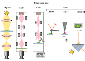
Types of microscopes illustrated by the principles of their beam paths
 
Evolution of spatial resolution achieved with optical, transmission (TEM) and aberration-corrected electron microscopes (ACTEM).
 
Microscopes can be separated into several different classes. One grouping is based on what interacts with the sample to generate the image, i.e., light or photons (optical microscopes), electrons (electron microscopes) or a probe (scanning probe microscopes). Alternatively, microscopes can be classified based on whether they analyze the sample via a scanning point (confocal optical microscopes, scanning electron microscopes and scanning probe microscopes) or analyze the sample all at once (wide field optical microscopes and transmission electron microscopes).

Wide field optical microscopes and transmission electron microscopes both use the theory of lenses (optics for light microscopes and electromagnet lenses for electron microscopes) in order to magnify the image generated by the passage of a wave transmitted through the sample, or reflected by the sample. The waves used are electromagnetic (in optical microscopes) or electron beams (in electron microscopes). Resolution in these microscopes is limited by the wavelength of the radiation used to image the sample, where shorter wavelengths allow for a higher resolution.

Scanning optical and electron microscopes, like the confocal microscope and scanning electron microscope, use lenses to focus a spot of light or electrons onto the sample then analyze the signals generated by the beam interacting with the sample. The point is then scanned over the sample to analyze a rectangular region. Magnification of the image is achieved by displaying the data from scanning a physically small sample area on a relatively large screen. These microscopes have the same resolution limit as wide field optical, probe, and electron microscopes.

Scanning probe microscopes also analyze a single point in the sample and then scan the probe over a rectangular sample region to build up an image. As these microscopes do not use electromagnetic or electron radiation for imaging they are not subject to the same resolution limit as the optical and electron microscopes described above.

Optical

The most common type of microscope (and the first invented) is the optical microscope. This is an optical instrument containing one or more lenses producing an enlarged image of a sample placed in the focal plane. Optical microscopes have refractive glass (occasionally plastic or quartz), to focus light on the eye or on to another light detector. Mirror-based optical microscopes operate in the same manner. Typical magnification of a light microscope, assuming visible range light, is up to 1250x with a theoretical resolution limit of around 0.250 micrometres or 250 nanometres. This limits practical magnification to ~1500x. Specialized techniques (e.g., scanning confocal microscopy, Vertico SMI) may exceed this magnification but the resolution is diffraction limited. The use of shorter wavelengths of light, such as ultraviolet, is one way to improve the spatial resolution of the optical microscope, as are devices such as the near-field scanning optical microscope

Sarfus is a recent optical technique that increases the sensitivity of a standard optical microscope to a point where it is possible to directly visualize nanometric films (down to 0.3 nanometre) and isolated nano-objects (down to 2 nm-diameter). The technique is based on the use of non-reflecting substrates for cross-polarized reflected light microscopy.

Ultraviolet light enables the resolution of microscopic features as well as the imaging of samples that are transparent to the eye. Near infrared light can be used to visualize circuitry embedded in bonded silicon devices, since silicon is transparent in this region of wavelengths.

In fluorescence microscopy many wavelengths of light ranging from the ultraviolet to the visible can be used to cause samples to fluoresce, which allows viewing by eye or with specifically sensitive cameras.

Unstained cells viewed by typical brightfield (left) compared to phase-contrast microscopy (right).

Phase-contrast microscopy is an optical microscopy illumination technique in which small phase shifts in the light passing through a transparent specimen are converted into amplitude or contrast changes in the image. The use of phase contrast does not require staining to view the slide. This microscope technique made it possible to study the cell cycle in live cells.

The traditional optical microscope has more recently evolved into the digital microscope. In addition to, or instead of, directly viewing the object through the eyepieces, a type of sensor similar to those used in a digital camera is used to obtain an image, which is then displayed on a computer monitor. These sensors may use CMOS or charge-coupled device (CCD) technology, depending on the application.

Digital microscopy with very low light levels to avoid damage to vulnerable biological samples is available using sensitive photon-counting digital cameras. It has been demonstrated that a light source providing pairs of entangled photons may minimize the risk of damage to the most light-sensitive samples. In this application of ghost imaging to photon-sparse microscopy, the sample is illuminated with infrared photons, each of which is spatially correlated with an entangled partner in the visible band for efficient imaging by a photon-counting camera.

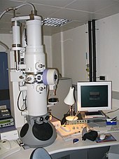
Modern transmission electron microscope

Electron

Transmission electron micrograph of a dividing cell undergoing cytokinesis

The two major types of electron microscopes are transmission electron microscopes (TEMs) and scanning electron microscopes (SEMs). They both have series of electromagnetic and electrostatic lenses to focus a high energy beam of electrons on a sample. In a TEM the electrons pass through the sample, analogous to basic optical microscopy. This requires careful sample preparation, since electrons are scattered strongly by most materials. The samples must also be very thin (50 – 100 nm) in order for the electrons to pass through it. Cross-sections of cells stained with osmium and heavy metals reveal clear organelle membranes and proteins such as ribosomes. With a 0.1 nm level of resolution, detailed views of viruses (20 – 300 nm) and a strand of DNA (2 nm in width) can be obtained. In contrast, the SEM has raster coils to scan the surface of bulk objects with a fine electron beam. Therefore, the specimen do not necessarily need to be sectioned, but require coating with a substance such as a heavy metal. This allows three-dimensional views of the surface of samples.

Scanning probe

The different types of scanning probe microscopes arise from the many different types of interactions that occur when a small probe is scanned over and interacts with a specimen. These interactions or modes can be recorded or mapped as function of location on the surface to form a characterization map. The three most common types of scanning probe microscopes are atomic force microscopes (AFM), near-field scanning optical microscopes (MSOM or SNOM, scanning near-field optical microscopy), and scanning tunneling microscopes (STM). An atomic force microscope has a fine probe, usually of silicon or silicon nitride, attached to a cantilever; the probe is scanned over the surface of the sample, and the forces that cause an interaction between the probe and the surface of the sample are measured and mapped. A near-field scanning optical microscope is similar to an AFM but its probe consists of a light source in an optical fiber covered with a tip that has usually an aperture for the light to pass through. The microscope can capture either transmitted or reflected light to measure very localized optical properties of the surface, commonly of a biological specimen. Scanning tunneling microscopes have a metal tip with a single apical atom; the tip is attached to a tube through which a current flows. The tip is scanned over the surface of a conductive sample until a tunneling current flows; the current is kept constant by computer movement of the tip and an image is formed by the recorded movements of the tip.

Leaf surface viewed by a scanning electron microscope.

Other types

Scanning acoustic microscopes use sound waves to measure variations in acoustic impedance. Similar to Sonar in principle, they are used for such jobs as detecting defects in the subsurfaces of materials including those found in integrated circuits. On February 4, 2013, Australian engineers built a "quantum microscope" which provides unparalleled precision.

Cooperative

From Wikipedia, the free encyclopedia ...