Search This Blog

Saturday, February 22, 2020

African trypanosomiasis

From Wikipedia, the free encyclopedia
African trypanosomiasis
Other namesSleeping sickness, African sleeping sickness
Trypanosoma sp. PHIL 613 lores.jpg
Trypanosoma forms in a blood smear
SpecialtyInfectious disease
SymptomsStage 1: Fevers, headaches, itchiness, joint pains
Stage 2: Trouble sleeping, confusion, poor coordination
Usual onset1–3 weeks post exposure
CausesTrypanosoma brucei spread by tsetse flies
Diagnostic methodBlood smear, lumbar puncture
MedicationPentamidine, suramin, eflornithine
PrognosisFatal without treatment
Frequency11,000
Deaths3,500

African trypanosomiasis, also known as African sleeping sickness or simply sleeping sickness, is an insect-borne parasitic disease of humans and other animals. It is caused by protozoa of the species Trypanosoma brucei. Humans are infected by two types, Trypanosoma brucei gambiense (TbG) and Trypanosoma brucei rhodesiense (TbR). TbG causes over 98% of reported cases. Both are usually transmitted by the bite of an infected tsetse fly and are most common in rural areas.

Initially, the first stage of the disease is characterized by fevers, headaches, itchiness, and joint pains, beginning one to three weeks after the bite. Weeks to months later the second stage begins with confusion, poor coordination, numbness, and trouble sleeping. Diagnosis is by finding the parasite in a blood smear or in the fluid of a lymph node. A lumbar puncture is often needed to tell the difference between first and second stage disease.

Prevention of severe disease involves screening the population at risk with blood tests for TbG. Treatment is easier when the disease is detected early and before neurological symptoms occur. Treatment of the first stage is with the medications pentamidine or suramin. Treatment of the second stage involves eflornithine or a combination of nifurtimox and eflornithine for TbG. While melarsoprol works for both stages, it is typically only used for TbR, due to serious side effects. Without treatment sleeping sickness typically results in death.

The disease occurs regularly in some regions of sub-Saharan Africa with the population at risk being about 70 million in 36 countries. An estimated 11,000 people are currently infected with 2,800 new infections in 2015. In 2015 it caused around 3,500 deaths, down from 34,000 in 1990. More than 80% of these cases are in the Democratic Republic of the Congo. Three major outbreaks have occurred in recent history: one from 1896 to 1906 primarily in Uganda and the Congo Basin and two in 1920 and 1970 in several African countries. It is classified as a neglected tropical disease. Other animals, such as cows, may carry the disease and become infected in which case it is known as animal trypanosomiasis.

Signs and symptoms

African trypanosomiasis symptoms occur in two stages. The first stage, known as the hemolymphatic phase, is characterized by fever, headaches, joint pains, and itching. Fever is intermittent, with attacks lasting from a day to a week, separated by intervals of a few days to a month or longer. Invasion of the circulatory and lymphatic systems by the parasites is associated with severe swelling of lymph nodes, often to tremendous sizes. Winterbottom's sign, the tell-tale swollen lymph nodes along the back of the neck, may appear. Occasionally, a chancre (red sore) will develop at the location of the tsetse fly bite. If left untreated, the disease overcomes the host's defenses and can cause more extensive damage, broadening symptoms to include anemia, endocrine, cardiac, and kidney dysfunctions.

The second phase of the disease, the neurological phase, begins when the parasite invades the central nervous system by passing through the blood–brain barrier. Disruption of the sleep cycle is a leading symptom of this stage and is the one that gave the disease the name 'sleeping sickness.' Infected individuals experience a disorganized and fragmented 24-hour rhythm of the sleep-wake cycle, resulting in daytime sleep episodes and nighttime periods of wakefulness.

Other neurological symptoms include confusion, tremor, general muscle weakness, hemiparesis, and paralysis of a limb. Parkinson-like movements might arise due to non-specific movement disorders and speech disorders. Individuals may also exhibit psychiatric symptoms such as irritability, psychotic reactions, aggressive behaviour, or apathy which can sometimes dominate the clinical diagnosis. Without treatment, the disease is invariably fatal, with progressive mental deterioration leading to coma, systemic organ failure, and death. An untreated infection with T. b. rhodesiense will cause death within months whereas an untreated infection with T. b. gambiense will cause death after several years. Damage caused in the neurological phase is irreversible.

Cause

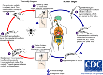
The life cycle of the Trypanosoma brucei parasites.

Trypanosoma brucei gambiense accounts for the majority of African trypanosomiasis cases, with humans as the main reservoir needed for the transmission, while Trypanosoma brucei rhodesiense is mainly zoonotic, with the occasional human infection. African Trypanosomiasis is dependent on the interaction of the parasite (trypanosome) with the tsetse flies (vector), as well as the host (human for Trypanosoma brucei gambiense, and animals for Trypanosoma brucei rhodesiense). The risk of contracting African Trypanosomiasis is dependent on coming in contact with an infected tsetse fly.

Trypanosoma brucei

There are two subspecies of the parasite that are responsible for starting the disease in humans. Trypanosoma brucei gambiense causes the diseases in west and central Africa, whereas Trypanosoma brucei rhodesiense has a limited geographical range and is responsible for causing the disease in east and southern Africa. In addition, a third subspecies of the parasite known as Trypanosoma brucei brucei is responsible for affecting animals but not humans.

Humans are the main reservoir for T. b. gambiense but this species can also be found in pigs and other animals. Wild game animals and cattle are the main reservoir of T. b. rhodesiense. These parasites primarily infect individuals in sub-Saharan Africa because that is where the vector (tsetse fly) is located. The two human forms of the disease also vary greatly in intensity. T. b. gambiense causes a chronic condition that can remain in a passive phase for months or years before symptoms emerge and the infection can last about 3 years before death occurs.

T. b. rhodesiense is the acute form of the disease, and death can occur within months since the symptoms emerge within weeks and it is more virulent and faster developing than T. b. gambiense. Furthermore, trypanosomes are surrounded by a coat that is composed of variant surface glycoproteins (VSG). These proteins act to protect the parasite from any lytic factors that are present in human plasma. The host's immune system recognizes the glycoproteins present on the coat of the parasite leading to the production of different antibodies (IgM and IgG).

These antibodies will then act to destroy the parasites that circulate around the blood. However, from the several parasites present in the plasma, a small number of them will experience changes in their surface coats resulting in the formation of new VSGs. Thus, the antibodies produced by the immune system will no longer recognize the parasite leading to proliferation until new antibodies are created to combat the novel VSGs. Eventually the immune system will no longer be able to fight off the parasite due to the constant changes in VSGs and infection will arise.

Vector

The tsetse fly (genus Glossina) is a large, brown, biting fly that serves as both a host and vector for the trypanosome parasites. While taking blood from a mammalian host, an infected tsetse fly injects metacyclic trypomastigotes into skin tissue. From the bite, parasites first enter the lymphatic system and then pass into the bloodstream. Inside the mammalian host, they transform into bloodstream trypomastigotes, and are carried to other sites throughout the body, reach other body fluids (e.g., lymph, spinal fluid), and continue to replicate by binary fission.

The entire life cycle of African trypanosomes is represented by extracellular stages. A tsetse fly becomes infected with bloodstream trypomastigotes when taking a blood meal on an infected mammalian host. In the fly's midgut, the parasites transform into procyclic trypomastigotes, multiply by binary fission, leave the midgut, and transform into epimastigotes. The epimastigotes reach the fly's salivary glands and continue multiplication by binary fission.

The entire life cycle of the fly takes about three weeks. In addition to the bite of the tsetse fly, the disease can be transmitted by:
  • Mother-to-child infection: the trypanosome can sometimes cross the placenta and infect the fetus.
  • Laboratories: accidental infections, for example, through the handling of blood of an infected person and organ transplantation, although this is uncommon.
  • Blood transfusion
  • Sexual contact (This may be possible)
Horse-flies (Tabanidae) and stable flies (Muscidae) possibly play a role in transmission of nagana (the animal form of sleeping sickness) and the human disease form.

Sleeping sickness Trypanosoma Distribution Vector
Chronic form T. brucei gambiense Western Africa G. palpalis G. tachinoides
G. fuscipes
G. morsitans
Acute form T. brucei rhodesiense Eastern Africa G. morsitans G. swynnertoni
G. pallidipes
G. fuscipes

Mechanism

Tryptophol is a chemical compound that induces sleep in humans. It is produced by the trypanosomal parasite in sleeping sickness.

Diagnosis

Two areas from a blood smear from a person with African trypanosomiasis, thin blood smear stained with Giemsa: Typical trypomastigote stages (the only stages found in people), with a posterior kinetoplast, a centrally located nucleus, an undulating membrane, and an anterior flagellum. The two Trypanosoma brucei subspecies that cause human trypanosomiasis, T. b. gambiense and T. b. rhodesiense, are indistinguishable morphologically. The trypanosomes' length range is 14 to 33 µm, Source: CDC

The gold standard for diagnosis is identification of trypanosomes in a sample by microscopic examination. Samples that can be used for diagnosis include chancre fluid, lymph node aspirates, blood, bone marrow, and, during the neurological stage, cerebrospinal fluid. Detection of trypanosome-specific antibodies can be used for diagnosis, but the sensitivity and specificity of these methods are too variable to be used alone for clinical diagnosis. Further, seroconversion occurs after the onset of clinical symptoms during a T. b. rhodesiense infection, so is of limited diagnostic use.

Trypanosomes can be detected from samples using two different preparations. A wet preparation can be used to look for the motile trypanosomes. Alternatively, a fixed (dried) smear can be stained using Giemsa's or Field's technique and examined under a microscope. Often, the parasite is in relatively low abundance in the sample, so techniques to concentrate the parasites can be used prior to microscopic examination. For blood samples, these include centrifugation followed by examination of the buffy coat; mini anion-exchange/centrifugation; and the quantitative buffy coat (QBC) technique. For other samples, such as spinal fluid, concentration techniques include centrifugation followed by examination of the sediment.

Three serological tests are also available for detection of the parasite: the micro-CATT, wb-CATT, and wb-LATEX. The first uses dried blood, while the other two use whole blood samples. A 2002 study found the wb-CATT to be the most efficient for diagnosis, while the wb-LATEX is a better exam for situations where greater sensitivity is required.

Prevention

Currently there are few medically related prevention options for African Trypanosomiasis (i.e. no vaccine exists for immunity). Although the risk of infection from a tsetse fly bite is minor (estimated at less than 0.1%), the use of insect repellants, wearing long-sleeved clothing, avoiding tsetse-dense areas, implementing bush clearance methods and wild game culling are the best options to avoid infection available for local residents of affected areas.

In July 2000, a resolution was passed to form the Pan African Tsetse and Trypanosomiasis Eradication Campaign (PATTEC). The campaign works to eradicate the tsetse vector population levels and subsequently the protozoan disease, by use of insecticide-impregnated targets, fly traps, insecticide-treated cattle, ultra-low dose aerial/ground spraying (SAT) of tsetse resting sites and the sterile insect technique (SIT). The use of SIT in Zanzibar proved effective in eliminating the entire population of tsetse flies but was expensive and is relatively impractical to use in many of the endemic countries afflicted with African trypanosomiasis.

A pilot program in Senegal has reduced the tsetse fly population by as much as 99% by introducing male flies which have been sterilized by exposure to gamma rays.

Regular active surveillance, involving detection and prompt treatment of new infections, and tsetse fly control is the backbone of the strategy used to control sleeping sickness. Systematic screening of at-risk communities is the best approach, because case-by-case screening is not practical in endemic regions. Systematic screening may be in the form of mobile clinics or fixed screening centres where teams travel daily to areas of high infection rates. Such screening efforts are important because early symptoms are not evident or serious enough to warrant people with gambiense disease to seek medical attention, particularly in very remote areas. Also, diagnosis of the disease is difficult and health workers may not associate such general symptoms with trypanosomiasis. Systematic screening allows early-stage disease to be detected and treated before the disease progresses, and removes the potential human reservoir. A single case of sexual transmission of West African sleeping sickness has been reported.

Treatment

First stage

The current treatment for first-stage disease is intravenous or intramuscular pentamidine for T. b. gambiense or intravenous suramin for T. b. rhodesiense.

Fexinidazole has been approved in the Democratic Republic Congo for T.b. gambiense sleeping sickness. Benefits include that it is taken by mouth and treats both stages of the disease.

Second stage

For T. b. gambiense a regimen involving the combination of nifurtimox and eflornithine, nifurtimox-eflornithine combination treatment (NECT), or eflornithine alone appear to be more effective and result in fewer side effects. These treatments may replace melarsoprol when available with the combination being first line. NECT has the benefit of requiring less injections of eflornithine.

Intravenous melarsoprol was previously the standard treatment for second-stage (neurological phase) disease and is effective for both types. Melarsoprol is the only treatment for second stage T. b. rhodesiense; however, it causes death in 5% of people who take it. Resistance to melarsoprol can occur.

Epidemiology

Deaths per 100,000 population due to African trypanosomiasis by country in 2002.

In 2010, it caused around 9,000 deaths, down from 34,000 in 1990. As of 2000, the disability-adjusted life-years (9 to 10 years) lost due to sleeping sickness are 2.0 million. From 2010-2014, there was an estimated 55 million people at risk for gambiense African Trypanosomiasis and over 6 million people at risk for rhodesiense African Trypanosomiasis. In 2014, the World Health Organization reported 3,797 cases of Human African Trypanosomiasis when the predicted number of cases were to be 5,000. The number of total reported cases in 2014 is an 86% reduction to the total number of cases reported in 2000.

The disease has been recorded as occurring in 37 countries, all in sub-Saharan Africa. It occurs regularly in southeast Uganda and western Kenya, and killed more than 48,000 Africans in 2008. The Democratic Republic of the Congo is the most affected country in the world, accounting for 75% of the Trypanosoma brucei gambiense cases. The population at risk being about 69 million with one third of this number being at a 'very high' to 'moderate' risk and the remaining two thirds at a 'low' to 'very low' risk. The number of people being affected by the disease has declined. At this rate, sleeping sickness elimination is a possibility. The World Health Organization plans to eradicate sleeping sickness by the year 2020.

Prognosis

If untreated, T. b. gambiense almost always results in death, with only a few individuals shown in a long-term 15 year follow-up to have survived after refusing treatment. T. b. rhodesiense, being a more acute and severe form of the disease, is consistently fatal if not treated. Disease progression greatly varies depending on disease form. For individuals which are infected by T. b. gambiense, which accounts for 98% of all of the reported cases, a person can be infected for months or even years without signs or symptoms until the advanced disease stage, where it is too late to be treated successfully. For individuals affected by T. b. rhodesiense, which accounts for 2% of all reported cases, symptoms appear within weeks or months of the infection. Disease progression is rapid and invades the central nervous system, causing death within a short amount of time.

History

In 1903, David Bruce recognized the tsetse fly as the arthropod vector.

The condition has been present in Africa for thousands of years. Because of a lack of travel between indigenous people, sleeping sickness in humans had been limited to isolated pockets. This changed after Arab slave traders entered central Africa from the east, following the Congo River, bringing parasites along. Gambian sleeping sickness travelled up the Congo River, and then further east.

An Arab writer of the 14th century left the following description in the case of a sultan of the Mali Kingdom: "His end was to be overtaken by the sleeping sickness (illat an-nawm) which is a disease that frequently befalls the inhabitants of these countries especially their chieftains. Sleep overtakes one of them in such a manner that it is hardly possible to awake him."

The British naval surgeon John Atkins described the disease on his return from West Africa in 1734:
"The Sleepy Distemper (common among the Negroes) gives no other previous Notice, than a want of Appetite 2 or 3 days before; their sleeps are sound, and Sense and Feeling very little; for pulling, drubbing or whipping will scarce stir up Sense and Power enough to move; and the Moment you cease beating the smart is forgot, and down they fall again into a state of Insensibility, drivling constantly from the Mouth as in deep salivation; breathe slowly, but not unequally nor snort. Young people are more subject to it than the old; and the Judgement generally pronounced is Death, the Prognostik seldom failing. If now and then one of them recovers, he certainly loses the little Reason he had, and turns Ideot..."
In 1901, a devastating epidemic erupted in Uganda, killing more than 250,000 people, including about two-thirds of the population in the affected lakeshore areas. According to The Cambridge History of Africa, "It has been estimated that up to half the people died of sleeping-sickness and smallpox in the lands on either bank of the lower river Congo."

The causative agent and vector were identified in 1903 by David Bruce, and the subspecies of the protozoa were differentiated in 1910. Bruce had earlier shown that T. brucei was the cause of a similar disease in horses and cattle that was transmitted by the tse-tse fly (Glossina morsitans).

The first effective treatment, atoxyl, an arsenic-based drug developed by Paul Ehrlich and Kiyoshi Shiga, was introduced in 1910, but blindness was a serious side effect.

Suramin was first synthesized by Oskar Dressel and Richard Kothe in 1916 for Bayer. It was introduced in 1920 to treat the first stage of the disease. By 1922, Suramin was generally combined with tryparsamide (another pentavalent organoarsenic drug), the first drug to enter the nervous system and be useful in the treatment of the second stage of the gambiense form. Tryparsamide was announced in the Journal of Experimental Medicine in 1919 and tested in the Belgian Congo by Louise Pearce of the Rockefeller Institute in 1920. It was used during the grand epidemic in West and Central Africa on millions of people and was the mainstay of therapy until the 1960s. American medical missionary Arthur Lewis Piper was active in using tryparsamide to treat sleeping sickness in the Belgian Congo in 1925.

Pentamidine, a highly effective drug for the first stage of the disease, has been used since 1937. During the 1950s, it was widely used as a prophylactic agent in western Africa, leading to a sharp decline in infection rates. At the time, eradication of the disease was thought to be at hand.

The organoarsenical melarsoprol (Arsobal) developed in the 1940s is effective for people with second-stage sleeping sickness. However, 3–10% of those injected have reactive encephalopathy (convulsions, progressive coma, or psychotic reactions), and 10–70% of such cases result in death; it can cause brain damage in those who survive the encephalopathy. However, due to its effectiveness, melarsoprol is still used today. Resistance to melarsoprol is increasing, and combination therapy with nifurtimox is currently under research.

Eflornithine (difluoromethylornithine or DFMO), the most modern treatment, was developed in the 1970s by Albert Sjoerdsma and underwent clinical trials in the 1980s. The drug was approved by the United States Food and Drug Administration in 1990. Aventis, the company responsible for its manufacture, halted production in 1999. In 2001, Aventis, in association with Médecins Sans Frontières and the World Health Organization, signed a long-term agreement to manufacture and donate the drug.

In addition to sleeping sickness, previous names have included negro lethargy, maladie du sommeil (Fr), Schlafkrankheit (Ger), African lethargy, and Congo trypanosomiasis.

Research

The genome of the parasite has been sequenced and several proteins have been identified as potential targets for drug treatment. Analysis of the genome also revealed the reason why generating a vaccine for this disease has been so difficult. T. brucei has over 800 genes that make proteins the parasite "mixes and matches" to evade immune system detection.

Using a genetically modified form of a bacterium that occurs naturally in the gut of the vectors is being studied as a method of controlling the disease.

Recent findings indicate that the parasite is unable to survive in the bloodstream without its flagellum. This insight gives researchers a new angle with which to attack the parasite.

Trypanosomiasis vaccines are undergoing research.

Additionally, the Drugs for Neglected Disease Initiative has contributed to the African sleeping sickness research by developing a compound called fexinidazole. This project was originally started in April 2007 and enrolled 749 people in the DRC and Central African Republic. The results showed efficacy and safety in both stages of the disease, both in adults and children ≥ 6 years old and weighing ≥ 20 kg. The European Medicines Agency approved it for first and second stage disease outside of Europe in November 2018. The treatment was approved in the DRC in December 2018.

Funding

For current funding statistics, human African trypanosomiasis is grouped with kinetoplastid infections. Kinetoplastids refer to a group of flagellate protozoa. Kinetoplastid infections include African sleeping sickness, Chagas' disease, and Leishmaniasis. All together, these three diseases accounted for 4.4 million disability adjusted life years (DALYs) and an additional 70,075 recorded deaths yearly. For kinetoplastid infections, the total global research and development funding was approximately $136.3 million in 2012. Each of the three diseases, African sleeping sickness, Chagas' disease, and Leishmaniasis each received approximately a third of the funding, which was about $36.8 million US dollars, $38.7 million US dollars, and $31.7 million US dollars, respectively.

For sleeping sickness, funding was split into basic research, drug discovery, vaccines, and diagnostics. The greatest amount of funding was directed towards basic research of the disease; approximately $21.6 million US dollars was directed towards that effort. As for therapeutic development, approximately $10.9 million was invested.

The top funder for kinetoplastid infection research and development are public sources. About 62% of the funding comes from high-income countries while 9% comes from low- and middle-income countries. High-income countries' public funding is the largest contributor to the neglected disease research effort. However, in recent years, funding from high-income countries has been steadily decreasing; in 2007, high-income countries provided 67.5% of the total funding whereas, in 2012, high-income countries public funds only provided 60% of the total funding for kinetoplastid infections. This downwards trend leaves a gap for other funders, such as philanthropic foundations and private pharmaceutical companies to fill.

Much of the progress that has been made in African sleeping sickness and neglected disease research as a whole is a result of the other non-public funders. One of these major sources of funding has come from foundations, which have increasingly become more committed to neglected disease drug discovery in the 21st century. In 2012, philanthropic sources provided 15.9% of the total funding. The Bill and Melinda Gates Foundation has been a leader in providing funding for neglected disease drug development. They have provided $444.1 million US dollars towards neglected disease research in 2012. To date, they have donated over $1.02 billion US dollars towards the neglected disease discovery efforts.

For kinetoplastid infections specifically, they have donated an average of $28.15 million US dollars annually between the years 2007 to 2011. They have labeled human African trypanosomiasis a high-opportunity target meaning it is a disease that presents the greatest opportunity for control, elimination, and eradication, through the development of new drugs, vaccines, public-health programs, and diagnostics. They are the second highest funding source for neglected diseases, immediately behind the US National Institutes of Health. At a time where public funding is decreasing and government grants for scientific research are harder to obtain, the philanthropic world has stepped in to push the research forward. 

Another important component of increased interest and funding has come from industry. In 2012, they contributed 13.1% total to the kinetoplastid research and development effort, and have additionally played an important role by contributing to public-private partnerships (PPP) as well as product-development partnerships (PDP). A public-private partnership is an arrangement between one or more public entities and one or more private entities that exists to achieve a specific health outcome or to produce a health product. The partnership can exist in numerous ways; they may share and exchange funds, property, equipment, human resources, and intellectual property. These public-private partnerships and product-development partnerships have been established to address challenges in pharmaceutical industry, especially related to neglected disease research. These partnerships can help increase the scale of the effort towards therapeutic development by using different knowledge, skills, and expertise from different sources. These types of partnerships have been shown to be more effective than industry or public groups working independently.
Amoebiasis

From Wikipedia, the free encyclopedia
 
Amoebiasis
Other namesAmoebic dysentery, amebiasis, entamoebiasis
Amebiasis LifeCycle.gif
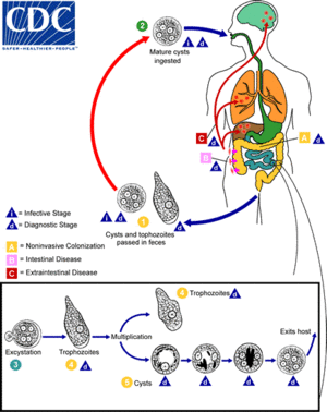
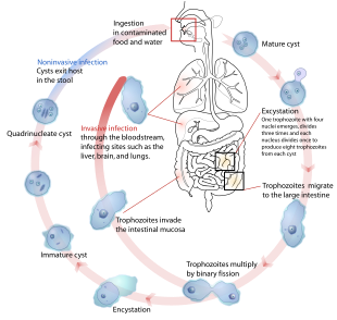
The life-cycle of various intestinal Entamoeba species
SpecialtyInfectious disease
SymptomsBloody diarrhea, abdominal pain
ComplicationsSevere colitis, colonic perforation, anemia
CausesAmoebas of the Entamoeba group
Diagnostic methodStool examination, antibodies in the blood
Differential diagnosisBacterial colitis
PreventionImproved sanitation
TreatmentTissue disease: metronidazole, tinidazole, nitazoxanide, dehydroemetine, chloroquine,
Intestinal infection: diloxanide furoate, iodoquinoline
Frequency>480 million

Amoebiasis, also known amoebic dysentery, is an infection caused by any of the amobae of the Entamoeba group. Symptoms are most common during infection by Entamoeba histolytica.

 Amoebiasis can be present with no, mild, or severe symptoms. Symptoms may include abdominal pain, diarrhea, or bloody diarrhea. Complications can include inflammation and ulceration of the colon with tissue death or perforation, which may result in peritonitis. People affected may develop anemia due to loss of blood.

Cysts of Entamoeba can survive for up to a month in soil or for up to 45 minutes under fingernails. Invasion of the intestinal lining results in bloody diarrhea. If the parasite reaches the bloodstream it can spread through the body, most frequently ending up in the liver where it can cause amoebic liver abscesses. Liver abscesses can occur without previous diarrhea. Diagnosis is typical by stool examination using a microscope, but may not reliably exclude infection or separate between specific types. An increased white blood cell count may be present in severe cases. The most accurate test is finding specific antibodies in the blood, but it may remain positive following treatment. Bacterial colitis can result in similar symptoms.

Prevention of amoebiasis is by improved sanitation, including separating food and water from faeces. There is no vaccine. There are two treatment options depending on the location of the infection. Amoebiasis in tissues is treated with either metronidazole, tinidazole, nitazoxanide, dehydroemetine or chloroquine, while luminal infection is treated with diloxanide furoate or iodoquinoline. Effective treatment against all stages of the disease may require a combination of medications. Infections without symptoms do not require treatment but infected individuals can spread the parasite to others and treatment can be considered. Treatment of other Entamoeba infections apart from E. histolytica is not needed.

Amoebiasis is present all over the world, though most cases occur in the developing world. About 480 million people are infected with amoebiasis and this results in the death of between 40,000–110,000 people a year. Most infections are now believed due to E. dispar. E. dispar is more common in certain areas and symptomatic cases may be less common than previously reported. The first case of amoebiasis was documented in 1875 and in 1891 the disease was described in detail, resulting in the terms amoebic dysentery and amoebic liver abscess. Further evidence from the Philippines in 1913 found that upon swallowing cysts of E. histolytica volunteers developed the disease.

Signs and symptoms

Most infected people, about 90%, are asymptomatic, but this disease has the potential to become serious. It is estimated that about 40,000 to 100,000 people worldwide die annually due to amoebiasis.

Infections can sometimes last for years if there is no treatment. Symptoms take from a few days to a few weeks to develop and manifest themselves, but usually it is about two to four weeks. Symptoms can range from mild diarrhea to dysentery with blood, coupled with intense abdominal pains. The blood comes from bleeding lesions created by the amoebae invading the lining of the colon. In about 10% of invasive cases the amoebae enter the bloodstream and may travel to other organs in the body. Most commonly this means the liver, as this is where blood from the intestine reaches first, but they can end up almost anywhere in the body.

Onset time is highly variable and the average asymptomatic infection persists for over a year. It is theorized that the absence of symptoms or their intensity may vary with such factors as strain of amoeba, immune response of the host, and perhaps associated bacteria and viruses.

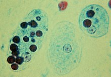
In asymptomatic infections, the amoeba lives by eating and digesting bacteria and food particles in the gut, a part of the gastrointestinal tract. It does not usually come in contact with the intestine itself due to the protective layer of mucus that lines the gut. Disease occurs when amoeba comes in contact with the cells lining the intestine. It then secretes the same substances it uses to digest bacteria, which include enzymes that destroy cell membranes and proteins. This process can lead to penetration and digestion of human tissues, resulting first in flask-shaped ulcerations in the intestine. Entamoeba histolytica ingests the destroyed cells by phagocytosis and is often seen with red blood cells (a process known as erythrophagocytosis) inside when viewed in stool samples. Especially in Latin America, a granulomatous mass (known as an amoeboma) may form in the wall of the ascending colon or rectum due to long-lasting immunological cellular response, and is sometimes confused with cancer.

The ingestion of one viable cyst may cause an infection.

Steroid therapy can lead to severe amoebic colitis in persons with asymptomatic or symptomatic E. histolytica infection. Severe amoebic colitis is associated with high mortality, and on average more than 50% with severe colitis die.

Cause

Amoebiasis is an infection caused by the amoeba Entamoeba histolytica. Likewise amoebiasis is sometimes incorrectly used to refer to infection with other amoebae, but strictly speaking it should be reserved for Entamoeba histolytica infection. Other amoebae infecting humans include:
Except for Dientamoeba, the parasites above are not thought to cause disease.

Transmission

Life-cycle of the Entamoeba histolytica

Amoebiasis is usually transmitted by the fecal-oral route, but it can also be transmitted indirectly through contact with dirty hands or objects as well as by anal-oral contact. Infection is spread through ingestion of the cyst form of the parasite, a semi-dormant and hardy structure found in feces. Any non-encysted amoebae, or trophozoites, die quickly after leaving the body but may also be present in stool: these are rarely the source of new infections. Since amoebiasis is transmitted through contaminated food and water, it is often endemic in regions of the world with limited modern sanitation systems, including México, Central America, western South America, South Asia, and western and southern Africa.

Amoebic dysentery is often confused with "traveler's diarrhea" because of its prevalence in developing nations. In fact, most traveler's diarrhea is bacterial or viral in origin.

Pathogenesis

Tissue damage caused by E. histolytica is a result of three main events, host cell death, inflammation, and parasite invasion. Abbreviations: EhMIF, E. histolytica macrophage migration inhibitory factor; MMP, matrix metalloproteinases.

Amoebiasis results from tissue destruction induced by the E. histolytica parasite. E. histolytica causes tissue damage by three main events: direct host cell killing, inflammation, and parasite invasion.

Diagnosis

With colonoscopy it is possible to detect small ulcers of between 3–5mm, but diagnosis may be difficult as the mucous membrane between these areas can look either healthy or inflamed. Trophozoites may be identified at the ulcer edge or within the tissue, using immunohistochemical staining with specific anti-E. histolytica antibodies.

Asymptomatic human infections are usually diagnosed by finding cysts shed in the stool. Various flotation or sedimentation procedures have been developed to recover the cysts from fecal matter and stains help to visualize the isolated cysts for microscopic examination. Since cysts are not shed constantly, a minimum of three stools are examined. In symptomatic infections, the motile form (the trophozoite) is often seen in fresh feces. Serological tests exist, and most infected individuals (with symptoms or not) test positive for the presence of antibodies. The levels of antibody are much higher in individuals with liver abscesses. Serology only becomes positive about two weeks after infection. More recent developments include a kit that detects the presence of amoeba proteins in the feces, and another that detects ameba DNA in feces. These tests are not in widespread use due to their expense.

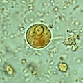
Microscopy is still by far the most widespread method of diagnosis around the world. However it is not as sensitive or accurate in diagnosis as the other tests available. It is important to distinguish the E. histolytica cyst from the cysts of nonpathogenic intestinal protozoa such as Entamoeba coli by its appearance. E. histolytica cysts have a maximum of four nuclei, while the commensal Entamoeba coli cyst has up to 8 nuclei. Additionally, in E. histolytica, the endosome is centrally located in the nucleus, while it is usually off-center in Entamoeba coli. Finally, chromatoidal bodies in E. histolytica cysts are rounded, while they are jagged in Entamoeba coli. However, other species, Entamoeba dispar and E. moshkovskii, are also commensals and cannot be distinguished from E. histolytica under the microscope. As E. dispar is much more common than E. histolytica in most parts of the world this means that there is a lot of incorrect diagnosis of E. histolytica infection taking place. The WHO recommends that infections diagnosed by microscopy alone should not be treated if they are asymptomatic and there is no other reason to suspect that the infection is actually E. histolytica. Detection of cysts or trophozoites stools under microscope may require examination of several samples over several days to determine if they are present, because cysts are shed intermittently and may not show up in every sample.

Typically, the organism can no longer be found in the feces once the disease goes extra-intestinal. Serological tests are useful in detecting infection by E. histolytica if the organism goes extra-intestinal and in excluding the organism from the diagnosis of other disorders. An Ova & Parasite (O&P) test or an E. histolytica fecal antigen assay is the proper assay for intestinal infections. Since antibodies may persist for years after clinical cure, a positive serological result may not necessarily indicate an active infection. A negative serological result, however, can be equally important in excluding suspected tissue invasion by E. histolytica.

Stool antigen detection tests have helped to overcome some of the limitations of stool microscopy. Antigen detection tests are easy to use, but they have variable sensitivity and specificity, especially in low-endemic areas.

Polymerase chain reaction (PCR) is considered the gold standard for diagnosis but remains underutilized.

Prevention

Specimen of the human intestine that was damaged by amebic ulcer.
 
To help prevent the spread of amoebiasis around the home :
To help prevent infection:
  • Avoid raw vegetables when in endemic areas, as they may have been fertilized using human feces.
  • Boil water or treat with iodine tablets.
  • Avoid eating street foods especially in public places where others are sharing sauces in one container
Good sanitary practice, as well as responsible sewage disposal or treatment, are necessary for the prevention of E. histolytica infection on an endemic level. E.histolytica cysts are usually resistant to chlorination, therefore sedimentation and filtration of water supplies are necessary to reduce the incidence of infection.

E. histolytica cysts may be recovered from contaminated food by methods similar to those used for recovering Giardia lamblia cysts from feces. Filtration is probably the most practical method for recovery from drinking water and liquid foods. E. histolytica cysts must be distinguished from cysts of other parasitic (but nonpathogenic) protozoa and from cysts of free-living protozoa as discussed above. Recovery procedures are not very accurate; cysts are easily lost or damaged beyond recognition, which leads to many falsely negative results in recovery tests.

Treatment

E. histolytica infections occur in both the intestine and (in people with symptoms) in tissue of the intestine and/or liver. Those with symptoms require treatment with two medications, an amoebicidal tissue-active agent and a luminal cysticidal agent. Individuals that are asymptomatic only need a luminal cysticidal agent.

Prognosis

Significance of Amebiasis

In the majority of cases, amoebas remain in the gastrointestinal tract of the hosts. Severe ulceration of the gastrointestinal mucosal surfaces occurs in less than 16% of cases. In fewer cases, the parasite invades the soft tissues, most commonly the liver. Only rarely are masses formed (amoebomas) that lead to intestinal obstruction.(Mistaken for Ca caecum and appendicular mass) Other local complications include bloody diarrhea, pericolic and pericaecal abscess.

Complications of hepatic amoebiasis includes subdiaphragmatic abscess, perforation of diaphragm to pericardium and pleural cavity, perforation to abdominal cavital (amoebic peritonitis) and perforation of skin (amoebiasis cutis)

Pulmonary amoebiasis can occur from liver lesions by spread through the blood or by perforation of pleural cavity and lung. It can cause lung abscess, pulmono pleural fistula, empyema lung and broncho pleural fistula. It can also reach the brain through blood vessels and cause amoebic brain abscess and amoebic meningoencephalitis. Cutaneous amoebiasis can also occur in skin around sites of colostomy wound, perianal region, region overlying visceral lesion and at the site of drainage of liver abscess.

Urogenital tract amoebiasis derived from intestinal lesion can cause amoebic vulvovaginitis (May's disease), rectovesicle fistula and rectovaginal fistula.

Entamoeba histolytica infection is associated with malnutrition and stunting of growth.

Epidemiology

Amoebiasis caused about 55,000 deaths worldwide in 2010, down from 68,000 in 1990. In older textbooks it is often stated that 10% of the world's population is infected with Entamoeba histolytica. It is now known that at least 90% of these infections are due to E. dispar. Nevertheless, this means that there are up to 50 million true E. histolytica infections and approximately seventy thousand die each year, mostly from liver abscesses or other complications. Although usually considered a tropical parasite, the first case reported (in 1875) was actually in St Petersburg in Russia, near the Arctic Circle. Infection is more common in warmer areas, but this is because of both poorer hygiene and the parasitic cysts surviving longer in warm moist conditions.

History

Amoebiasis was first described by Lösh in 1875, in northern Russia. The most dramatic incident in the US was the Chicago World's Fair outbreak in 1933, caused by contaminated drinking water. There were more than a thousand cases, with 98 deaths. It has been known since 1897 that at least one non-disease-causing species of Entamoeba existed (Entamoeba coli), but it was first formally recognized by the WHO in 1997 that E. histolytica was two species, despite this having first been proposed in 1925. In addition to the now-recognized E. dispar, evidence shows there are at least two other species of Entamoeba that look the same in humans: E. moshkovskii and Entamoeba bangladeshi. The reason these species haven't been differentiated until recently is because of the reliance on appearance.

Joel Connolly of the Chicago Bureau of Sanitary Engineering brought the outbreak to an end when he found that defective plumbing permitted sewage to contaminate drinking water. In 1998 there was an outbreak of amoebiasis in the Republic of Georgia. Between 26 May and 3 September 1998, 177 cases were reported, including 71 cases of intestinal amoebiasis and 106 probable cases of liver abscess. 

The Nicobarese people have attested to the medicinal properties found in Glochidion calocarpum, a plant common to India, saying that its bark and seed are most effective in curing abdominal disorders associated with amoebiasis.

Protozoa

From Wikipedia, the free encyclopedia
 
Clockwise from top left: Blepharisma japonicum, a ciliate; Giardia muris, a parasitic flagellate; Centropyxis aculeata, a testate (shelled) amoeba; Peridinium willei, a dinoflagellate; Chaos carolinense, a naked amoebozoan; Desmerella moniliformis, a choanoflagellate

Protozoa (also protozoan, plural protozoans) is an informal term for single-celled eukaryotes, either free-living or parasitic, which feed on organic matter such as other microorganisms or organic tissues and debris. Historically, the protozoa were regarded as "one-celled animals", because they often possess animal-like behaviors, such as motility and predation, and lack a cell wall, as found in plants and many algae. Although the traditional practice of grouping protozoa with animals is no longer considered valid, the term continues to be used in a loose way to identify single-celled organisms that can move independently and feed by heterotrophy.

In some systems of biological classification, Protozoa is a high-level taxonomic group. When first introduced in 1818, Protozoa was erected as a taxonomic class, but in later classification schemes it was elevated to a variety of higher ranks, including phylum, subkingdom and kingdom. In a series of classifications proposed by Thomas Cavalier-Smith and his collaborators since 1981, Protozoa has been ranked as a kingdom. The seven-kingdom scheme presented by Ruggiero et al. in 2015, places eight phyla under Kingdom Protozoa: Euglenozoa, Amoebozoa, Metamonada, Choanozoa sensu Cavalier-Smith, Loukozoa, Percolozoa, Microsporidia and Sulcozoa. Notably, this kingdom excludes several major groups of organisms traditionally placed among the protozoa, including the ciliates, dinoflagellates, foraminifera, and the parasitic apicomplexans, all of which are classified under Kingdom Chromista. Kingdom Protozoa, as defined in this scheme, does not form a natural group or clade, but a paraphyletic group or evolutionary grade, within which the members of Fungi, Animalia and Chromista are thought to have evolved.

History

Class Protozoa, order Infusoria, family Monades by Georg August Goldfuss, c. 1844

The word "protozoa" (singular protozoon or protozoan) was coined in 1818 by zoologist Georg August Goldfuss, as the Greek equivalent of the German Urthiere, meaning "primitive, or original animals" (ur- ‘proto-’ + Thier ‘animal’). Goldfuss created Protozoa as a class containing what he believed to be the simplest animals. Originally, the group included not only single-celled microorganisms but also some "lower" multicellular animals, such as rotifers, corals, sponges, jellyfish, bryozoa and polychaete worms. The term Protozoa is formed from the Greek words πρῶτος (prôtos), meaning "first", and ζῶα (zôa), plural of ζῶον (zôon), meaning "animal". The use of Protozoa as a formal taxon has been discouraged by some researchers, mainly because the term implies kinship with animals (Metazoa) and promotes an arbitrary separation of "animal-like" from "plant-like" organisms.

In 1848, as a result of advancements in cell theory pioneered by Theodor Schwann and Matthias Schleiden, the anatomist and zoologist C. T. von Siebold proposed that the bodies of protozoans such as ciliates and amoebae consisted of single cells, similar to those from which the multicellular tissues of plants and animals were constructed. Von Siebold redefined Protozoa to include only such unicellular forms, to the exclusion of all metazoa (animals). At the same time, he raised the group to the level of a phylum containing two broad classes of microorganisms: Infusoria (mostly ciliates and flagellated algae), and Rhizopoda (amoeboid organisms). The definition of Protozoa as a phylum or sub-kingdom composed of "unicellular animals" was adopted by the zoologist Otto Bütschli—celebrated at his centenary as the "architect of protozoology"—and the term came into wide use.

John Hogg's illustration of the Four Kingdoms of Nature, showing "Primigenal" as a greenish haze at the base of the Animals and Plants, 1860

As a phylum under Animalia, the Protozoa were firmly rooted in the old "two-kingdom" classification of life, according to which all living beings were classified as either animals or plants. As long as this scheme remained dominant, the protozoa were understood to be animals and studied in departments of Zoology, while photosynthetic microorganisms and microscopic fungi—the so-called Protophyta—were assigned to the Plants, and studied in departments of Botany.

Criticism of this system began in the latter half of the 19th century, with the realization that many organisms met the criteria for inclusion among both plants and animals. For example, the algae Euglena and Dinobryon have chloroplasts for photosynthesis, but can also feed on organic matter and are motile. In 1860, John Hogg argued against the use of "protozoa", on the grounds that "naturalists are divided in opinion—and probably some will ever continue so—whether many of these organisms, or living beings, are animals or plants." As an alternative, he proposed a new kingdom called Primigenum, consisting of both the protozoa and unicellular algae (protophyta), which he combined together under the name "Protoctista". In Hoggs's conception, the animal and plant kingdoms were likened to two great "pyramids" blending at their bases in the Kingdom Primigenum.

Six years later, Ernst Haeckel also proposed a third kingdom of life, which he named Protista. At first, Haeckel included a few multicellular organisms in this kingdom, but in later work he restricted the Protista to single-celled organisms, or simple colonies whose individual cells are not differentiated into different kinds of tissues.

Despite these proposals, Protozoa emerged as the preferred taxonomic placement for heterotrophic microorganisms such as amoebae and ciliates, and remained so for more than a century. In the course of the 20th century, however, the old "two kingdom" system began to weaken, with the growing awareness that fungi did not belong among the plants, and that most of the unicellular protozoa were no more closely related to the animals than they were to the plants. By mid-century, some biologists, such as Herbert Copeland, Robert H. Whittaker and Lynn Margulis, advocated the revival of Haeckel's Protista or Hogg's Protoctista as a kingdom-level eukaryotic group, alongside Plants, Animals and Fungi.[18] A variety of multi-kingdom systems were proposed, and Kingdoms Protista and Protoctista became well established in biology texts and curricula.

While many taxonomists have abandoned Protozoa as a high-level group, Thomas Cavalier-Smith has retained it as a kingdom in the various classifications he has proposed. As of 2015, Cavalier-Smith's Protozoa excludes several major groups of organisms traditionally placed among the protozoa, including the ciliates, dinoflagellates and foraminifera (all members of the SAR supergroup). In its current form, his kingdom Protozoa is a paraphyletic group which includes a common ancestor and most of its descendants, but excludes two important clades that branch within it: the animals and fungi.

Since the protozoa, as traditionally defined, can no longer be regarded as "primitive animals" the terms "protists", "Protista" or "Protoctista" are sometimes preferred. In 2005, members of the Society of Protozoologists voted to change its name to the International Society of Protistologists.

Characteristics

Size

Protozoa, as traditionally defined, range in size from as little as 1 micrometre to several millimetres, or more. Among the largest are the deep-sea–dwelling xenophyophores, single-celled foraminifera whose shells can reach 20 cm in diameter.

The ciliate Spirostomum ambiguum can attain 3 mm in length
 
Species Cell type Size in micrometres
Plasmodium falciparum malaria parasite, trophozoite phase 1-2
Massisteria voersi free-living cercozoan amoeboid 2.3–3
Bodo saltans free living kinetoplastid flagellate 5-8
Plasmodium falciparum malaria parasite, gametocyte phase 7-14
Trypanosoma cruzi parasitic kinetoplastid, Chagas disease 14-24
Entamoeba histolytica parasitic amoebozoan 15–60
Balantidium coli parasitic ciliate 50-100
Paramecium caudatum free-living ciliate 120-330
Amoeba proteus free-living amoebozoan 220–760
Noctiluca scintillans free-living dinoflagellate 700–2000
Syringammina fragilissima foraminiferan amoeboid up to 200000

Habitat

Free-living protozoans are common and often abundant in fresh, brackish and salt water, as well as other moist environments, such as soils and mosses. Some species thrive in extreme environments such as hot springs and hypersaline lakes and lagoons. All protozoa require a moist habitat; however, some can survive for long periods of time in dry environments, by forming resting cysts which enable them to remain dormant until conditions improve.

Parasitic and symbiotic protozoa live on or within other organisms, including vertebrates and invertebrates, as well as plants and other single-celled organisms. Some are harmless or beneficial to their host organisms; others may be significant causes of diseases, such as babesia, malaria and toxoplasmosis

Isotricha intestinalis, a ciliate present in the rumen of sheep.

Association between protozoan symbionts and their host organisms can be mutually beneficial. Flagellated protozoans such as Trichonympha and Pyrsonympha inhabit the guts of termites, where they enable their insect host to digest wood by helping to break down complex sugars into smaller, more easily digested molecules. A wide range of protozoans live commensally in the rumens of ruminant animals, such as cattle and sheep. These include flagellates, such as Trichomonas, and ciliated protozoa, such as Isotricha and Entodinium. The ciliate subclass Astomatia is composed entirely of mouthless symbionts adapted for life in the guts of annelid worms.

Feeding

All protozoans are heterotrophic, deriving nutrients from other organisms, either by ingesting them whole or consuming their organic remains and waste-products. Some protozoans take in food by phagocytosis, engulfing organic particles with pseudopodia (as amoebae do), or taking in food through a specialized mouth-like aperture called a cytostome. Others take in food by osmotrophy, absorbing dissolved nutrients through their cell membranes.

Parasitic protozoans use a wide variety of feeding strategies, and some may change methods of feeding in different phases of their life cycle. For instance, the malaria parasite Plasmodium feeds by pinocytosis during its immature trophozoite stage of life (ring phase), but develops a dedicated feeding organelle (cytostome) as it matures within a host's red blood cell.

Paramecium bursaria, a ciliate which derives some of its nutrients from algal endosymbionts in the genus Chlorella

Protozoa may also live as mixotrophs, supplementing a heterotrophic diet with some form of autotrophy. Some protozoa form close associations with symbiotic photosynthetic algae, which live and grow within the membranes of the larger cell and provide nutrients to the host. Others practice kleptoplasty, stealing chloroplasts from prey organisms and maintaining them within their own cell bodies as they continue to produce nutrients through photosynthesis. The ciliate Mesodinium rubrum retains functioning plastids from the cryptophyte algae on which it feeds, using them to nourish themselves by autotrophy. These, in turn, may be passed along to dinoflagellates of the genus Dinophysis , which prey on Mesodinium rubrum but keep the enslaved plastids for themselves. Within Dinophysis, these plastids can continue to function for months.

Motility

Organisms traditionally classified as protozoa are abundant in aqueous environments and soil, occupying a range of trophic levels. The group includes flagellates (which move with the help of whip-like structures called flagella), ciliates (which move by using hair-like structures called cilia) and amoebae (which move by the use of foot-like structures called pseudopodia). Some protozoa are sessile, and do not move at all.

Pellicle

Unlike plants, fungi and most types of algae, protozoans do not typically have a rigid cell wall, but are usually enveloped by elastic structures of membranes that permit movement of the cell. In some protozoans, such as the ciliates and euglenozoans, the cell is supported by a composite membranous envelope called the "pellicle". The pellicle gives some shape to the cell, especially during locomotion. Pellicles of protozoan organisms vary from flexible and elastic to fairly rigid. In ciliates and Apicomplexa, the pellicle is supported by closely packed vesicles called alveoli. In euglenids, it is formed from protein strips arranged spirally along the length of the body. Familiar examples of protists with a pellicle are the euglenoids and the ciliate Paramecium. In some protozoa, the pellicle hosts epibiotic bacteria that adhere to the surface by their fimbriae (attachment pili).

Resting cyst of ciliated protozoan Dileptus viridis.
 

Life cycle

Life cycle of parasitic protozoan, Toxoplasma gondii

Some protozoa have two-phase life cycles, alternating between proliferative stages (e.g., trophozoites) and dormant cysts. As cysts, protozoa can survive harsh conditions, such as exposure to extreme temperatures or harmful chemicals, or long periods without access to nutrients, water, or oxygen for periods of time. Being a cyst enables parasitic species to survive outside of a host, and allows their transmission from one host to another. When protozoa are in the form of trophozoites (Greek tropho = to nourish), they actively feed. The conversion of a trophozoite to cyst form is known as encystation, while the process of transforming back into a trophozoite is known as excystation.

All protozoans reproduce (not all) asexually by binary fission or multiple fission. Many protozoan species exchange genetic material by sexual means (typically, through conjugation); however, sexuality is generally decoupled from the process of reproduction, and does not immediately result in increased population.

Although meiotic sex is widespread among present day eukaryotes, it has, until recently, been unclear whether or not eukaryotes were sexual early in their evolution. Due to recent advances in gene detection and other techniques, evidence has been found for some form of meiotic sex in an increasing number of protozoans of ancient lineage that diverged early in eukaryotic evolution. Thus, such findings suggest that meiotic sex arose early in eukaryotic evolution. Examples of protozoan meiotic sexuality are described in the articles Amoebozoa, Giardia lamblia, Leishmania, Plasmodium falciparum biology, Paramecium, Toxoplasma gondii, Trichomonas vaginalis and Trypanosoma brucei.

Classification

Historically, the Protozoa were classified as "unicellular animals", as distinct from the Protophyta, single-celled photosynthetic organisms (algae) which were considered primitive plants. Both groups were commonly given the rank of phylum, under the kingdom Protista. In older systems of classification, the phylum Protozoa was commonly divided into several sub-groups, reflecting the means of locomotion. Classification schemes differed, but throughout much of the 20th century the major groups of Protozoa included:
With the emergence of molecular phylogenetics and tools enabling researchers to directly compare the DNA of different organisms, it became evident that, of the main sub-groups of Protozoa, only the ciliates (Ciliophora) formed a natural group, or monophyletic clade (that is, a distinct lineage of organisms sharing common ancestry). The other classes or subphyla of Protozoa were all polyphyletic groups composed of organisms that, despite similarities of appearance or way of life, were not necessarily closely related to one another. In the system of eukaryote classification currently endorsed by the International Society of Protistologists, members of the old phylum Protozoa have been distributed among a variety of supergroups.

Ecology

As components of the micro- and meiofauna, protozoa are an important food source for microinvertebrates. Thus, the ecological role of protozoa in the transfer of bacterial and algal production to successive trophic levels is important. As predators, they prey upon unicellular or filamentous algae, bacteria, and microfungi. Protozoan species include both herbivores and consumers in the decomposer link of the food chain. They also control bacteria populations and biomass to some extent.

Disease

Trophozoites of the amoebic dysentery pathogen Entamoeba histolytica with ingested human red blood cells (dark circles)


The protozoan Ophryocystis elektroscirrha is a parasite of butterfly larvae, passed from female to caterpillar. Severely infected individuals are weak, unable to expand their wings, or unable to eclose, and have shortened lifespans, but parasite levels vary in populations. Infection creates a culling effect, whereby infected migrating animals are less likely to complete the migration. This results in populations with lower parasite loads at the end of the migration. This is not the case in laboratory or commercial rearing, where after a few generations, all individuals can be infected.

Cooperative

From Wikipedia, the free encyclopedia ...