Search This Blog

Tuesday, March 31, 2020

Aldosterone

From Wikipedia, the free encyclopedia
Aldosterone
Aldosterone-2D-skeletal.svg
Names
IUPAC name
11β,21-Dihydroxy-3,20-dioxopregn-4-en-18-al
Identifiers
3D model (JSmol)
ChEBI
ChEMBL
ChemSpider
DrugBank
ECHA InfoCard 100.000.128
KEGG
MeSH Aldosterone
PubChem CID
UNII
Properties
C21H28O5
Molar mass 360.450 g·mol−1
Pharmacology
H02AA01 (WHO)
Except where otherwise noted, data are given for materials in their standard state (at 25 °C [77 °F], 100 kPa).

Aldosterone, the main mineralocorticoid hormone, is a steroid hormone produced by the zona glomerulosa of the adrenal cortex in the adrenal gland. It is essential for sodium conservation in the kidney, salivary glands, sweat glands and colon. It plays a central role in the homeostatic regulation of blood pressure, plasma sodium (Na+), and potassium (K+) levels. It does so primarily by acting on the mineralocorticoid receptors in the distal tubules and collecting ducts of the nephron. It influences the reabsorption of sodium and excretion of potassium (from and into the tubular fluids, respectively) of the kidney, thereby indirectly influencing water retention or loss, blood pressure and blood volume. When dysregulated, aldosterone is pathogenic and contributes to the development and progression of cardiovascular and kidney disease. Aldosterone has exactly the opposite function of the atrial natriuretic hormone secreted by the heart.

Aldosterone is part of the renin–angiotensin–aldosterone system. It has a plasma half-life of under 20 minutes. Drugs that interfere with the secretion or action of aldosterone are in use as antihypertensives, like lisinopril, which lowers blood pressure by blocking the angiotensin-converting enzyme (ACE), leading to lower aldosterone secretion. The net effect of these drugs is to reduce sodium and water retention but increase retention of potassium. In other words, these drugs stimulate the excretion of sodium and water in urine, while they block the excretion of potassium.

Another example is spironolactone, a potassium-sparing diuretic of the steroidal spirolactone group, which interferes with the aldosterone receptor (among others) leading to lower blood pressure by the mechanism described above.

Aldosterone was first isolated by Simpson and Tait in 1953.

Biosynthesis

The corticosteroids are synthesized from cholesterol within the zona glomerulosa of adrenal cortex. Most steroidogenic reactions are catalysed by enzymes of the cytochrome P450 family. They are located within the mitochondria and require adrenodoxin as a cofactor (except 21-hydroxylase and 17α-hydroxylase).
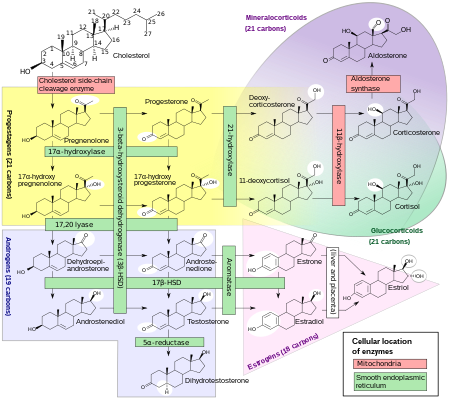
Aldosterone and corticosterone share the first part of their biosynthetic pathways. The last parts are mediated either by the aldosterone synthase (for aldosterone) or by the 11β-hydroxylase (for corticosterone). These enzymes are nearly identical (they share 11β-hydroxylation and 18-hydroxylation functions), but aldosterone synthase is also able to perform an 18-oxidation. Moreover, aldosterone synthase is found within the zona glomerulosa at the outer edge of the adrenal cortex; 11β-hydroxylase is found in the zona glomerulosa and zona fasciculata.
Steroidogenesis, showing aldosterone synthesis at upper-right corner.
Note: aldosterone synthase is absent in other sections of the adrenal gland.

Stimulation

Aldosterone synthesis is stimulated by several factors:
  • increase in the plasma concentration of angiotensin III, a metabolite of angiotensin II
  • increase in plasma angiotensin II, ACTH, or potassium levels, which are present in proportion to plasma sodium deficiencies. (The increased potassium level works to regulate aldosterone synthesis by depolarizing the cells in the zona glomerulosa, which opens the voltage-dependent calcium channels.) The level of angiotensin II is regulated by angiotensin I, which is in turn regulated by renin, a hormone secreted in the kidneys.
  • Serum potassium concentrations are the most potent stimulator of aldosterone secretion.
  • the ACTH stimulation test, which is sometimes used to stimulate the production of aldosterone along with cortisol to determine whether primary or secondary adrenal insufficiency is present. However, ACTH has only a minor role in regulating aldosterone production; with hypopituitarism there is no atrophy of the zona glomerulosa.
  • plasma acidosis
  • the stretch receptors located in the atria of the heart. If decreased blood pressure is detected, the adrenal gland is stimulated by these stretch receptors to release aldosterone, which increases sodium reabsorption from the urine, sweat, and the gut. This causes increased osmolarity in the extracellular fluid, which will eventually return blood pressure toward normal.
  • adrenoglomerulotropin, a lipid factor, obtained from pineal extracts. It selectively stimulates secretion of aldosterone.
The secretion of aldosterone has a diurnal rhythm.

Biological function

Aldosterone is the primary of several endogenous members of the class of mineralocorticoids in humans. Deoxycorticosterone is another important member of this class. Aldosterone tends to promote Na+ and water retention, and lower plasma K+ concentration by the following mechanisms:
  1. Acting on the nuclear mineralocorticoid receptors (MR) within the principal cells of the distal tubule and the collecting duct of the kidney nephron, it upregulates and activates the basolateral Na+/K+ pumps, which pumps three sodium ions out of the cell, into the interstitial fluid and two potassium ions into the cell from the interstitial fluid. This creates a concentration gradient which results in reabsorption of sodium (Na+) ions and water (which follows sodium) into the blood, and secreting potassium (K+) ions into the urine (lumen of collecting duct).
  2. Aldosterone upregulates epithelial sodium channels (ENaCs) in the collecting duct and the colon, increasing apical membrane permeability for Na+ and thus absorption.
  3. Cl is reabsorbed in conjunction with sodium cations to maintain the system's electrochemical balance.
  4. Aldosterone stimulates the secretion of K+ into the tubular lumen.
  5. Aldosterone stimulates Na+ and water reabsorption from the gut, salivary and sweat glands in exchange for K+.
  6. Aldosterone stimulates secretion of H+ via the H+/ATPase in the intercalated cells of the cortical collecting tubules
  7. Aldosterone upregulates expression of NCC in the distal convoluted tubule chronically and its activity acutely.
Aldosterone is responsible for the reabsorption of about 2% of filtered sodium in the kidneys, which is nearly equal to the entire sodium content in human blood under normal glomerular filtration rates.
Aldosterone, probably acting through mineralocorticoid receptors, may positively influence neurogenesis in the dentate gyrus.

Mineralocorticoid receptors

Steroid receptors are intracellular. The aldosterone mineralocorticoid receptor (MR) complex binds on the DNA to specific hormone response element, which leads to gene specific transcription. Some of the transcribed genes are crucial for transepithelial sodium transport, including the three subunits of the epithelial sodium channel (ENaC), the Na+/K+ pumps and their regulatory proteins serum and glucocorticoid-induced kinase, and channel-inducing factor, respectively.
The MR is stimulated by both aldosterone and cortisol, but a mechanism protects the body from excess aldosterone receptor stimulation by glucocorticoids (such as cortisol), which happen to be present at much higher concentrations than mineralocorticoids in the healthy individual. The mechanism consists of an enzyme called 11 β-hydroxysteroid dehydrogenase (11β-HSD). This enzyme co-localizes with intracellular adrenal steroid receptors and converts cortisol into cortisone, a relatively inactive metabolite with little affinity for the MR. Liquorice, which contains glycyrrhetinic acid, can inhibit 11β-HSD and lead to a mineralocorticoid excess syndrome.

Control of aldosterone release from the adrenal cortex

The renin–angiotensin system, showing role of aldosterone between the adrenal glands and the kidneys

Major regulators

The role of the renin–angiotensin system

Angiotensin is involved in regulating aldosterone and is the core regulation. Angiotensin II acts synergistically with potassium, and the potassium feedback is virtually inoperative when no angiotensin II is present. A small portion of the regulation resulting from angiotensin II must take lace indirectly from decreased blood flow through the liver due to constriction of capillaries. When the blood flow decreases so does the destruction of aldosterone by liver enzymes.
Although sustained production of aldosterone requires persistent calcium entry through low-voltage-activated Ca2+ channels, isolated zona glomerulosa cells are considered nonexcitable, with recorded membrane voltages that are too hyperpolarized to permit Ca2+ channels entry. However, mouse zona glomerulosa cells within adrenal slices spontaneously generate membrane potential oscillations of low periodicity; this innate electrical excitability of zona glomerulosa cells provides a platform for the production of a recurrent Ca2+ channels signal that can be controlled by angiotensin II and extracellular potassium, the 2 major regulators of aldosterone production. Voltage-gated Ca2+ channels have been detected in the zona glomerulosa of the human adrenal, which suggests that Ca2+ channel blockers may directly influence the adrenocortical biosynthesis of aldosterone in vivo.

The plasma concentration of potassium

The amount of aldosterone secreted is an indirect function of the serum potassium as probably determined by sensors in the carotid artery.

Adrenocorticotropic hormone

Adrenocorticotropic hormone (ACTH), a pituitary peptide, also has some stimulating effect on aldosterone, probably by stimulating the formation of deoxycorticosterone, a precursor of aldosterone. Aldosterone is increased by blood loss, pregnancy, and possibly by further circumstances such as physical exertion, endotoxin shock, and burns.

Miscellaneous regulators

The role of sympathetic nerves

The aldosterone production is also affected to one extent or another by nervous control, which integrates the inverse of carotid artery pressure, pain, posture, and probably emotion (anxiety, fear, and hostility) (including surgical stress). Anxiety increases aldosterone, which must have evolved because of the time delay involved in migration of aldosterone into the cell nucleus. Thus, there is an advantage to an animal's anticipating a future need from interaction with a predator, since too high a serum content of potassium has very adverse effects on nervous transmission.

The role of baroreceptors

Pressure-sensitive baroreceptors are found in the vessel walls of nearly all large arteries in the thorax and neck, but are particularly plentiful in the sinuses of the carotid arteries and in the arch of the aorta. These specialized receptors are sensitive to changes in mean arterial pressure. An increase in sensed pressure results in an increased rate of firing by the baroreceptors and a negative feedback response, lowering systemic arterial pressure. Aldosterone release causes sodium and water retention, which causes increased blood volume, and a subsequent increase in blood pressure, which is sensed by the baroreceptors. To maintain normal homeostasis these receptors also detect low blood pressure or low blood volume, causing aldosterone to be released. This results in sodium retention in the kidney, leading to water retention and increased blood volume.

The plasma concentration of sodium

Aldosterone levels vary as an inverse function of sodium intake as sensed via osmotic pressure. The slope of the response of aldosterone to serum potassium is almost independent of sodium intake. Aldosterone is increased at low sodium intakes, but the rate of increase of plasma aldosterone as potassium rises in the serum is not much lower at high sodium intakes than it is at low. Thus, potassium is strongly regulated at all sodium intakes by aldosterone when the supply of potassium is adequate, which it usually is in "primitive" diets.

Aldosterone feedback

Feedback by aldosterone concentration itself is of a nonmorphological character (that is, other than changes in the cells' number or structure) and is poor, so the electrolyte feedbacks predominate, short term.

Associated clinical conditions

Hyperaldosteronism is abnormally increased levels of aldosterone, while hypoaldosteronism is abnormally decreased levels of aldosterone.
A measurement of aldosterone in blood may be termed a plasma aldosterone concentration (PAC), which may be compared to plasma renin activity (PRA) as an aldosterone-to-renin ratio.

Hyperaldosteronism

Primary aldosteronism, also known as primary hyperaldosteronism, is characterized by the overproduction of aldosterone by the adrenal glands, when not a result of excessive renin secretion. It leads to arterial hypertension (high blood pressure) associated with hypokalemia, usually a diagnostic clue. Secondary hyperaldosteronism, on the other hand, is due to overactivity of the renin–angiotensin system.
Conn's syndrome is primary hyperaldosteronism caused by an aldosterone-producing adenoma.
Depending on cause and other factors, hyperaldosteronism can be treated by surgery and/or medically, such as by aldosterone antagonists.
The ratio of renin to aldosterone is an effective screening test to screen for primary hyperaldosteronism related to adrenal adenomas. It is the most sensitive serum blood test to differentiate primary from secondary causes of hyperaldosteronism. Blood obtained when the patient has been standing for more than 2 hours are more sensitive than those from when the patient is lying down. Before the test, individuals should not restrict salt and low potassium should be corrected before the test because it can suppress aldosterone secretion.

Hypoaldosteronism

An ACTH stimulation test for aldosterone can help in determining the cause of hypoaldosteronism, with a low aldosterone response indicating a primary hypoaldosteronism of the adrenals, while a large response indicating a secondary hypoaldosteronism.
Additional images

Polycystic kidney disease

From Wikipedia, the free encyclopedia
 
Polycystic kidney disease
Other namesKidney - polycystic
Polycystic kidneys, gross pathology CDC PHIL.png
Severely affected polycystic kidneys removed at time of transplantation
SpecialtyNephrology
SymptomsAbdominal pain
TypesADPKD and ARPKD
Diagnostic methodMRI, CT scan, Ultrasound
TreatmentAntihypertensives, Life style management

Polycystic kidney disease (PKD or PCKD, also known as polycystic kidney syndrome) is a genetic disorder in which the renal tubules become structurally abnormal, resulting in the development and growth of multiple cysts within the kidney. These cysts may begin to develop in utero, in infancy, in childhood, or in adulthood. Cysts are non-functioning tubules filled with fluid pumped into them, which range in size from microscopic to enormous, crushing adjacent normal tubules and eventually rendering them non-functional as well.

PKD is caused by abnormal genes which produce a specific abnormal protein; this protein has an adverse effect on tubule development. PKD is a general term for two types, each having their own pathology and genetic cause: autosomal dominant polycystic kidney disease (ADPKD) and autosomal recessive polycystic kidney disease (ARPKD). The abnormal gene exists in all cells in the body; as a result, cysts may occur in the liver, seminal vesicles, and pancreas. This genetic defect can also cause aortic root aneurysms, and aneurysms in the circle of Willis cerebral arteries, which if they rupture, can cause a subarachnoid hemorrhage.

Diagnosis may be suspected from one, some, or all of the following: new onset flank pain or red urine; a positive family history; palpation of enlarged kidneys on physical exam; an incidental finding on abdominal sonogram; or an incidental finding of abnormal kidney function on routine lab work (BUN, serum creatinine, or eGFR). Definitive diagnosis is made by abdominal CT exam.

Complications include hypertension due to the activation of the renin–angiotensin–aldosterone system (RAAS), frequent cyst infections, urinary bleeding, and declining renal function. Hypertension is treated with angiotensin converting enzyme inhibitors (ACEIs) or angiotensin receptor blockers (ARBs). Infections are treated with antibiotics. Declining renal function is treated with renal replacement therapy (RRT): dialysis and/or transplantation. Management from the time of the suspected or definitive diagnosis is by a board-certified nephrologist.

Signs and symptoms

Signs and symptoms include high blood pressure, headaches, abdominal pain, blood in the urine, and excessive urination. Other symptoms include pain in the back, and cyst formation (renal and other organs).

Cause

PKD is caused by abnormal genes which produce a specific abnormal protein which has an adverse effect on tubule development. PKD is a general term for two types, each having their own pathology and genetic cause: autosomal dominant polycystic kidney disease (ADPKD) and autosomal recessive polycystic kidney disease (ARPKD).

Autosomal dominant

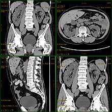
CT scan showing autosomal dominant polycystic kidney disease
 
Cartoon of autosomal dominant polycystic kidney disease with normal kidney inset to right of diagram
 
Cartoon of autosomal recessive polycystic kidney disease with normal kidney inset to right of diagram

Autosomal dominant polycystic kidney disease (ADPKD) is the most common of all the inherited cystic kidney diseases with an incidence of 1:500 live births. Studies show that 10% of end-stage kidney disease (ESKD) patients being treated with dialysis in Europe and the U.S. were initially diagnosed and treated for ADPKD.

Genetic mutations in any of the three genes PKD1, PKD2, and PKD3 have similar phenotypical presentations.
  • Gene PKD1 is located on chromosome 16 and codes for a protein involved in regulation of cell cycle and intracellular calcium transport in epithelial cells, and is responsible for 85% of the cases of ADPKD.
  • A group of voltage-linked cation channels, with inward selectivity for K>Na>>Ca and outward selectivity for Ca2+ ≈ Ba2+ > Na+ ≈ K+, are coded for by PKD2 on chromosome 4
  • PKD3 recently appeared in research papers as a postulated third gene. Fewer than 10% of cases of ADPKD appear in non-ADPKD families. Cyst formation begins in utero from any point along the nephron, although fewer than 5% of nephrons are thought to be involved. As the cysts accumulate fluid, they enlarge, separate entirely from the nephron, compress the neighboring kidney parenchyma, and progressively compromise kidney function.

Autosomal recessive

Autosomal recessive polycystic kidney disease (ARPKD) (OMIM #263200) is the lesser common of the two types of PKD, with an incidence of 1:20,000 live births and is typically identified in the first few weeks after birth. Unfortunately, the kidneys are often underdeveloped resulting in a 30% death rate in newborns with ARPKD. PKHD1 is involved.

Mechanism

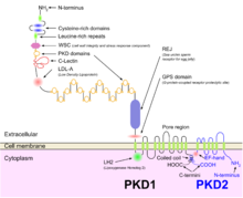
PKD1 and PKD2

Both autosomal dominant and autosomal recessive polycystic kidney disease cyst formation are tied to abnormal cilia-mediated signaling. The polycystin-1 and polycystin-2 proteins appear to be involved in both autosomal dominant and recessive polycystic kidney disease due to defects in both proteins. Both proteins have communication with calcium channel proteins, and causes reduction in resting (intracellular) calcium and endoplasmic reticulum storage of calcium.

The disease is characterized by a ‘second hit’ phenomenon, in which a mutated dominant allele is inherited from a parent, with cyst formation occurring only after the normal, wild-type gene sustains a subsequent second genetic ‘hit’, resulting in renal tubular cyst formation and disease progression.

PKD results from defects in the primary cilium, an immotile, hair-like cellular organelle present on the surface of most cells in the body, anchored in the cell body by the basal body. In the kidney, primary cilia have been found to be present on most cells of the nephron, projecting from the apical surface of the renal epithelium into the tubule lumen. The cilia were believed to bend in the urine flow, leading to changes in signalling, however this has since been shown to be an experimental error (the bending of cilia was an artifact of focal plane compensation, and also the actual effect on micturition by severe hypertension and cardiac arrest) and that bending of cilia does not contribute to alterations in Ca flux. While it is not known how defects in the primary cilium lead to cyst development, it is thought to possibly be related to disruption of one of the many signaling pathways regulated by the primary cilium, including intracellular calcium, Wnt/β-catenin, cyclic adenosine monophosphate (cAMP), or planar cell polarity (PCP). Function of the primary cilium is impaired, resulting in disruption of a number of intracellular signaling cascades which produce differentiation of cystic epithelium, increased cell division, increased apoptosis, and loss of resorptive capacity.

Diagnosis

Polycystic kidney disease can be ascertained via a CT scan of abdomen, as well as, an MRI and ultrasound of the same area. A physical exam/test can reveal enlarged liver, heart murmurs and elevated blood pressure

Natural history

Most cases progress to bilateral disease in adulthood.

Treatment

Chr 11 FISH-mapped BACs from CGAP

There is no FDA-approved treatment. However, recent research indicates that mild to moderate dietary restrictions slow the progression of autosomal dominant polycystic kidney disease (ADPKD) in mice.

If and when the disease progresses enough in a given case, the nephrologist or other practitioner and the patient will have to decide what form of renal replacement therapy will be used to treat end-stage kidney disease (kidney failure, typically stage 4 or 5 of chronic kidney disease).

That will either be some form of dialysis, which can be done at least two different ways at varying frequencies and durations (whether it is done at home or in the clinic depends on the method used and the patient's stability and training) and eventually, if they are eligible because of the nature and severity of their condition and if a suitable match can be found, unilateral or bilateral kidney transplantation.

A Cochrane Review study of autosomal dominant polycystic kidney disease made note of the fact that it is important at all times, while avoiding antibiotic resistance, to control infections of the cysts in the kidneys, and if affected, the liver, when needed for a certain duration to combat infection, by using, "bacteriostatic and bacteriocidal drugs".

Prognosis

ADPKD individuals might have a normal life; conversely, ARPKD can cause kidney dysfunction and can lead to kidney failure by the age of 40–60. ADPKD1 and ADPKD2 are very different, in that ADPKD2 is much milder.

Currently, there are no therapies proven effective to prevent the progression of ADPKD.

Epidemiology

PKD is one of the most common hereditary diseases in the United States, affecting more than 600,000 people. It is the cause of nearly 10% of all end-stage renal disease. It equally affects men, women, and all races.[20] PKD occurs in some animals as well as humans.[21][22]

Nephrology

From Wikipedia, the free encyclopedia
Nephrology
KidneyStructures PioM.svg
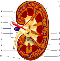
A human kidney (click on image for description).
SystemUrinary
Significant diseasesHypertension, Kidney cancer
Significant testsKidney biopsy, Urinalysis
SpecialistNephrologist
GlossaryGlossary of medicine

Nephrology (from Greek nephros "kidney", combined with the suffix -logy, "the study of") is a specialty of medicine and pediatric medicine that concerns itself with the kidneys: the study of normal kidney function and kidney disease, the preservation of kidney health, and the treatment of kidney disease, from diet and medication to renal replacement therapy (dialysis and kidney transplantation).

Nephrology also studies systemic conditions that affect the kidneys, such as diabetes and autoimmune disease; and systemic diseases that occur as a result of kidney disease, such as renal osteodystrophy and hypertension. A physician who has undertaken additional training and become certified in nephrology is called a nephrologist.

The term "nephrology" was first used in about 1960. Before then, the specialty was usually referred to as "kidney medicine."

Scope

Nephrology concerns the diagnosis and treatment of kidney diseases, including electrolyte disturbances and hypertension, and the care of those requiring renal replacement therapy, including dialysis and renal transplant patients. The word 'dialysis' is from the mid 19th century: via Latin from the Greek word 'dialusis'; from 'dialuein' (split, separate), from 'dia' (apart) and 'luein' (set free). In other words, dialysis replaces the primary (excretory) function of the kidney, which separates (and removes) excess toxins and water from the blood, placing them in the urine.

Many diseases affecting the kidney are systemic disorders not limited to the organ itself, and may require special treatment. Examples include acquired conditions such as systemic vasculitides (e.g. ANCA vasculitis) and autoimmune diseases (e.g., lupus), as well as congenital or genetic conditions such as polycystic kidney disease

Patients are referred to nephrology specialists after a urinalysis, for various reasons, such as acute kidney failure, chronic kidney disease, hematuria, proteinuria, kidney stones, hypertension, and disorders of acid/base or electrolytes.

Nephrologist

A nephrologist is a physician who specializes in the care and treatment of kidney disease. Nephrology requires additional training to become an expert with advanced skills. Nephrologists may provide care to people without kidney problems and may work in general/internal medicine, transplant medicine, immunosuppression management, intensive care medicine, clinical pharmacology, perioperative medicine, or pediatric nephrology.

Nephrologists may further sub-specialise in dialysis, kidney transplantation, chronic kidney disease, cancer-related kidney diseases (Onconephrology), procedural nephrology or other non-nephrology areas as described above.

Procedures a nephrologist may perform include native kidney and transplant kidney biopsy, dialysis access insertion (temporary vascular access lines, tunnelled vascular access lines, peritoneal dialysis access lines), fistula management (angiographic or surgical fistulogram and plasty), and bone biopsy. Bone biopsies are now unusual.

Training

India
To become a nephrologist in India, one has to complete an MBBS (5 and1/2 years) degree, followed by a MD/DNB (3 years) either in medicine or paediatrics, followed by a DM/DNB (3 years) course in either nephrology or paediatric nephrology.

Australia and New Zealand

Nephrology training in Australia and New Zealand typically includes completion of a medical degree (Bachelor of Medicine, Bachelor of Surgery: 4–6 years), internship (1 year), Basic Physician Training (3 years minimum), successful completion of the Royal Australasian College of Physicians written and clinical examinations, and Advanced Physician Training in Nephrology (2–3 years). The training pathway is overseen and accredited by the Royal Australasian College of Physicians. Increasingly, nephrologists may additionally complete of a post-graduate degree (usually a PhD) in a nephrology research interest (3–4 years). Finally, all Australian and New Zealand nephrologists participate in career-long professional and personal development through the Royal Australasian College of Physicians and other bodies such as the Australian and New Zealand Society of Nephrology and the Transplant Society of Australia and New Zealand.

United Kingdom

In the United Kingdom, nephrology (often called renal medicine) is a subspecialty of general medicine. A nephrologist has completed medical school, foundation year posts (FY1 and FY2) and core medical training (CMT), specialist training (ST) and passed the Membership of the Royal College of Physicians (MRCP) exam before competing for a National Training Number (NTN) in renal medicine. The typical Specialty Training (when they are called a registrar, or a ST) is five years and leads to a Certificate of Completion of Training (CCT) in both renal medicine and general (internal) medicine. In that five years, they usually rotate yearly between hospitals on a region (known as a deanery). They are then accepted on to the Specialist Register of the General Medical Council (GMC). Specialty trainees often interrupt their clinical training to obtain research degrees (MD/PhD). After achieving CCT, the registrar (ST) may apply for a permanent post as Consultant in Renal Medicine. Subsequently, some Consultants practice nephrology alone. Others work in this area, and in Intensive Care (ICU) , or General (Internal) or Acute Medicine.

United States

Nephrology training can be accomplished through one of two routes. The first pathway is through an internal medicine pathway leading to an Internal Medicine/Nephrology specialty, and sometimes known as "adult nephrology". The second pathway is through Pediatrics leading to a speciality in Pediatric Nephrology. In the United States, after medical school adult nephrologists complete a three-year residency in internal medicine followed by a two-year (or longer) fellowship in nephrology. Complementary to an adult nephrologist, a pediatric nephrologist will complete a three-year pediatric residency after medical school or a four-year Combined Internal Medicine and Pediatrics residency. This is followed by a three-year fellowship in Pediatic Nephrology. Once training is satisfactorily completed, the physician is eligible to take the American Board of Internal Medicine (ABIM) or American Osteopathic Board of Internal Medicine (AOBIM) nephrology examination. Nephrologists must be approved by one of these boards. To be approved, the physician must fulfill the requirements for education and training in nephrology in order to qualify to take the board's examination. If a physician passes the examination, then he or she can become a nephrology specialist. Typically, nephrologists also need two to three years of training in an ACGME or AOA accredited fellowship in nephrology. Nearly all programs train nephrologists in continuous renal replacement therapy; fewer than half in the United States train in the provision of plasmapheresis. Only pediatric trained physicians are able to train in pediatric nephrology, and internal medicine (adult) trained physicians may enter general (adult) nephrology fellowships.

Diagnosis

History and physical examination are central to the diagnostic workup in nephrology. The history typically includes the present illness, family history, general medical history, diet, medication use, drug use and occupation. The physical examination typically includes an assessment of volume state, blood pressure, heart, lungs, peripheral arteries, joints, abdomen and flank. A rash may be relevant too, especially as an indicator of autoimmune disease. 

Examination of the urine (urinalysis) allows a direct assessment for possible kidney problems, which may be suggested by appearance of blood in the urine (haematuria), protein in the urine (proteinuria), pus cells in the urine (pyuria) or cancer cells in the urine. A 24-hour urine collection used to be used to quantify daily protein loss (see proteinuria), urine output, creatinine clearance or electrolyte handling by the renal tubules. It is now more common to measure protein loss from a small random sample of urine. 

Basic blood tests can be used to check the concentration of hemoglobin, white count, platelets, sodium, potassium, chloride, bicarbonate, urea, creatinine, albumin, calcium, magnesium, phosphate, alkaline phosphatase and parathyroid hormone (PTH) in the blood. All of these may be affected by kidney problems. The serum creatinine concentration is the most important blood test as it is used to estimate the function of the kidney, called the creatinine clearance or estimated glomerular filtration rate (GFR). 

It is good idea for patients with longterm kidney disease to know an up-to-date list of medications, and their latest blood tests, especially the blood creatinine level. In the United Kingdom, blood tests can monitored online by the patient, through a website called RenalPatientView. 

More specialized tests can be ordered to discover or link certain systemic diseases to kidney failure such as infections (hepatitis B, hepatitis C), autoimmune conditions (systemic lupus erythematosus, ANCA vasculitis), paraproteinemias (amyloidosis, multiple myeloma) and metabolic diseases (diabetes, cystinosis). 

Structural abnormalities of the kidneys are identified with imaging tests. These may include Medical ultrasonography/ultrasound, computed axial tomography (CT), scintigraphy (nuclear medicine), angiography or magnetic resonance imaging (MRI)

In certain circumstances, less invasive testing may not provide a certain diagnosis. Where definitive diagnosis is required, a biopsy of the kidney (renal biopsy) may be performed. This typically involves the insertion, under local anaesthetic and ultrasound or CT guidance, of a core biopsy needle into the kidney to obtain a small sample of kidney tissue. The kidney tissue is then examined under a microscope, allowing direct visualization of the changes occurring within the kidney. Additionally, the pathology may also stage a problem affecting the kidney, allowing some degree of prognostication. In some circumstances, kidney biopsy will also be used to monitor response to treatment and identify early relapse. A transplant kidney biopsy may also be performed to look for rejection of the kidney.

Treatment

Treatments in nephrology can include medications, blood products, surgical interventions (urology, vascular or surgical procedures), renal replacement therapy (dialysis or kidney transplantation) and plasma exchange. Kidney problems can have significant impact on quality and length of life, and so psychological support, health education and advanced care planning play key roles in nephrology.

Chronic kidney disease is typically managed with treatment of causative conditions (such as diabetes), avoidance of substances toxic to the kidneys (nephrotoxins like radiologic contrast and non-steroidal anti-inflammatory drugs), antihypertensives, diet and weight modification and planning for end-stage kidney failure. Impaired kidney function has systemic effects on the body. An erythropoetin stimulating agent (ESA) may be required to ensure adequate production of red blood cells, activated vitamin D supplements and phosphate binders may be required to counteract the effects of kidney failure on bone metabolism, and blood volume and electrolyte disturbance may need correction. Diuretics (such as furosemide) may be used to correct fluid overload, and alkalis (such as sodium bicarbonate) can be used to treat metabolic acidosis. 

Auto-immune and inflammatory kidney disease, such as vasculitis or transplant rejection, may be treated with immunosuppression. Commonly used agents are prednisone, mycophenolate, cyclophosphamide, ciclosporin, tacrolimus, everolimus, thymoglobulin and sirolimus. Newer, so-called "biologic drugs" or monoclonal antibodies, are also used in these conditions and include rituximab, basiliximab and eculizumab. Blood products including intravenous immunoglobulin and a process known as plasma exchange can also be employed.

When the kidneys are no longer able to sustain the demands of the body, end-stage kidney failure is said to have occurred. Without renal replacement therapy, death from kidney failure will eventually result. Dialysis is an artificial method of replacing some kidney function to prolong life. Renal transplantation replaces kidney function by inserting into the body a healthier kidney from an organ donor and inducing immunologic tolerance of that organ with immunosuppression. At present, renal transplantation is the most effective treatment for end-stage kidney failure although its worldwide availability is limited by lack of availability of donor organs. Generally speaking, kidneys from living donors are 'better' than those from deceased donors, as they last longer. 

Most kidney conditions are chronic conditions and so long term followup with a nephrologist is usually necessary. In the United Kingdom, care may be shared with the patient's primary care physician, called a General Practitioner (GP).

Organizations

The world's first society of nephrology was the French 'Societe de Pathologie Renale'. Its first president was Jean Hamburger, and its first meeting was in Paris in February 1949. In 1959, Hamburger also founded the 'Société de Néphrologie', as a continuation of the older society. The UK's Renal Association was founded in 1950; the second society of nephrologists. Its first president was Arthur Osman. Its first meeting was on 30 March 1950 in London. The Società di Nefrologia Italiana was founded in 1957 and was the first national society to incorporate the phrase nephrologia (or nephrology) into its name.

The word 'nephrology' appeared for the first time in a conference, on 1–4 September 1960 at the "Premier Congrès International de Néphrologie" in Evian and Geneva, the first meeting of the International Society of Nephrology (ISN, International Society of Nephrology). The first day (1.9.60) was in Geneva and the next three (2–4.9.60) were in Evian, France. The early history of the ISN is described by Robinson and Richet in 2005 and the later history by Barsoum in 2011. The ISN is the largest global society representing medical professionals engaged in advancing kidney care worldwide. 

In the USA, founded in 1964, the National Kidney Foundation is a national organization representing patients and professionals who treat kidney diseases. Founded in 1966, the American Society of Nephrology (ASN) is the world’s largest professional society devoted to the study of kidney disease. The American Nephrology Nurses' Association (ANNA), founded in 1969, promotes excellence in and appreciation of nephrology nursing to make a positive difference for patients with kidney disease. The American Association of Kidney Patients (AAKP) is a non-profit, patient-centric group focused on improving the health and well-being of CKD and dialysis patients. The National Renal Administrators Association (NRAA), founded in 1977, is a national organization that represents and supports the independent and community-based dialysis providers. The American Kidney Fund directly provides financial support to patients in need, as well as participating in health education and prevention efforts. ASDIN (American Society of Diagnostic and Interventional Nephrology) is the main organization of interventional nephrologists. Other organizations include CIDA, VASA etc. which deal with dialysis vascular access. The Renal Support Network (RSN) is a nonprofit, patient-focused, patient-run organization that provides non-medical services to those affected by chronic kidney disease (CKD).

In the United Kingdom, UK National Kidney Federation and Kidney Care UK (previously known as British Kidney Patient Association, BKPA) represent patients, and the Renal Association represents renal physicians and works closely with the National Service Framework for kidney disease. 

There is as well an International Office in Brussels, Belgium.

Classical radicalism

From Wikipedia, the free encyclopedia https://en.wikipedia.org/wiki/Cla...