Search This Blog

Thursday, June 13, 2024

Viral evolution

From Wikipedia, the free encyclopedia
(Redirected from Virus evolution)

Viral evolution is a subfield of evolutionary biology and virology that is specifically concerned with the evolution of viruses. Viruses have short generation times, and many—in particular RNA viruses—have relatively high mutation rates (on the order of one point mutation or more per genome per round of replication). Although most viral mutations confer no benefit and often even prove deleterious to viruses, the rapid rate of viral mutation combined with natural selection allows viruses to quickly adapt to changes in their host environment. In addition, because viruses typically produce many copies in an infected host, mutated genes can be passed on to many offspring quickly. Although the chance of mutations and evolution can change depending on the type of virus (e.g., double stranded DNA, double stranded RNA, single strand DNA), viruses overall have high chances for mutations.

Viral evolution is an important aspect of the epidemiology of viral diseases such as influenza (influenza virus), AIDS (HIV), and hepatitis (e.g. HCV). The rapidity of viral mutation also causes problems in the development of successful vaccines and antiviral drugs, as resistant mutations often appear within weeks or months after the beginning of a treatment. One of the main theoretical models applied to viral evolution is the quasispecies model, which defines a viral quasispecies as a group of closely related viral strains competing within an environment.

Origins

Three classical hypotheses

Viruses are ancient. Studies at the molecular level have revealed relationships between viruses infecting organisms from each of the three domains of life, suggesting viral proteins that pre-date the divergence of life and thus infecting the last universal common ancestor. This indicates that some viruses emerged early in the evolution of life, and that they have probably arisen multiple times. It has been suggested that new groups of viruses have repeatedly emerged at all stages of evolution, often through the displacement of ancestral structural and genome replication genes.

There are three classical hypotheses on the origins of viruses and how they evolved:

  • Virus-first hypothesis: Viruses evolved from complex molecules of protein and nucleic acid before cells first appeared on earth. By this hypothesis, viruses contributed to the rise of cellular life. This is supported by the idea that all viral genomes encode proteins that do not have cellular homologs. The virus-first hypothesis has been dismissed by some scientists because it violates the definition of viruses, in that they require a host cell to replicate.
  • Reduction hypothesis (degeneracy hypothesis): Viruses were once small cells that parasitized larger cells. This is supported by the discovery of giant viruses with similar genetic material to parasitic bacteria. However, the hypothesis does not explain why even the smallest of cellular parasites do not resemble viruses in any way.
  • Escape hypothesis (vagrancy hypothesis): Some viruses evolved from bits of DNA or RNA that "escaped" from the genes of larger organisms. This does not explain the structures that are unique to viruses and are not seen anywhere in cells. It also does not explain the complex capsids and other structures of virus particles.

Virologists are in the process of re-evaluating these hypotheses.

Later hypotheses

  • Coevolution hypothesis (Bubble Theory): At the beginning of life, a community of early replicons (pieces of genetic information capable of self-replication) existed in proximity to a food source such as a hot spring or hydrothermal vent. This food source also produced lipid-like molecules self-assembling into vesicles that could enclose replicons. Close to the food source replicons thrived, but further away the only non-diluted resources would be inside vesicles. Therefore, evolutionary pressure could push replicons along two paths of development: merging with a vesicle, giving rise to cells; and entering the vesicle, using its resources, multiplying and leaving for another vesicle, giving rise to viruses.
  • Chimeric-origins hypothesis: Based on the analyses of the evolution of the replicative and structural modules of viruses, a chimeric scenario for the origin of viruses was proposed in 2019. According to this hypothesis, the replication modules of viruses originated from the primordial genetic pool, although the long course of their subsequent evolution involved many displacements by replicative genes from their cellular hosts. By contrast, the genes encoding major structural proteins evolved from functionally diverse host proteins throughout the evolution of the virosphere. This scenario is distinct from each of the three traditional scenarios but combines features of the Virus-first and Escape hypotheses.

One of the problems for studying viral origins and evolution is the high rate of viral mutation, particularly the case in RNA retroviruses like HIV/AIDS. A recent study based on comparisons of viral protein folding structures, however, is offering some new evidence. Fold Super Families (FSFs) are proteins that show similar folding structures independent of the actual sequence of amino acids, and have been found to show evidence of viral phylogeny. The proteome of a virus, the viral proteome, still contains traces of ancient evolutionary history that can be studied today. The study of protein FSFs suggests the existence of ancient cellular lineages common to both cells and viruses before the appearance of the 'last universal cellular ancestor' that gave rise to modern cells. Evolutionary pressure to reduce genome and particle size may have eventually reduced viro-cells into modern viruses, whereas other coexisting cellular lineages eventually evolved into modern cells. Furthermore, the long genetic distance between RNA and DNA FSFs suggests that the RNA world hypothesis may have new experimental evidence, with a long intermediary period in the evolution of cellular life.

Definitive exclusion of a hypothesis on the origin of viruses is difficult to make on Earth given the ubiquitous interactions between viruses and cells, and the lack of availability of rocks that are old enough to reveal traces of the earliest viruses on the planet. From an astrobiological perspective, it has therefore been proposed that on celestial bodies such as Mars not only cells but also traces of former virions or viroids should be actively searched for: possible findings of traces of virions in the apparent absence of cells could provide support for the virus-first hypothesis.

Evolution

Time-line of paleoviruses in the human lineage

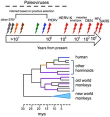
Viruses do not form fossils in the traditional sense, because they are much smaller than the finest colloidal fragments forming sedimentary rocks that fossilize plants and animals. However, the genomes of many organisms contain endogenous viral elements (EVEs). These DNA sequences are the remnants of ancient virus genes and genomes that ancestrally 'invaded' the host germline. For example, the genomes of most vertebrate species contain hundreds to thousands of sequences derived from ancient retroviruses. These sequences are a valuable source of retrospective evidence about the evolutionary history of viruses, and have given birth to the science of paleovirology.

The evolutionary history of viruses can to some extent be inferred from analysis of contemporary viral genomes. The mutation rates for many viruses have been measured, and application of a molecular clock allows dates of divergence to be inferred.

Viruses evolve through changes in their RNA (or DNA), some quite rapidly, and the best adapted mutants quickly outnumber their less fit counterparts. In this sense their evolution is Darwinian. The way viruses reproduce in their host cells makes them particularly susceptible to the genetic changes that help to drive their evolution. The RNA viruses are especially prone to mutations. In host cells there are mechanisms for correcting mistakes when DNA replicates and these kick in whenever cells divide. These important mechanisms prevent potentially lethal mutations from being passed on to offspring. But these mechanisms do not work for RNA and when an RNA virus replicates in its host cell, changes in their genes are occasionally introduced in error, some of which are lethal. One virus particle can produce millions of progeny viruses in just one cycle of replication, therefore the production of a few "dud" viruses is not a problem. Most mutations are "silent" and do not result in any obvious changes to the progeny viruses, but others confer advantages that increase the fitness of the viruses in the environment. These could be changes to the virus particles that disguise them so they are not identified by the cells of the immune system or changes that make antiviral drugs less effective. Both of these changes occur frequently with HIV.

Phylogenetic tree showing the relationships of morbilliviruses of different species

Many viruses (for example, influenza A virus) can "shuffle" their genes with other viruses when two similar strains infect the same cell. This phenomenon is called genetic shift, and is often the cause of new and more virulent strains appearing. Other viruses change more slowly as mutations in their genes gradually accumulate over time, a process known as antigenic drift.

Through these mechanisms new viruses are constantly emerging and present a continuing challenge in attempts to control the diseases they cause. Most species of viruses are now known to have common ancestors, and although the "virus first" hypothesis has yet to gain full acceptance, there is little doubt that the thousands of species of modern viruses have evolved from less numerous ancient ones. The morbilliviruses, for example, are a group of closely related, but distinct viruses that infect a broad range of animals. The group includes measles virus, which infects humans and primates; canine distemper virus, which infects many animals including dogs, cats, bears, weasels and hyaenas; rinderpest, which infected cattle and buffalo; and other viruses of seals, porpoises and dolphins. Although it is not possible to prove which of these rapidly evolving viruses is the earliest, for such a closely related group of viruses to be found in such diverse hosts suggests the possibility that their common ancestor is ancient.

Bacteriophage

Escherichia virus T4 (phage T4) is a species of bacteriophage that infects Escherichia coli bacteria. It is a double-stranded DNA virus in the family Myoviridae. Phage T4 is an obligate intracellular parasite that reproduces within the host bacterial cell and its progeny are released when the host is destroyed by lysis. The complete genome sequence of phage T4 encodes about 300 gene products. These virulent viruses are among the largest, most complex viruses that are known and one of the best studied model organisms. They have played a key role in the development of virology and molecular biology. The numbers of reported genetic homologies between phage T4 and bacteria and between phage T4 and eukaryotes are similar suggesting that phage T4 shares ancestry with both bacteria and eukaryotes and has about equal similarity to each. Phage T4 may have diverged in evolution from a common ancestor of bacteria and eukaryotes or from an early evolved member of either lineage. Most of the phage genes showing homology with bacteria and eukaryotes encode enzymes acting in the ubiquitous processes of DNA replication, DNA repair, recombination and nucleotide synthesis. These processes likely evolved very early. The adaptive features of the enzymes catalyzing these early processes may have been maintained in the phage T4, bacterial, and eukaryotic lineages because they were established well-tested solutions to basic functional problems by the time these lineages diverged.

Transmission

Viruses have been able to continue their infectious existence due to evolution. Their rapid mutation rates and natural selection has given viruses the advantage to continue to spread. One way that viruses have been able to spread is with the evolution of virus transmission. The virus can find a new host through:

  • Droplet transmission- passed on through body fluids (sneezing on someone)
    • An example is the influenza virus
  • Airborne transmission- passed on through the air (brought in by breathing)
    • An example would be how viral meningitis is passed on
  • Vector transmission- picked up by a carrier and brought to a new host
    • An example is viral encephalitis
  • Waterborne transmission- leaving a host, infecting the water, and being consumed in a new host
    • Poliovirus is an example for this
  • Sit-and-wait-transmission- the virus is living outside a host for long periods of time
    • The smallpox virus is also an example for this

Virulence, or the harm that the virus does on its host, depends on various factors. In particular, the method of transmission tends to affect how the level of virulence will change over time. Viruses that transmit through vertical transmission (transmission to the offspring of the host) will evolve to have lower levels of virulence. Viruses that transmit through horizontal transmission (transmission between members of the same species that don't have a parent-child relationship) will usually evolve to have a higher virulence.

Veterinary virology

From Wikipedia, the free encyclopedia
Bluetongue virus

Veterinary virology is the study of viruses in non-human animals. It is an important branch of veterinary medicine.

Rhabdoviruses

Vesicular stomatitis virus (VSV), the prototypical rhabdovirus

Rhabdoviruses are a diverse family of single stranded, negative sense RNA viruses that infect a wide range of hosts, from plants and insects, to fish and mammals. The Rhaboviridae family consists of six genera, two of which, cytorhabdoviruses and nucleorhabdoviruses, only infect plants. Novirhabdoviruses infect fish, and vesiculovirus, lyssavirus and ephemerovirus infect mammals, fish and invertebrates. The family includes pathogens such as rabies virus, vesicular stomatitis virus and potato yellow dwarf virus that are of public health, veterinary, and agricultural significance.

Foot-and-mouth disease virus

Foot-and-mouth disease virus (FMDV) is a member of the Aphthovirus genus in the Picornaviridae family and is the cause of foot-and-mouth disease in pigs, cattle, sheep and goats. It is a non-enveloped, positive strand, RNA virus. FMDV is a highly contagious virus. It enters the body through inhalation.

Pestiviruses

Pestiviruses have a single stranded, positive-sense RNA genomes. They cause Classical swine fever (CSF) and Bovine viral diarrhea(BVD). Mucosal disease is a distinct, chronic persistent infection, whereas BVD is an acute infection.

Arteriviruses

Arteriviruses are small, enveloped, animal viruses with an icosahedral core containing a positive-sense RNA genome. The family includes equine arteritis virus (EAV), porcine reproductive and respiratory syndrome virus (PRRSV), lactate dehydrogenase elevating virus (LDV) of mice and simian haemorrhagic fever virus (SHFV).

Coronaviruses

Coronaviruses are enveloped viruses with a positive-sense RNA genome and with a nucleocapsid of helical symmetry. They infect the upper respiratory and gastrointestinal tract of mammals and birds. They are the cause of a wide range of diseases in cats, dog, pigs, rodents, cattle and humans. Transmission is by the faecal-oral route.

Toroviruses

Torovirus is a genus of viruses within the family Coronaviridae, subfamily Torovirinae that primarily infect vertebrates and include Berne virus of horses and Breda virus of cattle. They cause gastroenteritis in mammals, including humans but rarely.

Influenza

Influenza is caused by RNA viruses of the family Orthomyxoviridae and affects birds and mammals.

Avian influenza

Wild aquatic birds are the natural hosts for a large variety of influenza A viruses. Occasionally viruses are transmitted from this reservoir to other species and may then cause devastating outbreaks in domestic poultry or give rise to human influenza pandemics.

Swine influenza

Bluetongue virus

Bluetongue virus (BTV), a member of Orbivirus genus within the Reoviridae family causes serious disease in livestock (sheep, goat, cattle). It is non-enveloped, double-stranded RNA virus. The genome is segmented.

Circoviruses

Circoviruses are small single-stranded DNA viruses. There are two genera: gyrovirus, with one species called chicken anemia virus; and circovirus, which includes porcine circovirus types 1 and 2, psittacine beak and feather disease virus, pigeon circovirus, canary circovirus, goose circovirus.

Herpesviruses

Herpesviruses are ubiquitous pathogens infecting a variety of animals, including humans. Hosts include many economically important species such as abalone, oysters, salmon, poultry (avian infectious laryngotracheitis, Marek's disease), cattle (bovine malignant catarrhal fever), dogs, goats, horses, cats (feline viral rhinotracheitis), and pigs (pseudorabies). Infections may be severe and may result in fatalities or reduced productivity. Therefore, outbreaks of herpesviruses in livestock cause significant financial losses and are an important area of study in veterinary virology.

African swine fever virus

African swine fever virus (ASFV) is a large double-stranded DNA virus which replicates in the cytoplasm of infected cells and is the only member of the Asfarviridae family. The virus causes a lethal haemorraghic disease in domestic pigs. Some strains can cause death of animals within as little as a week after infection. In other species, the virus causes no obvious disease. ASFV is endemic to sub-Saharan Africa and exists in the wild through a cycle of infection between ticks and wild pigs, bushpigs and warthogs.

Retroviruses

Retroviruses are established pathogens of veterinary importance. They are generally a cause of cancer or immune deficiency.

Flaviviruses

Flaviviruses constitute a family of linear, single-stranded RNA(+) viruses. Flaviviruses include the West Nile virus, dengue virus, Tick-borne Encephalitis Virus, Yellow Fever Virus, and several other viruses. Many flavivirus species can replicate in both mammalian and insect cells. Most flaviviruses are arthropod borne and multiply in both vertebrates and arthropods. The viruses in this family that are of veterinary importance include Japanese encephalitis virus, St. Louis encephalitis virus, West Nile virus, Israel turkey meningoencephalomyelitis virus, Sitiawan virus, Wesselsbron virus, yellow fever virus and the tick-borne flaviviruses e.g. louping ill virus.

Paramyxoviruses

Paramyxoviruses are a diverse family of non-segmented negative strand RNA viruses that include many highly pathogenic viruses affecting humans, animals, and birds. These include canine distemper virus (dogs), phocine distemper virus (seals), cetacean morbillivirus (dolphins and porpoises) Newcastle disease virus (birds) and rinderpest virus (cattle). Some paramyxoviruses such as the henipaviruses are zoonotic pathogens, occurring primarily in an animal hosts, but also able to infect humans.

Parvovirus

Parvoviruses are linear, non-segmented single-stranded DNA viruses, with an average genome size of 5000 nucleotides. They are classified as group II viruses in Baltimore classification of viruses. Parvoviruses are among the smallest viruses (hence the name, from Latin parvus meaning small) and are 18–28 nm in diameter.

Parvoviruses can cause disease in some animals, including starfish and humans. Because the viruses require actively dividing cells to replicate, the type of tissue infected varies with the age of the animal. The gastrointestinal tract and lymphatic system can be affected at any age, leading to vomiting, diarrhea and immunosuppression but cerebellar hypoplasia is only seen in cats that were infected in the womb or at less than two weeks of age, and disease of the myocardium is seen in puppies infected between the ages of three and eight weeks.

Relationship between telomeres and longevity

Telomeres at the end of a chromosome.

The relationship between telomeres and longevity and changing the length of telomeres is one of the new fields of research on increasing human lifespan and even human immortality. Telomeres are sequences at the ends of chromosomes that shorten with each cell division and determine the lifespan of cells. The telomere was first discovered by biologist Hermann Joseph Muller in the early 20th century. However, experiments by Elizabeth Blackburn, Carol Greider, and Jack Szostak in the 1980s led to the successful discovery of telomerase (the enzyme responsible for maintaining telomere length) and a better understanding of telomeres.

Telomeres play essential roles in the stability and control of cell division. Telomeres protect chromosomes from deterioration and fusion with neighboring chromosomes and act as a buffer zone, preventing the loss of essential genetic information during cell division.

It is predicted that the knowledge of methods to increase the length of cell telomeres (Stem cell and quasi-stem cells, control the regeneration and rebuilding of different tissues of the body) will pave the way for increasing human lifespan. Examining telomeres is one of the most important fields of research related to aging. It is also very important to investigate the mechanisms of maintaining telomerase, cell cleansing (old cells that accumulate in tissues and sometimes cause cancer and inflammation) and the production of new cells in long-lived organisms. However, this idea faces major challenges such as increased cancer incidence, immune system problems, and unwanted long-term consequences.

Telomere and Telomerase

In the early 1970s, Alexey Olovnikov first recognized that chromosomes cannot completely duplicate their ends during cell division. This is known as the "end replication problem". Olovnikov proposed that every time a cell divides, a part of the DNA sequence is lost, and if this loss reaches a certain level, cell division will stop at the end. According to his "marginotomy" theory, there are sequences at the end of the DNA (telomeres) that are placed in tandem repeats and create a buffer zone that determines the number of divisions a particular cell can undergo.

Many organisms have a ribonucleoprotein enzyme called telomerase, which is responsible for adding repetitive nucleotide sequences to the ends of DNA. Telomerase replicates the telomere head and does not require ATP. In most multicellular eukaryotic organisms, telomerase is active only in germ cells, some types of stem cells such as embryonic stem cells, and certain white blood cells. Telomerase can be reactivated and telomeres restored to the embryonic state by somatic cell nuclear transfer. The continuous shortening of telomeres with each replication in somatic (body) cells may play a role in aging and in cancer prevention. This is because telomeres act as a kind of "delayed fuse" and eventually run out after a certain number of cell divisions. This action results in the loss of vital genetic information from the cell's chromosome after multiple divisions. Research on telomerase is extremely important in understanding its role in maintaining telomere length and its potential implications for aging and cancer.

Challenges

While telomeres play an important role in cellular senescence, the intricate biological details of telomeres still require further investigation. The complex interactions between telomeres, different proteins and the cellular environment must be fully understood in order to develop precise and safe interventions to change it. Understanding the long-term effects of telomere extension on the body is complex and risky. Prediction of long-term consequences, including potential unanticipated side effects or interactions with other cellular processes, requires thorough and long-term investigation.

Disturbances in cell division processes and mutations in DNA are among the most important causes of cancer. Cancer cells develop mechanisms to increase their lifespan.

Increased risk of cancer

One of the major concerns associated with telomere lengthening is the potential for increased cancer risk. Telomeres naturally shorten with each cell division and act as a tumor suppressor mechanism. Extending telomeres can allow cells to divide more and increase the risk of uncontrolled cell growth and cancer development. A study conducted by Johns Hopkins University challenged the idea that long telomeres prevent aging. Rather than protecting cells from aging, long telomeres help cells with age-related mutations last longer. This problem prepares the conditions for the occurrence of various types of cancer, and people with longer cell telomeres showed more signs of suffering from types of cancer such as Melanoma and Lymphoma.

Telomere length balance

Achieving balance in telomere length is challenging. While extended telomeres can reverse some aspects of cellular aging, excessively long telomeres may lead to cellular instability and dysfunction. It is important to strike the right balance to avoid unintended consequences.

Old cells and telomere dysfunction

Telomere dysfunction during cellular aging (a state in which cells do not divide but are metabolically active) affects the health of the body. Preventing telomere shortening without clearing old cells may lead to the accumulation of these cells in the body and contribute to age-related diseases and tissue dysfunction.

Intertissue differences

Different tissues of the human body may react differently to changes in telomeres. Telomere length is different in different tissues and cell types of the body. Developing a general telomere lengthening strategy that is effective in all tissues is a complex task; Also, understanding how different types of cells, organs and systems react to telomere manipulation is very important for developing safe and effective interventions.

Effects on the immune system

The immune system plays an important role in monitoring and destroying abnormal or cancerous cells. Telomere extension may affect the immune system's ability to recognize and eliminate cells with long telomeres, potentially compromising immune surveillance. It is very important to ensure the ability of the immune system to effectively identify and fight against pathogens and abnormal cells.

Interferon

From Wikipedia, the free encyclopedia
Interferon type I (α/β/δ...)
The molecular structure of human interferon-alpha (PDB: 1RH2​)
Identifiers
SymbolInterferons
PfamPF00143
InterProIPR000471
SMARTSM00076
PROSITEPDOC00225
CATH1au0
SCOP21au1 / SCOPe / SUPFAM
CDDcd00095

Available protein structures:
Interferon type II (γ)
The three-dimensional structure of human interferon gamma (PDB: 1HIG​)
Identifiers
SymbolIFN-gamma
PfamPF00714
InterProIPR002069
CATH1d9cA00
SCOP2d1d9ca_ / SCOPe / SUPFAM

Available protein structures:
Interferon type III (λ)
Identifiers
SymbolIL28A
PfamPF15177
InterProIPR029177
CATH3og6A00

Available protein structures:

Interferons (IFNs, /ˌɪntərˈfɪərɒn/ IN-tər-FEER-on) are a group of signaling proteins made and released by host cells in response to the presence of several viruses. In a typical scenario, a virus-infected cell will release interferons causing nearby cells to heighten their anti-viral defenses.

IFNs belong to the large class of proteins known as cytokines, molecules used for communication between cells to trigger the protective defenses of the immune system that help eradicate pathogens. Interferons are named for their ability to "interfere" with viral replication by protecting cells from virus infections. However, virus-encoded genetic elements have the ability to antagonize the IFN response, contributing to viral pathogenesis and viral diseases. IFNs also have various other functions: they activate immune cells, such as natural killer cells and macrophages, and they increase host defenses by up-regulating antigen presentation by virtue of increasing the expression of major histocompatibility complex (MHC) antigens. Certain symptoms of infections, such as fever, muscle pain and "flu-like symptoms", are also caused by the production of IFNs and other cytokines.

More than twenty distinct IFN genes and proteins have been identified in animals, including humans. They are typically divided among three classes: Type I IFN, Type II IFN, and Type III IFN. IFNs belonging to all three classes are important for fighting viral infections and for the regulation of the immune system.

Types of interferon

Based on the type of receptor through which they signal, human interferons have been classified into three major types.

  • Interferon type I: All type I IFNs bind to a specific cell surface receptor complex known as the IFN-α/β receptor (IFNAR) that consists of IFNAR1 and IFNAR2 chains. The type I interferons present in humans are IFN-α, IFN-β, IFN-ε, IFN-κ and IFN-ω. Interferon beta (IFN-β) can be produced by all nucleated cells when they recognize that a virus has invaded them. The most prolific producers of IFN-α and IFN-β are plasmacytoid dendritic cells circulating in the blood. Monocytes and macrophages can also produce large amounts of type I interferons when stimulated by viral molecular patterns. The production of type I IFN-α is inhibited by another cytokine known as Interleukin-10. Once released, type I interferons bind to the IFN-α/β receptor on target cells, which leads to expression of proteins that will prevent the virus from producing and replicating its RNA and DNA. Overall, IFN-α can be used to treat hepatitis B and C infections, while IFN-β can be used to treat multiple sclerosis.
  • Interferon type II (IFN-γ in humans): This is also known as immune interferon and is activated by Interleukin-12. Type II interferons are also released by cytotoxic T cells and type-1 T helper cells. However, they block the proliferation of type-2 T helper cells. The previous results in an inhibition of Th2 immune response and a further induction of Th1 immune response. IFN type II binds to IFNGR, which consists of IFNGR1 and IFNGR2 chains.
  • Interferon type III: Signal through a receptor complex consisting of IL10R2 (also called CRF2-4) and IFNLR1 (also called CRF2-12). Although discovered more recently than type I and type II IFNs, recent information demonstrates the importance of Type III IFNs in some types of virus or fungal infections.

In general, type I and II interferons are responsible for regulating and activating the immune response. Expression of type I and III IFNs can be induced in virtually all cell types upon recognition of viral components, especially nucleic acids, by cytoplasmic and endosomal receptors, whereas type II interferon is induced by cytokines such as IL-12, and its expression is restricted to immune cells such as T cells and NK cells.

Function

All interferons share several common effects: they are antiviral agents and they modulate functions of the immune system. Administration of Type I IFN has been shown experimentally to inhibit tumor growth in animals, but the beneficial action in human tumors has not been widely documented. A virus-infected cell releases viral particles that can infect nearby cells. However, the infected cell can protect neighboring cells against a potential infection of the virus by releasing interferons. In response to interferon, cells produce large amounts of an enzyme known as protein kinase R (PKR). This enzyme phosphorylates a protein known as eIF-2 in response to new viral infections; the phosphorylated eIF-2 forms an inactive complex with another protein, called eIF2B, to reduce protein synthesis within the cell. Another cellular enzyme, RNAse L—also induced by interferon action—destroys RNA within the cells to further reduce protein synthesis of both viral and host genes. Inhibited protein synthesis impairs both virus replication and infected host cells. In addition, interferons induce production of hundreds of other proteins—known collectively as interferon-stimulated genes (ISGs)—that have roles in combating viruses and other actions produced by interferon. They also limit viral spread by increasing p53 activity, which kills virus-infected cells by promoting apoptosis. The effect of IFN on p53 is also linked to its protective role against certain cancers.

Another function of interferons is to up-regulate major histocompatibility complex molecules, MHC I and MHC II, and increase immunoproteasome activity. All interferons significantly enhance the presentation of MHC I dependent antigens. Interferon gamma (IFN-gamma) also significantly stimulates the MHC II-dependent presentation of antigens. Higher MHC I expression increases presentation of viral and abnormal peptides from cancer cells to cytotoxic T cells, while the immunoproteasome processes these peptides for loading onto the MHC I molecule, thereby increasing the recognition and killing of infected or malignant cells. Higher MHC II expression increases presentation of these peptides to helper T cells; these cells release cytokines (such as more interferons and interleukins, among others) that signal to and co-ordinate the activity of other immune cells.

Interferons can also suppress angiogenesis by down regulation of angiogenic stimuli deriving from tumor cells. They also suppress the proliferation of endothelial cells. Such suppression causes a decrease in tumor angiogenesis, a decrease in its vascularization and subsequent growth inhibition. Interferons, such as interferon gamma, directly activate other immune cells, such as macrophages and natural killer cells.

Induction of interferons

Production of interferons occurs mainly in response to microbes, such as viruses and bacteria, and their products. Binding of molecules uniquely found in microbes—viral glycoproteins, viral RNA, bacterial endotoxin (lipopolysaccharide), bacterial flagella, CpG motifs—by pattern recognition receptors, such as membrane bound toll like receptors or the cytoplasmic receptors RIG-I or MDA5, can trigger release of IFNs. Toll Like Receptor 3 (TLR3) is important for inducing interferons in response to the presence of double-stranded RNA viruses; the ligand for this receptor is double-stranded RNA (dsRNA). After binding dsRNA, this receptor activates the transcription factors IRF3 and NF-κB, which are important for initiating synthesis of many inflammatory proteins. RNA interference technology tools such as siRNA or vector-based reagents can either silence or stimulate interferon pathways. Release of IFN from cells (specifically IFN-γ in lymphoid cells) is also induced by mitogens. Other cytokines, such as interleukin 1, interleukin 2, interleukin-12, tumor necrosis factor and colony-stimulating factor, can also enhance interferon production.

Downstream signaling

By interacting with their specific receptors, IFNs activate signal transducer and activator of transcription (STAT) complexes; STATs are a family of transcription factors that regulate the expression of certain immune system genes. Some STATs are activated by both type I and type II IFNs. However each IFN type can also activate unique STATs.

STAT activation initiates the most well-defined cell signaling pathway for all IFNs, the classical Janus kinase-STAT (JAK-STAT) signaling pathway. In this pathway, JAKs associate with IFN receptors and, following receptor engagement with IFN, phosphorylate both STAT1 and STAT2. As a result, an IFN-stimulated gene factor 3 (ISGF3) complex forms—this contains STAT1, STAT2 and a third transcription factor called IRF9—and moves into the cell nucleus. Inside the nucleus, the ISGF3 complex binds to specific nucleotide sequences called IFN-stimulated response elements (ISREs) in the promoters of certain genes, known as IFN stimulated genes ISGs. Binding of ISGF3 and other transcriptional complexes activated by IFN signaling to these specific regulatory elements induces transcription of those genes. A collection of known ISGs is available on Interferome, a curated online database of ISGs (www.interferome.org); Additionally, STAT homodimers or heterodimers form from different combinations of STAT-1, -3, -4, -5, or -6 during IFN signaling; these dimers initiate gene transcription by binding to IFN-activated site (GAS) elements in gene promoters. Type I IFNs can induce expression of genes with either ISRE or GAS elements, but gene induction by type II IFN can occur only in the presence of a GAS element.

In addition to the JAK-STAT pathway, IFNs can activate several other signaling cascades. For instance, both type I and type II IFNs activate a member of the CRK family of adaptor proteins called CRKL, a nuclear adaptor for STAT5 that also regulates signaling through the C3G/Rap1 pathway. Type I IFNs further activate p38 mitogen-activated protein kinase (MAP kinase) to induce gene transcription. Antiviral and antiproliferative effects specific to type I IFNs result from p38 MAP kinase signaling. The phosphatidylinositol 3-kinase (PI3K) signaling pathway is also regulated by both type I and type II IFNs. PI3K activates P70-S6 Kinase 1, an enzyme that increases protein synthesis and cell proliferation; phosphorylates ribosomal protein s6, which is involved in protein synthesis; and phosphorylates a translational repressor protein called eukaryotic translation-initiation factor 4E-binding protein 1 (EIF4EBP1) in order to deactivate it.

Interferons can disrupt signaling by other stimuli. For example, interferon alpha induces RIG-G, which disrupts the CSN5-containing COP9 signalosome (CSN), a highly conserved multiprotein complex implicated in protein deneddylation, deubiquitination, and phosphorylation. RIG-G has shown the capacity to inhibit NF-κB and STAT3 signaling in lung cancer cells, which demonstrates the potential of type I IFNs.

Viral resistance to interferons

Many viruses have evolved mechanisms to resist interferon activity. They circumvent the IFN response by blocking downstream signaling events that occur after the cytokine binds to its receptor, by preventing further IFN production, and by inhibiting the functions of proteins that are induced by IFN. Viruses that inhibit IFN signaling include Japanese Encephalitis Virus (JEV), dengue type 2 virus (DEN-2), and viruses of the herpesvirus family, such as human cytomegalovirus (HCMV) and Kaposi's sarcoma-associated herpesvirus (KSHV or HHV8). Viral proteins proven to affect IFN signaling include EBV nuclear antigen 1 (EBNA1) and EBV nuclear antigen 2 (EBNA-2) from Epstein-Barr virus, the large T antigen of Polyomavirus, the E7 protein of Human papillomavirus (HPV), and the B18R protein of vaccinia virus. Reducing IFN-α activity may prevent signaling via STAT1, STAT2, or IRF9 (as with JEV infection) or through the JAK-STAT pathway (as with DEN-2 infection). Several poxviruses encode soluble IFN receptor homologs—like the B18R protein of the vaccinia virus—that bind to and prevent IFN interacting with its cellular receptor, impeding communication between this cytokine and its target cells. Some viruses can encode proteins that bind to double-stranded RNA (dsRNA) to prevent the activity of RNA-dependent protein kinases; this is the mechanism reovirus adopts using its sigma 3 (σ3) protein, and vaccinia virus employs using the gene product of its E3L gene, p25. The ability of interferon to induce protein production from interferon stimulated genes (ISGs) can also be affected. Production of protein kinase R, for example, can be disrupted in cells infected with JEV. Some viruses escape the anti-viral activities of interferons by gene (and thus protein) mutation. The H5N1 influenza virus, also known as bird flu, has resistance to interferon and other anti-viral cytokines that is attributed to a single amino acid change in its Non-Structural Protein 1 (NS1), although the precise mechanism of how this confers immunity is unclear. The relative resistance of hepatitis C virus genotype I to interferon-based therapy has been attributed in part to homology between viral envelope protein E2 and host protein kinase R, a mediator of interferon-induced suppression of viral protein translation, although mechanisms of acquired and intrinsic resistance to interferon therapy in HCV are polyfactorial.

Coronavirus response

Coronaviruses evade innate immunity during the first ten days of viral infection. In the early stages of infection, SARS-CoV-2 induces an even lower interferon type I (IFN-I) response than SARS-CoV, which itself is a weak IFN-I inducer in human cells. SARS-CoV-2 limits the IFN-III response as well. Reduced numbers of plasmacytoid dendritic cells with age is associated with increased COVID-19 severity, possibly because these cells are substantial interferon producers.

Ten percent of patients with life-threatening COVID-19 have autoantibodies against type I interferon.

Delayed IFN-I response contributes to the pathogenic inflammation (cytokine storm) seen in later stages of COVID-19 disease. Application of IFN-I prior to (or in the very early stages of) viral infection can be protective, as can treatment with pegylated IFN-λIII,[42] which should be validated in randomized clinical trials.

Interferon therapy

Three vials filled with human leukocyte interferon

Diseases

Interferon beta-1a and interferon beta-1b are used to treat and control multiple sclerosis, an autoimmune disorder. This treatment may help in reducing attacks in relapsing-remitting multiple sclerosis and slowing disease progression and activity in secondary progressive multiple sclerosis.

Interferon therapy is used (in combination with chemotherapy and radiation) as a treatment for some cancers. This treatment can be used in hematological malignancy, such as in leukemia and lymphomas including hairy cell leukemia, chronic myeloid leukemia, nodular lymphoma, and cutaneous T-cell lymphoma. Patients with recurrent melanomas receive recombinant IFN-α2b.

Both hepatitis B and hepatitis C can be treated with IFN-α, often in combination with other antiviral drugs. Some of those treated with interferon have a sustained virological response and can eliminate hepatitis virus in the case of hepatitis C. The most common strain of hepatitis C virus (HCV) worldwide—genotype I— can be treated with interferon-α, ribavirin and protease inhibitors such as telaprevir, boceprevir or the nucleotide analog polymerase inhibitor sofosbuvir. Biopsies of patients given the treatment show reductions in liver damage and cirrhosis. Control of chronic hepatitis C by IFN is associated with reduced hepatocellular carcinoma. A single nucleotide polymorphism (SNP) in the gene encoding the type III interferon IFN-λ3 was found to be protective against chronic infection following proven HCV infection and predicted treatment response to interferon-based regimens. The frequency of the SNP differed significantly by race, partly explaining observed differences in response to interferon therapy between European-Americans and African-Americans.

Unconfirmed results suggested that interferon eye drops may be an effective treatment for people who have herpes simplex virus epithelial keratitis, a type of eye infection. There is no clear evidence to suggest that removing the infected tissue (debridement) followed by interferon drops is an effective treatment approach for these types of eye infections. Unconfirmed results suggested that the combination of interferon and an antiviral agent may speed the healing process compared to antiviral therapy alone.

When used in systemic therapy, IFNs are mostly administered by an intramuscular injection. The injection of IFNs in the muscle or under the skin is generally well tolerated. The most frequent adverse effects are flu-like symptoms: increased body temperature, feeling ill, fatigue, headache, muscle pain, convulsion, dizziness, hair thinning, and depression. Erythema, pain, and hardness at the site of injection are also frequently observed. IFN therapy causes immunosuppression, in particular through neutropenia and can result in some infections manifesting in unusual ways.

Drug formulations

Pharmaceutical forms of interferons
Generic name Brand name
Interferon alfa Multiferon
Interferon alpha 2a Roferon A
Interferon alpha 2b Intron A/Reliferon/Uniferon
Human leukocyte Interferon-alpha (HuIFN-alpha-Le) Multiferon
Interferon beta 1a, liquid form Rebif
Interferon beta 1a, lyophilized Avonex
Interferon beta 1a, biogeneric (Iran) Cinnovex
Interferon beta 1b Betaseron / Betaferon
Interferon gamma 1b Actimmune
PEGylated interferon alpha 2a Pegasys
PEGylated interferon alpha 2a (Egypt) Reiferon Retard
PEGylated interferon alpha 2b PegIntron
Ropeginterferon alfa-2b Besremi
PEGylated interferon alpha 2b plus ribavirin (Canada) Pegetron

Several different types of interferons are approved for use in humans. One was first approved for medical use in 1986. For example, in January 2001, the Food and Drug Administration (FDA) approved the use of PEGylated interferon-alpha in the USA; in this formulation, PEGylated interferon-alpha-2b (Pegintron), polyethylene glycol is linked to the interferon molecule to make the interferon last longer in the body. Approval for PEGylated interferon-alpha-2a (Pegasys) followed in October 2002. These PEGylated drugs are injected once weekly, rather than administering two or three times per week, as is necessary for conventional interferon-alpha. When used with the antiviral drug ribavirin, PEGylated interferon is effective in treatment of hepatitis C; at least 75% of people with hepatitis C genotypes 2 or 3 benefit from interferon treatment, although this is effective in less than 50% of people infected with genotype 1 (the more common form of hepatitis C virus in both the U.S. and Western Europe). Interferon-containing regimens may also include protease inhibitors such as boceprevir and telaprevir.

There are also interferon-inducing drugs, notably tilorone that is shown to be effective against Ebola virus.

History

Sidney Pestka of Rutgers University, seen here receiving the National Medal of Technology.

Interferons were first described in 1957 by Alick Isaacs and Jean Lindenmann at the National Institute for Medical Research in London; the discovery was a result of their studies of viral interference. Viral interference refers to the inhibition of virus growth caused by previous exposure of cells to an active or a heat-inactivated virus. Isaacs and Lindenmann were working with a system that involved the inhibition of the growth of live influenza virus in chicken embryo chorioallantoic membranes by heat-inactivated influenza virus. Their experiments revealed that this interference was mediated by a protein released by cells in the heat-inactivated influenza virus-treated membranes. They published their results in 1957 naming the antiviral factor they had discovered interferon. The findings of Isaacs and Lindenmann have been widely confirmed and corroborated in the literature.

Furthermore, others may have made observations on interferons before the 1957 publication of Isaacs and Lindenmann. For example, during research to produce a more efficient vaccine for smallpox, Yasu-ichi Nagano and Yasuhiko Kojima—two Japanese virologists working at the Institute for Infectious Diseases at the University of Tokyo—noticed inhibition of viral growth in an area of rabbit-skin or testis previously inoculated with UV-inactivated virus. They hypothesised that some "viral inhibitory factor" was present in the tissues infected with virus and attempted to isolate and characterize this factor from tissue homogenates. Independently, Monto Ho, in John Enders's lab, observed in 1957 that attenuated poliovirus conferred a species specific anti-viral effect in human amniotic cell cultures. They described these observations in a 1959 publication, naming the responsible factor viral inhibitory factor (VIF). It took another fifteen to twenty years, using somatic cell genetics, to show that the interferon action gene and interferon gene reside in different human chromosomes. The purification of human beta interferon did not occur until 1977. Y.H. Tan and his co-workers purified and produced biologically active, radio-labeled human beta interferon by superinducing the interferon gene in fibroblast cells, and they showed its active site contains tyrosine residues. Tan's laboratory isolated sufficient amounts of human beta interferon to perform the first amino acid, sugar composition and N-terminal analyses. They showed that human beta interferon was an unusually hydrophobic glycoprotein. This explained the large loss of interferon activity when preparations were transferred from test tube to test tube or from vessel to vessel during purification. The analyses showed the reality of interferon activity by chemical verification. The purification of human alpha interferon was not reported until 1978. A series of publications from the laboratories of Sidney Pestka and Alan Waldman between 1978 and 1981, describe the purification of the type I interferons IFN-α and IFN-β. By the early 1980s, genes for these interferons had been cloned, adding further definitive proof that interferons were responsible for interfering with viral replication.[81] Gene cloning also confirmed that IFN-α was encoded by a family of many related genes.[82] The type II IFN (IFN-γ) gene was also isolated around this time.[83]

Interferon was first synthesized manually at Rockefeller University in the lab of Dr. Bruce Merrifield, using solid phase peptide synthesis, one amino acid at a time. He later won the Nobel Prize in chemistry. Interferon was scarce and expensive until 1980, when the interferon gene was inserted into bacteria using recombinant DNA technology, allowing mass cultivation and purification from bacterial cultures or derived from yeasts. Interferon can also be produced by recombinant mammalian cells. Before the early 1970s, large scale production of human interferon had been pioneered by Kari Cantell. He produced large amounts of human alpha interferon from large quantities of human white blood cells collected by the Finnish Blood Bank. Large amounts of human beta interferon were made by superinducing the beta interferon gene in human fibroblast cells.

Cantell's and Tan's methods of making large amounts of natural interferon were critical for chemical characterisation, clinical trials and the preparation of small amounts of interferon messenger RNA to clone the human alpha and beta interferon genes. The superinduced human beta interferon messenger RNA was prepared by Tan's lab for Cetus. to clone the human beta interferon gene in bacteria and the recombinant interferon was developed as 'betaseron' and approved for the treatment of MS. Superinduction of the human beta interferon gene was also used by Israeli scientists to manufacture human beta interferon.

Hoplite

From Wikipedia, the free encyclopedia https://en.wikipedia.org/wiki/Hoplite ...