Search This Blog

Thursday, February 23, 2023

Mechanical ventilation

From Wikipedia, the free encyclopedia

Mechanical ventilation
Servo I Ventilator.jpg
Servo-u Ventilator
ICD-993.90 96.7
MeSHD012121
OPS-301 code8-71

Mechanical ventilation, assisted ventilation or intermittent mandatory ventilation (IMV), is the medical term for using a machine called a ventilator to fully or partially provide artificial ventilation. Mechanical ventilation helps move air into and out of the lungs, with the main goal of helping the delivery of oxygen and removal of carbon dioxide. Mechanical ventilation is used for many reasons, including to protect the airway due to mechanical or neurologic cause, to ensure adequate oxygenation, or to remove excess carbon dioxide from the lungs. Various healthcare providers are involved with the use of mechanical ventilation and people who require ventilators are typically monitored in an intensive care unit.

Mechanical ventilation is termed invasive if it involves an instrument to create an airway that is placed inside the trachea. This is done through an endotracheal tube or nasotracheal tube.

For non-invasive ventilation in people who are conscious, face or nasal masks are used.

The two main types of mechanical ventilation include positive pressure ventilation where air is pushed into the lungs through the airways, and negative pressure ventilation where air is pulled into the lungs. There are many specific modes of mechanical ventilation, and their nomenclature has been revised over the decades as the technology has continually developed.

History

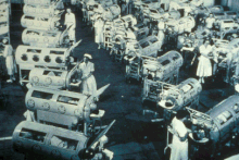
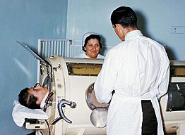
Hospital staff examine a patient in an Iron lung tank respirator during the polio epidemic. The machine creates a negative pressure around the thoracic cavity, thereby causing air to rush into the lungs to equalize intrapulmonary pressure.

The Greek physician Galen may have been the first to describe mechanical ventilation: "If you take a dead animal and blow air through its larynx [through a reed], you will fill its bronchi and watch its lungs attain the greatest distention." In the 1600s, Robert Hooke conducted experiments on dogs to demonstrate this concept. Vesalius too describes ventilation by inserting a reed or cane into the trachea of animals. These experiments predate the discovery of oxygen and its role in respiration. In 1908, George Poe demonstrated his mechanical respirator by asphyxiating dogs and seemingly bringing them back to life. These experiments all demonstrate positive pressure ventilation.

To achieve negative pressure ventilation, there must be a sub-atmospheric pressure to draw air into the lungs. This was first achieved in the late 19th century when John Dalziel and Alfred Jones independently developed tank ventilators, in which ventilation was achieved by placing a patient inside a box that enclosed the body in a box with sub-atmospheric pressures. This machine came to be known colloquially as the Iron lung, which went through many iterations of development. The use of the iron lung became widespread during the polio epidemic of the 1900s.

Early ventilators were control style with no support breaths integrated into them and were limited to an inspiration to expiration ration of 1:1. In the 1970s, intermittent mandatory ventilation was introduced as well as synchronized intermittent mandatory ventilation. These styles of ventilation had control breaths that patients could breath between.

Uses

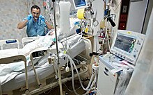
Respiratory therapist (RT) examining a mechanically ventilated patient in an intensive care unit. RTs participate in the optimization of ventilation management, adjustment, and weaning.

Mechanical ventilation is indicated when a patient's spontaneous breathing is inadequate to maintain life. It may be indicated in anticipation of imminent respiratory failure, acute respiratory failure, acute hypoxemia, or prophylactically. Because mechanical ventilation serves only to provide assistance for breathing and does not cure a disease, the patient's underlying condition should be identified and treated in order to liberate them from the ventilator.

Common specific medical indications for mechanical ventilation include:

Mechanical ventilation is typically used as a short-term measure. It may, however, be used at home or in a nursing or rehabilitation institution for patients that have chronic illnesses that require long-term ventilatory assistance.

Risks and complications

Mechanical ventilation is often a life-saving intervention, but carries potential complications. A common complication of positive pressure ventilation stemming directly from the ventilator settings include volutrauma and barotrauma. Others include pneumothorax, subcutaneous emphysema, pneumomediastinum, and pneumoperitoneum. Another well-documented complication is ventilator-associated lung injury which presents as acute respiratory distress syndrome. Other complications include diaphragm atrophy, decreased cardiac output, and oxygen toxicity. One of the primary complications that presents in patients mechanically ventilated is acute lung injury (ALI)/acute respiratory distress syndrome (ARDS). ALI/ARDS are recognized as significant contributors to patient morbidity and mortality.

In many healthcare systems, prolonged ventilation as part of intensive care is a limited resource. For this reason, decisions to commence and remove ventilation may raise ethical debate and often involve legal orders such as do-not-resuscitate orders.

Mechanical ventilation is often associated with many painful procedures and the ventilation itself can be uncomfortable. For infants who require opioids for pain, the potential side effects of opioids include problems with feeding, gastric and intestinal mobility problems, the potential for opioid dependence, and opioid tolerance.

Withdrawal from mechanical ventilation

Timing of withdrawal from mechanical ventilation—also known as weaning—is an important consideration. People who require mechanical ventilation should have their ventilation considered for withdrawal if they are able to support their own ventilation and oxygenation, and this should be assessed continuously. There are several objective parameters to look for when considering withdrawal, but there are no specific criteria that generalizes to all patients.

The Rapid Shallow Breathing Index (RSBI, the ratio of respiratory frequency to tidal volume (f/VT), previously referred to as the "Yang Tobin Index" or "Tobin Index" after Dr. Karl Yang and Prof. Martin J. Tobin of Loyola University Medical Center) is one of the best studied and most commonly used weaning predictors, with no other predictor having been shown to be superior. It was described in a prospective cohort study of mechanically ventilated patients which found that a RSBI > 105 breaths/min/L was associated with weaning failure, while a RSBI < 105 breaths/min/L.

Spontaneous breathing trials are conducted to assess the likelihood of a patient being able to maintain stability and breath on their own without the ventilator. This is done by changing the mode to one where they have to trigger breaths and ventilatory support is only given to compensate for the added resistance of the endotracheal tube.

A cuff leak test is done to detect if there is airway edema to show the chances of post-extubation stridor. This is done by deflating to the cuff to check if air begins leaking around the endotracheal tube.

Physiology

The function of the lungs is to provide gas exchange via oxygenation and ventilation. This phenomenon of respiration involves the physiologic concepts of air flow, tidal volume, compliance, resistance, and dead space. Other relevant concepts include alveolar ventilation, arterial PaCO2, alveolar volume, and FiO2. Alveolar ventilation is the amount of gas per unit of time that reaches the alveoli and becomes involved in gas exchange. PaCO2 is the partial pressure of carbon dioxide of arterial blood, which determines how well carbon dioxide is able to move out of the body. Alveolar volume is the volume of air entering and leaving the alveoli per minute. Mechanical dead space is another important parameter in ventilator design and function, and is defined as the volume of gas breathed again as the result of use in a mechanical device.

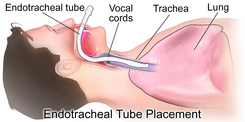
Image of endotracheal tube placement required to connect a patient's physiologic airway to the ventilator.

Due to the anatomy of the human pharynx, larynx, and esophagus and the circumstances for which ventilation is needed, additional measures are required to secure the airway during positive-pressure ventilation in order to allow unimpeded passage of air into the trachea and avoid air passing into the esophagus and stomach. The common method is by insertion of a tube into the trachea. Intubation, which provides a clear route for the air can be either an endotracheal tube, inserted through the natural openings of mouth or nose, or a tracheostomy inserted through an artificial opening in the neck. In other circumstances simple airway maneuvers, an oropharyngeal airway or laryngeal mask airway may be employed. If non-invasive ventilation or negative-pressure ventilation is used, then an airway adjunct is not needed.

Pain medicine such as opioids are sometimes used in adults and infants who require mechanical ventilation. For preterm or full term infants who require mechanical ventilation, there is no strong evidence to prescribe opioids or sedation routinely for these procedures, however, some select infants requiring mechanical ventilation may require pain medicine such as opioids. It is not clear if clonidine is safe or effective to be used as a sedative for preterm and full term infants who require mechanical ventilation.

When 100% oxygen (1.00 FiO
2
) is used initially for an adult, it is easy to calculate the next FiO
2
to be used, and easy to estimate the shunt fraction. The estimated shunt fraction refers to the amount of oxygen not being absorbed into the circulation. In normal physiology, gas exchange of oxygen and carbon dioxide occurs at the level of the alveoli in the lungs. The existence of a shunt refers to any process that hinders this gas exchange, leading to wasted oxygen inspired and the flow of un-oxygenated blood back to the left heart, which ultimately supplies the rest of the body with de-oxygenated blood. When using 100% oxygen, the degree of shunting is estimated as 700 mmHg - measured PaO
2
. For each difference of 100 mmHg, the shunt is 5%. A shunt of more than 25% should prompt a search for the cause of this hypoxemia, such as mainstem intubation or pneumothorax, and should be treated accordingly. If such complications are not present, other causes must be sought after, and positive end-expiratory pressure (PEEP) should be used to treat this intrapulmonary shunt. Other such causes of a shunt include:

Technique

Modes

Mechanical ventilation utilizes several separate systems for ventilation referred to as the mode. Modes come in many different delivery concepts but all conventional positive pressure ventilators modes fall into one of two categories; volume-cycled or pressure-cycled. A relatively new ventilation mode is flow-controlled ventilation (FCV). FCV is a fully dynamic mode without significant periods of 'no flow'. It is based on creating a stable gas flow into or out of the patient’s lungs to generate an inspiration or expiration, respectively. This results in linear increases and decreases in intratracheal pressure. In contrast to conventional modes of ventilation, there are no abrupt drop intrathoracic pressure drops, because of the controlled expiration. Further, this mode allows to use thin endotracheal tubes (~2 - 10 mm inner diameter) to ventilate a patient as expiration is actively supported. In general, the selection of which mode of mechanical ventilation to use for a given patient is based on the familiarity of clinicians with modes and the equipment availability at a particular institution.

Types of Ventilation

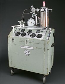
Carl Gunnar Engström invented in 1950 one of the first intermittent positive pressure ventilator, which delivers air straight into the lungs using an endotracheal tube placed into the windpipe.

Positive pressure

The design of the modern positive-pressure ventilators were based mainly on technical developments by the military during World War II to supply oxygen to fighter pilots in high altitude. Such ventilators replaced the iron lungs as safe endotracheal tubes with high-volume/low-pressure cuffs were developed. The popularity of positive-pressure ventilators rose during the polio epidemic in the 1950s in Scandinavia and the United States and was the beginning of modern ventilation therapy. Positive pressure through manual supply of 50% oxygen through a tracheostomy tube led to a reduced mortality rate among patients with polio and respiratory paralysis. However, because of the sheer amount of man-power required for such manual intervention, mechanical positive-pressure ventilators became increasingly popular.

Positive-pressure ventilators work by increasing the patient's airway pressure through an endotracheal or tracheostomy tube. The positive pressure allows air to flow into the airway until the ventilator breath is terminated. Then, the airway pressure drops to zero, and the elastic recoil of the chest wall and lungs push the tidal volume — the breath-out through passive exhalation.

Negative pressure

Negative pressure mechanical ventilators are produced in small, field-type and larger formats. The prominent design of the smaller devices is known as the cuirass, a shell-like unit used to create negative pressure only to the chest using a combination of a fitting shell and a soft bladder. In recent years this device has been manufactured using various-sized polycarbonate shells with multiple seals, and a high-pressure oscillation pump in order to carry out biphasic cuirass ventilation. Its main use has been in patients with neuromuscular disorders that have some residual muscular function. The latter, larger formats are in use, notably with the polio wing hospitals in England such as St Thomas' Hospital in London and the John Radcliffe in Oxford.

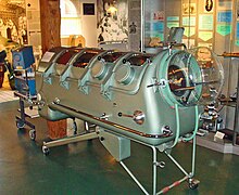
The larger units have their origin in the iron lung, also known as the Drinker and Shaw tank, which was developed in 1928 by J.H Emerson Company and was one of the first negative-pressure machines used for long-term ventilation. It was refined and used in the 20th century largely as a result of the polio epidemic that struck the world in the 1940s. The machine is, in effect, a large elongated tank, which encases the patient up to the neck. The neck is sealed with a rubber gasket so that the patient's face (and airway) are exposed to the room air. While the exchange of oxygen and carbon dioxide between the bloodstream and the pulmonary airspace works by diffusion and requires no external work, air must be moved into and out of the lungs to make it available to the gas exchange process. In spontaneous breathing, a negative pressure is created in the pleural cavity by the muscles of respiration, and the resulting gradient between the atmospheric pressure and the pressure inside the thorax generates a flow of air. In the iron lung by means of a pump, the air is withdrawn mechanically to produce a vacuum inside the tank, thus creating negative pressure. This negative pressure leads to expansion of the chest, which causes a decrease in intrapulmonary pressure, and increases flow of ambient air into the lungs. As the vacuum is released, the pressure inside the tank equalizes to that of the ambient pressure, and the elastic recoil of the chest and lungs leads to passive exhalation. However, when the vacuum is created, the abdomen also expands along with the lung, cutting off venous flow back to the heart, leading to pooling of venous blood in the lower extremities. The patients can talk and eat normally, and can see the world through a well-placed series of mirrors. Some could remain in these iron lungs for years at a time quite successfully.

Some of the problems with the full body design were such as being unable to control the inspiratory to expiratory ratio and the flow rate. This design also caused blood pooling in the legs.

Intermittent abdominal pressure ventilator

Another type is the intermittent abdominal pressure ventilator that applies pressure externally via an inflated bladder, forcing exhalation, sometimes termed exsufflation. The first such apparatus was the Bragg-Paul Pulsator. The name of one such device, the Pneumobelt made by Puritan Bennett has to a degree become a generic name for the type.

Oscillator

3100A Oscillator

The most commonly used high frequency ventilator and only one approved in the United States is the 3100A from Vyaire Medical. It works by using very small tidal volumes by setting amplitude and a high rate set in hertz. This type of ventilation is primarily used in neonates and pediatric patients who are failing conventional ventilation.

High Frequency Jet Ventilation

The first type of high frequency ventilator made for neonates and the only jet type is made by Bunnell Incorporated. It works in conjunction with a separate CMV ventilator to add pulses of air to the control breaths and PEEP.

Neonatal Jet ventilator

Monitoring

One of the main reasons why a patient is admitted to an ICU is for delivery of mechanical ventilation. Monitoring a patient in mechanical ventilation has many clinical applications: Enhance understanding of pathophysiology, aid with diagnosis, guide patient management, avoid complications, and assess trends.

In ventilated patients, pulse oximetry is commonly used when titrating FIO2. A reliable target of Spo2 is greater than 95%.

The total PEEP in the patient can be determined by doing an expiratory hold on the ventilator. If this is higher than the set PEEP, this indicates air trapping.

The plateau pressure can be found by doing an inspiratory hold. This shows the actual pressure the patient's lungs are experiencing.

Loops can be used to see what is occurring in the patient's lungs. These include flow-volume and pressure-volume loops. They can show changes in compliance and resistance.

Functional Residual Capacity can be determined when using the GE Carestation.

Modern ventilators have advanced monitoring tools. There are also monitors that work independently of the ventilator which allow for measuring patients after the ventilator has been removed, such as a Tracheal tube test.

Types of ventilators

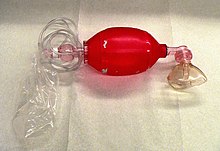
SMART BAG MO Bag-Valve-Mask Resuscitator

Ventilators come in many different styles and method of giving a breath to sustain life. There are manual ventilators such as bag valve masks and anesthesia bags that require the users to hold the ventilator to the face or to an artificial airway and maintain breaths with their hands. Mechanical ventilators are ventilators not requiring operator effort and are typically computer-controlled or pneumatic-controlled. Mechanical ventilators typically require power by a battery or a wall outlet (DC or AC) though some ventilators work on a pneumatic system not requiring power. There are a variety of technologies available for ventilation, falling into two main (and then lesser categories), the two being the older technology of negative-pressure mechanisms, and the more common positive-pressure types.

Common positive-pressure mechanical ventilators include:

  1. Transport ventilators—These ventilators are small and more rugged, and can be powered pneumatically or via AC or DC power sources.
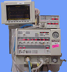
  2. Intensive-care ventilators—These ventilators are larger and usually run on AC power (though virtually all contain a battery to facilitate intra-facility transport and as a back-up in the event of a power failure). This style of ventilator often provides greater control of a wide variety of ventilation parameters (such as inspiratory rise time). Many ICU ventilators also incorporate graphics to provide visual feedback of each breath.
  3. Neonatal ventilators (bubble CPAP, HFJV, HFOV)—Designed with the preterm neonate in mind, these are a specialized subset of ICU ventilators that are designed to deliver smaller volumes and pressures to these patients. These may be conventional or high frequency types.
  4. Positive airway pressure ventilators (PAP) — These ventilators are specifically designed for non-invasive ventilation. This includes ventilators for use at home for treatment of chronic conditions such as sleep apnea or COPD and in the ICU setting.

Breath delivery mechanisms

Trigger

The trigger, either flow or pressure, is what causes a breath to be delivered by a mechanical ventilator. Breaths may be triggered by a patient taking their own breath, a ventilator operator pressing a manual breath button, or based on the set respiratory rate.

Cycle

The cycle is what causes the breath to transition from the inspiratory phase to the exhalation phase. Breaths may be cycled by a mechanical ventilator when a set time has been reached, or when a preset flow or percentage of the maximum flow delivered during a breath is reached depending on the breath type and the settings. Breaths can also be cycled when an alarm condition such as a high pressure limit has been reached.

Limit

Limit is how the breath is controlled. Breaths may be limited to a set maximum pressure or volume.

Breath exhalation

Exhalation in mechanical ventilation is almost always completely passive. The ventilator's expiratory valve is opened, and expiratory flow is allowed until the baseline pressure (PEEP) is reached. Expiratory flow is determined by patient factors such as compliance and resistance.

Artificial airways as a connection to the ventilator

There are various procedures and mechanical devices that provide protection against airway collapse, air leakage, and aspiration:

  • Face mask — In resuscitation and for minor procedures under anaesthesia, a face mask is often sufficient to achieve a seal against air leakage. Airway patency of the unconscious patient is maintained either by manipulation of the jaw or by the use of nasopharyngeal or oropharyngeal airway. These are designed to provide a passage of air to the pharynx through the nose or mouth, respectively. Poorly fitted masks often cause nasal bridge ulcers, a problem for some patients. Face masks are also used for non-invasive ventilation in conscious patients. A full-face mask does not, however, provide protection against aspiration. Non-invasive ventilation can be considered for epidemics of COVID-19 where sufficient invasive ventilation capacity is not available (or in some milder cases), but pressurized protection suits for caregivers are recommended due to the risks of poorly fitting masks emitting contaminating aerosols.
  • Tracheal intubation is often performed for mechanical ventilation of hours to weeks duration. A tube is inserted through the nose (nasotracheal intubation) or mouth (orotracheal intubation) and advanced into the trachea. In most cases, tubes with inflatable cuffs are used for protection against leakage and aspiration. Intubation with a cuffed tube is thought to provide the best protection against aspiration. Tracheal tubes inevitably cause pain and coughing. Therefore, unless a patient is unconscious or anaesthetized for other reasons, sedative drugs are usually given to provide tolerance of the tube. Other disadvantages of tracheal intubation include damage to the mucosal lining of the nasopharynx or oropharynx and subglottic stenosis.
  • Supraglottic airway — a supraglottic airway (SGA) is any airway device that is seated above and outside the trachea, as an alternative to endotracheal intubation. Most devices work via masks or cuffs that inflate to isolate the trachea for oxygen delivery. Newer devices feature esophageal ports for suctioning or ports for tube exchange to allow intubation. Supraglottic airways differ primarily from tracheal intubation in that they do not prevent aspiration. After the introduction of the laryngeal mask airway (LMA) in 1998, supraglottic airway devices have become mainstream in both elective and emergency anesthesia. There are many types of SGAs available including the esophageal-tracheal combitube (ETC), laryngeal tube (LT), and the obsolete esophageal obturator airway (EOA).
  • Cricothyrotomy — Patients requiring emergency airway management, in whom tracheal intubation has been unsuccessful, may require an airway inserted through a surgical opening in the cricothyroid membrane. This is similar to a tracheostomy but a cricothyrotomy is reserved for emergency access.
  • Tracheostomy — When patients require mechanical ventilation for several weeks, a tracheostomy may provide the most suitable access to the trachea. A tracheostomy is a surgically created passage into the trachea. Tracheostomy tubes are well tolerated and often do not necessitate any use of sedative drugs. Tracheostomy tubes may be inserted early during treatment in patients with pre-existing severe respiratory disease, or in any patient expected to be difficult to wean from mechanical ventilation, i.e., patients with little muscular reserve.
  • Mouthpiece — Less common interface, does not provide protection against aspiration. There are lipseal mouthpieces with flanges to help hold them in place if patient is unable.
Wednesday, February 22, 2023

Iron lung

From Wikipedia, the free encyclopedia
Iron lung
Iron lung CDC.jpg
An Emerson iron lung
ICD-9-CM93.99
MeSHD015919

An iron lung is a type of negative pressure ventilator (NPV), a mechanical respirator which encloses most of a person's body, and varies the air pressure in the enclosed space, to stimulate breathing. It assists breathing when muscle control is lost, or the work of breathing exceeds the person's ability. Need for this treatment may result from diseases including polio and botulism and certain poisons (for example, barbiturates, tubocurarine).

The use of iron lungs is largely obsolete in modern medicine, as more modern breathing therapies have been developed, and due to the eradication of polio in most of the world. However, in 2020, the COVID-19 pandemic revived some interest in the device as a cheap, readily-producible substitute for positive-pressure ventilators, which were feared to be outnumbered by patients potentially needing temporary artificially assisted respiration.

Design and function

Iron lung cylinder (black), patient head exposed through sealed opening. Diaphragm (yellow) mechanically extends/retracts, varying cylinder air pressure, causing patient chest to expand (inhale) (top) and contract (exhaling) (bottom)

The iron lung is typically a large horizontal cylinder, in which a person is laid, with their head protruding from a hole in the end of the cylinder, so that their full head (down to their voice box) is outside the cylinder, exposed to ambient air, and the rest of their body sealed inside the cylinder, where air pressure is continuously cycled up and down, to stimulate breathing.

To cause the patient to inhale, air is pumped out of the cylinder, causing a slight vacuum, which causes the patient's chest and abdomen to expand (drawing air from outside the cylinder, through the patient's exposed nose or mouth, into their lungs). Then, for the patient to exhale, the air inside the cylinder is compressed slightly (or allowed to equalize to ambient room pressure), causing the patient's chest and abdomen to partially collapse, forcing air out of the lungs, as the patient exhales the breath through their exposed mouth and nose, outside the cylinder.

Examples of the device include the Drinker respirator, the Emerson respirator, and the Both respirator. Iron lungs can be either manually or mechanically powered but normally are powered by an electric motor linked to a flexible pumping diaphragm (commonly opposite the end of the cylinder from the patient's head). Larger "room-sized" iron lungs were also developed, allowing for simultaneous ventilation of several patients (each with their heads protruding from sealed openings in the outer wall), with sufficient space inside for a nurse or a respiratory therapist to be inside the sealed room, attending the patients.

Smaller, single-patient versions of the iron lung include the so-called cuirass ventilator (named for the cuirass, a torso-covering body armor). The cuirass ventilator encloses only the patient's torso, or chest and abdomen, but otherwise operates essentially the same as the original, full-sized iron lung. A lightweight variation on the cuirass ventilator is the jacket ventilator or poncho or raincoat ventilator, which uses a flexible, impermeable material (such as plastic or rubber) stretched over a metal or plastic frame over the patient's torso.

Method and use

An Iron Lung ward, as mocked-up for a film, circa 1953

Humans, like most mammals, breathe by negative pressure breathing: the rib cage expands and the diaphragm contracts, expanding the chest cavity. This causes the pressure in the chest cavity to decrease, and the lungs expand to fill the space. This, in turn, causes the pressure of the air inside the lungs to decrease (it becomes negative, relative to the atmosphere), and air flows into the lungs from the atmosphere: inhalation. When the diaphragm relaxes, the reverse happens and the person exhales. If a person loses part or all of the ability to control the muscles involved, breathing becomes difficult or impossible.

Invention and early use

Initial development

Iron lung from the 1950s in the Gütersloh Town Museum. In Germany, fewer than a dozen of these breathing machines are available to the public.

In 1670, English scientist John Mayow came up with the idea of external negative pressure ventilation. Mayow built a model consisting of bellows and a bladder to pull in and expel air. The first negative pressure ventilator was described by British physician John Dalziel in 1832. Successful use of similar devices was described a few years later. Early prototypes included a hand-operated bellows-driven "Spirophore" designed by Dr Woillez of Paris (1876), and an airtight wooden box designed specifically for the treatment of polio by Dr Stueart of South Africa (1918). Stueart's box was sealed at the waist and shoulders with clay and powered by motor-driven bellows.

Drinker and Shaw tank

A Drinker iron lung displayed at the chapel of Netley Hospital, 2018

The first of these devices to be widely used however was developed in 1928 by Drinker and Shaw of the United States. The iron lung, often referred to in the early days as the "Drinker respirator", was invented by Philip Drinker (1894–1972) and Louis Agassiz Shaw Jr., professors of industrial hygiene at the Harvard School of Public Health. The machine was powered by an electric motor with air pumps from two vacuum cleaners. The air pumps changed the pressure inside a rectangular, airtight metal box, pulling air in and out of the lungs. The first clinical use of the Drinker respirator on a human was on October 12, 1928, at the Boston Children's Hospital in the US. The subject was an eight-year-old girl who was nearly dead as a result of respiratory failure due to polio. Her dramatic recovery, within less than a minute of being placed in the chamber, helped popularize the new device.

Variations

Boston manufacturer Warren E. Collins began production of the iron lung that year. Although it was initially developed for the treatment of victims of coal gas poisoning, it was most famously used in the mid-20th century for the treatment of respiratory failure caused by poliomyelitis.

Danish physiologist August Krogh, upon returning to Copenhagen in 1931 from a visit to New York where he saw the Drinker machine in use, constructed the first Danish respirator designed for clinical purposes. Krogh's device differed from Drinker's in that its motor was powered by water from the city pipelines. Krogh also made an infant respirator version.

In 1931, John Haven Emerson (1906–1997) introduced an improved and less expensive iron lung. The Emerson iron lung had a bed that could slide in and out of the cylinder as needed, and the tank had portal windows which allowed attendants to reach in and adjust limbs, sheets, or hot packs. Drinker and Harvard University sued Emerson, claiming he had infringed on patent rights. Emerson defended himself by making the case that such lifesaving devices should be freely available to all. Emerson also demonstrated that every aspect of Drinker's patents had been published or used by others at earlier times. Since an invention must be novel to be patentable, prior publication/use of the invention meant it was not novel and therefore unpatentable. Emerson won the case, and Drinker's patents were declared invalid.

The United Kingdom's first iron lung was designed in 1934 by Robert Henderson, an Aberdeen doctor. Henderson had seen a demonstration of the Drinker respirator in the early 1930s and built a device of his own upon his return to Scotland. Four weeks after its construction, the Henderson respirator was used to save the life of a 10-year-old boy from New Deer, Aberdeenshire, who had poliomyelitis. Despite this success, Henderson was reprimanded for secretly using hospital facilities to build the machine.

Both respirator

A Both cabinet respirator being used to treat a patient at the 110th Australian Military Hospital in 1943
 

The Both respirator, a negative pressure ventilator, was invented in 1937 when Australia's epidemic of poliomyelitis created an immediate need for more ventilating machines to compensate for respiratory paralysis. Although the Drinker model was effective and saved lives, its widespread use was hindered by the fact that the machines were very large, heavy (about 750 lbs or 340 kg), bulky, and expensive. In the US, an adult machine cost about $2000 in 1930, and £2000 delivered to Melbourne in 1936. The cost in Europe in the mid-1950s was around £1500. Consequently, there were few of the Drinker devices in Australia and Europe.

The South Australia Health Department asked Adelaide brothers Edward and Don Both to create an inexpensive "iron lung". Biomedical engineer Edward Both designed and developed a cabinet respirator made of plywood that worked similarly to the Drinker device, with the addition of a bi-valved design which allowed temporary access to the patient's body. Far cheaper to make (only £100) than the Drinker machine, the Both Respirator also weighed less and could be constructed and transported more quickly. Such was the demand for the machines that they were often used by patients within an hour of production.

Both-Nuffield iron lung display at the Thackray Medical Museum, Leeds. Pictures show assembly at the Morris motor works

Visiting London in 1938 during another polio epidemic, Both produced additional respirators there which attracted the attention of William Morris (Lord Nuffield), a British motor manufacturer and philanthropist. Nuffield, intrigued by the design, financed the production of approximately 1700 machines at his car factory in Cowley, and donated them to hospitals throughout all parts of Britain and the British Empire. Soon, the Both-Nuffield respirators were able to be produced by the thousand at about one-thirteenth the cost of the American design. By the early 1950s, there were over 700 Both-Nuffield iron lungs in the United Kingdom, but only 50 Drinker devices.

Polio epidemic

Staff in a Rhode Island hospital examine a patient in an iron lung tank respirator during a polio epidemic in Rhode Island in 1960. The iron lung encased the thoracic cavity in an air-tight chamber used to create negative pressure around the thoracic cavity, thereby causing air to enter the lungs to equalize intrapulmonary pressure.

Rows of iron lungs filled hospital wards at the height of the polio outbreaks of the 1940s and 1950s, helping children, and some adults, with bulbar polio and bulbospinal polio. A polio patient with a paralyzed diaphragm would typically spend two weeks inside an iron lung while recovering.

Modern development and usage

Polio vaccination programs have virtually eradicated new cases of poliomyelitis in the developed world. Because of this, and the development of modern ventilators, and widespread use of tracheal intubation and tracheotomy, the iron lung has mostly disappeared from modern medicine. In 1959, 1,200 people were using tank respirators in the United States, but by 2004 that number had decreased to just 39.

By 2014, only 10 people were left with an iron lung.

Replacement

Positive pressure ventilation systems are now more common than negative pressure systems. Positive pressure ventilators work by blowing air into the patient's lungs via intubation through the airway; they were used for the first time in Blegdams Hospital, Copenhagen, Denmark, during a polio outbreak in 1952. It proved a success and soon superseded the iron lung throughout Europe.

The iron lung now has a marginal place in modern respiratory therapy. Most patients with paralysis of the breathing muscles use modern mechanical ventilators that push air into the airway with positive pressure. These are generally efficacious and have the advantage of not restricting patients' movements or caregivers' ability to examine the patients as significantly as an iron lung does.

Continued use

Despite the advantages of positive ventilation systems, negative pressure ventilation is a truer approximation of normal physiological breathing and results in a more normal distribution of air in the lungs. It may also be preferable in certain rare conditions, such as central hypoventilation syndrome, in which failure of the medullary respiratory centers at the base of the brain results in patients having no autonomic control of breathing. At least one reported polio patient, Dianne Odell, had a spinal deformity that caused the use of mechanical ventilators to be contraindicated.

At least a few patients today still use the older machines, often in their homes, despite the occasional difficulty of finding replacement parts.

Joan Headley of Post-Polio Health International said that as of May 28, 2008, about 30 patients in the U.S. were still using an iron lung. That figure may be inaccurately low; Houston alone had 19 iron lung patients living at home in 2008.

Martha Mason of Lattimore, North Carolina died on May 4, 2009, after spending 60 of her 72 years in an iron lung.

On October 30, 2009, June Middleton of Melbourne, Australia, who had been entered in the Guinness Book of Records as the person who spent the longest time in an iron lung, died aged 83, having spent more than 60 years in her iron lung.

In 2013, the Post-Polio Health International (PHI) organizations estimated that only six to eight iron lung users were in the United States; as of 2017 its executive director knew of none. Press reports then emerged, however, of at least three (perhaps the last three) users of such devices, sparking interest amongst those in the makerspace community such as Naomi Wu in the manufacture of the obsolete components, particularly the gaskets. One is retired lawyer Paul Alexander, 77, of Dallas.

In 2021, the National Public Radio programs Radio Diaries and All Things Considered gave a report on Martha Lillard, one of the last two Americans depending on the daily use of an iron lung, which she had been using since 1953. In her audio interview, she reported that she was having problems obtaining replacement parts to keep her machine working properly.

COVID-19 pandemic

In early 2020, reacting to the COVID-19 pandemic, to address the urgent global shortage of modern ventilators (needed for patients with advanced, severe COVID-19), some enterprises developed prototypes of new, readily-producible versions of the iron lung. These developments included:

History of polio

From Wikipedia, the free encyclopedia
 
An Egyptian stele thought to represent a person with polio. 18th Dynasty (1403–1365 BC).

The history of polio (poliomyelitis) infections began during prehistory. Although major polio epidemics were unknown before the 20th century, the disease has caused paralysis and death for much of human history. Over millennia, polio survived quietly as an endemic pathogen until the 1900s when major epidemics began to occur in Europe. Soon after, widespread epidemics appeared in the rest of the world. By 1910, frequent epidemics became regular events throughout the developed world primarily in cities during the summer months. At its peak in the 1940s and 1950s, polio would paralyze or kill over half a million people worldwide every year.

The fear and the collective response to these epidemics would give rise to extraordinary public reaction and mobilization spurring the development of new methods to prevent and treat the disease and revolutionizing medical philanthropy. Although the development of two polio vaccines has eliminated wild poliomyelitis in all but two countries (Afghanistan and Pakistan), the legacy of poliomyelitis remains in the development of modern rehabilitation therapy and in the rise of disability rights movements worldwide.

Early history

Ancient Egyptian paintings and carvings depict otherwise healthy people with withered limbs, and children walking with canes at a young age. It is theorized that the Roman Emperor Claudius was stricken as a child, and this caused him to walk with a limp for the rest of his life. Perhaps the earliest recorded case of poliomyelitis is that of Sir Walter Scott. In 1773, Scott was said to have developed "a severe teething fever which deprived him of the power of his right leg". At the time, polio was not known to medicine. A retrospective diagnosis of polio is considered to be strong due to the detailed account Scott later made, and the resultant lameness of his right leg had an important effect on his life and writing.

The symptoms of poliomyelitis have been described by many names. In the early nineteenth century the disease was known variously as: Dental Paralysis, Infantile Spinal Paralysis, Essential Paralysis of Children, Regressive Paralysis, Myelitis of the Anterior Horns, Tephromyelitis (from the Greek tephros, meaning "ash-gray") and Paralysis of the Morning. In 1789 the first clinical description of poliomyelitis was provided by the British physician Michael Underwood—he refers to polio as "a debility of the lower extremities". The first medical report on poliomyelitis was by Jakob Heine, in 1840; he called the disease Lähmungszustände der unteren Extremitäten ("Paralysis of the lower Extremities"). Karl Oskar Medin was the first to empirically study a poliomyelitis epidemic in 1890. This work, and the prior classification by Heine, led to the disease being known as Heine-Medin disease.

Epidemics

Major polio epidemics were unknown before the 20th century; localized paralytic polio epidemics began to appear in Europe and the United States around 1900. The first report of multiple polio cases was published in 1843 and described an 1841 outbreak in Louisiana. A fifty-year gap occurs before the next U.S. report—a cluster of 26 cases in Boston in 1893. The first recognized U.S. polio epidemic occurred the following year in Vermont with 132 total cases (18 deaths), including several cases in adults. Numerous epidemics of varying magnitude began to appear throughout the country; by 1907 approximately 2,500 cases of poliomyelitis were reported in New York City.

This cardboard placard was placed in windows of residences where patients were quarantined due to poliomyelitis. Violating the quarantine order or removing the placard was punishable by a fine of up to US$100 in 1909 (equivalent to $3,016 in 2021).

Polio was a plague. One day you had a headache and an hour later you were paralyzed. How far the virus crept up your spine determined whether you could walk afterward or even breathe. Parents waited fearfully every summer to see if it would strike. One case turned up and then another. The count began to climb. The city closed the swimming pools and we all stayed home, cooped indoors, shunning other children. Summer seemed like winter then.

Richard Rhodes, A Hole in the World

On Saturday, June 17, 1916, an official announcement of the existence of an epidemic polio infection was made in Brooklyn, New York. That year, there were 27,363 cases and 7,130 deaths due to polio in the United States, with over 2,000 deaths in New York City alone. The names and addresses of individuals with confirmed polio cases were published daily in the press, their houses were identified with placards, and their families were quarantined. Dr. Hiram M. Hiller, Jr. was one of the physicians in several cities who realized what they were dealing with, but the nature of the disease remained largely a mystery. The 1916 epidemic caused widespread panic and thousands fled the city to nearby mountain resorts; movie theaters were closed, meetings were canceled, public gatherings were almost nonexistent, and children were warned not to drink from water fountains, and told to avoid amusement parks, swimming pools, and beaches. From 1916 onward, a polio epidemic appeared each summer in at least one part of the country, with the most serious occurring in the 1940s and 1950s. In the epidemic of 1949, 42,173 cases were reported in the United States and 2,720 deaths from the disease occurred. Canada and the United Kingdom were also affected.

Prior to the 20th century, polio infections were rarely seen in infants before 6 months of age, and most cases occurred in children 6 months to 4 years of age. Young children who contract polio generally develop only mild symptoms, but as a result they become permanently immune to the disease. In developed countries during the late 19th and early 20th centuries, improvements were being made in community sanitation, including improved sewage disposal and clean water supplies. Better hygiene meant that infants and young children had fewer opportunities to encounter and develop immunity to polio. Exposure to poliovirus was therefore delayed until late childhood or adult life, when it was more likely to take the paralytic form.

In children, paralysis due to polio occurs in one in 1,000 cases, while in adults, paralysis occurs in one in 75 cases. By 1950, the peak age incidence of paralytic poliomyelitis in the United States had shifted from infants to children aged 5 to 9 years; about one-third of the cases were reported in persons over 15 years of age. Accordingly, the rate of paralysis and death due to polio infection also increased during this time. In the United States, the 1952 polio epidemic was the worst outbreak in the nation's history, and is credited with heightening parents' fears of the disease and focusing public awareness on the need for a vaccine. Of the 57,628 cases reported that year, 3,145 died and 21,269 were left with mild to disabling paralysis.

Historical treatments

In the early 20th century—in the absence of proven treatments—a number of odd and potentially dangerous polio treatments were suggested. In John Haven Emerson's A Monograph on the Epidemic of Poliomyelitis (Infantile Paralysis) in New York City in 1916 one suggested remedy reads:

Give oxygen through the lower extremities, by positive electricity. Frequent baths using almond meal, or oxidising the water. Applications of poultices of Roman chamomile, slippery elm, arnica, mustard, cantharis, amygdalae dulcis oil, and of special merit, spikenard oil and Xanthoxolinum. Internally use caffeine, Fl. Kola, dry muriate of quinine, elixir of cinchone, radium water, chloride of gold, liquor calcis and wine of pepsin.

Following the 1916 epidemics and having experienced little success in treating polio patients, researchers set out to find new and better treatments for the disease. Between 1917 and the early 1950s, several therapies were explored in an effort to prevent deformities, including hydrotherapy and electrotherapy.

In 1939, Albert Sabin reported that "In the experiments reported in the present communication it was found that vitamin C, both natural and synthetic preparations, had no effect on the course of experimental poliomyelitis induced by nasal instillation of the virus."

Surgical treatments such as nerve grafting, tendon lengthening, tendon transfers, and limb lengthening and shortening were used extensively during this time. Patients with residual paralysis were treated with braces and taught to compensate for lost function with the help of calipers, crutches and wheelchairs. The use of devices such as rigid braces and body casts, which tended to cause muscle atrophy due to the limited movement of the user, were also touted as effective treatments. Massage and passive motion exercises were also used to treat patients with polio. Most of these treatments proved to be of little therapeutic value, however several effective supportive measures for the treatment of polio did emerge during these decades including the iron lung, an anti-polio antibody serum, and a treatment regimen developed by Sister Elizabeth Kenny.

Iron lung

This iron lung was donated to the CDC by the family of Barton Hebert of Covington, Louisiana, who had used the device from the late 1950s until his death in 2003.

The first iron lung used in the treatment of polio was invented by Philip Drinker, Louis Agassiz Shaw, and James Wilson at Harvard, and tested October 12, 1928, at Children's Hospital, Boston. The original Drinker iron lung was powered by an electric motor attached to two vacuum cleaners, and worked by changing the pressure inside the machine. When the pressure is lowered, the chest cavity expands, trying to fill this partial vacuum. When the pressure is raised the chest cavity contracts. This expansion and contraction mimics the physiology of normal breathing. The design of the iron lung was subsequently improved by using a bellows attached directly to the machine, and John Haven Emerson modified the design to make production less expensive. The Emerson Iron Lung was produced until 1970. Other respiratory aids were used, such as the Bragg-Paul Pulsator and the "rocking bed" for patients with less critical breathing difficulties.

During the polio epidemics, the iron lung saved many thousands of lives, but the machine was large, cumbersome and very expensive: in the 1930s, an iron lung cost about $1,500—about the same price as the average home. The cost of running the machine was also prohibitive, as patients were encased in the metal chambers for months, years and sometimes for life. Even with an iron lung, the fatality rate for patients with bulbar polio exceeded 90%.

These drawbacks led to the development of more modern positive-pressure ventilators and the use of positive-pressure ventilation by tracheostomy. Positive pressure ventilators reduced mortality in bulbar patients from 90% to 20%. In the Copenhagen epidemic of 1952, large numbers of patients were ventilated by hand ("bagged") by medical students and anyone else on hand because of the large number of bulbar polio patients and the small number of ventilators available.

Passive immunotherapy

In 1950 William Hammon at the University of Pittsburgh isolated serum, containing antibodies against poliovirus, from the blood of polio survivors. The serum, Hammon believed, would prevent the spread of polio and to reduce the severity of disease in polio patients. Between September 1951 and July 1952 nearly 55,000 children were involved in a clinical trial of the anti-polio serum. The results of the trial were promising; the serum was shown to be about 80% effective in preventing the development of paralytic poliomyelitis, and protection was shown to last for 5 weeks if given under tightly controlled circumstances. The serum was also shown to reduce the severity of the disease in patients who developed polio.

The large-scale use of antibody serum to prevent and treat polio had a number of drawbacks, however, including the observation that the immunity provided by the serum did not last long, and the protection offered by the antibody was incomplete, that re-injection was required during each epidemic outbreak, and that the optimal time frame for administration was unknown. The antibody serum was widely administered, but obtaining the serum was an expensive and time-consuming process, and the focus of the medical community soon shifted to the development of a polio vaccine.

Kenny regimen

Early management practices for paralyzed muscles emphasized the need to rest the affected muscles and suggested that the application of splints would prevent tightening of muscle, tendons, ligaments, or skin that would prevent normal movement. Many paralyzed polio patients lay in plaster body casts for months at a time. This prolonged casting often resulted in atrophy of both affected and unaffected muscles.

In 1940, Sister Elizabeth Kenny, an Australian bush nurse from Queensland, arrived in North America and challenged this approach to treatment. In treating polio cases in rural Australia between 1928 and 1940, Kenny had developed a form of physical therapy that—instead of immobilizing affected limbs—aimed to relieve pain and spasms in polio patients through the use of hot, moist packs to relieve muscle spasm and early activity and exercise to maximize the strength of unaffected muscle fibers and promote the neuroplastic recruitment of remaining nerve cells that had not been killed by the virus. Sister Kenny later settled in Minnesota where she established the Sister Kenny Rehabilitation Institute, beginning a world-wide crusade to advocate her system of treatment. Slowly, Kenny's ideas won acceptance, and by the mid-20th century had become the hallmark for the treatment of paralytic polio. In combination with antispasmodic medications to reduce muscular contractions, Kenny's therapy is still used in the treatment of paralytic poliomyelitis.

In 2009 as part of the Q150 celebrations, the Kenny regimen for polio treatment was announced as one of the Q150 Icons of Queensland for its role as an iconic "innovation and invention".

Vaccine development

People in Columbus, Georgia, awaiting polio vaccination during the early days of the National Polio Immunization Program.

In 1935 Maurice Brodie, a research assistant at New York University and William Hallock Park of the New York City Department of Health, attempted to produce a polio vaccine, procured from virus in ground up monkey spinal cords, and killed by formaldehyde. Brodie first tested the vaccine on himself and several of his assistants. He then gave the vaccine to three thousand children. Many developed allergic reactions, but none of the children developed an immunity to polio. During the late 1940s and early 1950s, a research group, headed by John Enders at the Boston Children's Hospital, successfully cultivated the poliovirus in human tissue. This significant breakthrough ultimately allowed for the development of the polio vaccines. Enders and his colleagues, Thomas H. Weller and Frederick C. Robbins, were recognized for their labors with the Nobel Prize in 1954.

Two vaccines are used throughout the world to combat polio. The first was developed by Jonas Salk, first tested in 1952 using the HeLa cell, and announced to the world by Salk on April 12, 1955. The Salk vaccine, or inactivated poliovirus vaccine (IPV), consists of an injected dose of killed poliovirus. In 1954, the vaccine was tested for its ability to prevent polio; its field trials grew to be the largest medical experiment in history. In 1955, it was chosen for use throughout the United States. By 1957, following mass immunizations promoted by the March of Dimes, the annual number of polio cases in the United States was reduced, from a peak of nearly 58,000 cases, to 5,600 cases.

Eight years after Salk's success, Albert Sabin developed an oral polio vaccine (OPV) using live but weakened (attenuated) virus. Human trials of Sabin's vaccine began in 1957 and it was licensed in 1962. Following the development of oral polio vaccine, a second wave of mass immunizations led to a further decline in the number of cases: by 1961, only 161 cases were recorded in the United States. The last cases of paralytic poliomyelitis caused by endemic transmission of poliovirus in the United States were in 1979, when an outbreak occurred among the Amish in several Midwestern states.

Legacy

Early in the twentieth century polio became one of the most feared diseases of the developed world. The disease hit without warning and required long quarantine periods during which parents were separated from children: it was impossible to tell who would get the disease and who would be spared. The consequences of the disease left polio survivors marked for life, leaving behind vivid images of wheelchairs, crutches, leg braces, breathing devices, and deformed limbs. However, polio changed not only the lives of those who survived it, but also affected profound cultural changes: the emergence of grassroots fund-raising campaigns that would revolutionize medical philanthropy, the rise of rehabilitation therapy and, through campaigns for the social and civil rights of disabled people, polio survivors helped to spur the modern disability rights movement.

In addition, the occurrence of polio epidemics led to a number of public health innovations. One of the most widespread was the proliferation of "no spitting" ordinances in the United States and elsewhere.

Philanthropy

In 1921 Franklin D. Roosevelt became totally and permanently paralyzed from the waist down. Although the paralysis (whether from poliomyelitis, as diagnosed at the time, or from Guillain–Barré syndrome) had no cure at the time, Roosevelt, who had planned a life in politics, refused to accept the limitations of his disease. He tried a wide range of therapies, including hydrotherapy in Warm Springs, Georgia (see below). In 1938 Roosevelt helped to found the National Foundation for Infantile Paralysis (now known as the March of Dimes), that raised money for the rehabilitation of people with paralytic polio, and was instrumental in funding the development of polio vaccines. The March of Dimes changed the way it approached fund-raising. Rather than soliciting large contributions from a few wealthy individuals, the March of Dimes sought small donations from millions of individuals. Its hugely successful fund-raising campaigns collected hundreds of millions of dollars—more than all of the U.S. charities at the time combined (with the exception of the Red Cross). By 1955 the March of Dimes had invested $25.5 million in research; funding both Jonas Salk's and Albert Sabin's vaccine development; the 1954–55 field trial of vaccine, and supplies of free vaccine for thousands of children.

In 1952, during the worst recorded epidemic, 3,145 people in the United States died from polio.

Rehabilitation therapy

A physical therapist assists two polio-stricken children while they exercise their lower limbs.

Prior to the polio scares of the twentieth century, most rehabilitation therapy was focused on treating injured soldiers returning from war. The disabling effects of polio led to heightened awareness and public support of physical rehabilitation, and in response a number of rehabilitation centers specifically aimed at treating polio patients were opened, with the task of restoring and building their remaining strength and teaching new, compensatory skills to large numbers of newly paralyzed individuals.

In 1926, Franklin Roosevelt, convinced of the benefits of hydrotherapy, bought a resort at Warm Springs, Georgia, where he founded the first modern rehabilitation center for treatment of polio patients which still operates as the Roosevelt Warm Springs Institute for Rehabilitation.

The cost of polio rehabilitation was often more than the average family could afford, and more than 80% of the nation's polio patients would receive funding through the March of Dimes. Some families also received support through philanthropic organizations such as the Ancient Arabic Order of the Nobles of the Mystic Shrine fraternity, which established a network of pediatric hospitals in 1919, the Shriners Hospitals for Children, to provide care free of charge for children with polio.

Disability rights movement

As thousands of polio survivors with varying degrees of paralysis left the rehabilitation hospitals and went home, to school and to work, many were frustrated by a lack of accessibility and discrimination they experienced in their communities. In the early twentieth century the use of a wheelchair at home or out in public was a daunting prospect as no public transportation system accommodated wheelchairs and most public buildings including schools, were inaccessible to those with disabilities. Many children left disabled by polio were forced to attend separate institutions for "crippled children" or had to be carried up and down stairs.

As people who had been paralyzed by polio matured, they began to demand the right to participate in the mainstream of society. Polio survivors were often in the forefront of the disability rights movement that emerged in the United States during the 1970s, and pushed legislation such as the Rehabilitation Act of 1973 which protected qualified individuals from discrimination based on their disability, and the Americans with Disabilities Act of 1990. Other political movements led by polio survivors include the Independent Living and Universal design movements of the 1960s and 1970s.

Polio survivors are one of the largest disabled groups in the world. The World Health Organization estimates that there are 10 to 20 million polio survivors worldwide. In 1977, the National Health Interview Survey reported that there were 254,000 people living in the United States who had been paralyzed by polio. According to local polio support groups and doctors, some 40,000 polio survivors with varying degrees of paralysis live in Germany, 30,000 in Japan, 24,000 in France, 16,000 in Australia, 12,000 in Canada and 12,000 in the United Kingdom.

Child abandonment

From Wikipedia, the free encyclopedia https://en.wikipedia.org/wiki/Child_abandonment ...