Search This Blog

Friday, March 20, 2015

Central nervous system


From Wikipedia, the free encyclopedia

Central nervous system
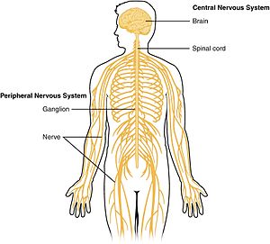
1201 Overview of Nervous System.jpg
Schematic diagram showing the central nervous system in pink, peripheral in yellow
Details
Latin Systema nervosum centrale
pars centralis systematis nervosi[1]
Identifiers
TA A14.1.00.001
FMA 55675
Anatomical terminology

The central nervous system (CNS) is the part of the nervous system consisting of the brain and spinal cord. The central nervous system is so named because it integrates information it receives from, and coordinates and influences the activity of, all parts of the bodies of bilaterally symmetric animals — that is, all multicellular animals except sponges and radially symmetric animals such as jellyfish, and it contains the majority of the nervous system. Arguably, many consider the retina[2] and the optic nerve (2nd cranial nerve),[3][4] as well as the olfactory nerves (1st) and olfactory epithelium[5] as parts of the CNS, synapsing directly on brain tissue without intermediate ganglia. Following this classification the olfactory epithelium is the only central nervous tissue in direct contact with the environment, which opens up for therapeutic treatments. [5] The CNS is contained within the dorsal cavity, with the brain in the cranial cavity and the spinal cord in the spinal cavity. In vertebrates, the brain is protected by the skull, while the spinal cord is protected by the vertebrae, both enclosed in the meninges.[6]

Structure

The central nervous system consists of the two major structures: the brain and spinal cord. The brain is encased in the skull, and protected by the cranium.[7] The spinal cord is continuous with the brain and lies caudaly to the brain,[8] and is protected by the vertebra.[7] The spinal cord reaches from the base of the skull, continues through[7] or starting below[9] the foramen magnum,[7] and terminates roughly level with the first or second lumbar vertebra,[8][9] occupying the upper sections of the vertebral canal.[4]

White and gray matter

Dissection of a brain with labels showing the clear division between white and gray matter.

Microscopically, there are differences between the neurons and tissue of the central nervous system and the peripheral nervous system.[citation needed] The central nervous system is divided in white and gray matter.[8] This can also be seen macroscopically on brain tissue. The white matter consists of axons and oligodendrocytes, while the gray matter chiefly consists of neurons. Both tissues include a number of glial cells (although the white matter contains more), which are often referred to as supporting cells of the central nervous system. Different forms of glial cells have different functions, some acting almost as scaffolding for neuroblasts to climb during neurogenesis such as bergmann glia, while others such as microglia are a specialized form of macrophage, involved in the immune system of the brain as well as the clearance of various metabolites from the brain tissue.[4] Astrocytes may be involved with both clearance of metabolites as well as transport of fuel and various beneficial substances to neurons from the capillaries of the brain. Upon CNS injury astrocytes will proliferate, causing gliosis, a form of neuronal scar tissue, lacking in functional neurons.[4]

The brain (cerebrum as well as midbrain and hindbrain) consists of a cortex, composed of neuron-bodies constituting gray matter, while internally there is more white matter that form tracts and commissures. Apart from cortical gray matter there is also subcortical gray making up a large number of different nuclei.[8]

Spinal cord

From and to the spinal cord are projections of the peripheral nervous system in the form of spinal nerves (sometimes segmental nerves[7]). The nerves connect the spinal cord with skin, joints, muscles etc. and allow for the transmission of efferent motor as well as afferent sensory signals and stimuli.[8] This allows for voluntary and involuntary motions of muscles, as well as the perception of senses. All in all 31 spinal nerves project from the brain stem,[8] some forming plexa as they branch out, such as the brachial plexa, sacral plexa etc.[7] Each spinal nerve will carry both sensory and motor signals, but the nerves synapse at different regions of the spinal cord, either from the periphery to sensory relay neurons that relay the information to the CNS or from the CNS to motor neurons, which relay the information out.[8]

Diagram of the columns and of the course of the fibers in the spinal cord. Sensory synapses occur in the dorsal spinal cord (above in this image), and motor nerves leave through the ventral (as well as lateral) horns of the spinal cord as seen below in the image.

Different ways in which the central nervous system can be activated without engaging the cortex, and making us aware of the actions. The above example shows the process in which the pupil dilates during dim light, activating neurons in the spinal cord. The second example shows the constriction of the pupil as a result of the activation of the Eddinger-Westphal nucleus (a cerebral ganglion).

The spinal cord relays information up to the brain through spinal tracts through the "final common pathway"[8] to the thalamus and ultimately to the cortex. Not all information is relayed to the cortex, and does not reach our immediate consciousness, but is instead transmitted only to the thalamus which sorts and adapts accordingly. This in turn may explain why we are not constantly aware of all aspects of our surroundings.[citation needed]

Cranial nerves

Apart from the spinal cord, there are also peripheral nerves of the PNS that synapse through intermediaries or ganglia directly on the CNS. These 12 nerves exist in the head and neck region and are called cranial nerves. Cranial nerves bring information to the CNS to and from the face, as well as to certain muscles (such as the trapezius muscle, which is innervated by accessory nerves[7] as well as certain cervical spinal nerves).[7]

Two pairs of cranial nerves; the olfactory nerves and the optic nerves[2] are often considered structures of the central nervous system. This is because they do not synapse first on peripheral ganglia, but directly on central nervous neurons. The olfactory epithelium is significant in that it consists of central nervous tissue expressed in direct contact to the environment, allowing for administration of certain pharmaceuticals and drugs. [5]
Image showing the way Schwann cells myelinate periferal nerves.
A neuron of the central nervous system, myelinated by an oligodendrocyte
Myelinated peripheral nerve at top, central nervous neuron at bottom

Brain

Rostrally to the spinal cord lies the brain.[8] The brain makes up the largest portion of the central nervous system, and is often the main structure referred to when speaking of the nervous system. The brain is the major functional unit of the central nervous system. While the spinal cord has certain processing ability such as that of spinal locomotion and can process reflexes, the brain is the major processing unit of the nervous system.[citation needed]

Brainstem

The brainstem consists of the medulla, the pons and the midbrain. The medulla can be referred to as an extension of the spinal cord, and its organization and functional properties are similar to those of the spinal cord.[8] The tracts passing from the spinal cord to the brain pass through here.[8]
Regulatory functions of the medulla nuclei include control of the blood pressure and breathing. Other nuclei are involved in balance, taste, hearing and control of muscles of the face and neck.[8]

The next structure rostral to the medulla is the pons, which lies on the ventral anterior side of the brainstem. Nuclei in the pons include pontine nuclei which work with the cerebellum and transmit information between the cerebellum and the cerebral cortex.[8] In the dorsal posterior pons lie nuclei that have to do with breathing, sleep and taste.[8]

The midbrain (or mesencephalon) is situated above and rostral to the pons, and includes nuclei linking distinct parts of the motor system, among others the cerebellum, the basal ganglia and both cerebral hemispheres. Additionally parts of the visual and auditory systems are located in the mid brain, including control of automatic eye movements.[8]

The brainstem at large provides entry and exit to the brain for a number of pathways for motor and autonomic control of the face and neck through cranial nerves,[8] and autonomic control of the organs is mediated by the tenth cranial (vagus) nerve.[4] A large portion of the brainstem is involved in such autonomic control of the body. Such functions may engage the heart, blood vessels, pupillae, among others.[8]

The brainstem also hold the reticular formation, a group of nuclei involved in both arousal and alertness.[8]

Cerebellum

The cerebellum lies posteriorly or dorsally to the pons. The cerebellum is composed of several dividing fissures and lobes. Its function includes posture and coordination of movements of eyes, limbs as well as that of the head. 
Further it is involved in motion that has been learned and perfected though practice, and will adapt to new learned movements.[8] Despite its previous classification as a motor structure, the cerebellum also displays connections to areas of the cerebral cortex involved in language as well as cognitive functions. These connections have been recently shown through anatomical studies, such as fMRI and PET.[8]The body of the cerebellum holds more neurons than any other structure of the brain including that of the larger cerebrum (or cerebral hemispheres), but is also more extensively understood than other structures of the brain, and includes fewer types of different neurons.[8] It handles and processes sensory stimuli, motor information as well as balance information from the vestibular organ.[8]

Diencephalon

The two structures of the diencephalon worth noting are the thalamus and the hypothalamus. The thalamus acts as a linkage between incoming pathways from the peripheral nervous system as well as the optical nerve (though it does not receive input from the olfactory nerve) to the cerebral hemispheres. Previously it was considered only a "relay station", but it is engaged in the sorting of information that will reach cerebral hemispheres (neocortex).[8]
Apart from its function of sorting information from the periphery, the thalamus also connects the cerebellum and basal ganglia with the cerebrum. In common with the aforementioned reticular system the thalamus is involved in wakefullness and consciousness, such as though the SCN.[8]

The hypothalamus engages in functions of a number of primitive emotions or feelings such as hunger, thirst and maternal bonding. This is regulated partly through control of secretion of hormones from the pituitary gland.
Additionally the hypothalamus plays a role in motivation and many other behaviors of the individual.[8]

Cerebrum

The cerebrum of cerebral hemispheres make up the largest portion of the human brain. Various structures combine forming the cerebral hemispheres, among others, the cortex, basal ganglia, amygdala and hippocampus. The hemispheres together control a large portion of the functions of the human brain such as emotion, memory, perception and motor functions. Apart from this the cerebral hemispheres stand for the cognitive capabilities of the brain.[8]
Connecting each of the hemispheres is the corpus callosum as well as several additional commissures.[8] One of the most important parts of the cerebral hemispheres is the cortex, made up of gray matter covering the surface of the brain. Functionally, the cerebral cortex is involved in planning and carrying out of everyday tasks.[8]

The hippocampus is involved in storage of memories, the amygdala plays a role in perception and communication of emotion, while the basal ganglia play a major role in the coordination of voluntary movement.[8]

Difference from the peripheral nervous system


A map over the different structures of the nervous systems in the body, showing the CNS, PNS, and ENS.

This differentiates the central nervous system from the peripheral nervous system, which consists of neurons, axons and Schwann cells. Oligodendrocytes and Schwann cells have similar functions in the central and peripheral nervous system respectively. Both act to add myelin sheaths to the axons, which acts as a form of insulation allowing for better and faster proliferation of electrical signals along the nerves. Axons in the central nervous system are often very short (barely a few millimeters) and do not need the same degree of isolation as peripheral nerves do. Some peripheral nerves can be over 1m in length, such as the nerves to the big toe. To ensure signals move at sufficient speed, myelination is needed.

The way in which the Schwann cells and oligodendrocytes myelinate nerves differ. A Schwann cell usually myelinates a single axon, completely surrounding it. Sometimes they may myelinate many axons, especially when in areas of short axons.[7] Oligodendrocytes usually myelinate several axons. They do this by sending out thin projections of their cell membrane which envelop and enclose the axon.

Development

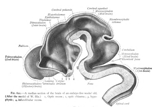
Central nervous system seen in a median section of a five week old embryo.
Central nervous system seen in a median section of a 3 month old embryo.
Top; CNS as seen in a median section of a 5 week old embryo.
Bottom; CNS seen in a median section of a 3 month old embryo.

During early development of the vertebrate embryo, a longitudinal groove on the neural plate gradually deepens and the ridges on either side of the groove (the neural folds) become elevated, and ultimately meet, transforming the groove into a closed tube, the ectodermal wall of which forms the rudiment of the nervous system. This tube initially differentiates into three vesicles (pockets): the prosencephalon at the front, the mesencephalon, and, between the mesencephalon and the spinal cord, the rhombencephalon. (By six weeks in the human embryo) the prosencephalon then divides further into the telencephalon and diencephalon; and the rhombencephalon divides into the metencephalon and myelencephalon.

As a vertebrate grows, these vesicles differentiate further still. The telencephalon differentiates into, among other things, the striatum, the hippocampus and the neocortex, and its cavity becomes the first and second ventricles. Diencephalon elaborations include the subthalamus, hypothalamus, thalamus and epithalamus, and its cavity forms the third ventricle. The tectum, pretectum, cerebral peduncle and other structures develop out of the mesencephalon, and its cavity grows into the mesencephalic duct (cerebral aqueduct). The metencephalon becomes, among other things, the pons and the cerebellum, the myelencephalon forms the medulla oblongata, and their cavities develop into the fourth ventricle.
Central
nervous
system
Brain Prosencephalon Telencephalon Rhinencephalon, Amygdala, Hippocampus, Neocortex, Basal ganglia, Lateral ventricles
Diencephalon Epithalamus, Thalamus, Hypothalamus, Subthalamus, Pituitary gland, Pineal gland, Third ventricle
Brain stem Mesencephalon Tectum, Cerebral peduncle, Pretectum, Mesencephalic duct
Rhombencephalon Metencephalon Pons, Cerebellum
Myelencephalon Medulla oblongata
Spinal cord

Evolution

Lancelets or amphioxus are regarded as similar to the archetypal vertebrate form, and possess to true brain.
A neuron of the central nervous system, myelinated by an oligodendrocyte
Traditional spindle diagram of the evolution of the vertebrates at class level.
Top: the lancelet, regarded an archetypal vertebrate, lacking a true brain. Middle: an early vertebrate. Bottom: spindle diagram of the evolution of vertebrates.

Planarians, members of the phylum Platyhelminthes (flatworms), have the simplest, clearly defined delineation of a nervous system into a central nervous system (CNS) and a peripheral nervous system (PNS).[10][11] Their primitive brains, consisting of two fused anterior ganglia, and longitudinal nerve cords form the CNS; the laterally projecting nerves form the PNS. A molecular study found that more than 95% of the 116 genes involved in the nervous system of planarians, which includes genes related to the CNS, also exist in humans.[12] Like planarians, vertebrates have a distinct CNS and PNS, though more complex than those of planarians.

The CNS of chordates differs from that of other animals in being placed dorsally in the body, above the gut and notochord/spine.[13] The basic pattern of the CNS is highly conserved throughout the different species of vertebrates and during evolution. The major trend that can be observed is towards a progressive telencephalisation: the telencephalon of reptiles is only an appendix to the large olfactory bulb, while in mammals it makes up most of the volume of the CNS. In the human brain, the telencephalon covers most of the diencephalon and the mesencephalon. Indeed, the allometric study of brain size among different species shows a striking continuity from rats to whales, and allows us to complete the knowledge about the evolution of the CNS obtained through cranial endocasts.

Mammals – which appear in the fossil record after the first fishes, amphibians, and reptiles – are the only vertebrates to possess the evolutionarily recent, outermost part of the cerebral cortex known as the neocortex.[14] The neocortex of monotremes (the duck-billed platypus and several species of spiny anteaters) and of marsupials (such as kangaroos, koalas, opossums, wombats, and Tasmanian devils) lack the convolutions – gyri and sulci – found in the neocortex of most placental mammals (eutherians).[15] Within placental mammals, the size and complexity of the neocortex increased over time. The area of the neocortex of mice is only about 1/100 that of monkeys, and that of monkeys is only about 1/10 that of humans.[14] In addition, rats lack convolutions in their neocortex (possibly also because rats are small mammals), whereas cats have a moderate degree of convolutions, and humans have quite extensive convolutions.[14] Extreme convolution of the neocortex is found in dolphins, possibly related to their complex echolocation.

Clinical significance

Diseases

There are many central nervous system diseases and conditions, including infections of the central nervous system such as encephalitis and poliomyelitis, early-onset neurological disorders including ADHD and autism, late-onset neurodegenerative diseases such as Alzheimer's disease, Parkinson's disease, and essential tremor, autoimmune and inflammatory diseases such as multiple sclerosis and acute disseminated encephalomyelitis, genetic disorders such as Krabbe's disease and Huntington's disease, as well as amyotrophic lateral sclerosis and adrenoleukodystrophy. Lastly, cancers of the central nervous system can cause severe illness and, when malignant, can have very high mortality rates.
Specialty professional organizations recommend that neurological imaging of the brain be done only to answer a specific clinical question and not as routine screening.[16]

Fighting pollution, together: California teams up with Quebec on cap-and-trade


Fighting pollution, together: California teams up with Quebec on cap-and-trade


18 Mar 2015 at 10:20 ET                   
Businessman looking at scene of polluted city (Shutterstock) California raised $630 million by selling pollution allowances one Wednesday last month, each of them entitling the buyer to harm the climate with a ton of carbon dioxide. Most of the money that was raised during the quarterly allowance auction will be used to reduce customers’ electricity bills; the rest will go to environmental projects. That same Wednesday, the Canadian province of Quebec raised $150 million doing the exact same thing.
The timing was no coincidence. The auction was held jointly. It was the result of the historic linking of two cap-and-trade programs operating in different nations, thousands of miles apart. A refinery in Richmond, Calif., can now buy its pollution allowances from Quebec. In lieu of buying an allowance needed to release a ton of carbon dioxide, a power plant operating on the banks of Quebec’s Saint Lawrence River could buy an offset from a Central Valley dairy farm that kept an equivalent amount of methane out of the atmosphere.
Experts say fresh linkages between the two cap-and-trade programs — one of them among the biggest, the other on the small side — may serve as a template for the gradual emergence of what could eventually become a global carbon market. That market could be formed by linking together the dozens of smaller markets being established worldwide, from Kazakhstan to Tokyo to Mexico.
Such a global market could help humanity ratchet down its annual levels of climate pollution, by aggressively dialing back the overall pollution cap over time, and pushing up the price of fossil fuels as the costs of renewables tumble. That would help nations deliver on ambitious pledges that could be made during upcoming U.N. climate talks, including in Paris in December.
“Linkage is going to be a very important element of implementing the 2015 Paris agreement,” Harvard University economics professor Robert Stavins, an expert on carbon markets and international climate talks, said. “California is going to be an important model.”
The full results of the February joint auction were published Tuesday, showing 84 million allowances were sold for a little over $12 apiece, with about 20 million of those sold by utilities. It was the second auction to be held jointly by the two cap-and-trade programs, and the first to be held without any mid-auction software-related technical glitches. Both cap-and-trade programs began operating in 2013, and they were linked last year, following years of planning.
“There was a wall between our two programs,” Michael Gibbs, assistant executive officer at the California Air Resources Board, said. “After linking became effective, the wall came down.”
California has a bigger economy than Quebec, and its cap on greenhouse gas pollution is far larger. That’s why it sold four times more allowances during the February auction than did Quebec, and why it raised four times the revenue.
“These are instruments that are issued by the jurisdictions, and the total equals the emissions cap,” Gibbs said. “The number of allowances issued over time is declining, which is how you ensure that the emissions decline.”
Washington state and Ontario are among the North American states and provinces considering forming their own cap-and-trade programs. These, along with similar programs on other continents, could eventually be linked with California’s and Quebec’s.
According to classical economic theory, costs of climate action fall as the size of a carbon market increases, which is what happens when two cap-and-trade systems are linked. That helps explain why analysts have concluded it would be cheaper for American states to work together to comply with upcoming power plant pollution rules, such as by creating and linking carbon markets, than if each of the states tried to comply with the new rules alone.
Originally published at Climate Central

Guess what? More people are living in peace now. Just look at the numbers


John Gray is not just wrong but flat-earth wrong. Conflict dominates the headlines, yet the most destructive form of war has effectively ceased to exist
US and Soviet soldiers celebrating their victory over the Nazis.


US and Soviet soldiers celebrating their victory over the Nazis. ‘Even the recent uptick from the wars in Iraq and Syria has not brought the world anywhere near the death rates of the preceding decades.' Photograph: Misha Japaridze/AP
Has the world seen moral progress? The answer should not depend on whether one has a sunny or a morose temperament. Everyone agrees that life is better than death, health better than sickness, prosperity better than privation, freedom better than tyranny, peace better than war. All of these can be measured, and the results plotted over time. If they go up, that’s progress.
For John Gray, this is a big problem. As a part of his campaign against reason, science and Enlightenment humanism, he insists that the strivings of humanity over the centuries have left us no better off. This dyspepsia was hard enough to sustain when Gray first expressed it in the teeth of obvious counterexamples such as the abolition of human sacrifice, chattel slavery and public torture-executions. But as scholars have increasingly measured human flourishing, they have found that Gray is not just wrong but howlingly, flat-earth, couldn’t-be-more-wrong wrong. The numbers show that after millennia of near-universal poverty and despotism, a steadily growing proportion of humankind is surviving infancy and childbirth, going to school, voting in democracies, living free of disease, enjoying the necessities of modern life and surviving to old age.

And more people are living in peace. In the 1980s several military scholars noticed to their astonishment that the most destructive form of armed conflict – wars among great powers and developed states – had effectively ceased to exist. At the time this “long peace” could have been dismissed as a random lull, but it has held firm for an additional three decades.

Then came another pleasant surprise. Starting in the 1990s, political scientists such as Joshua Goldstein, who kept track of ongoing wars of all kinds, including civil wars and wars among smaller and poorer countries, noticed that the list kept getting shorter. Research institutes in Oslo and Uppsala compiled datasets of global battle deaths since 1946, and their plots showed an unmistakable downward trend. The per-capita death rate fell more than tenfold between the peak of the second world war and the Korean war, and then plunged an additional hundredfold by the mid-2000s. Even the recent uptick from the wars in Iraq and Syria has not brought the world anywhere near the death rates of the preceding decades. Other datasets show steep declines in genocides and other mass killings. The declines are precipitous enough that they don’t depend on precise body counts: the estimates could be off by 25%, 100%, or 250% and the decline would still be there.

In a recent Guardian article, Gray tries to shoo away these pesky facts, which he disingenuously calls a “new orthodoxy”. Far from being orthodox, the discoveries are typically greeted with incredulity and sometimes furious denial, because most people fall prey to a cognitive illusion and assess the world from headlines rather than data. As long as violence has not vanished altogether, there will always be enough explosions and gunfire to fill the news, while the vastly greater portion of the planet in which people live boringly peaceful lives is reporter-free and invisible. Only by systematically tallying wars and war deaths and plotting them over time can one reach a defensible conclusion about global trends.

Oblivious to this logical point, Gray indiscriminately enumerates every violent episode of the past century he can think of, including recent ones that killed a handful of people or none at all. But his laundry list shows only that rates of violence have not fallen to zero, not that they have remained unchanged. And it certainly doesn’t support the preposterously melodramatic claim that the advanced societies of western Europe – the safest places in the history of our species – are “terrains of violent conflict” in which “peace and war [are] fatally blurred”.

Equally innumerate is his observation that while the cold war superpowers never met on the battlefield, they supported proxies in civil wars. Civil wars are far less destructive than wars between great powers, and even civil wars went into decline with the end of the cold war a quarter-century ago. The numbers matter: the difference between a war with 8,500,000 battle deaths (like the first world war) and a war with 5,000 deaths (like eastern Ukraine) is 8,495,000 human beings who get to work, play, and love rather than rot in their graves.

Gray tries to wave off the battle death numbers by repeating the legend that during the 20th century the ratio of military to civilian deaths flipped from 9:1 to 1:9. In my book The Better Angels of Our Nature I noted that this meme originated in a counting error and has been debunked many times. Throughout history wars have displaced, and frequently have targeted, civilians. No one knows how the ratio has changed, but when battle deaths decline a thousandfold it hardly matters. A great-power war kills massive numbers of soldiers and civilians, a small war kills vastly fewer of each, and in the many parts of the world that see no wars at all, the number of civilian war deaths must be zero. More attention to the maths would also have disabused Gray of the gambler’s fallacy, which leads him to believe that major war is cyclical and due for a return. The data shows that wars are patterned at random, though with a probability that can change over time.

In a last diversionary tactic, Gray serves up a prolix disquisition on Aztec obsidian mirrors, which is somehow meant to show that quantification is like “sorcery”, “auguries”, “amulets”, and “prayer wheels”. But the inescapable fact is that whenever you use the words “more”, “less”, “rise”, or “fall” you are making a claim about numbers. If you then refuse to look at them, no one should take your claims seriously.

Lymphatic system


From Wikipedia, the free encyclopedia

Lymphatic System
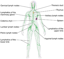
Blausen 0623 LymphaticSystem Female.png
Human lymphatic system
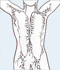
Lymphatic system.png
Details
Latin systema lymphoideum
Identifiers
TA A13.0.00.000
FMA 74594
Anatomical terminology

The lymphatic system is part of the circulatory system, comprising a network of lymphatic vessels that carry a clear fluid called lymph (from Latin lympha meaning water[1]) directionally towards the heart. The lymphatic system was first described in the seventeenth century independently by Olaus Rudbeck and Thomas Bartholin. Unlike the cardiovascular system the lymphatic system is not a closed system. The human circulatory system processes an average of 20 litres of blood per day through capillary filtration which removes plasma while leaving the blood cells. Roughly 17 litres of the filtered plasma get reabsorbed directly into the blood vessels, while the remaining 3 litres are left behind in the interstitial fluid. One of the main functions of the lymph system is to provide an accessory return route to the blood for the surplus 3 litres.[2]

The other main function is that of defense in the immune system. Lymph is very similar to blood plasma but contains lymphocytes and other white blood cells. It also contains waste products and debris of cells together with bacteria and protein. Associated organs composed of lymphoid tissue are the sites of lymphocyte production. Lymphocytes are concentrated in the lymph nodes. The spleen and the thymus are also lymphoid organs of the immune system. The tonsils are lymphoid organs that are also associated with the digestive system. Lymphoid tissues contain lymphocytes, and also contain other types of cells for support.[3] The system also includes all the structures dedicated to the circulation and production of lymphocytes (the primary cellular component of lymph), which also includes the bone marrow, and the lymphoid tissue associated with the digestive system.[4]

The blood does not come into direct contact with the parenchymal cells and tissues in the body, but constituents of the blood first exit the microvascular exchange blood vessels to become interstitial fluid, which comes into contact with the parenchymal cells of the body. Lymph is the fluid that is formed when interstitial fluid enters the initial lymphatic vessels of the lymphatic system. The lymph is then moved along the lymphatic vessel network by either intrinsic contractions of the lymphatic passages or by extrinsic compression of the lymphatic vessels via external tissue forces (e.g., the contractions of skeletal muscles), or by lymph hearts in some animals. The organization of lymph nodes and drainage follows the organization of the body into external and internal regions; therefore, the lymphatic drainage of the head, limbs, and body cavity walls follows an external route, and the lymphatic drainage of the thorax, abdomen, and pelvic cavities follows an internal route.[5] Eventually, the lymph vessels empty into the lymphatic ducts, which drain into one of the two subclavian veins, near their junction with the internal jugular veins.

Structure


Lymphatic system

The lymphatic system consists of lymphatic organs, a conducting network of lymphatic vessels, and the circulating lymph.

The tertiary lymphoid tissue typically contains far fewer lymphocytes, and assumes an immune role only when challenged with antigens that result in inflammation. It achieves this by importing the lymphocytes from blood and lymph.[6])

The thymus and the bone marrow constitute the primary lymphoid organs involved in the production and early clonal selection of lymphocyte tissues. Bone marrow is responsible for both the creation of T cells and the production and maturation of B cells. From the bone marrow, B cells immediately join the blood system and travel to secondary lymphoid organs in search of pathogens. T cells, on the other hand, travel from the bone marrow to the thymus, where they are allowed to develop further. Mature T cells join B cells in search of pathogens. The other 95% of T cells begin a process of apoptosis (programmed cell death).

The central or primary lymphoid organs generate lymphocytes from immature progenitor cells.

Secondary or peripheral lymphoid organs, which include lymph nodes and the spleen, maintain mature naive lymphocytes and initiate an adaptive immune response. The peripheral lymphoid organs are the sites of lymphocyte activation by antigens. Activation leads to clonal expansion and affinity maturation. Mature lymphocytes recirculate between the blood and the peripheral lymphoid organs until they encounter their specific antigen.

Secondary lymphoid tissue provides the environment for the foreign or altered native molecules (antigens) to interact with the lymphocytes. It is exemplified by the lymph nodes, and the lymphoid follicles in tonsils, Peyer's patches, spleen, adenoids, skin, etc. that are associated with the mucosa-associated lymphoid tissue (MALT).

In the gastrointestinal wall the appendix has mucosa resembling that of the colon, but here it is heavily infiltrated with lymphocytes.

Thymus


The thymus is a primary lymphoid organ and is the site where T cells, the lymphocytes of the adaptive immune system, mature.

Spleen

The spleen synthesizes antibodies in its white pulp and removes antibody-coated bacteria and antibody-coated blood cells by way of blood and lymph node circulation. A study published in 2009 using mice found that the spleen contains, in its reserve, half of the body's monocytes within the red pulp.[7] These monocytes, upon moving to injured tissue (such as the heart), turn into dendritic cells and macrophages while promoting tissue healing.[7][8][9] The spleen is a center of activity of the mononuclear phagocyte system and can be considered analogous to a large lymph node, as its absence causes a predisposition to certain infections.
Like the thymus, the spleen has only efferent lymphatic vessels. Both the short gastric arteries and the splenic artery supply it with blood.[10]

The germinal centers are supplied by arterioles called penicilliary radicles.[11]

Up to the fifth month of prenatal development the spleen creates red blood cells. After birth the bone marrow is solely responsible for hematopoiesis. As a major lymphoid organ and a central player in the reticuloendothelial system, the spleen retains the ability to produce lymphocytes. The spleen stores red blood cells and lymphocytes. It can store enough blood cells to help in an emergency. Up to 25% of lymphocytes can be stored at any one time.[12]

Lymph nodes

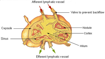
A lymph node showing afferent and efferent lymphatic vessels

A lymph node is an organized collection of lymphoid tissue, through which the lymph passes on its way back to the blood. Lymph nodes are located at intervals along the lymphatic system. Several afferent lymph vessels bring in lymph, which percolates through the substance of the lymph node, and is then drained out by an efferent lymph vessel. There are between five and six hundred lymph nodes in the human body, many of which are grouped in clusters in different regions as in the underarm and abdominal areas.

The substance of a lymph node consists of lymphoid follicles in an outer portion called the "cortex." The inner portion of the node is called the "medulla," which is surrounded by the cortex on all sides except for a portion known as the "hilum." The hilum presents as a depression on the surface of the lymph node, which makes the otherwise spherical lymph node, bean-shaped or ovoid. The efferent lymph vessel directly emerges from the lymph node here. The arteries and veins supplying the lymph node with blood enter and exit through the hilum.

There is a region of the lymph node called the paracortex that immediately surrounds the medulla. Unlike the cortex, which has mostly immature T cells, or thymocytes, the paracortex has a mixture of immature and mature T cells. Lymphocytes enter the lymph nodes through specialised high endothelial venules found in the paracortex.

A lymph follicle is a dense collection of lymphocytes, the number, size and configuration of which change in accordance with the functional state of the lymph node. For example, the follicles expand significantly when encountering a foreign antigen. The selection of B cells also called B lymphocytes, occurs in the germinal center of the lymph nodes.

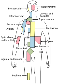
Lymph nodes are particularly numerous in the mediastinum in the chest, neck, pelvis, axilla (armpit), inguinal (groin) region, and in association with the blood vessels of the intestines.[4]

Other lymphoid tissue


Regional lymph nodes

Lymphoid tissue associated with the lymphatic system is concerned with immune functions in defending the body against infections and the spread of tumors. It consists of connective tissue formed of reticular fibers, with various types of leukocytes, (white blood cells), mostly lymphocytes enmeshed in it, through which the lymph passes.[13]
Regions of the lymphoid tissue that are densely packed with lymphocytes are known as lymphoid follicles. Lymphoid tissue can either be structurally well organized as lymph nodes or may consist of loosely organized lymphoid follicles known as the mucosa-associated lymphoid tissue.

Lymphatics

Lymph capillaries in the tissue spaces

The lymphatic vessels also sometimes called lymph vessels, conduct lymph between different parts of the body. They include the tubular vessels of the lymph capillaries, the collecting lymphatic vessels, the right lymphatic duct and the thoracic duct, also called the left lymphatic duct. The lymph vessels transport lymph back to the blood ultimately replacing the volume lost from the blood during the formation of the interstitial fluid. These vessels are also called the lymphatic channels or simply lymphatics.[14]

The lymphatics are responsible for maintaining the balance of the body fluids. Its network of capillaries and collecting lymphatic vessels, work to efficiently drain and transport extravasated fluid back to the cardiovascular system. Fluid, along with proteins and antigens are captured and returned to the blood circulation. There are numerous intraluminal valves in place to ensure a unidirectional flow without reflux.[15] Two valve systems are used to achieve this one directional flow—a primary and a secondary valve system.[16] The capillaries are blind ended and the valves at the ends of capillaries use specialised junctions together with anchoring filaments to allow a unidirectional flow to the primary vessels. The collecting lymphatics are, however, seen to act to propel the lymph through to the blood circulation by the combined actions of the intraluminal valves and lymphatic muscle cells.[17]

Development

Lymphatic tissues begin to develop by the end of the fifth week of embryonic development. Lymphatic vessels develop from lymph sacs that arise from developing veins, which are derived from mesoderm.

The first lymph sacs to appear are the paired jugular lymph sacs at the junction of the internal jugular and subclavian veins. From the jugular lymph sacs, lymphatic capillary plexuses spread to the thorax, upper limbs, neck and head. Some of the plexuses enlarge and form lymphatic vessels in their respective regions. Each jugular lymph sac retains at least one connection with its jugular vein, the left one developing into the superior portion of the thoracic duct.

The next lymph sac to appear is the unpaired retroperitoneal lymph sac at the root of the mesentery of the intestine. It develops from the primitive vena cava and mesonephric veins. Capillary plexuses and lymphatic vessels spread from the retroperitoneal lymph sac to the abdominal viscera and diaphragm. The sac establishes connections with the cisterna chyli but loses its connections with neighboring veins.

The last of the lymph sacs, the paired posterior lymph sacs, develop from the iliac veins. The posterior lymph sacs produce capillary plexuses and lymphatic vessels of the abdominal wall, pelvic region, and lower limbs. The posterior lymph sacs join the cisterna chyli and lose their connections with adjacent veins.

With the exception of the anterior part of the sac from which the cisterna chyli develops, all lymph sacs become invaded by mesenchymal cells and are converted into groups of lymph nodes.

The spleen develops from mesenchymal cells between layers of the dorsal mesentery of the stomach. The thymus arises as an outgrowth of the third pharyngeal pouch.

Function

The lymphatic system has multiple interrelated functions:[18]

Function of the fatty acid transport system

Lymph vessels called lacteals are present in the lining of the gastrointestinal tract, predominantly in the small intestine. While most other nutrients absorbed by the small intestine are passed on to the portal venous system to drain via the portal vein into the liver for processing, fats (lipids) are passed on to the lymphatic system to be transported to the blood circulation via the thoracic duct. (There are exceptions, for example medium-chain triglycerides (MCTs) are fatty acid esters of glycerol that passively diffuse from the GI tract to the portal system.) The enriched lymph originating in the lymphatics of the small intestine is called chyle. The nutrients that are released to the circulatory system are processed by the liver, having passed through the systemic circulation.

Clinical significance

The study of lymphatic drainage of various organs is important in diagnosis, prognosis, and treatment of cancer. The lymphatic system, because of its physical proximity to many tissues of the body, is responsible for carrying cancerous cells between the various parts of the body in a process called metastasis. The intervening lymph nodes can trap the cancer cells. If they are not successful in destroying the cancer cells the nodes may become sites of secondary tumors.

Lymphadenopathy

Lymphadenopathy refers to one or more enlarged lymph nodes. Small groups or individually enlarged lymph nodes are generally reactive in response to infection or inflammation. This is called local lymphadenopathy. When many lymph nodes in different areas of the body are involved, this is called generalised lymphadenopathy. Generalised lymphadenopathy may be caused by infections such as infectious mononucleosis, tuberculosis and HIV, connective tissue diseases such as SLE and rheumatoid arthritis, and cancers, including both cancers of tissue within lymph nodes, discussed below, and metastasis of cancerous cells from other parts of the body, that have arrived via the lymphatic system.[19]

Lymphedema

Lymphedema is the swelling caused by the accumulation of lymph, which may occur if the lymphatic system is damaged or has malformations. It usually affects limbs, though the face, neck and abdomen may also be affected. In an extreme state, called elephantiasis the edema progresses to the extent that the skin becomes thick with an appearance similar to the skin on elephant limbs.[20]
Causes are unknown in most cases, but sometimes there is a previous history of severe infection, usually caused by a parasitic disease, such as lymphatic filariasis.

Lymphangiomatosis is a disease involving multiple cysts or lesions formed from lymphatic vessels.

Lymphedema can also occur after surgical removal of cancerous lymph nodes in the armpit (causing the arm to swell due to poor lymphatic drainage) or groin (causing swelling of the leg). Treatment is by massage, and is not permanent.

Cancer


Cancer of the lymphatic system can be primary or secondary. Lymphoma refers to cancer that arises from lymphatic tissue. Lymphoid leukemias and lymphomas are now considered to be tumors of the same type of cell lineage. They are called "leukemia" when in the blood or marrow and "lymphoma" when in lymphatic tissue. They are grouped together under the name "lymphoid malignancy".[21]

Lymphoma is generally considered as Hodgkin's lymphoma and non-Hodgkin lymphoma. Hodgkin's lymphoma is characterised by a particular type of cell, called a Reed–Sternberg cell, visible under microscope. It is associated with past infection with the Epstein-Barr Virus, and generally causes a painless "rubbery" lymphadenopathy. It is staged, using Ann Arbor staging. Chemotherapy generally involves the ABVD and may also involved radiotherapy.[22] Non-Hodgkin's lymphoma is a cancer characterised by increased proliferation of B-cells or T-cells, generally occurs in an older age group than Hodgkin's lymphoma, It is treated according to whether it is high-grade or low-grade, and carries a poorer prognosis than Hodgkin's lymphoma.[22]

Lymphangiosarcoma is a malignant soft tissue tumor, whereas lymphangioma is a benign tumor occurring frequently in association with Turner syndrome. Lymphangioleiomyomatosis is a benign tumor of the smooth muscles of the lymphatics that occurs in the lungs.

History

Hippocrates, in 5th century BC, was one of the first people to mention the lymphatic system. In his work On Joints, he briefly mentioned the lymph nodes in one sentence. Rufus of Ephesus, a Roman physician, identified the axillary, inguinal and mesenteric lymph nodes as well as the thymus during the 1st to 2nd century AD.[23] The first mention of lymphatic vessels was in 3rd century BC by Herophilos, a Greek anatomist living in Alexandria, who incorrectly concluded that the "absorptive veins of the lymphatics," by which he meant the lacteals (lymph vessels of the intestines), drained into the hepatic portal veins, and thus into the liver.[23] The findings of Ruphus and Herophilos were further propagated by the Greek physician Galen, who described the lacteals and mesenteric lymph nodes which he observed in his dissection of apes and pigs in the 2nd century AD.[23][24]

In the mid 16th century, Gabriele Falloppio (discoverer of the fallopian tubes), described what are now known as the lacteals as "coursing over the intestines full of yellow matter."[23] In about 1563 Bartolomeo Eustachi, a professor of anatomy, described the thoracic duct in horses as vena alba thoracis.[23] The next breakthrough came when in 1622 a physician, Gaspare Aselli, identified lymphatic vessels of the intestines in dogs and termed them venae alba et lacteae, which is now known as simply the lacteals. The lacteals were termed the fourth kind of vessels (the other three being the artery, vein and nerve, which was then believed to be a type of vessel), and disproved Galen's assertion that chyle was carried by the veins. But, he still believed that the lacteals carried the chyle to the liver (as taught by Galen).[25] He also identified the thoracic duct but failed to notice its connection with the lacteals.[23] This connection was established by Jean Pecquet in 1651, who found a white fluid mixing with blood in a dog's heart. He suspected that fluid to be chyle as its flow increased when abdominal pressure was applied. He traced this fluid to the thoracic duct, which he then followed to a chyle-filled sac he called the chyli receptaculum, which is now known as the cisternae chyli; further investigations led him to find that lacteals' contents enter the venous system via the thoracic duct.[23][25] Thus, it was proven convincingly that the lacteals did not terminate in the liver, thus disproving Galen's second idea: that the chyle flowed to the liver.[25] Johann Veslingius drew the earliest sketches of the lacteals in humans in 1647.[24]

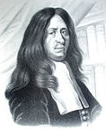
The idea that blood recirculates through the body rather than being produced anew by the liver and the heart was first accepted as a result of works of William Harvey—a work he published in 1628. In 1652, Olaus Rudbeck (1630–1702), a Swede, discovered certain transparent vessels in the liver that contained clear fluid (and not white), and thus named them hepatico-aqueous vessels. He also learned that they emptied into the thoracic duct, and that they had valves.[25] He announced his findings in the court of Queen Christina of Sweden, but did not publish his findings for a year,[26] and in the interim similar findings were published by Thomas Bartholin, who additionally published that such vessels are present everywhere in the body, not just in the liver. He is also the one to have named them "lymphatic vessels."[25] This had resulted in a bitter dispute between one of Bartholin's pupils, Martin Bogdan,[27] and Rudbeck, whom he accused of plagiarism.[26]

Galen's ideas prevailed in medicine until the 17th century. It was believed that blood was produced by the liver from chyle contaminated with ailments by the intestine and stomach, to which various spirits were added by other organs, and that this blood was consumed by all the organs of the body. This theory required that the blood be consumed and produced many times over. Even in the 17th century, his ideas were defended by some physicians.[24]
Alexander Monro, of the University of Edinburgh Medical School, was the first to describe the function of the
lymphatic system in detail.[28]

Etymology

The adjective used for the lymph-transporting system is "lymphatic." The adjective used for the tissues where lymphocytes are formed is "lymphoid."

Lymphatic comes from the Latin word lymphaticus, meaning "connected to water."

Cryogenics

From Wikipedia, the free encyclopedia https://en.wikipedia.org/wiki/Cryogenics...