Search This Blog

Sunday, May 6, 2018

Epigenetic clock

From Wikipedia, the free encyclopedia

An epigenetic clock is a type of a molecular age estimation method based on DNA methylation levels. Pre-eminent examples for epigenetic clocks are Horvath's clock[1][2][3][4], which applies to all human tissues/cells, and Hannum's clock [5], which applies to blood.

History

The strong effects of age on DNA methylation levels have been known since the late 1960s.[6] A vast literature describes sets of CpGs whose DNA methylation levels correlate with age, e.g.[7][8][9][10][11]. The first robust demonstration that DNA methylation levels in saliva could generate accurate age predictors was published by a UCLA team including Steve Horvath in 2011 (Bocklandt et al 2011) [12]. The labs of Trey Ideker and Kang Zhang at the University of California San Diego published the Hannum epigenetic clock (Hannum 2013) [5], which consisted of 71 markers that accurately estimate age based on blood methylation levels. The first multi-tissue epigenetic clock, Horvath's epigenetic clock, was developed by Steve Horvath, a professor of human genetics and of biostatistics at UCLA (Horvath 2013)[1][3]. Horvath spent over 4 years collecting publicly available Illumina DNA methylation data and identifying suitable statistical methods.[13] The personal story behind the discovery was featured in Nature.[14] The age estimator was developed using 8,000 samples from 82 Illumina DNA methylation array datasets, encompassing 51 healthy tissues and cell types. The major innovation of Horvath's epigenetic clock lies in its wide applicability: the same set of 353 CpGs and the same prediction algorithm is used irrespective of the DNA source within the organism, i.e. it does not require any adjustments or offsets.[1] This property allows one to compare the ages of different areas of the human body using the same aging clock.

Relationship to a cause of biological aging

It is not yet known what exactly is measured by DNA methylation age. Horvath hypothesized that DNA methylation age measures the cumulative effect of an epigenetic maintenance system but details are unknown. The fact that DNA methylation age of blood predicts all-cause mortality in later life [15][16][17][18] strongly suggests that it relates to a process that causes aging.[19] However, it is unlikely that the 353 clock CpGs are special or play a direct causal role in the aging process.[1] Rather, the epigenetic clock captures an emergent property of the epigenome.

Motivation for biological clocks

In general, biological aging clocks and biomarkers of aging are expected to find many uses in biological research since age is a fundamental characteristic of most organisms. Accurate measures of biological age (biological aging clocks) could be useful for
Overall, biological clocks are expected to be useful for studying what causes aging and what can be done against it.

Properties of Horvath's clock

The clock is defined as an age estimation method based on 353 epigenetic markers on the DNA. The 353 markers measure DNA methylation of CpG dinucleotides. Estimated age ("predicted age" in mathematical usage), also referred to as DNA methylation age, has the following properties: first, it is close to zero for embryonic and induced pluripotent stem cells; second, it correlates with cell passage number; third, it gives rise to a highly heritable measure of age acceleration; and, fourth, it is applicable to chimpanzee tissues (which are used as human analogs for biological testing purposes). Organismal growth (and concomitant cell division) leads to a high ticking rate of the epigenetic clock that slows down to a constant ticking rate (linear dependence) after adulthood (age 20).[1] The fact that DNA methylation age of blood predicts all-cause mortality in later life even after adjusting for known risk factors [15][16] suggests that it relates to a process that causes aging. Similarly, markers of physical and mental fitness are associated with the epigenetic clock (lower abilities associated with age acceleration).[20]

Salient features of Horvath's epigenetic clock include its high accuracy and its applicability to a broad spectrum of tissues and cell types. Since it allows one to contrast the ages of different tissues from the same subject, it can be used to identify tissues that show evidence of accelerated age due to disease.

Statistical approach

The basic approach is to form a weighted average of the 353 clock CpGs, which is then transformed to DNAm age using a calibration function. The calibration function reveals that the epigenetic clock has a high ticking rate until adulthood, after which it slows to a constant ticking rate. Using the training data sets, Horvath used a penalized regression model (Elastic net regularization) to regress a calibrated version of chronological age on 21,369 CpG probes that were present both on the Illumina 450K and 27K platform and had fewer than 10 missing values. DNAm age is defined as estimated ("predicted") age. The elastic net predictor automatically selected 353 CpGs. 193 of the 353 CpGs correlate positively with age while the remaining 160 CpGs correlate negatively with age. R software and a freely available web-based tool can be found at the following webpage.[21]

Accuracy

The median error of estimated age is 3.6 years across a wide spectrum of tissues and cell types .[1] The epigenetic clock performs well in heterogeneous tissues (for example, whole blood, peripheral blood mononuclear cells, cerebellar samples, occipital cortex, buccal epithelium, colon, adipose, kidney, liver, lung, saliva, uterine cervix, epidermis, muscle) as well as in individual cell types such as CD4 T cells, CD14 monocytes, glial cells, neurons, immortalized B cells, mesenchymal stromal cells.[1] However, accuracy depends to some extent on the source of the DNA.

Comparison with other biological clocks

The epigenetic clock leads to a chronological age prediction that has a Pearson correlation coefficient of r=0.96 with chronological age (Figure 2 in [1]). Thus the age correlation is close to its maximum possible correlation value of 1. Other biological clocks are based on a) telomere length, b) p16INK4a expression levels (also known as INK4a/ARF locus),[22] and c) microsatellite mutations.[23] The correlation between chronological age and telomere length is r=−0.51 in women and r=−0.55 in men.[24] The correlation between chronological age and expression levels of p16INK4a in T cells is r=0.56.[25] p16INK4a expression levels only relate to age in T cells, a type of white blood cells.[citation needed] The microsatellite clock measures not chronological age but age in terms of elapsed cell divisions within a tissue.[citation needed]

Applications of Horvath's clock

By contrasting DNA methylation age (estimated age) with chronological age, one can define measures of age acceleration. Age acceleration can be defined as the difference between DNA methylation age and chronological age. Alternatively, it can be defined as the residual that results from regressing DNAm age on chronological age. The latter measure is attractive because it does not correlate with chronological age. A positive/negative value of epigenetic age acceleration suggests that the underlying tissue ages faster/slower than expected.

Genetic studies of epigenetic age acceleration

The broad sense heritability (defined via Falconer's formula) of age acceleration of blood from older subjects is around 40% but it appears to be much higher in newborns.[1] Similarly, the age acceleration of brain tissue (prefrontal cortex) was found to be 41% in older subjects.[26] Genome-wide association studies of cerebellar age acceleration have identified several SNPs at a genomewide significance level.[27][28] Gene and SNP sets found by genome-wide association analysis of epigenetic age acceleration exhibit significant overlap with those of Alzheimer’s disease, age-related macular degeneration, and Parkinson’s disease.[27][28]

Female breast tissue is older than expected

DNAm age is higher than chronological age in female breast tissue that is adjacent to breast cancer tissue.[1] Since normal tissue which is adjacent to other cancer types does not exhibit a similar age acceleration effect, this finding suggests that normal female breast tissue ages faster than other parts of the body.[1] Similarly, normal breast tissue samples from women without cancer have been found to be substantially older than blood samples collected from the same women at the same time PMID 28364215.

Cancer tissue

Cancer tissues show both positive and negative age acceleration effects. For most tumor types, no significant relationship can be observed between age acceleration and tumor morphology (grade/stage).[1][2] On average, cancer tissues with mutated TP53 have a lower age acceleration than those without it.[1] Further, cancer tissues with high age acceleration tend to have fewer somatic mutations than those with low age acceleration.[1][2] Age acceleration is highly related to various genomic aberrations in cancer tissues. Somatic mutations in estrogen receptors or progesterone receptors are associated with accelerated DNAm age in breast cancer.[1] Colorectal cancer samples with a BRAF (V600E) mutation or promoter hypermethylation of the mismatch repair gene MLH1 are associated with an increased age acceleration.[1] Age acceleration in glioblastoma multiforme samples is highly significantly associated with certain mutations in H3F3A.[1] One study suggests that the epigenetic age of blood tissue may be prognostic of lung cancer incidence.[29]

Obesity and metabolic syndrome

The epigenetic clock was used to study the relationship between high body mass index (BMI) and the DNA methylation ages of human blood, liver, muscle and adipose tissue.[30] A significant correlation (r=0.42) between BMI and epigenetic age acceleration could be observed for the liver. A much larger sample size (n=4200 blood samples) revealed a weak but statistically significant correlation (r=0.09) between BMI and intrinsic age acceleration of blood PMID 28198702. The same large study found that various biomarkers of metabolic syndrome (glucose-, insulin-, triglyceride levels, C-reactive protein, waist-to-hip ratio) were associated with epigenetic age acceleration in blood PMID 28198702. Conversely, high levels of the good cholesterol HDL were associated with a lower epigenetic aging rate of blood PMID 28198702.

Trisomy 21 (Down syndrome)

Down Syndrome (DS) entails an increased risk of many chronic diseases that are typically associated with older age. The clinical manifestations of accelerated aging suggest that trisomy 21 increases the biological age of tissues, but molecular evidence for this hypothesis has been sparse. According to the epigenetic clock, trisomy 21 significantly increases the age of blood and brain tissue (on average by 6.6 years).[31]

Alzheimer's disease related neuropathology

Epigenetic age acceleration of the human prefrontal cortex was found to be correlated with several neuropathological measurements that play a role in Alzheimer's disease [26] Further, it was found to be associated with a decline in global cognitive functioning, and memory functioning among individuals with Alzheimer's disease.[26] The epigenetic age of blood relates to cognitive functioning in the elderly.[20] Overall, these results strongly suggest that the epigenetic clock lends itself for measuring the biological age of the brain.

Cerebellum ages slowly

It has been difficult to identify tissues that seem to evade aging due to the lack of biomarkers of tissue age that allow one to contrast compare the ages of different tissues. An application of epigenetic clock to 30 anatomic sites from six centenarians and younger subjects revealed that the cerebellum ages slowly: it is about 15 years younger than expected in a centenarian.[32] This finding might explain why the cerebellum exhibits fewer neuropathological hallmarks of age related dementias compared to other brain regions. In younger subjects (e.g. younger than 70), brain regions and brain cells appear to have roughly the same age.[1][32] Several SNPs and genes have been identified that relate to the epigenetic age of the cerebellum [27]

Huntington's disease

Huntington's disease has been found to increase the epigenetic aging rates of several human brain regions.[33]

Centenarians age slowly

The offspring of semi-supercentenarians (subjects who reached an age of 105–109 years) have a lower epigenetic age than age-matched controls (age difference=5.1 years in blood) and centenarians are younger (8.6 years) than expected based on their chronological age.[18]

HIV infection

Infection with the Human Immunodeficiency Virus-1 (HIV) is associated with clinical symptoms of accelerated aging, as evidenced by increased incidence and diversity of age-related illnesses at relatively young ages. But it has been difficult to detect an accelerated aging effect on a molecular level. An epigenetic clock analysis of human DNA from HIV+ subjects and controls detected a significant age acceleration effect in brain (7.4 years) and blood (5.2 years) tissue due to HIV-1 infection.[34] These results are consistent with an independent study that also found an age advancement of 5 years in blood of HIV patients and a strong effect of the HLA locus.[35]

Parkinson's disease

A large-scale study suggests that the blood of Parkinson's disease subjects exhibits (relatively weak) accelerated aging effects.[36]

Developmental disorder: syndrome X

Children with a very rare disorder known as syndrome X maintain the façade of persistent toddler-like features while aging from birth to adulthood. Since the physical development of these children is dramatically delayed, these children appear to be a toddler or at best a preschooler. According to an epigenetic clock analysis, blood tissue from syndrome X cases is not younger than expected.[37]

Menopause accelerates epigenetic aging

The following results strongly suggest that the loss of female hormones resulting from menopause accelerates the epigenetic aging rate of blood and possibly that of other tissues.[38] First, early menopause has been found to be associated with an increased epigenetic age acceleration of blood.[38] Second, surgical menopause (due to bilateral oophorectomy) is associated with epigenetic age acceleration in blood and saliva. Third, menopausal hormone therapy, which mitigates hormonal loss, is associated with a negative age acceleration of buccal cells (but not of blood cells).[38] Fourth, genetic markers that are associated with early menopause are also associated with increased epigenetic age acceleration in blood.[38]

Cellular senescence versus epigenetic aging

A confounding aspect of biological aging is the nature and role of senescent cells. It is unclear whether the three major types of cellular senescence, namely replicative senescence, oncogene-induced senescence and DNA damage-induced senescence are descriptions of the same phenomenon instigated by different sources, or if each of these is distinct, and how they are associated with epigenetic aging. Induction of replicative senescence (RS) and oncogene-induced senescence (OIS) were found to be accompanied by epigenetic aging of primary cells but senescence induced by DNA damage was not, even though RS and OIS activate the cellular DNA damage response pathway.[39] These results highlight the independence of cellular senescence from epigenetic aging. Consistent with this, telomerase-immortalised cells continued to age (according to the epigenetic clock) without having been treated with any senescence inducers or DNA-damaging agents, re-affirming the independence of the process of epigenetic ageing from telomeres, cellular senescence, and the DNA damage response pathway. Although the uncoupling of senescence from cellular aging appears at first sight to be inconsistent with the fact that senescent cells contribute to the physical manifestation of organism ageing, as demonstrated by Baker et al., where removal of senescent cells slowed down aging.[40] However, the epigenetic clock analysis of senescence suggests that cellular senescence is a state that cells are forced into as a result of external pressures such as DNA damage, ectopic oncogene expression and exhaustive proliferation of cells to replenish those eliminated by external/environmental factors.[39] These senescent cells, in sufficient numbers, will probably cause the deterioration of tissues, which is interpreted as organism ageing. However, at the cellular level, aging, as measured by the epigenetic clock, is distinct from senescence. It is an intrinsic mechanism that exists from the birth of the cell and continues. This implies that if cells are not shunted into senescence by the external pressures described above, they would still continue to age. This is consistent with the fact that mice with naturally long telomeres still age and eventually die even though their telomere lengths are far longer than the critical limit, and they age prematurely when their telomeres are forcibly shortened, due to replicative senescence. Therefore, cellular senescence is a route by which cells exit prematurely from the natural course of cellular aging.[39]

Effect of sex and race/ethnicity

Men age faster than women according to epigenetic age acceleration in blood, brain, saliva, and many other tissues. [41] The epignetic clock method applies to all examined racial/ethnic groups in the sense that DNAm age is highly correlated with chronological age. But ethnicity can be associated with epigenetic age acceleration.[41] For example, the blood of Hispanics and the Tsimané ages more slowly than that of other populations which might explain the Hispanic mortality paradox.[41]

Rejuvenation effect due to stem cell transplantation in blood

Hematopoietic stem cell transplantation, which transplants these cells from a young donor to an older recipient, rejuvenates the epigenetic age of blood to that of the donor PMID 28550187. However, graft-versus-host disease is associated with increased DNA methyhlation age PMID 28550187.

Progeria

Adult progeria also known as Werner syndrome is associated with epigenetic age acceleration in blood.[42]

Biological mechanism behind the epigenetic clock

Despite the fact that biomarkers of ageing based on DNA methylation data have enabled accurate age estimates for any tissue across the entire life course, the precise biological mechanism behind the epigenetic clock is currently unknown[43]. However, epigenetic biomarkers may help to address long-standing questions in many fields, including the central question: why do we age? The following explanations have been proposed in the literature.

Possible explanation 1: Epigenomic maintenance system

Horvath hypothesized that his clock arises from a methylation footprint left by an epigenomic maintenance system.[1]

Possible explanation 2: Unrepaired DNA damages

Endogenous DNA damages occur frequently including about 50 double-strand DNA breaks per cell cycle[44] and about 10,000 oxidative damages per day (see DNA damage (naturally occurring)). During repair of double-strand breaks many epigenetic alterations are introduced, and in a percentage of cases epigenetic alterations remain after repair is completed, including increased methylation of CpG island promoters.[45][46][47] Similar, but usually transient epigenetic alterations were recently found during repair of oxidative damages caused by H2O2, and it was suggested that occasionally these epigenetic alterations may also remain after repair.[48] These accumulated epigenetic alterations may contribute to the epigenetic clock. Accumulation of epigenetic alterations may parallel the accumulation of un-repaired DNA damages that are proposed to cause aging (see DNA damage theory of aging).

Other age estimators based on DNA methylation levels

Several other age estimators have been described in the literature.

1) Weidner et al. (2014) describe an age estimator for DNA from blood that uses only three CpG sites of genes hardly affected by aging (cg25809905 in integrin, alpha 2b (ITGA2B); cg02228185 in aspartoacylase (ASPA) and cg17861230 in phosphodiesterase 4C, cAMP specific (PDE4C)).[49] The age estimator by Weidener et al. (2014) applies only to blood. Even in blood this sparse estimator is far less accurate than Horvath's epigenetic clock (Horvath 2014) when applied to data generated by the Illumina 27K or 450K platforms. [50] But the sparse estimator was developed for pyrosequencing data and is highly cost effective. [51]

2) Hannum et al. (2013) [5] report several age estimators: one for each tissue type. Each of these estimators requires covariate information (e.g. gender, body mass index, batch). The authors mention that each tissue led to a clear linear offset (intercept and slope). Therefore, the authors had to adjust the blood-based age estimator for each tissue type using a linear model. When the Hannum estimator is applied to other tissues, it leads to a high error (due to poor calibration) as can be seen from Figure 4A in Hannum et al. (2013). Hannum et al. adjusted their blood-based age estimator (by adjusting the slope and the intercept term) in order to apply it to other tissue types. Since this adjustment step removes differences between tissue, the blood-based estimator from Hannum et al. cannot be used to compare the ages of different tissues/organs. In contrast, a salient characteristic of the epigenetic clock is that one does not have to carry out such a calibration step:[1] it always uses the same CpGs and the same coefficient values. Therefore, Horvath's epigenetic clock can be used to compare the ages of different tissues/cells/organs from the same individual. While the age estimators from Hannum et al. cannot be used to compare the ages of different normal tissues, they can be used to compare the age of a cancerous tissue with that of a corresponding normal (non-cancerous) tissue. Hannum et al. reported pronounced age acceleration effects in all cancers. In contrast, Horvath's epigenetic clock [2][52] reveals that some cancer types (e.g. triple negative breast cancers or uterine corpus endometrial carcinoma) exhibit negative age acceleration, i.e. cancer tissue can be much younger than expected. An important difference relates to additional covariates. Hannum's age estimators make use of covariates such as gender, body mass index, diabetes status, ethnicity, and batch. Since new data involve different batches, one cannot apply it directly to new data. However, the authors present coefficient values for their CpGs in Supplementary Tables which can be used to define an aggregate measure that tends to be strongly correlated with chronological age but may be poorly calibrated (i.e. lead to high errors).

Comparison of the 3 age predictors described in A) Horvath (2013),[1] B) Hannum (2013),[53] and C) Weidener (2014),[54] respectively. The x-axis depicts the chronological age in years whereas the y-axis shows the predicted age. The solid black line corresponds to y=x. These results were generated in an independent blood methylation data set that was not used in the construction of these predictors (data generated in Nov 2014).
3.) Giuliani et al. identify genomic regions whose DNA methylation level correlates with age in human teeth. They propose the evaluation of DNA methylation at ELOVL2, FHL2, and PENK genes in DNA recovered from both cementum and pulp of the same modern teeth.[55] They wish to apply this method also to historical and relatively ancient human teeth.

In a multicenter benchmarking study 18 research groups from three continents compared all promising methods for analyzing DNA methylation in the clinic and identified the most accurate methods, having concluded that epigenetic tests based on DNA methylation are a mature technology ready for broad clinical use.[56]

Other species

Wang et al., (in mice livers)[57] and Petkovich et al.,(based on mice blood DNA methylation profiles)[58] examined whether mice and humans experience similar patterns of change in the methylome with age. They found that mice treated with lifespan-extending interventions (surch as calorie restriction or dietary rapamycin) were significantly younger in epigenetic age than their untreated, wild-type age-matched controls. Mice age predictors also detects the longevity effects of gene knockouts, and rejuvenation of fibroblast-derived iPSCs.

Mice multi-tissue age predictor based on DNA methylation at 329 unique CpG sites reached a median absolute error of less than 4 weeks (~5% of lifespan). An attempt to use the human clock sites in mouse for age predictions showed that human clock is not fully conserved in mouse.[59] Differences between human and mouse clocks suggests that epigenetic clocks need to be trained specifically for different species.[60]

Changes to DNA methylation patterns have great potential for age estimation and biomarker search in domestic and wild animals.[61]

The Written History of a Neuron

Brain cells record their activity in gene expression, new study finds.

Image: jxfzsy/iStock
Original link:  https://hms.harvard.edu/news/written-history-neuron?utm_source=facebook&utm_medium=social&utm_campaign=hms-facebook-general

From burning your palm on a hot pan handle to memorizing the name of a new acquaintance, “anytime you experience something, your neurons are active,” says Kelsey Tyssowski, a graduate student in genetics at Harvard Medical School.

Different experiences stimulate different patterns of activity in brain cells. Researchers want to track these activity patterns to better understand how the brain makes sense of the world, but they’ve been limited by the transient nature of the activity and by the tiny fraction of neurons they’re able to study at once—only a few thousand out of an estimated 100 billion.

A new study by Tyssowski, HMS graduate student Nicholas DeStefino and colleagues promises to change that.

Studying cells in a dish and lab mice, the researchers reported April 19 in Neuron that bits of a neuron’s activity record can be reconstructed by analyzing its gene expression pattern—the particular genes that are active in the cell.
Specifically, the researchers found that expression patterns reflect how long a neuron fired in response to a stimulus.

“The longer a neuron’s activity persists, the more genes are turned on by it,” explained Jesse Gray, assistant professor of genetics at HMS and co-senior author of the paper, along with Ramendra Saha of the University of California, Merced, and Serena Dudek of the National Institute of Environmental Health Sciences.

Because gene expression is easier to measure across many neurons than neuronal activity, linking the two should now allow researchers to “analyze the activity patterns of tens of thousands of neurons in a single experiment,” said Gray.

That, in turn, promises to enable more comprehensive research into how the brain works, particularly how it forms memories.

An illuminating experiment

Tyssowski made her first discoveries in the Gray lab using neurons in a dish.

As she investigated how neuronal activity leads to gene activation, she found that exposing the cells to a brief stimulus turned on genes that responded quickly, while a sustained stimulus turned on both fast-response and slow-response genes.

“There’s an elegance to what Kelsey found, an unexpected simplicity to nature,” said Gray. “The faster a gene is able to be turned on, the more likely it is to be turned on by brief activity. That makes intuitive sense, but we hadn’t known that that’s how it works.”

The results were intriguing, but a critical question loomed, said Tyssowski: “Does this happen in an actual brain?”

Together with Jin Hyung Cho, a research fellow in genetics in the Gray lab, Tyssowski teamed up with Mark Andermann, HMS associate professor of medicine, and research technician Crista Carty, both at Beth Israel Deaconess Medical Center, to confirm the findings in mice.

The scientists used an established experimental method in which mice are housed in the dark for a while to clear out any residual gene expression related to light exposure; then they turned on lights near the cages for either a few minutes or a longer time.

The team recorded the activity of light-sensing neurons in the mice’s visual cortex, a brain region that handles vision. To the researchers’ delight, Tyssowski’s initial findings remained true: Brief light exposure turned on fast-response genes, and longer exposure turned on both fast- and slow-response genes.

Given this consistent outcome, the researchers wondered: Would it be possible to estimate the duration of an earlier exposure simply by looking at a neuron’s gene expression?

The answer, it seemed, was yes. Tyssowski successfully trained a computer to look at gene expression patterns in neurons from the mouse experiment and guess whether they had undergone brief or sustained light exposure.

Two-thirds of the puzzle still remain. Now that they’ve figured out the relationship between the duration of neuronal activity and changes in gene expression, researchers can explore the dynamics of two other major types of neuronal activity variation: frequency of firing and “burstiness,” periods of rapid firing with long gaps in between.

Thinking, fast and slow

The group’s findings could improve understanding of the connection between what Gray calls the “fast computers” and “slow computers” inside neurons that convert sensory experiences into thoughts and actions.

“The fast computer, which performs electrical and chemical computations in milliseconds, acts in the moment to elicit rapid actions that determine whether we get eaten or not,” said Gray.

The slower computer uses the genome to perform computations over hours or days.

“It helps store memories that make it easier to avoid getting eaten the next time we encounter the same predator,” said Gray.

Decoding the relationship between these computers could help solve the puzzle of how the slow computer completes two of its major tasks: storing information following new experiences and reining in neuronal overactivity.

“One of my hopes is that our study sparks more interest in the connection between fast and slow, which we call the coupling map,” he said. “We hope our fellow neuroscientists will think about this map more, because we think it’s important for understanding how the brain operates.”

Artist’s rendition of how the “slow computer” uses gene expression to process electrochemical information from the “fast computer.” Illustrations: Anastasia Nizhnik and Kelsey TyssowskiArtist’s rendition of how the “slow computer” uses geneexpression to process electrochemical information fromthe “fast computer.” Illustrations: Anastasia Nizhnik andKelsey Tyssowski


This research was supported by the NIH (grants R01 MH101528-01, R01 MH116223-01, R01 DK109930, Z01 ES100221, R00 MH096941 and New Innovator Award DP2 DK105570), the Canadian Institute of Health Research, the Giovanni Armenise-Harvard Foundation, the National Science Foundation Graduate Research Fellowship Program, a McKnight Scholar Award, a Harvard Brain Science Initiative Bipolar Disorder Seed Grant, the Kaneb family and Kent and Liz Dauten.

Chronobiology

From Wikipedia, the free encyclopedia
 
Overview, including some physiological parameters, of the human circadian rhythm ("biological clock").

Chronobiology is a field of biology that examines periodic (cyclic) phenomena in living organisms and their adaptation to solar- and lunar-related rhythms.[1] These cycles are known as biological rhythms. Chronobiology comes from the ancient Greek χρόνος (chrónos, meaning "time"), and biology, which pertains to the study, or science, of life. The related terms chronomics and chronome have been used in some cases to describe either the molecular mechanisms involved in chronobiological phenomena or the more quantitative aspects of chronobiology, particularly where comparison of cycles between organisms is required.

Chronobiological studies include but are not limited to comparative anatomy, physiology, genetics, molecular biology and behavior of organisms within biological rhythms mechanics.[1] Other aspects include epigenetics, development, reproduction, ecology and evolution.

Description

The variations of the timing and duration of biological activity in living organisms occur for many essential biological processes. These occur (a) in animals (eating, sleeping, mating, hibernating, migration, cellular regeneration, etc.), (b) in plants (leaf movements, photosynthetic reactions, etc.), and in microbial organisms such as fungi and protozoa. They have even been found in bacteria, especially among the cyanobacteria (aka blue-green algae, see bacterial circadian rhythms). The most important rhythm in chronobiology is the circadian rhythm, a roughly 24-hour cycle shown by physiological processes in all these organisms. The term circadian comes from the Latin circa, meaning "around" and dies, "day", meaning "approximately a day." It is regulated by circadian clocks.

The circadian rhythm can further be broken down into routine cycles during the 24-hour day:[2]
  • Diurnal, which describes organisms active during daytime
  • Nocturnal, which describes organisms active in the night
  • Crepuscular, which describes animals primarily active during the dawn and dusk hours (ex: white-tailed deer, some bats)
While circadian rhythms are defined as endogenously regulated, other biological cycles may be regulated by exogenous signals. In some cases, multi-trophic systems may exhibit rhythms driven by the circadian clock of one of the members (which may also be influenced or reset by external factors). The endogenous plant cycles may regulate the activity of the bacterium by controlling availability of plant-produced photosynthate.

Many other important cycles are also studied, including:
Within each cycle, the time period during which the process is more active is called the acrophase.[3] When the process is less active, the cycle is in its bathyphase or trough phase. The particular moment of highest activity is the peak or maximum; the lowest point is the nadir. How high (or low) the process gets is measured by the amplitude.

History

A circadian cycle was first observed in the 18th century in the movement of plant leaves by the French scientist Jean-Jacques d'Ortous de Mairan.[4] In 1751 Swedish botanist and naturalist Carl Linnaeus (Carl von Linné) designed a flower clock using certain species of flowering plants. By arranging the selected species in a circular pattern, he designed a clock that indicated the time of day by the flowers that were open at each given hour. For example, among members of the daisy family, he used the hawk's beard plant which opened its flowers at 6:30 am and the hawkbit which did not open its flowers until 7 am.[5]

The 1960 symposium at Cold Spring Harbor Laboratory laid the groundwork for the field of chronobiology.[6]

It was also in 1960 that Patricia DeCoursey invented the phase response curve, one of the major tools used in the field since.

Franz Halberg of the University of Minnesota, who coined the word circadian, is widely considered the "father of American chronobiology." However, it was Colin Pittendrigh and not Halberg who was elected to lead the Society for Research in Biological Rhythms in the 1970s. Halberg wanted more emphasis on the human and medical issues while Pittendrigh had his background more in evolution and ecology. With Pittendrigh as leader, the Society members did basic research on all types of organisms, plants as well as animals. More recently it has been difficult to get funding for such research on any other organisms than mice, rats, humans[7][8] and fruit flies.

Recent developments

More recently, light therapy and melatonin administration have been explored by Alfred J. Lewy (OHSU), Josephine Arendt (University of Surrey, UK) and other researchers as a means to reset animal and human circadian rhythms. Additionally, the presence of low-level light at night accelerates circadian re-entrainment of hamsters of all ages by 50%; this is thought to be related to simulation of moonlight.[9]

Humans can have a propensity to be morning people or evening people; these behavioral preferences are called chronotypes for which there are various assessment questionnaires and biological marker correlations.[10]

In the second half of 20th century, substantial contributions and formalizations have been made by Europeans such as Jürgen Aschoff and Colin Pittendrigh, who pursued different but complementary views on the phenomenon of entrainment of the circadian system by light (parametric, continuous, tonic, gradual vs. nonparametric, discrete, phasic, instantaneous, respectively[11]).

There is also a food-entrainable biological clock, which is not confined to the suprachiasmatic nucleus. The location of this clock has been disputed. Working with mice, however, Fuller et al. concluded that the food-entrainable clock seems to be located in the dorsomedial hypothalamus. During restricted feeding, it takes over control of such functions as activity timing, increasing the chances of the animal successfully locating food resources.[12]

Other fields

Chronobiology is an interdisciplinary field of investigation. It interacts with medical and other research fields such as sleep medicine, endocrinology, geriatrics, sports medicine, space medicine and photoperiodism.[13][14][15]

In spite of the similarity of the name to legitimate biological rhythms, the theory and practice of biorhythms is a classic example of pseudoscience. It attempts to describe a set of cyclic variations in human behavior based on a person's birth date. It is not a part of chronobiology.[16]

Pineal gland

From Wikipedia, the free encyclopedia
 
Pineal gland
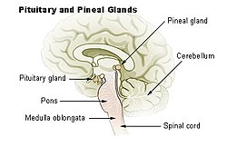
Illu pituitary pineal glands.jpg
Diagram of pituitary and pineal glands in the human brain
Details
Precursor Neural ectoderm, roof of diencephalon
Artery posterior cerebral artery
Identifiers
Latin glandula pinealis
MeSH D010870
NeuroNames 297
NeuroLex ID birnlex_1184
TA A11.2.00.001
FMA 62033
Pineal gland or epiphysis (in red in back of the brain). Expand the image to an animated version

The pineal gland, also known as the conarium or epiphysis cerebri, is a small endocrine gland in the vertebrate brain. The pineal gland produces melatonin, a serotonin-derived hormone which modulates sleep patterns in both circadian and seasonal cycles. The shape of the gland resembles a pine cone, hence its name. The pineal gland is located in the epithalamus, near the center of the brain, between the two hemispheres, tucked in a groove where the two halves of the thalamus join.[1][2]

Nearly all vertebrate species possess a pineal gland. The most important exception is a primitive vertebrate, the hagfish. Even in the hagfish, however, there may be a "pineal equivalent" structure in the dorsal diencephalon.[3] The lancelet Branchiostoma lanceolatum, the nearest existing relative to vertebrates, also lacks a recognizable pineal gland.[4] The lamprey (another primitive vertebrate), however, does possess one.[4] A few more developed vertebrates lost pineal glands over the course of their evolution.[5]

The results of various scientific research in evolutionary biology, comparative neuroanatomy and neurophysiology, have explained the phylogeny of the pineal gland in different vertebrate species. From the point of view of biological evolution, the pineal gland represents a kind of atrophied photoreceptor. In the epithalamus of some species of amphibians and reptiles, it is linked to a light-sensing organ, known as the parietal eye, which is also called the pineal eye or third eye.[6]

René Descartes believed the pineal gland to be the "principal seat of the soul". Academic philosophy among his contemporaries considered the pineal gland as a neuroanatomical structure without special metaphysical qualities; science studied it as one endocrine gland among many. However, the pineal gland continues to have an exalted status in the realm of pseudoscience.[7]

Structure

The pineal gland is a midline brain structure that is unpaired. It takes its name from its pine-cone shape.[8] The gland is reddish-gray and about the size of a grain of rice (5–8 mm) in humans. The pineal gland, also called the pineal body, is part of the epithalamus, and lies between the laterally positioned thalamic bodies and behind the habenular commissure. It is located in the quadrigeminal cistern near to the corpora quadrigemina.[9] It is also located behind the third ventricle and is bathed in cerebrospinal fluid supplied through a small pineal recess of the third ventricle which projects into the stalk of the gland.[10]

Blood supply

Unlike most of the mammalian brain, the pineal gland is not isolated from the body by the blood–brain barrier system;[11] it has profuse blood flow, second only to the kidney,[12] supplied from the choroidal branches of the posterior cerebral artery.

Nerve supply

The pineal gland receives a sympathetic innervation from the superior cervical ganglion. A parasympathetic innervation from the pterygopalatine and otic ganglia is also present.[13] Further, some nerve fibers penetrate into the pineal gland via the pineal stalk (central innervation). Also, neurons in the trigeminal ganglion innervate the gland with nerve fibers containing the neuropeptide PACAP.

Microanatomy

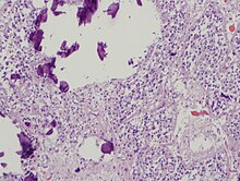
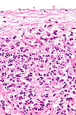
Pineal gland parenchyma with calcifications.
Micrograph of a normal pineal gland – very high magnification.
Micrograph of a normal pineal gland – intermediate magnification.

The pineal body consists in humans of a lobular parenchyma of pinealocytes surrounded by connective tissue spaces. The gland's surface is covered by a pial capsule.

The pineal gland consists mainly of pinealocytes, but four other cell types have been identified. As it is quite cellular (in relation to the cortex and white matter), it may be mistaken for a neoplasm.[14]

Cell type Description
Pinealocytes The pinealocytes consist of a cell body with 4–6 processes emerging. They produce and secrete melatonin. The pinealocytes can be stained by special silver impregnation methods. Their cytoplasm is lightly basophilic. With special stains, pinealocytes exhibit lengthy, branched cytoplasmic processes that extend to the connective septa and its blood vessels.
Interstitial cells Interstitial cells are located between the pinealocytes. They have elongated nuclei and a cytoplasm that is stained darker than that of the pinealocytes.
Perivascular phagocyte Many capillaries are present in the gland, and perivascular phagocytes are located close to these blood vessels. The perivascular phagocytes are antigen presenting cells.
Pineal neurons In higher vertebrates neurons are usually located in the pineal gland. However, this is not the case in rodents.
Peptidergic neuron-like cells In some species, neuronal-like peptidergic cells are present. These cells might have a paracrine regulatory function.

In some parts of the brain and in particular the pineal gland, there are calcium structures, the number of which increases with age, called corpora arenacea (or "acervuli," or "brain sand"). Chemical analysis shows that they are composed of calcium phosphate, calcium carbonate, magnesium phosphate, and ammonium phosphate.[15] In 2002, deposits of the calcite form of calcium carbonate were described.[16] Calcium and phosphorus[17] deposits in the pineal gland have been linked with aging.

Development

The human pineal gland grows in size until about 1–2 years of age, remaining stable thereafter,[18][19] although its weight increases gradually from puberty onwards.[20][21] The abundant melatonin levels in children are believed to inhibit sexual development, and pineal tumors have been linked with precocious puberty. When puberty arrives, melatonin production is reduced.[citation needed]

Symmetry

In the zebrafish the pineal gland does not straddle the midline but shows a left-sided bias. In humans, functional cerebral dominance is accompanied by subtle anatomical asymmetry.[22][23][24]

Function

The primary function of the pineal gland is to produce melatonin. Melatonin has various functions in the central nervous system, the most important of which is to help modulate sleep patterns. Melatonin production is stimulated by darkness and inhibited by light.[25][26] Light sensitive nerve cells in the retina detect light and send this signal to the suprachiasmatic nucleus (SCN), synchronizing the SCN to the day-night cycle. Nerve fibers then relay the daylight information from the SCN to the paraventricular nuclei (PVN), then to the spinal cord and via the sympathetic system to superior cervical ganglia (SCG), and from there into the pineal gland.

The compound pinoline is also claimed to be produced in the pineal gland; it is one of the beta-carbolines.[27] This claim is subject to some controversy.

Regulation of the pituitary gland

Studies on rodents suggest that the pineal gland influences the pituitary gland's secretion of the sex hormones, follicle-stimulating hormone (FSH), and luteinizing hormone (LH). Pinealectomy performed on rodents produced no change in pituitary weight, but caused an increase in the concentration of FSH and LH within the gland.[28] Administration of melatonin did not return the concentrations of FSH to normal levels, suggesting that the pineal gland influences pituitary gland secretion of FSH and LH through an undescribed transmitting molecule.[28]

The pineal gland contains receptors for the regulatory neuropeptide, endothelin-1,[29] which, when injected in picomolar quantities into the lateral cerebral ventricle, causes a calcium-mediated increase in pineal glucose metabolism.[30]

Drug metabolism

Studies on rodents suggest that the pineal gland may influence the actions of recreational drugs, such as cocaine,[31] and antidepressants, such as fluoxetine (Prozac),[32] and that its hormone melatonin can protect against neurodegeneration.[33]

Regulation of bone metabolism

Studies in mice suggest that the pineal-derived melatonin regulates new bone deposition. Pineal-derived melatonin mediates its action on the bone cells through MT2 receptors. This pathway could be a potential new target for osteoporosis treatment as the study shows the curative effect of oral melatonin treatment in a postmenopausal osteoporosis mouse model.[34]

Clinical significance

Calcification

Calcification of the pineal gland is typical in young adults, and has been observed in children as young as two years of age.[35] Calcium and phosphorus deposits in the pineal gland have been correlated with aging.[17] By old age, the pineal gland contains about the same amount of fluoride as teeth.[36] Pineal fluoride and pineal calcium are correlated.[36]

The calcified gland is often seen in skull X-Rays.[35] Calcification rates vary widely by country and correlate with an increase in age, with calcification occurring in an estimated 40% of Americans by age seventeen.[35] Calcification of the pineal gland is largely associated with corpora arenacea, also known as "brain sand".

It seems that the internal secretions of the pineal gland inhibit the development of the reproductive glands, because, in cases where it is severely damaged in children, the result is accelerated development of the sexual organs and the skeleton.[37]

Some studies show that the degree of pineal gland calcification is significantly higher in patients with Alzheimer's disease vs. other types of dementia.[38] Pineal gland calcification may contribute to the pathogenesis of Alzheimer's disease and may reflect an absence of crystallization inhibitors.[38] Calcification of the pineal gland has also been found to be closely associated to certain types of migraines as well as cluster headaches.[39][36]

Tumours

Tumours of the pineal gland are called pinealomas. These tumours are rare and 50% to 70% are germinomas that arise from sequestered embryonic germ cells. Histologically they are similar to testicular seminomas and ovarian dysgerminomas.[40]

A pineal tumour can compress the superior colliculi and pretectal area of the dorsal midbrain, producing Parinaud's syndrome. Pineal tumours also can cause compression of the cerebral aqueduct, resulting in a noncommunicating hydrocephalus. Other manifestations are the consequence of their pressure effects and consist of visual disturbances, headache, mental deterioration, and sometimes dementia-like behaviour.[41]

These neoplasms are divided into three categories, pineoblastomas, pineocytomas, and mixed tumours, based on their level of differentiation, which, in turn, correlates with their neoplastic aggressiveness.[42] The clinical course of patients with pineocytomas is prolonged, averaging up to several years.[43] The position of these tumours makes them very difficult to remove surgically.

Other animals

Most living vertebrates have pineal glands. It's likely that the common ancestor of all vertebrates had a pair of photosensory organs on the top of its head, similar to the arrangement in modern lampreys.[44] Some extinct Devonian fishes have two parietal foramina in their skulls,[45][46] suggesting an ancestral bilaterality of parietal eyes. The parietal eye and the pineal gland of living tetrapods are probably the descendants of the left and right parts of this organ, respectively.[47]

During embryonic development, the parietal eye and the pineal organ of modern lizards[48] and tuataras[49] form together from a pocket formed in the brain ectoderm. The loss of parietal eyes in many living tetrapods is supported by developmental formation of a paired structure that subsequently fuses into a single pineal gland in developing embryos of turtles, snakes, birds, and mammals.[50]

The pineal organs of mammals fall into one of three categories based on shape. Rodents have more structurally-complex pineal glands than other mammals.[51]

Crocodilians and some tropical lineages of mammals (some xenarthrans [sloths], pangolins, sirenians [manatees & dugongs], and some marsupials [sugar gliders]) have lost both their parietal eye and their pineal organ.[52][53][51] Polar mammals, such as walruses and some seals, possess unusually large pineal glands.[52]

All amphibians have a pineal organ, but some frogs and toads also have what is called a "frontal organ", which is essentially a parietal eye.[54]

Pinealocytes in many non-mammalian vertebrates have a strong resemblance to the photoreceptor cells of the eye. Evidence from morphology and developmental biology suggests that pineal cells possess a common evolutionary ancestor with retinal cells.[55]

Pineal cytostructure seems to have evolutionary similarities to the retinal cells of the lateral eyes.[55] Modern birds and reptiles express the phototransducing pigment melanopsin in the pineal gland. Avian pineal glands are thought to act like the suprachiasmatic nucleus in mammals.[56] The structure of the pineal eye in modern lizards and tuatara is analogous to the cornea, lens, and retina of the lateral eyes of vertebrates.[50]

In most vertebrates, exposure to light sets off a chain reaction of enzymatic events within the pineal gland that regulates circadian rhythms.[57] In humans and other mammals, the light signals necessary to set circadian rhythms are sent from the eye through the retinohypothalamic system to the suprachiasmatic nuclei (SCN) and the pineal gland.

The fossilized skulls of many extinct vertebrates have a pineal foramen (opening), which in some cases is larger than that of any living vertebrate.[58] Although fossils seldom preserve deep-brain soft anatomy, the brain of the Russian fossil bird Cerebavis cenomanica from Melovatka, about 90 million years old, shows a relatively large parietal eye and pineal gland.[59]

Society and culture

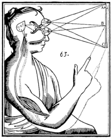
Diagram of the operation of the pineal gland for Descartes in the Treatise of Man (figure published in the edition of 1664)

Seventeenth-century philosopher and scientist René Descartes was highly interested in anatomy and physiology. He discussed the pineal gland both in his first book, the Treatise of Man (written before 1637, but only published posthumously 1662/1664), and in his last book, The Passions of the Soul (1649) and he regarded it as "the principal seat of the soul and the place in which all our thoughts are formed."[7] In the Treatise of Man, Descartes described conceptual models of man, namely creatures created by God, which consist of two ingredients, a body and a soul.[7][60] In the Passions, Descartes split man up into a body and a soul and emphasized that the soul is joined to the whole body by "a certain very small gland situated in the middle of the brain's substance and suspended above the passage through which the spirits in the brain's anterior cavities communicate with those in its posterior cavities". Descartes attached significance to the gland because he believed it to be the only section of the brain to exist as a single part rather than one-half of a pair. Most of Descartes's basic anatomical and physiological assumptions were totally mistaken, not only by modern standards, but also in light of what was already known in his time.[7][61]

The notion of a "pineal-eye" is central to the philosophy of the French writer Georges Bataille, which is analyzed at length by literary scholar Denis Hollier in his study Against Architecture. In this work Hollier discusses how Bataille uses the concept of a "pineal-eye" as a reference to a blind-spot in Western rationality, and an organ of excess and delirium.[62] This conceptual device is explicit in his surrealist texts, The Jesuve and The Pineal Eye.[63]

In the late 19th century Madame Blavatsky (who founded theosophy) identified the pineal gland with the Hindu concept of the third eye, or the Ajna chakra. This association is still popular today.[7]

Rick Strassman, an author and Clinical Associate Professor of Psychiatry at the University of New Mexico School of Medicine, has theorised that the human pineal gland is capable of producing the hallucinogen N,N-dimethyltryptamine (DMT) under certain circumstances.[64] In 2013 he and other researchers first reported DMT in the pineal gland microdialysate of rodents.[65]

In the short story "From Beyond" by H. P. Lovecraft, a scientist creates an electronic device that emits a resonance wave, which stimulates an affected person's pineal gland, thereby allowing her or him to perceive planes of existence outside the scope of accepted reality, a translucent, alien environment that overlaps our own recognized reality. It was adapted as a film of the same name in 1986. The 2013 horror film, Banshee Chapter is heavily influenced by this short story.

History

The secretory activity of the pineal gland is only partially understood. Its location deep in the brain suggested to philosophers throughout history that it possesses particular importance. This combination led to its being regarded as a "mystery" gland with mystical, metaphysical, and occult theories surrounding its perceived functions.

The pineal gland was originally believed to be a "vestigial remnant" of a larger organ. In 1917, it was known that extract of cow pineals lightened frog skin. Dermatology professor Aaron B. Lerner and colleagues at Yale University, hoping that a substance from the pineal might be useful in treating skin diseases, isolated and named the hormone melatonin in 1958.[66] The substance did not prove to be helpful as intended, but its discovery helped solve several mysteries such as why removing the rat's pineal accelerated ovary growth, why keeping rats in constant light decreased the weight of their pineals, and why pinealectomy and constant light affect ovary growth to an equal extent; this knowledge gave a boost to the then new field of chronobiology.[67]

Quantum cryptography

From Wikipedia, the free encyclopedia https://en.wikipedia.org/wiki/Quantum_crypto...